STRUCTURAL ANALYSIS AND MOLECULAR DYNAMICS STUDY OF PHB SYNTHASE
6
0
0
Full text
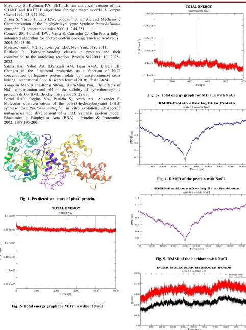
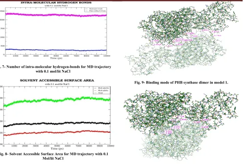
(2) T. Femlin Blessia et al. IRJP 2012, 3 (2) water. The number of water molecules added was 40113. Before the dynamics simulation, internal constraints were relaxed by energy minimization for 10 pico second. After the minimization, position restraint dynamics was run for 100 pico second, holding the target molecule stable, but allowing the water to settle around it. A 5 ns long production MD run was performed after the equilibration. During the MD run, the LINCS algorithm16 was used to constrain the lengths of hydrogen containing bonds; the waters were restrained using the SETTLE algorithm17. The simulations were run under NPT conditions, using Berendsen’s coupling algorithm to keep the temperature and the pressure constant (P = 1 bar, τP = 0.5 ps; T = 300° K; τT = 0.1 ps). The coordinates were saved every 0.5 ps. B) 10 nano second molecular dynamics run with 0.1 mol/lit NaCl Next, another set of 10 ns molecular dynamics simulation was carried out by adding 86 Na+ and 78 Cl- ions, corresponding to a salt concentration of 0.1 mol/lit, by replacing the water molecules at random positions. Molecular dynamics trajectory analysis The trajectories of 5 nano second MD simulation without NaCl and 10 nano second MD simulation with NaCl were analyzed using various tools available in GROMACS. The convergence of thermodynamic parameters, such as temperature, pressure, potential and kinetic energy were analyzed to check the quality of the MD simulation. The convergence was also checked in terms of the structure, through the root mean square deviation (RMSD) against the starting structure. The inter-molecular hydrogen bonds, intra-molecular hydrogen bonds and Solvent Accessible Surface Area (SASA) were also analyzed to have a clear idea about the structure. The role of NaCl in the structural stabilization of PHB synthase was studied by analyzing the number of hydrogen bonds and salt bridges in the low energy frames of 5 ns MD trajectory without NaCl and 10 ns MD trajectory with NaCl. Protein-protein docking The PHB synthase protein is active in the dimeric state18. So, protein-protein (PHB synthase-PHB synthase) docking was performed for the PHB synthase protein, using 'Cluspro 2.0' server19. The docking results were analyzed, for the interactions using Maestro (Schrodinger, Version 9.2)20. RESULTS Sequence analysis PSI-BLAST shows that the protein has two domains: the first one belongs to phaC_N superfamily and the second is esterase lipase Superfamily. The multiple sequence alignment of Chromobacterium violaceum PHB synthase with class I, II and III PHA synthases revealed that the conserved catalytic aspartic acid, histidine and cysteine are located at residues 447, 477 and 291 respectively. So, the proposed catalytic triad residues are CYS 291, HIS 477 and ASP 447. The residue participating in PHB synthase protein-ligand (3hydroxybutyryl coenzyme A) linkage is predicted as CYS 302. Tertiary structure prediction by threading approach and loop refinement Out of 5 models generated by I-TASSER, the model with best confidence score was selected for further analysis. Fig. 1 shows the best model generated by I-TASSER. The analysis of stereochemistry of the predicted PHB synthase. structure using PROCHECK AND VERIFY_3D confirms the quality of the three dimensional structure. Molecular dynamics simulation of PHB synthase The molecular dynamics simulation (the first 5 ns without adding NaCl and the second 10 ns with the addition of 0.1 mol/lit NaCl) carried out for the PHB synthase protein, gave an overall idea about some of its structural and functional parameters. The 5 ns MD trajectory without NaCl did not show any significant results. So the following discussion focuses mainly on the different parameters of 10 ns MD trajectory. A) Convergence in terms of energy The total energy of PHB synthase protein is stable throughout the MD simulation without NaCl, with values between -1.46e+06 KJ mol-1 to -1.45 e+06 KJ mol-1. The total energy of the PHB synthase protein throughout the MD run with 0.1 mol/lit NaCl has lower energy and is slightly fluctuating within the range -1.49e+06 KJ mol-1to 1.51e+06 KJ mol-1 . The plots showing the total energy throughout the trajectory for both MD run without NaCl and MD run with 0.1 mol/lit NaCl are displayed in Fig. 2 and Fig. 3 respectively. The temperature and pressure is also stable throughout the MD simulation. The MD run with NaCl prefers slightly higher temperature, due to the presence of NaCl ions in the system. B) Convergence in terms of structure The convergence of the structure was checked in terms of RMSD. The time evolution of the RMSD with respect to the initial structure provides a measurement of convergence of the dynamic properties of the protein. Each frame in the trajectory was superimposed on the reference frame using least-squares fit method. The RMSD of the whole protein was calculated. Analysis of the 10 ns MD simulation with 0.1 mol/lit NaCl showed that the RMSD values of the MD trajectory frames (whole protein) with respect to the initial structure lies between the range 0 - 1.3 nm. The RMSD from the initial starting structure is unstable. So the structure may still be progressing towards its equilibrium state. Fig. 4 describes the graph showing the RMSD of the protein (all atoms) with respect to the initial structure. Analysis of the 10 ns MD simulation with 0.1 mol/lit NaCl also shows that the RMSD of the MD trajectory frames of protein backbone lies between the range 0 -1.25 nm. The RMSD of the backbone has a slightly lower value than the RMSD of the whole protein. This is the result of excluding the, often flexible, side chain atoms. However, with increasing distance, the amount of conformations available also increases. The graph showing the RMSD of the protein backbone with respect to the starting structure is displayed in Fig. 5. C) Hydrogen bonds Inter-molecular hydrogen bonds The number of inter-molecular hydrogen bonds in the MD trajectory with 0.1 mol/lit NaCl ranges from 1225-1700. It is well known from the previous findings21 that intermolecular hydrogen bonds play a major role in stabilization of the molecule. The more number of inter-molecular interactions in the 10 ns MD trajectory with NaCl corresponds to its greater stability and lower energy. The plot showing the number of inter-molecular hydrogen bonds present in the protein in each 0.5 ps of 10 ns MD run with 0.1 mol/lit NaCl is shown in Fig. 6. Page 252.
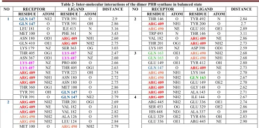
(3) T. Femlin Blessia et al. IRJP 2012, 3 (2) Intra-molecular hydrogen bonds The number of intra-molecular interactions in the protein molecule is maintained in the range 2250-2425, in the 10 ns simulation with 0.1 mol/lit NaCl. The plot showing the number of intra-molecular hydrogen bonds present in the protein in each 0.5 ps of 10 ns MD run with 0.1 mol/lit NaCl is displayed in Fig. 7. D) Solvent Accessible Surface Area The SASA graph (Fig. 8) for the MD run with NaCl shows the hydrophilic area, hydrophobic area, and the total area of the protein in each frame (each 0.5 ps). The values are almost stable throughout the simulation. Role of NaCl in structural stabilization The low energy frames from the molecular dynamics trajectory (both 5 ns MD simulation without NaCl and 10 ns MD simulation with 0.1 mol/lit NaCl) were selected. A comparative study between the low energy frames of 10 ns MD trajectory with NaCl and 5 ns MD trajectory without NaCl was performed, based on the number of hydrogen bonds and salt bridges, to study the role of NaCl in structural stabilization of PHB synthase. Table 1 shows the energy, number of salt bridges and number of hydrogen bonds in the selected frames of 5 ns MD trajectory without NaCl and 10 ns MD trajectory with NaCl respectively. It is apparent from Table 1 that the number of salt bridges and number of hydrogen bonds is low in the MD trajectory with NaCl. Addition of NaCl decreases electrostatic repulsion, which is the reason for the low number of hydrogen bonds observed in 10 ns MD trajectory with NaCl22. Protein-protein docking As the PHB synthase protein is active only in dimeric state, protein-protein docking was performed. Ten top scoring dimer models obtained from Cluspro server was intensively analyzed for their inter-molecular interactions. The interactions between the homo-dimer in the best four models are displayed in Table 2. Analysis of these docked dimer models reveals that GLN 147, GLN 163, ARG 409, LYS 487 and ARG 490 are highly participating in the protein-protein interaction. The Fig. 9 and Fig. 10 shows the binding mode of the PHA synthase in dimeric state in the two best models obtained from cluspro protein-protein docking server. DISCUSSION In the absence of the structural information of PHB synthase, protein structure prediction using bioinformatics methods helped us to have an overall idea about the protein structure. The molecular dynamics calculation reveals that the protein has stable energy. Inter-molecular hydrogen bond and intra-molecular hydrogen bond analysis clearly shows that the high number of hydrogen bonds in 10 ns MD trajectory with NaCl is responsible for its energy stability23. Other parameters like temperature, pressure, SASA, etc. are also stable throughout the simulation. RMSD analysis from the MD trajectory reveals the greater difference in the protein structure throughout the MD simulation. This clearly says that the structure may still be progressing towards the equilibrium state. Analysis of the low energy frames from MD trajectory with NaCl and MD trajectory without NaCl proves that NaCl plays an important role in modulating the electrostatic interactions. Previous research in PHA synthase from other organisms, revealed the catalytic triad residues and the residue participating in PHB synthase protein-ligand (3HB-CoA) linkage24. The sequence alignment of the. Chromobacterium violaceum PHB synthase sequence with the sequences of PHA synthase from other organisms suggests that the catalytic triad residues in Chromobacterium violaceum PHB synthase are CYS 291, ASP 447 and HIS 477. The sequence alignment also reveals that the amino acid in PHB synthase which links with the ligand 3HB-CoA (3-hydroxybutyryl coenzyme A) is CYS 302. The protein-protein interface prediction and proteinprotein docking results reveals the important residues participating in the dimerization. CONCLUSION Tertiary structure prediction of the phaC sequence gave a clear insight about its structural and functional properties. Sequence alignment of Chromobacterium violaceum PHB synthase with the PHA synthase sequences from other species suggested the catalytic triad residues in Chromobacterium violaceum PHB synthase and the residue which links PHB synthase to 3HB-CoA ligand. Molecular dynamics simualtions revealed that the structure is stable in terms of energy, but there is a greater deviation in RMSD. Molecular dynamics study also proves that NaCl plays an important role in modulating electrostatic interactions. Protein-protein interaction studies revealed the binding mode of PHB synthase during its dimerization. REFERENCES 1. Gao D, Maehara A, Yamane T, Ueda S. Identification of the intracellularpolyhydroxyalkanoate depolymerase gene of Paracoccus denitrificans and some properties of the gene product. FEMS Microbiol Lett 2001; 196: 159-164. Steinbuchel A, Schlegel HG. Physiology and molecular genetics of 2. poly(beta-hydroxy-alkanoic acid) synthesis in Alcaligenes eutrophus. Mol Microbiol 1991; 5: 535-542. 3. Bonartsev AP, Myshkina VL, Nikolaeva DA, Furina EK, Makhina TA, Livshits VA, et al. Biosynthesis, biodegradation, and application of poly(3-hydroxybutyrate) and its copolymers - natural polyesters produced by diazotrophic bacteria. Communicating Current Research and Educational Topics and Trends in Applied Microbiology 2007; Méndez-Vilas A (Ed.), 295-307. 4. Rehm BHA, Steinbuchel A. Biochemical and genetic analysis of PHA synthases and other proteins required for PHA synthesis. Int. J. Biol. Macromol 1999; 25: 3-19. 5. Chadi. Energy-minimization approach to the atomic geometry of semiconductor surfaces. Phys Rev Lett 1978; 41: 1062-1065. 6. Alder BJ, Wainwright TE. Studies in Molecular Dynamics. I. General Method. J Chem Phys 1959; 31: 459-461. Altschul SF, Gish W, Miller W, Myers EW, Lipman, DJ. Basic local 7. alignment search tool. J Mol Biol 1990; 215: 403-410. Thompson JD, Higgins DG, Gibson TJ. CLUSTAL W: improving the 8. sensitivity of progressive multiple sequence alignment through sequence weighting, position-specific gap penalties and weight matrix choice. Nuc Acids Res 1994; 22: 4673-4680. 9. Zhang Y. I-TASSER: Fully automated protein structure prediction in CASP8. Proteins 2009; 9: 100-113. 10. Zhang Y. I-TASSER server for protein 3D structure prediction. BMC Bioinformatics 2008; 9: 40-45. 11. Eswar N, Marti-Renom MA, Webb B, Madhusudhan MS, Eramian D, Shen M, et al. Comparative Protein Structure Modeling with MODELLER. Curr Prot in Bioin 2006; 15: 561-563. 12. Laskowski RA, MacArthur MW, Moss DS, Thornton JM. PROCHECK: a program to check the stereochemical quality of protein structures. J Appl Crystallogr 1993; 26: 283-291. 13. Bowie JU, Lüthy R, Eisenberg D. A method to identify protein sequences that fold into a known three-dimensional structure. Science 1991; 253: 164-170. 14. Luthy R, Bowie JU, Eisenberg D. Assessment of protein models with three-dimensional profiles. Nature 1992; 6364: 83-85. 15. Lindahl E, Hess B, Van der Spoel D. GROMACS 3.0: a package for molecular simulation and trajectory analysis. J Mol Model 2001; 7: 306-309. 16. Hess B, Bekker H, Berendsen HJC, Fraaije JGEM. LINCS: a linear constraint solver for molecular simulations. J Comput Chem 1997; 18: 1463-1472.. Page 253.
(4) T. Femlin Blessia et al. IRJP 2012, 3 (2) 17. Miyamoto S, Kollman PA. SETTLE: an analytical version of the SHAKE and RATTLE algorithms for rigid water models. J Comput Chem 1992; 13: 952-962. 18. Zhang S, Yasuo T, Lenz RW, Goodwin S. Kinetic and Mechanistic Characterization of the Polyhydroxybutyrate Synthase from Ralstonia eutropha”. Biomacromolecules 2000; 1: 244-251. 19. Comeau SR, Gatchell DW, Vajda S, Camacho CJ. ClusPro: a fully automated algorithm for protein-protein docking. Nucleic Acids Res 2004; 20: 45-50. 20. Maestro, version 9.2, Schrodinger, LLC, New York, NY, 2011. 21. Raffaele R. Hydrogen-bonding classes in proteins and their contribution to the unfolding reaction. Protein Sci 2001; 10: 2075– 2082. 22. Salma HA, Nahid AA, ElShazali AM, Isam AMA, Elfadil EB. Changes in the functional properties as a function of NaCl concentration of legumes protein isolate by transglutaminase cross linking. International Food Research Journal 2010; 17: 817-824. 23. Yong-Jin Mao, Xiang-Rong Sheng, Xian-Ming Pan, The effects of NaCl concentration and pH on the stability of hyperthermophilic protein Ssh10b. BMC Biochemistry 2007; 8: 28-33. 24. Bernd HAR, Regina VA, Patricia S, Amro AA, Alexander S. Molecular characterization of the poly(3-hydroxybutyrate) (PHB) synthase from Ralstonia eutropha: in vitro evolution, site-specific mutagenesis and development of a PHB synthase protein model. Biochimica et Biophysica Acta (BBA) - Proteins & Proteomics 2002; 1598:195-200.. Fig. 3- Total energy graph for MD run with NaCl. Fig. 4- RMSD of the protein with NaCl.. Fig. 1- Predicted structure of phaC protein.. Fig. 5- RMSD of the backbone with NaCl. Fig. 2- Total energy graph for MD run without NaCl. Fig. 6- Number of inter-molecular hydrogen-bonds for MD trajectory with 0.1 mol/lit NaCl. Page 254.
(5) T. Femlin Blessia et al. IRJP 2012, 3 (2). Fig. 7- Number of intra-molecular hydrogen-bonds for MD trajectory with 0.1 mol/lit NaCl Fig. 9- Binding mode of PHB synthase dimer in model 1.. Fig. 8- Solvent Accessible Surface Area for MD trajectory with 0.1 Mol/lit NaCl Fig. 10- Binding mode of PHB synthase dimer in model 2. Table 1- Comparative study of hydrogen bonds and salt bridges in the selected frames of MD trajectory with NaCl and without NaCl MD Trajectory NO OF SALT TIME STEPS (ps) ENERGY (KJ/mol) NO OF HYDROGEN BONDS BRIDGES 2551.800049 -1459524.000000 29 85 Without NaCl 3061.000244 -1459534.000000 31 83 4688.200195 -1459485.500000 29 84 4824.200000 -1459595.500000 24 89 -1459262.250000 30 88 4895.000000 With 0.1 mol/lit NaCl 533.600037 -1505461.875000 10 78 -1505370.625000 11 67 1064.600098 2298.600098 -1505213.125000 10 76 8140.200195 -1510120.000000 11 58 -1510140.250000 11 61 8142.800293 Low energy frames selected from MD trajectory without NaCl and MD trajectory with NaCl were analyzed for the number of salt bridges and number of hydrogen bonds. This study clearly shows that the electrostatic repulsion caused by the addition of Na and Cl ions is the reason for its minimum energy.. Page 255.
(6) T. Femlin Blessia et al. IRJP 2012, 3 (2) Table 2- Inter-molecular interactions of the dimer PHB synthase in balanced state RECEPTOR LIGAND DISTANCE NO RECEPTOR LIGAND DISTANCE RESIDUE ATOM RESIDUE ATOM RESIDUE ATOM RESIDUE ATOM NE2 TYR 391 O 2.9 THR 146 O TYR 492 N 2.84 1 GLN 147 2 TYR 391 OH 2.86 TYR 200 O 2.7 O NH1 ARG 409 GLN 147 LEU 181 O ILE 413 N 3.16 NE GLU 101 OE1 2.89 ARG 490 MET 100 O PHE 361 N 3.43 TRP 493 N THR 146 O 3.11 ASN 180 OD1 NH1 2.60 VAL 182 O NE 2.86 ARG 409 ARG 409 NH2 NH2 GLN 410 OE1 2.75 THR 201 OG1 2.73 ARG 409 ARG 409 LYS 179 NZ SER 363 OG 3.03 LYS 105 NZ ASP 398 OD1 2.59 THR 405 OG1 NZ 2.47 GLN 163 OE1 NH2 2.68 LYS 487 3 ARG 490 ASN 367 OD1 NZ 2.60 GLN 163 O NH1 2.68 LYS 487 ARG 490 NZ PRO 400 O 2.66 GLU 149 OE1 TYR 412 OH 2.80 LYS 487 NZ NE THR 405 OG1 2.63 GLN 147 O 2.73 LYS 487 ARG 409 NE NH1 TYR 223 OH 2.74 LYS 164 O 2.70 ARG 409 ARG 490 NH1 NH2 O ASN 180 O 2.72 2.67 ARG 409 ARG 490 GLN 163 NH2 NH1 ASN 180 O 2.75 GLU 149 OE2 2.70 ARG 409 ARG 409 OG1 MET 100 O 2.86 NH1 GLY 148 O 2.62 THR 360 ARG 409 TYR 391 O NH2 OH 2.83 ALA 143 O 2.71 GLN 147 ARG 409 NE2 NH2 TYR 391 O 2.79 ILE 144 O 2.67 ARG 409 GLN 147 4 NH2 THR 201 OG1 2.69 ARG 445 NH2 GLU 336 OE1 2.74 2 ARG 409 VAL 182 O 2.81 SER 453 OG GLU 329 OE2 2.86 NE ARG 409 NH2 VAL 182 O 2.82 HIS 448 ND1 ALA 337 O 2.89 ARG 409 NH2 ALA 126 O 2.95 GLU 329 OE2 TYR 456 OH 2.83 ARG 490 NH2 LEU 124 O 2.84 GLU 336 OE1 ARG 445 NE 2.90 ARG 490 MET 100 O NH2 2.79 ARG 490 GLN 147, GLN 163, ARG 409, LYS 487 and ARG 490 are highly participating in the protein-protein interaction which clearly shows that these residues are very important in the dimerization of the protein. NO. Source of support: Nil, Conflict of interest: None Declared. Page 256.
(7)
Figure
Related documents
It is emphasized that the anomalous electric dipole moment of the pointlike electron (AEDM) is fundamentally different from the quantum field type electric dipole moment of
B.: Congenital atresia of the inferior vena cava, common iliac veins, and left innominate vein; a case with extensive development of superficial venous col-
Regarding electric arc slag furnace, it is found that when they are treated in a similar way to the cement (M2), their properties are better than those obtained with the reference
Due to the continued interest and debate in this area, we conducted a scoping systematic literature review and meta-analysis of all observational studies
Remarkable Boron Delivery Of iRGD-Modified Polymeric Nanoparticles For Boron Neutron Capture Therapy
To assess the effect of boron accumulation in tumor using tumor vascular normalization, B16F10 tumor-bearing BALB/c nude mice following either PBS or Endostar admin- istration
In the following, the event selection used in the three analyses will be presented (Section 2) before describing the SM background estimation techniques (Section 3).. The last
The paper examines the role of security agencies in cubing election violence in Nigeria using Kebbi state as a
We also performed unilateral orchidectomy as well as scrotal ablation to prevent recurrence of hernia, seroma formation and to preserve the unaffected testicle.
