R E S E A R C H
Open Access
How to describe a cryptic species? Practical
challenges of molecular taxonomy
Katharina M Jörger
1,2*and Michael Schrödl
1,2Abstract
Background:Molecular methods of species delineation are rapidly developing and widely considered as fast and efficient means to discover species and face the‘taxonomic impediment’ in times of biodiversity crisis. So far, however, this form of DNA taxonomy frequently remains incomplete, lacking the final step of formal species description, thus enhancing rather than reducing impediments in taxonomy. DNA sequence information contributes valuable diagnostic characters and–at least for cryptic species–could even serve as the backbone of a taxonomic description. To this end solutions for a number of practical problems must be found, including a way in which molecular data can be presented to fulfill the formal requirements every description must meet. Multi-gene barcoding and a combined molecular species delineation approach recently revealed a radiation of at least 12 more or less cryptic species in the marine meiofaunal slug genusPontohedyle(Acochlidia, Heterobranchia). All identified candidate species are well delimited by a consensus across different methods based on mitochondrial and nuclear markers.
Results:The detailed microanatomical redescription ofPontohedyle verrucosaprovided in the present paper does not reveal reliable characters for diagnosing even the two major clades identified within the genus on molecular data. We thus characterize three previously validPontohedylespecies based on four genetic markers
(mitochondrial cytochrome c oxidase subunit I, 16S rRNA, nuclear 28S and 18S rRNA) and formally describe nine cryptic new species (P. kepiisp. nov.,P. jonisp. nov.,P. neridaesp. nov.,P. liliaesp. nov.,P. wiggisp. nov.,P. wenzli
sp. nov.,P. peteryallisp. nov.,P. martynovisp. nov.,P. yurihookerisp. nov.) applying molecular taxonomy, based on diagnostic nucleotides in DNA sequences of the four markers. Due to the minute size of the animals, entire specimens were used for extraction, consequently the holotype is a voucher of extracted DNA (‘DNA-type’). We used the Character Attribute Organization System (CAOS) to determine diagnostic nucleotides, explore the dependence on input data and data processing, and aim for maximum traceability in our diagnoses for future research. Challenges, pitfalls and necessary considerations for applied DNA taxonomy are critically evaluated.
Conclusions:To describe cryptic species traditional lines of evidence in taxonomy need to be modified. DNA sequence information, for example, could even serve as the backbone of a taxonomic description. The present contribution demonstrates that few adaptations are needed to integrate into traditional taxonomy novel
diagnoses based on molecular data. The taxonomic community is encouraged to join the discussion and develop a quality standard for molecular taxonomy, ideally in the form of an automated final step in molecular species delineation procedures.
* Correspondence:[email protected] 1Mollusca Section, SNSB-Bavarian State Collection of Zoology, Münchhausenstr 21, 81247 München, Germany
2Department Biology II, Ludwig-Maximilians-University, Großhaderner Str. 2, 82152 Planegg-Martinsried, Germany
Background
Species boundaries are frequently hard to delimit based on morphology only, a fact which has called for integrative taxonomy, including additional sources of information such as molecular data, biogeography, behavior and ecol-ogy [1,2]. Founding a species description on a variety of characters from different, independent datasets is generally regarded as best practice [3]. When species are considered as independently evolving lineages [4], different lines of evidence (e.g., from morphology, molecules, ecology or dis-tribution) are additive to each other and no line is neces-sarily exclusive nor need different lines obligatory be used in combination [3,5]. Taxonomists are urged to discrimin-ate characters according to their quality and suitability for species delineation, rather than to just add more and more data [5]. The specifics of the taxon in question will guide the way to the respective set(s) of characters that will pro-vide the best backbone for the diagnosis. In cases of pseudo-cryptic species (among which morphological differ-ences can be detected upon re-examining lineages sepa-rated e.g. on molecular data) or of fully cryptic species (that morphology fails to delimit), the traditional lines of evidence have to be modified by using, e.g., molecular in-formation to break out of the‘taxonomic circle’[6,7].
Cryptic species are a common phenomenon throughout the metazoan taxa, and can be found in all sorts of habitats and biogeographic zones [8-10]. Groups characterized by poor dispersal abilities (e.g., most meiofaunal organisms or animals inhabiting special regions where direct developers predominate, such as Antarctica), are especially prone to cryptic speciation [11,12]. Uncovering these cryptic species is fundamental for the understanding of evolutionary pro-cesses, historical biogeography, ecology, and also to conser-vation approaches, as distribution ranges that are smaller than initially assumed mean a higher risk of local extinc-tion [8,10]. The lack of morphological characters to distin-guish cryptic species should not lead to considerable parts of biological diversity remaining unaddressed.
The utility of DNA barcoding and molecular species de-lineation approaches to uncover cryptic lineages has been demonstrated by numerous studies (e.g., [11,13-19]). Unfor-tunately, inconsistencies in terminology associated with the interface between sequence data and taxonomy have led to confusion and various criticisms [6,20]. First of all, one needs to distinguish between species identification via mo-lecular data (DNA barcoding in its strict sense) and species discovery [6,21,22]. While species identification is a primary technical application, species delimitation requires means of molecular species delineation that is either distance, tree or character based [6,23]. Under ideal circumstances suffi-cient material is collected from different populations across the entire distribution area of a putative group of cryptic species. Using population genetics the distribution of haplo-types can be analyzed and different, genetically isolated
lineages can be detected [24]. Population genetic ap-proaches are, however, not always feasible with animals that are rare or hard to collect, which might actually be a com-mon phenomenon across faunas of most marine ecosys-tems [25-28]. Derived from barcoding initiatives, threshold based species delimitation became the method of choice, aiming for the detection of a‘barcoding gap’between intra-and interspecific variation [29-31]. This approach has been criticized, however, due to its sensitivity to the degree of sampling, the general arbitrariness of fixed or relative thresholds, and to frequent overlap between intra- and in-terspecific variation [6,32,33]. In the recently developed Automatic Barcode Gap Discovery (ABGD) [34], progress has been made in avoiding the dependence of a priori defined species hypotheses in threshold based approaches, but reservations remain concerning the concept of a barcoding gap [25]. Several independent delineation tools exist, e.g. using haplotype networks based on statistical par-simony [35], maximum likelihood approaches applying the General Mixed Yule-Coalescent model [36,37], or Bayesian species delineation [38,39]. Empirical research currently compares the powers of these different tools on real datasets [25,32,40]. The effect of the inclusion of single-tons in analyses is considered as most problematic [25]. At the present stage of knowledge, independent approaches allowing cross-validation between the different methods of molecular species delineation and other sources of infor-mation (morphology, biogeography, behavioral traits) seem the most reliable way of delimiting cryptic species [25].
The second inconsistency in terminology concerns us-ages of ‘DNA taxonomy’. Originally, DNA taxonomy was proposed to revolutionize taxonomy by generally founding descriptions on sequence data and overthrowing the Linnaean binominal system [41]. Alternatively, it was suggested as a concept of clustering DNA barcodes into MOTUs [42]. Since then, however, it has been applied as an umbrella term for barcoding, molecular species delineation, and including molecular data in species descriptions (see e.g., [13,14,20,36,43,44]). In a strict sense, one cannot speak of molecular taxonomy if the process of species discovery is not followed by formal species description (i.e. there are two steps to a taxonomic process: species discovery (delimitation) and attributing them with formal diagnoses and names.) Taxonomy remains incom-plete if species hypotheses new to science are flagged as merely putative by provisional rather than fully established scientific names. For practical reasons and journal re-quirements, most studies on molecular species delineation postpone formal descriptions of the discovered species (e.g., [13,14,25,33,36,40,43-46]), and then rarely carry them out later. DNA barcoding and molecular species delinea-tion are promoted as fast and efficient ways to face the
species richness in the middle of a biodiversity crisis [7,47,48]. However, keeping discovered entities formally unrecognized does not solve the taxonomic challenges but adds to them by creating parallel worlds populated by numbered MOTUs, OTUs or candidate species. In many cases the discovered taxa remain inapplicable to future research, thus denying the scientific community this taxonomic service, e.g. for species inventories or conservation attempts. Without formal description or a testable hypothesis, i.e. a differential diagnosis, 1) the discovered species might not be properly documented or vouchered by specimens deposited at Natural History Museums; and 2) their reproducibility can be hindered and confusion caused by different numbering systems. A deterrent example of the proliferation of informal epithets circulating as‘nomina nuda’(i.e. species which lack formal diagnoses and deposited vouchers) in the literature is given by the ‘ten species in one’ Astraptes fulgerator complex [31,49]. Thus, we consider it as all but indispens-able for DNA taxonomy to take the final step and formalize the successfully discovered molecular lineages.
The transition from species delimitation to species de-scription is the major task to achieve. Nearly ten years after the original proposal of DNA taxonomy [41], revolutioniz-ing traditional taxonomy has found little acceptance in the taxonomic community, as most authors agree that there is no need for overthrowing the Linnaean System. Conse-quently, the challenge is to integrate DNA sequence infor-mation in the current taxonomic system. Several studies have attempted to include DNA data in taxonomic descrip-tions, albeit in various non-standardized ways; see the re-view by Goldstein and DeSalle ([21]; box 3): In some cases, DNA sequence information is simply added to the taxo-nomic description (in the form of GenBank numbers or pure sequence data), without evaluating and reporting diag-nostic features [21]. Others rely on sequence information for the description, either reporting results of species delin-eation approaches, e.g. raw distance measurements or model based assumptions, or extracting diagnostic charac-ters from their molecular datasets. There still is a consensus that species descriptions should be character based [50] (but see the Discussion below for attempts at model based taxonomy), and that tree or distance based methods fail to extract diagnostic characters [6]. Character based ap-proaches, like the Characteristic Attribute Organization Sys-tem (CAOS), are suggested as an efficient and reliable way of defining species barcodes based on discrete nucleotide substitution, and these established diagnostics from DNA sequences can be used directly for species descriptions as molecular taxonomic characters [51,52]. Yet, the application of CAOS or similar tools requires an evaluation of how to select and present molecular synapomorphies and how to formalize procedures to create a‘best practice’linking DNA sequence information to existing taxonomy [20].
In the present study, we formally describe the candi-date species of minute mesopsammic sea slugs in the genusPontohedyleGolikov & Starobogatov (Acochlidia, Heterobranchia) discovered by Jörger et al. [25]. This cryptic radiation was uncovered in a global sampling ap-proach with multi-gene and multiple-method molecular species delineation [25]. The initially identified 12 MOTUs, nine of which do not correspond to described species, are considered as species [following 4] resulting from a conser-vative minimum consensus approach applying different methods of molecular species delineation [25]. The authors demonstrated that traditional taxonomic characters (exter-nal morphology, spicules and radula features) are insuffi-cient to delineate cryptic Pontohedyle species [25]. To evaluate the power of more advanced histological and mi-croanatomical data, we first provide a detailed computer based 3D redescription of the anatomy of Pontohedyle verrucosa (Challis, 1970) and additional histological semi-thin sections ofP. kepiisp. nov. In the absence of reliable diagnostic characters from morphology and microanatomy, we then rely on DNA sequence data as the backbone for our species descriptions. For the three previously valid Pontohedyle species we extract diagnostic characters using the Character Attribute Organization System (CAOS) based on four standard markers (mitochondrial cytochrome c oxi-dase subunit I, 16S rRNA, and nuclear 18S rRNA and 28S rRNA). In addition, nine new species are formally described on molecular characteristics and evidence from other data sources. Various approaches to the practical challenges for molecular driven taxonomy–such as critical consideration of the quality of the alignment, detection of diagnostic nu-cleotides and their presentation aiming for maximum trace-ability in future studies–are tested and critically evaluated.
Results
Evaluation of putative morphological characters
The radulae of eight species were investigated using SEM (see Figures 1 and 2). Radulae of P. neridae sp. nov., P. martynovisp. nov. andP. yurihookerisp. nov. were not re-covered whole from molecular preparations, and thus were unavailable for further examination [25]. The radula of P. wiggi sp. nov. could only be observed under the light-microscope, but not successfully transferred to a SEM stub. All radulae are hook-shaped with a longer dorsal and a shorter ventral ramus, typical for Acochlidia. Radula formulas are 38–53 × 1.1.1, lateral plates are curved
rectangular, and the rhachidian tooth is triangular and bears a central cusp and typically three smaller lateral denticles. Most radulae bear one pointed denticle centrally on the anterior margin of each lateral plate and a corre-sponding notch on the posterior side. Only the radula ofP. kepiisp. nov. and P. verrucosacan be clearly dis-tinguished from the others by the absence of this den-ticle and the more curved lateral teeth (see Figure 1A and [25], Figure 1D,E). Uniquely, P. verrucosa bears five lateral denticles next to the central cusp of the Figure 1External morphology (living specimens) and radula characteristics (SEM micrographs) inPontohedylespecies (part 1).
rhachidian tooth [25]; inP. liliaesp. nov. a tiny fourth denticle borders the central cusp (see * in Figure 1C).
Previous phylogenetic analyses [25] recovered a deep split into twoPontohedyle clades: the P. milaschewitchii clade and the P. verrucosa clade. This is supported by novel analyses in a larger phylogenetic framework and additionally including a second nuclear marker (18S rRNA) (own unpublished data). Since no detailed histological account exists of any representative from the large P. verrucosaclade, we redescribeP. verrucosa(based on ZSM
Mol-20071833, 20071837 and 20100548), supplementing the original description with detailed information of the previously undescribed nervous and reproductive systems. The central nervous system (cns) ofP. verrucosa lies prepharyngeal and shows an epiathroid condition. It consists of paired rhinophoral, cerebral, pleural, pedal and buccal ganglia and three unpaired ganglia on the visceral nerve cord, tentatively identified as left parietal ganglion, median fused visceral and subintestinal ganglion and right fused parietal and supraintestinal ganglion (Figure 3A). An Figure 2External morphology (living specimens) and radula characteristics (SEM micrographs) inPontohedylespecies (part 2).
osphradial ganglion or gastro-oesophagial ganglia were not detected. Anterior and lateral to the cerebral ganglia are masses of accessory ganglia. Due to the retracted condition of all examined specimens, tissues are highly condensed and no separation in different complexes of accessory gan-glia could be detected. Attached to the pedal gangan-glia are large monostatolith statocysts. Oval, unpigmented globules are located in an antero-ventral position of the cerebral gan-glia, interpreted as the remainder of eyes (see Figure 3B).
P. verrucosais a gonochoristic species. The three sec-tioned specimens include two males and one female. The male reproductive system is comprised of gonad, ampulla, postampullary sperm duct, prostatic vas defer-ens, ciliated (non-glandular) vas deferdefer-ens, genital open-ing and a small ciliated ‘subepidermal’duct leading to a second genital opening anterodorsally of the mouth opening (Figure 3C). The sac-like gonad is relatively small and bears few irregular distributed spermatozoa. The large tubular ampulla emerges from the gonad with-out a detectable preampullary sperm duct; it is loosely filled with irregularly distributed spermatozoa (Figure 3D). The ampulla leads into a short, narrow ciliated post-ampullary duct widening into the large tubular prostatic vas deferens (staining pink in methylene-blue sections, Figure 3D). Close to the male genital opening, the duct loses its glandular appearance and bears cilia. The primary genital opening is located on the right side of the body at the visceral hump and close to the transition with the head-foot complex. Next to the genital opening, the anterior vas deferens splits off as an inconspicuous subepithelial ciliated duct that leads anteriorly on the right side of the head foot complex. It terminates in a second genital opening between the oral tentacles anterodorsally from the mouth opening.
The female reproductive system consists of gonad, nidamental glands and oviduct (Figure 3E) and a genital opening located on the right side, in the posterior part of the visceral hump (not visible in Figure 3E, due to the retracted stage of the individual). The gonad is sac-like and bears one large vitellogenic egg (see Figure 3F) and several developing oocytes. Three histologically dif-ferentiated tube-like nidamental glands could be detected with a supposedly continuous lumen and with an epithe-lium bearing cilia. From proximal to distal these glands are identified as albumen gland (cells filled with dark blue stained granules), membrane gland (pinkish, vacuolated secretory cells) and winding mucus gland (secretory cells stained pink-purple). In its proximal part the distal oviduct shows a similar histology as the mucous gland, but then loses its glandular appearance. The epithelium of the distal oviduct bears long, densely arranged cilia.
Additional notable histological features are numerous dark-blue-stained epidermal gland cells (see e.g., arrow-head in Figure 3D) and refracting fusiform structures in
the digestive gland (see Figure 3B). An additional series of histological semi-thin sections of Pontohedyle kepii sp. nov. was sectioned and brief investigation revealed no variation in the major organization of the organ sys-tems inPontohedyleas described herein and in previous studies [55,56].
Remarks on the presentation of molecular characters
Diagnostic characters for each species of Pontohedyle were extracted using the ‘Characteristic Attribute Organization System’ (CAOS) [51,57,58]. We define diagnostic characters as single pure characters, i.e. unique character states that respectively occur in all in-vestigated specimens in a single Pontohedyle species but in none of the specimens of its congeners. As add-itional information single heterogeneous pure charac-ters (i.e., different character states present within the species but absent from the congeners) are reported (for further details on the chosen approach see the Material and methods and Discussion sections). Posi-tions refer to the position of the diagnostic nucleotide within the respective alignment (see Additional files 1, 2, 3, 4, 5 and 6). Where alignment positions differ from those in the deposited sequences, positions within the sequence of the holotype or in another reference se-quence are also provided.
Taxonomy ofPontohedyle
Family: Microhedylidae Odhner, 1938 [59]
Genus:PontohedyleGolikov & Starobogatov, 1972 [60] Synonymy:MancohedyleRankin, 1979;Gastrohedyle
Rankin, 1979;MaraunibinaRankin, 1979
Type species (by subsequent designation):Pontohedyle milaschewitchii(Kowalevsky, 1901) [61]
Phylogenetic analyses of the genus Pontohedyle [25] confirmed earlier assumptions, that the three genera established by Rankin [62] (see above) present junior synonyms ofPontohedyle.
system with accessory ganglia at cerebral nerves anter-ior to the cns. Sexes separate, male reproductive system aphallic, sperm transferred via spermatophores.
Molecular diagnosis of the genusPontohedyle, based on the sequences analyzed herein (Table 1) and on sequences from a set of outgroups including all acochlidian genera
Table 1 DNA sequence data analyzed in the present study to determine diagnostic nucleotides inPontohedyle
Species Museums number DNA
voucher
GenBank accession numbers
18S rRNA 28S rRNA 16S rRNA COI
P. milaschewitchii ZSM Mol 20071381 AB34404214 - JQ410926 JQ410925 JQ410897
ZSM Mol 20080054 AB34404241 HQ168435 JF828043 HQ168422
-ZSM Mol 20080055 AB34404239 - - JQ410927
-ZSM Mol 20080925 - - - JQ410928 HQ168459
ZSM Mol 20080953 AB35081832 KC984282 - JQ410929 JQ410898
P. brasilensis SI-CBC20 10KJ01-E03 AB34500510 KC984283 JQ410941 JQ410940
-SI-CBC20 10KJ01-B07 AB34402082 - JQ410943 JQ410942
-SI-CBC20 10KJ01-D07 AB34500513 - JQ410944 -
-SI-CBC20 10KJ01-B09 AB34402031 - JQ410946 JQ410945 JQ410904
SI-CBC20 10KJ01-C09 AB34500576 - JQ410948 JQ410947 JQ410905
SI-CBC20 10KJ01-A10 AB34402026 - - JQ410949
-SI-CBC20 10KJ02-E01 AB34402030 - JQ410950 -
-ZSM Mol 20110723 AB34402034 KC984284 JQ410952 JQ410951 JQ410906
ZSM Mol 20110722 AB34402086 KC984285 JQ410932 JQ410931 JQ410900
ZSM Mol 20090198 AB35081813 KC984286 JQ410936 JQ410935
-P. verrucosa ZSM Mol 20071820 AB34404223 KC984287 JQ410978 JQ410977 JQ410920
ZSM Mol 20080176 AB34404286 - JQ410980 JQ410979 JQ410921
ZSM Mol 20071135 AB34404221 KC984288 JQ410971 JQ410970 JQ410914
ZSM Mol 20100388 AB34500547 - - - JQ410916
ZSM Mol 20100389 AB34402044 - JQ410974 - JQ410917
ZSM Mol 20100390 AB34402070 - JQ410975 - JQ410918
ZSM Mol 20100391 AB34500531 KC984289 - JQ410976 JQ410919
Pontohedyle kepiisp. nov. ZSM Mol 20081013 AB35081769 KC984290 JQ410967 JQ410966 JQ410912
Pontohedyle jonisp. nov. ZSM Mol 20090197 AB34858164 KC984291 JQ410934 JQ410933 JQ410901
SI-CBC20 10KJ01-D05 AB34402049 KC984292 - JQ410937 JQ410902
SI-CBC20 10KJ01-C08 AB34402065 - JQ410939 JQ410938 JQ410903
Pontohedyle neridaesp.nov. AM C. 476062.001 AB34500497 - JQ410986 JQ410985 JQ410922
Pontohedyle liliaesp.nov. ZSM Mol 20090471 AB35081802 KC984293 JQ410954 JQ410953
-ZSM Mol 20090472 AB35081838 - JQ410956 JQ410955
-Pontohedyle wiggisp.nov. ZSM Mol 20100595 AB34402059 - JQ410960 JQ410959 JQ410908
ZSM Mol 20100596 AB34402001 - - JQ410961 JQ410909
ZSM Mol 20100597 AB34500571 - JQ410963 JQ410962 JQ410910
ZSM Mol 20100603 AB34402020 - JQ410965 JQ410964 JQ410911
Pontohedyle wenzlisp.nov. ZSM Mol 20100592 AB34402021 KC984294 JQ410958 JQ410957 JQ410907
AM C. 476051.001 AB34402037 KC984295 JQ410982 JQ410981
-ZSM Mol 20081014 AB35081827 KC984296 JQ410969 JQ410968 JQ410913
ZSM Mol 20100379 AB34500521 KC984297 JQ410973 JQ410972 JQ410915
Pontohedyle peteryallisp. nov. ZSM Mol 20071133 AB34404268 KC984298 - JQ410930 JQ410899
Pontohedyle martynovisp. nov. AM C. 476054.001 AB34402062 - JQ410984 JQ410983
-Pontohedyle yurihookerisp. nov. ZSM Mol 20080565 AB34402000 KC984299 JQ410987 -
for which data are available [63,64]. Positions refer to the alignments in Additional files 1 and 2, and to the reference sequences of P. milaschewitchii, ZSM Mol 20080054 (GenBank HQ168435 and JF828043) from Croatia, Mediterranean Sea (confirmed to be conspecific with material collected at the type locality in molecular spe-cies delineation approaches [25]). Molecular diagnosis is given in Table 2.
Pontohedyle milaschewitchii(Kowalevsky, 1901) [61]
Hedyle milaschewitchiiKowalevsky, 1901: p. 19–20 [61]
Pontohedyle milaschewitchii(Kowalevsky)–Golikov & Starobogatov [60]
Mancohedyle milaschewitchii(Kowalevsky)–Rankin (1979: p. 100) [62]
Pontohedyle milatchevitchi(Kowalevsky)–Vonnemann et al. (2005: p. 3) [65]; Göbbeler & Klussmann-Kolb (2011: p. 122) [66].
Type locality: Black Sea, bay of St George monastery near Sevastopol, Crimean Peninsula, Ukraine.
Type material: To our knowledge no type material remains. Nevertheless we refrain from designating a neotype, as there is no taxonomic need, i.e. no possibil-ity of confusion in the species' area of distribution.
Distribution and habitat: Reported from the Black Sea and numerous collecting sites throughout the Medi-terranean e.g. [55,61,67,68]; marine, interstitial, subtidal 1–30 m, coarse sand.
Molecular diagnosis is given in Table 3.
ZSM Mol 20071381 (recollected at the type locality, see Figure 4) serves as the reference sequence, unless the sequence could not be successfully amplified. Then sequences (indicated below) from material from the Mediterranean serve as reference sequences (conspecifity was confirmed in a previous molecular species delineation approach 25]). Diagnostic characters in 18S rRNA were determined based on ZSM Mol 20080054 (GenBank HQ168435 = reference sequence) and ZSM Mol 20080953 (GenBank KC984282); in nuclear 28S rRNA based on ZSM Mol 20071381 (GenBank JQ410926) and ZSM Mol 20080054 (GenBank JF828043 = reference sequence), in mitochondrial 16S rRNA based on ZSM Mol 20071381 (GenBank JQ410925), ZSM Mol 20080054 (GenBank HQ168422), ZSM Mol 20080055 (GenBank JQ410927), ZSM Mol 20080925 (GenBank JQ410928) and ZSM Mol 20080953 (GenBank JQ410929), in mitochondrial COI based on ZSM Mol 20071381 (GenBank JQ410827), ZSM Mol 20080925 (GenBank HQ168459) and ZSM Mol 20080953 (GenBank JQ410898).
Pontohedyle verrucosa(Challis, 1970) [53]
Microhedyle verrucosaChallis, 1970: pp. 37–38 [53]
Pontohedyle verrucosa(Challis)–Wawra (1987: p. 139) [69]
Maraunibina verrucosa(Challis)–Rankin (1979: p. 102) [62]
Type locality: Coarse, clean shell sand, a little above low water at neap tide, near southern end of Maraunibina Island, Marau Sound, East Guadalcanal, Solomon Islands.
Type material: According to Challis [53] in the Nat-ural History Museum, London, and the Dominion Mu-seum, Wellington, New Zealand. Own investigations revealed that the type material of Challis never arrived at the Natural History Museum, London and visiting the Museum of New Zealand Te Papa Tongarewa (former Dominion Museum), we were unable to lo-cate any of her types. Thus, at current stage of Table 2 Molecular diagnostic characters ofPontohedyle
Marker Diagnostic characters with position
in alignment (in reference sequence)
18S rRNA 165 (168), G; 1358 (1365), A; 1360 (1367), T; 1371 (1378), T; 1514 (1521), T
28S rRNA 260, C; 576, T; 622, T
Table 3 Molecular diagnostic characters ofPontohedyle milaschewitchii
Marker Diagnostic characters with position in alignment (in reference sequence)
Heterogeneous single pure positions
18S rRNA 159, C; 164 (165), G
-28S rRNA 329 (324), T
-16S rRNA 8, G; 26, A; 145 (146), C; 203 (209), A; 243 (274), G; 275 (306), T; 290 (321), T; 333 (363), A; 352 (382), T
351 (381), T (G in ZSM Mol 20080953, position 381)
COI 11, C; 25, C; 58, T; 160, C; 272, A; 273,G; 319, T; 352, G; 371, G; 376, G; 397, A; 451, A; 476, C; 495, G; 496, G; 520, C
-knowledge, type material might only remain in her private collection. We refrain from designating a neo-type because we were unable to recollect at the neo-type locality (see below).
Distribution and habitat: Reported from Indonesia and the Solomon Islands [25,53]; marine, interstitial, intertidal, coarse sand.
Sequenced material: In a collecting trip to the Solomon Islands, we were unfortunately unable to rec-ollect at the type locality (Maraunibina Island, East Guadalcanal), but successfully recollected in Komimbo Bay (West Guadalcanal), a locality, from which the de-scribing author noted similar ecological parameters and recorded several meiofaunal slug species occur-ring at both sites [53,70] Additional material was collected at different collecting sites in Indonesia (see Figure 4).
Molecular diagnosis is given in Table 4.
ZSM Mol 20071820 (from Komimbo Bay, East Guadalcanal, Solomon Islands) serves as the reference sequence. Diagnostic characters in nuclear 18S rRNA were determined based on ZSM Mol 20071820 (GenBank KC984287), ZSM Mol 20071135 (GenBank KC984288) and ZSM Mol 20100391 (GenBank KC984289), in nuclear 28S rRNA based on ZSM Mol 20071820 (GenBank JQ410978), ZSM Mol 20080176 (GenBank JQ410980), ZSM Mol 20071135 (GenBank JQ410971), ZSM Mol 20100389 (GenBank JQ410974) and ZSM Mol 20100390 (GenBank JQ410975), in mitochondrial 16S rRNA based on ZSM Mol 20071820 (GenBank JQ410977), ZSM Mol 20080176 (GenBank JQ410979), ZSM Mol 20071135 (GenBank JQ410970) and ZSM Mol 20100391 (GenBank JQ410976) and in mitochondrial COIbased on ZSM Mol 20071820 (GenBank JQ410920), ZSM Mol 20080176 (GenBank JQ410921), ZSM Mol 20071135 (GenBank JQ410914), ZSM Mol 20100388 (GenBank JQ410916),
Table 4 Molecular diagnostic characters ofPontohedyle verrucosa
Marker Diagnostic characters with position in
alignment (in reference sequence)
Heterogeneous single pure positions
18S rRNA -
-28S rRNA 597 (605), T; 604 (612), G
-16S rRNA 235, deletion; 243 (266), C; 249 (272), T; 330 (352), C
ZSM Mol 20100389 (GenBank JQ410917), ZSM Mol 20100390 (GenBank JQ410918) and ZSM Mol 20100391 (GenBank JQ410919).
Pontohedyle brasilensis(Rankin, 1979)
Microhedyle milaschewitchii(Kowalevsky)–sensu
Marcus (1953: pp. 219–220) [71]
Gastrohedyle brasilensisRankin, 1979: p. 101 [62]
Pontohedyle milaschewitchii(Kowalevsky)–sensu
Jörger et al. (2007) [56],partim: all Western Atlantic specimens.
Type locality: Shell gravel, intertidal, Vila, Ilhabela, São Paulo, Brazil.
Type material:No type material remaining in Marcus’ collection (pers. comm. Luiz Simone). We nevertheless refrain from designating a neotype, since we lack mater-ial from the type locality.
Distribution and habitat: Caribbean Sea to southern Brazil [25,72]; marine, interstitial, intertidal to subtidal, coarse sand and shell gravel.
Sequenced material: Despite a series of recollecting attempts at the type locality and its vicinity in the past five years, we were unable to recollect any specimen of Pontohedyle in Southern Brazil. Our reference sequence refers to the southern-most specimen of a Western At-lantic Pontohedyle clade (see Figure 4), herein assigned to P. brasilensis (see Discussion). Additional material was collected at different collecting sites in the Carib-bean (see Figure 4 for collecting sites and Figure 2C for photograph of a living specimen and SEM of radula).
Molecular diagnosis is given in Table 5.
Diagnostic characters in nuclear 18S rRNA were deter-mined based on ZSM Mol 20110722 from Pernambuco, Brazil (GenBank KC984285 = reference sequence), ZSM Mol 20110723 (GenBank KC984284), SI-CBC2010KJ01-E03 (GenBank KC984283), ZSM Mol 20080198 (Gen Bank KC984286), in nuclear 28S rRNA based on ZSM Mol 20110722 (GenBank JQ410932); ZSM Mol 20090198 from St. Lucia Caribbean (GenBank JQ410936 = reference sequence); SI-CBC2010KJ01-E03 (GenBank JQ410941); SI-CBC2010KJ01-B07 (GenBank JQ410943), SI-CBC2010 KJ01-D07 (GenBank JQ410944); SI-CBC2010KJ01-B09 (GenBank JQ410946), SI-CBC2010KJ01-C09 (GenBank JQ410948), SI-CBC2010KJ02-E01(GenBank JQ410950), ZSM Mol 20110723 (GenBank JQ410952); in mitochon-drial 16S rRNA based on ZSM Mol 20110722 (GenBank JQ410931 = reference sequence); ZSM Mol 20090198 (GenBank JQ410935); SI-CBC2010KJ01-E03 (GenBank JQ410940); SI-CBC2010KJ01-B07 (GenBank JQ410942), SI-CBC2010KJ01-B09 (GenBank JQ410945), SI-CBC2010 KJ01-C09 (GenBank JQ410947), SI-CBC2010KJ01-A10 (GenBank JQ410949), ZSM Mol 20110723 (GenBank JQ410951) and in mitochondrial COI based on ZSM Mol 20110722 (GenBank JQ410900 = reference se-quence); SI-CBC2010KJ01-B09 (GenBank JQ410904); SI-CBC2010KJ01-C09 (GenBank JQ410905); ZSM Mol 20110723 (GenBank JQ410906).
Descriptions of newPontohedylespecies Pontohedyle kepii sp. nov.
Pontohedyle sp.1 (MOTU I) in [25]
Types: Holotype: DNA voucher (extracted DNA in buffer, stored deep frozen at -80°C) ZSM Mol 20081013
Table 5 Molecular diagnostic characters ofPontohedyle brasilensis
Marker Diagnostic characters with position in alignment (in reference sequence)
Heterogeneous single pure positions
18S rRNA 164, T; 213 (225), G; 1693 (1706), T
-28S rRNA 648 (654), A; 653 (659), T; 678, deletion, 679 (684), T; 683 (688), T; 704 (709), C; 801 (806), T
564 (570), T (in SI-CBC2010KJ01-B09 and ZSM 20090198: A); 793 (798) , C (in SI-CBC2010KJ02-E01: T, position 682)
16S rRNA 1, T; 11, deletion; 18 (17), A ; 80 (81), T; 102 (103), G; 107 (108), T; 131, G; 142, C; 172 (173), C; 182 (184), A; 210 (212), A; 214, deletion; 288 (306), G; 308 (325), C; 359 (376), C; 369 (386), G
-COI 4, G; 16, C; 40, C; 44, G; 46, G; 68, G; 97, C; 101, C; 102, C; 167, G; 169, C; 170, T; 197, A; 202, G; 217, A; 227, G; 228, C; 239, T; 272, G; 287, A; 295, G; 310, C; 332, T; 351, deletion; 352, deletion; 353, deletion; 357 (354), A; 358( 355), G; 365 (362), T; 372 (369), T; 387 (384), C; 434 (431), G; 456 (453), G; 457 (454), G; 467 (464), G; 482 (479), T; 483 (480), G; 497(494), C; 499 (496), T; 512 (509), T; 518 (515), A; 529 (526), A; 535 (532), G; 542 (539), T; 543 (540), C; 566 (563), C; 619 (616), G; 635 (632), G
70, A (in ZSM Mol 20110722, G); 205, T (in ZSM Mol 20110722, C); 517, T (in ZSM Mol 20110722, C);
COI (AA) 4, I; 15, A; 23, V; 32, T; 34, P; 56, V; 57, L; 66, I; 76, A; 80, L; 91, A; 96, M; 111, L; 118, E; 119, deletion; 124 (123), F; 129 (128), A; 145 (144), V; 152 (151), W; 156 (155), A; 161 (160), W; 171 (170), L; 173 (172), I; 176 (175), L; 189 (188), L; 212 (211), V
-(DNA bank accession number AB35081769). Paratypes: two specimens fixed in 96% ethanol were lost during DNA extraction. Two specimens fixed in glutaralde-hyde and embedded in epoxy resin (ZSM 20080877 and 20080977). ZSM 20080877 sectioned at 1 μm. One add-itional specimen dissolved for radula preparation, SEM stub with radula available (ZSM Mol 20131101). All material collected at type locality.
Type locality: S 8°13′59“, E 117°28′32“; Pulau Moyo, Nusa Tengarra, Indonesia, Flores Sea, Indo Pacific (see Figure 4).
ZooBank registration: urn:lsid:zoobank.org:act:694022 A2-BE21-4082-8CFD-A66094740A95
Etymology: Named after our good friend and long-time diving companion, Klaus-Peter (‘Kepi’) Schaaf, who assisted us in collecting sand samples during diving in Indonesia.
Distribution and habitat:Currently known from type locality only; marine, interstitial, subtidal 5–6 m, coarse coral sand.
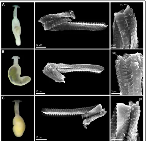
Description:morphologically with diagnostic charac-ters of the genus Pontohedyle (see Figure 1A). Radula formula 1-1-1, rhachidian tooth with three lateral cusps, lateral plate smooth without denticle (Figure 1A).
Molecular diagnosis is given in Table 6.
Positions of the diagnostic characters refer to the sequence of the holotype. Diagnostic characters in nu-clear 18S rRNA were determined based on GenBank KC984290, in 28S rRNA based on GenBank JQ410967, in mitochondrial 16S rRNA based on GenBank JQ410966, and in mitochondrial COI based on GenBank JQ410912.
Pontohedyle jonisp. nov.
Pontohedylesp. 2 (MOTU II) in [25]
Types: Holotype: DNA voucher (extracted DNA in buffer) ZSM Mol 20090197 (DNA bank accession num-ber AB34858164). Paratype: one specimen fixed in 96% ethanol, collected with the holotype.
Type locality: N 14°3′34.56”, W 60°58′18.24”; near Castries, St. Lucia, Central America, Caribbean Sea, West Atlantic Ocean (see Figure 4).
Additional material: DNA voucher (extracted DNA in buffer) SI-CBC2010KJ01-D05 (DNAbank at ZSM AB34402049) and SEM preparation of radula (ZSM Mol 20131102) from N 16°48′13.44“, W 88°4′36.9“, and DNA voucher (extracted DNA in buffer) SI-CBC2010KJ01-C08 (DNAbank AB34402065) from N 16°48′7.62“, W 88°4′36.42“ both Carrie Bow Cay, Belize, Central America, Caribbean Sea, West Atlantic Ocean.
ZooBank registration: urn:lsid:zoobank.org:act:73AA C79D-5A43-40E4-B0D6-0329CAAA2AA0
Etymology: Named after Dr. Jon Norenburg to honor his efforts and enthusiasm for meiofaunal research and to thank him for his support for uncovering the largely unknown Caribbean meiofauna.
Distribution and habitat: Currently known from the Caribbean Sea (St. Vincent and Belize), type lo-cality subtidal, 2–3 m depth, sand patches between seagrass, coarse sand. Additional material also sub-tidal, 14–15 m, sand patches between corals, coarse sand.
Description: morphologically with diagnostic charac-ters of the genusPontohedyle. Radula formula 48 × 1-1-1, rhachidian tooth with 3 lateral cusps, lateral plate with one pointed denticle (see Figure 1B).
Molecular diagnosis is given in Table 7. Table 6 Molecular diagnostic characters ofPontohedyle
kepiisp. nov.
Marker Diagnostic characters with position in alignment (in reference sequence)
18S rRNA 199 (182), G; 202 (185), C; 203, deletion; 204, deletion; 206, deletion; 254 (244), T; 707 (697), T; 1355 (1345), A; 1356 (1346), C
28S rRNA 410 (439), T; 419 (448), C; 719 (754), G; 867 (902), C
16S rRNA 11, T; 184 (189), A; 187 (192), C; 239 (267), A; 242, deletion; 243, deletion; 244, deletion; 294 (324), G; 302 (328), G
COI 49, A; 79, T; 118, C; 148, C; 160, A; 193, G; 292, G; 331, G; 466, T; 494, G; 583, G; 628, A; 638, C
COI (AA) 165, D
Table 7 Molecular diagnostic characters ofPontohedyle jonisp. nov.
Marker Diagnostic characters with position in alignment (in reference sequence) Heterogeneous single pure positions
18S rRNA 207 (215), T; 209 (217), T; 256 (263), A
-28S rRNA 443 (446), A; 547 (556), T; 868 (873), A
16S rRNA 44 (47), C; 122 (125), T; 141 (144), A; 142 (145), G; 143 (146), G; 146, G; 152 (157), A; 182 (188), T; 236 (252), A; 259 (284), C
181 (187), T (in SI-CBC20 10KJ01-C08, C at position 187)
The sequences retrieved from the holotype ZSM Mol 20090197 serve as reference sequences. Diagnostic char-acters in nuclear 18S rRNA were determined based on ZSM Mol 20090197 (GenBank KC984291) and SI-CBC2010KJ01-D05 (GenBank KC984292), in nuclear 28S rRNAbased on ZSM Mol 20090197 (GenBank JQ410934) and SI-CBC2010KJ01-C08 (GenBank JQ410939), in mitochondrial 16S rRNA based on ZSM Mol 20090197 (GenBank JQ410933), SI-CBC2010KJ01-D05 (GenBank JQ410937) and SI-CBC2010KJ01-C08 (GenBank JQ41 0938), and in mitochondrial COI based on ZSM Mol 20090197 (GenBank JQ410901), SI-CBC2010KJ01-D05 (GenBank JQ410902) and SI-CBC2010KJ01-C08 (GenBank JQ410903).
Pontohedyle neridaesp. nov.
Pontohedylesp. 3 (MOTU III) in [25]
Types: Holotype: DNA voucher (extracted DNA in buffer, stored deep frozen at -80°C) AM C. 476062.001 (DNA bank accession number at ZSM AB34500497). Paratype: one specimen fixed in 5% formalin and embed-ded in epoxy resin (AM C.476063.001), collected with the holotype.
Type locality: S 17°32′50.172”, W 149°46′35.4”; Motu Iti, Moorea, Oceania, Central Pacific Ocean (see Figure 4).
ZooBank registration: urn:lsid:zoobank.org:act:BE3E 7920-5451-429D-95E4-C8D2F859C7CB
Etymology: Named after our friend and colleague, Dr. Nerida Wilson, with a big ‘thank you’ for ac-tively sharing with us the fascination for interstitial Acochlidia.
Distribution and habitat: Known from type locality only; subtidal 3-4 m, fine to medium coral sand.
Description:Morphologically with diagnostic charac-ters of the genus Pontohedyle. Radula characteristics unknown.
Molecular diagnosis is given in Table 8.
The sequences retrieved from the holotype serve as reference sequences. Diagnostic characters in nuclear 28S rRNA were determined based onAM C. 476062.001 (GenBank JQ410986), in mitochondrial 16S rRNA based on AM C. 476062.001 (GenBank JQ410985), and in mi-tochondrial COI based on AM C. 476062.001 (GenBank JQ410922).
Pontohedyle liliaesp. nov.
Pontohedylesp. 4 (MOTU IV) in [25]
Types:Holotype: DNA voucher (extracted DNA in buf-fer, stored deep frozen at -80°C) ZSM Mol 20090471 (DNA bank accession number AB35081802). Paratypes (all collected with the holotype): DNA voucher (extracted DNA in buffer) ZSM Mol 20090472 (DNA bank accession number AB35081838), one additional specimen used for radula preparation, SEM stub with radula available (ZSM Mol 20131103).
Type locality: N 24°11′50“, E 35°38′26“ (approxima-tion from Google Earth), Sha’ab Malahi, Egypt, Africa, Red Sea (see Figure 4).
ZooBank registration: urn:lsid:zoobank.org:act:2711E 3E5-1D1D-41B0-B919-7D7E690FD525
Etymology:Named after Reinhilde (‘Lili’) Schmid, our friend and diving companion, who assisted us during sand collecting in Egypt and shares our fascination for this world of little creatures.
Distribution and habitat: Known from type locality only; subtidal 20 m, relatively fine coral sand.
Description: Morphologically with diagnostic charac-ters of the genusPontohedyle. Radula formula 45 × 1-1-1, rhachidian tooth with three (to four) lateral cusps, lateral plate with one pointed denticle (Figure 1C). Eyes clearly visibly externally, monaxone spicules in accumu-lation between oral tentacles and irregular all over the body.
Molecular diagnosis is given in Table 9.
Table 8 Molecular diagnostic characters ofPontohedyle neridaesp. nov.
Marker Diagnostic characters with position
in alignment (in reference sequence)
28S rRNA 61 (57), G; 522 (518), A
16S rRNA 11, G; 121 (123), T; 145 (147), T; 147 (149), G; 252 (276), C; 263 (286), T; 330 (352), G; 336 (358), G
COI 46, C; 151, C; 169, G; 220, A; 277, C; 278, T; 289, T; 391, C; 397, G; 421, C; 479, T; 505, A; 601, C
Table 9 Molecular diagnostic characters ofPontohedyle liliaesp. nov.
Marker Diagnostic characters with position in alignment (in reference sequence)
18S rRNA 33, C; 40, C; 54, G; 117, T; 129, T; 146 (147), C; 149 (150), T; 186 (187), C; 214 (223), A; 215 (224), C; 623 (631), T; 663 (673), T; 677 (687), C; 841 (853), G; 959 (971), G; 1028 (1040), T; 1030 (1042), C; 1348 (1360), A; 1363 (1375), T
28S rRNA 34 (30), C; 63 (59), C; 536 (532), T; 537 (533), G; 542, deletion; 555 (554), G; 590 (589), T; 642 (641), C; 643 (642), T; 658 (657), A; 671 (670), C; 696 (695), A; 827, G; 837, C; 902 (904), C
The sequences retrieved from the holotype (ZSM Mol 20100471) serve as reference sequences. Diagnostic char-acters in nuclear 18S rRNA were determined based on ZSM Mol 20100471 (GenBank KC984293), in nuclear 28S rRNA based on ZSM Mol 20100471 (GenBank JQ410954) and ZSM Mol 20100472 (GenBank JQ410956), and in mitochondrial 16S rRNA based on ZSM Mol 20100471 (GenBank JQ410953) and ZSM Mol 20100472 (GenBank JQ410955).
Pontohedyle wiggisp. nov.
Pontohedylesp. 5 (MOTU V) in [25]
Types: Holotype: DNA voucher (extracted DNA in buffer) ZSM Mol-20100595 (DNA bank accession number AB34402059). Paratypes (all collected with the holotype): DNA voucher (extracted DNA in buffer) ZSM Mol-20100596 (DNA bank AB34402001), ZSM Mol 20100597 (DNA bank AB34500571), ZSM Mol 20100603 (DNA bank AB34402020); one specimen fixed in glutaraldehyde and embedded in epoxy resin (ZSM Mol 20100598).
Type locality:N 7°36′15“, E 98°22′37“, Ko Raccha Yai, Phuket, Thailand, Andaman Sea, Indian Ocean (see Figure 4).
ZooBank registration: urn:lsid:zoobank.org:act:808E5 62E-0E1A-4D79-BB2C-1377B3734F86
Etymology: Named in memory of Ludwig (‘Wigg’) Demharter, a malacologist friend, passionate diver,‘fun researcher’, and for many years a supporter of the ZSM and the second author's working group.
Distribution and habitat:Known from the type local-ity only; marine, interstitial between sand grains, rela-tively fine coral sand, subtidal 6–7 m depth, sandy slope among patches of corals.
Description: Morphologically with diagnostic char-acters of the genusPontohedyle. Radula formula 1-1-1, lateral plate with one pointed denticle (as in P. milas chewitchii). Eyes visibly externally, monaxone spicules present.
Molecular diagnosis is given in Table 10.
The sequences retrieved from the holotype (ZSM Mol 20090595) serve as reference sequences. Diagnostic characters in nuclear 28S rRNA were determined based on ZSM Mol 20100595 (GenBank: JQ410960), ZSM Mol 20100597 (GenBank: JQ410963), ZSM Mol 20100603 (GenBank: JQ410965), in mitochondrial 16S rRNA based on ZSM Mol 20100595 (GenBank: JQ410959), ZSM Mol 20100596 (GenBank: JQ410961), ZSM Mol 20100597 (GenBank: JQ410962), ZSM Mol 20100603 (GenBank: JQ410964), and in mitochondrial COI based on ZSM Mol 20100595 (GenBank: JQ410908), ZSM Mol 20100596 (GenBank: JQ410909), ZSM Mol 20100597 (GenBank: JQ410910), ZSM Mol 20100603 (GenBank: JQ410911).
Pontohedyle wenzlisp. nov.
Pontohedylesp. 6 (MOTU VIII) in [25]
Types: Holotype: DNA voucher (extracted DNA in buffer) ZSM Mol 20100379 (DNA bank accession num-ber AB34500521).
Type locality:N 1°27′53“, E 125°13′48“, Lembeh Strait, Sulawesi, Indonesia, Banda Sea, West Pacific Ocean (see Figure 4).
Additional material DNA voucher (extracted DNA in buffer) ZSM Mol 20081014 (DNA bank accession num-ber AB35081827) and one specimen used for SEM preparation of radula (available at ZSM Mol 20131105), locality S 8°23′58“, E 119°18′56“, Pulau Banta, Nusa Tengarra, Indonesia Flores Sea, Indo-Pacific. DNA vou-cher (extracted DNA in buffer) ZSM 20100592 (DNA bank AB34402021), locality N 7°36′15“, E 98°22′37“, Ko Raccha Yai, Phuket, Thailand, Andaman Sea, Indian Ocean. DNA voucher (extracted DNA in buffer) AM C. 476051.001 (DNA bank AB34402037) and one specimen fixed in 5% formalin and embedded in epoxy resin (AM C.476050.001), locality S 17°28′33.96”, W 149°49′51.6”, E of Cook’s Bay Pass, Moorea, Oceania, Central Pacific.
Note: Most species delineation approaches suggested ZSM 20100592, and some also AM C. 476051.001, as an independently evolving lineage [25]. Due to the conserva-tive consensus approach, these specimens were included in the described species. Future analyses might show that their separation as independent species is warranted.
ZooBank registration: urn:lsid:zoobank.org:act:558E C548-1FB3-4B00-B248-4424CA7B098C
Etymology:Named after Alexander Wenzl, for his sup-port during the development of this manuscript and his interest for meiofaunal research.
Distribution and habitat:Known from Indonesia, with putative distribution across the Indo-Pacific and Central Pacific; marine, subtidal (3–22 m), interstitial, coarse sand and shell grid.
Table 10 Molecular diagnostic characters ofPontohedyle wiggisp. nov.
Marker Diagnostic characters with position
in alignment (in reference sequence)
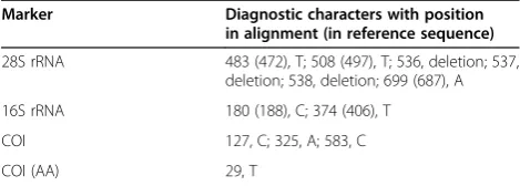
28S rRNA 483 (472), T; 508 (497), T; 536, deletion; 537, deletion; 538, deletion; 699 (687), A
16S rRNA 180 (188), C; 374 (406), T
COI 127, C; 325, A; 583, C
Description:Morphologically with diagnostic characters of the genusPontohedyle, eyes clearly visible externally (see Figure 2B, picture of living holotype). Radula 43 × 1-1-1, rhachidian tooth with three lateral cusps, lateral plate with pointed denticle (like inP. milaschewitchii).
Molecular diagnosis is given in Table 11.
The sequences retrieved from the holotype (ZSM Mol 20100379) serve as reference sequences. Diagnostic char-acters in nuclear 18S rRNA were determined based on ZSM Mol 20100379 (GenBank KC984297), ZSM Mol 20081014 (GenBank KC984296), ZSM Mol 20100592 (GenBank KC984294), AM C. 476051.001 (GenBank KC984295), in nuclear 28S rRNA based on ZSM Mol 20100379 (GenBank JQ410973), ZSM Mol 20081014 (GenBank JQ410969), ZSM Mol 20100592 (GenBank JQ410958), AM C. 476051.001 (GenBank JQ410982), in mitochondrial 16S rRNA based ZSM Mol 20100379 (GenBank JQ410972), ZSM Mol 20081014 (GenBank JQ410968), ZSM Mol 20100592 (GenBank JQ410957), AM C. 476051.001 (GenBank JQ410981), and in mito-chondrial COI based on ZSM Mol 20100379 (GenBank JQ410915), ZSM Mol 20081014 (GenBank JQ410913), ZSM Mol 20100592 (GenBank JQ410907).
Pontohedyle peteryallisp. nov.
Pontohedylesp. 7 (MOTU VII) in [25]
Types:Holotype: DNA voucher (extracted DNA in buf-fer) ZSM Mol 20071133 (DNA bank accession number AB34404268). Paratypes (all collected with the holotype): eight specimens preserved in 96% ethanol (ZSM Mol 20070827); four in 75% ethanol (ZSM Mol 20070827), sixteen specimens fixed in glutaraldehyde, post-fixed in osmium and embedded in epoxy resin (ZSM Mol 20080453–60; ZSM Mol 20080462–69). SEM stub with radula available (ZSM Mol 20131104).
Type locality:N 04°47′46”, W 02°10′06”, MiaMia, Ghana, Africa, Gulf of Guinea, East Atlantic Ocean (see Figure 4).
Additional material: six specimens in 75% Ethanol col-lected at Nzema Cape, Ghana, Africa, Gulf of Guinea, East Atlantic Ocean; conspecifity still needs to be confirmed via barcoding.
ZooBank registration: urn:lsid:zoobank.org:act:B25E5 0F7-F0D2-4842-B6C3-5A79EA784A0C
Etymology: Named for our friend and malacologist, Peter (‘Pete’) Ryall, who invited us to explore sea slugs right in front of his MiaMia home.
Distribution and habitat:Currently only known from the Ghana West Coast around MiaMia, marine, intersti-tial, subtidal 2-3 m, fine sand.
Description: Morphologically with diagnostic char-acters of the genus Pontohedyle. Radula 42 × 1-1-1, rhachidian tooth with three lateral cusps, lateral plate with pointed denticle (like in P. milaschewitchii), see Figure 2A.
Molecular diagnosis is given in Table 12.
The sequences retrieved from the holotype (ZSM Mol 20071133) serve as reference sequences. Diagnostic characters in nuclear 18S rRNA were determined based on GenBank KC984298, in mitochondrial 16S rRNA based GenBank JQ410930 and in mitochondrial COI based on GenBank JQ410899.
Table 11 Molecular diagnostic characters ofPontohedyle wenzlisp. nov.
Marker Diagnostic characters with position in alignment (in reference sequence) Heterogeneous single pure positions
18S rRNA 771 (791), T; 772 (792), T
-28S rRNA 449 (455), C; 539 (545), A
-16S rRNA 36, G; 41, T; 84 (88), A; 143 (147), A; 144 (148), A; 161 (167), T; 176 (182), A; 194 (201), T; 207 (214), A; 256 (296), C; 258 (298), A; 269 (309), T; 295, deletion; 331 (369), A; 340 (378), A
332 (370), A (ZSM Mol 20081014, G at position 370)
COI 181, A; 218, G; 219, T; 296, T; 383, C; 430, T; 593, A
-COI (AA) 73, V; 94, F; 122, A; 198, I
-Table 12 Molecular diagnostic characters ofPontohedyle peteryallisp. nov.
Marker Diagnostic characters with position in alignment (in reference sequence)
18S rRNA 160, C; 164, C
COI 14, T; 23, A; 48, C; 68, A; 76, C; 81, T; 83, A; 95, T; 101, A; 102, G; 140, A; 141, C; 167, A; 187, C; 209, C; 232, C; 280, A; 286, C; 293, A; 294, G; 357, C; 358, A; 361, A; 365, A; 373, A; 433, C; 448, G; 467, A; 468, T; 487, T; 503, T; 504, G; 512, A; 535, C; 556, C; 574, A; 586, C; 628, C; 634, C
Pontohedyle martynovisp. nov.
Pontohedylesp. 8 (MOTU IX) in [25]
Types: Holotype: DNA voucher (extracted DNA in buffer) AM C. 476054.001 (DNA bank accession number at ZSM AB34402062). Paratype: one specimen fixed in 5% formalin embedded in epoxy resin (AM C.476053.001), collected together with the holotype.
Type locality:S 17°28′17”, W 149°48′42”, E of Cook’s Bay Pass, Moorea, Oceania, Central Pacific Ocean (see Figure 4).
ZooBank registration: urn:lsid:zoobank.org:act:9431E 4B8-EAF3-4E29-9993-BCD7C52928C6
Etymology: Named to thank our Russian friend and taxonomist, Alexander (‘Sasha’) Martynov, for collecting acochlidians for us in many places, including Pontohe dyle milaschewitchiiat its type locality.
Distribution and habitat: Known from type locality only; marine, interstitial, subtidal 18–20 m, coarse sand, shell grid and rubble.
Description:Morphologically with diagnostic characters of the genusPontohedyle. Radula characteristics unknown.
Molecular diagnosis is given in Table 13.
The sequences retrieved from the holotype (AM C. 476054.001) serve as reference sequences. Diagnostic char-acters in nuclear 28S rRNA were determined based on GenBank JQ410984, and in mitochondrial 16S rRNA based on GenBank JQ410983.
Pontohedyle yurihookerisp. nov.
Pontohedylesp. 9 (MOTU X) in [25]
Types:Holotype: DNA voucher (extracted DNA in buf-fer) ZSM Mol 20080565 (DNA bank accession number AB34402000).
Type locality: S 3°58′55”, W 80° 59′10”, Punta Sal, Peru, South America, East Pacific Ocean (see Figure 4).
ZooBank registration: urn:lsid:zoobank.org:act:9B858 AA5-59FA-4505-AE94-FB2EA27FBEF6
Etymology:Named for our Peruvian friend and mar-ine biologist, Yuri Hooker, who jomar-ined us during a great diving expedition to explore the Peruvian sea slug fauna.
Distribution and habitat: Known from type lo-cality only; marine, interstitial, subtidal (8 m), coarse sand.
Description: Morphologically with diagnostic char-acters of the genusPontohedyle. Radula characteristics unkown.
Molecular diagnosis is given in Table 14.
The sequences retrieved from the holotype (ZSM Mol 20080565) serve as reference sequences. Diagnostic char-acters in nuclear 18S rRNA were determined based on GenBank KC984299, and in nuclear 28S rRNA based on GenBank JQ410987.
Discussion
Cryptic species challenging traditional taxonomy
Largely due to the development of molecular methods, research on cryptic species has increased over the past two decades [8,9], demonstrating their commonness across Metazoan taxa, though with random or non-random dis-tribution among taxa and biomes still to be investigated [9,10]. Several recent studies have underlined that there is a large deficit in alpha taxonomy and that the diver-sity of marine invertebrates and especially meiofaunal animals might be much higher than expected, partly caused by high proportions of cryptic species e.g., [11,13,14,25,73-75]. Rather than global, amphi-Oceanic, circum-tropical or otherwise wide ranging, the distribu-tion areas of the biological meiofaunal species involved may be regional and their ecology more specialized [12,25,76]. At an initial stage of molecular and ecological exploration, cryptic meiofauna is potentially threatened by global change and cannot effectively be included in conservation approaches.
In traditional taxonomy, most species descriptions are based on morphological and anatomical characters. Morphological species delineation, however, can fail to Table 13 Molecular diagnostic characters ofPontohedyle
martynovisp. nov.
Marker Diagnostic characters with position in alignment (in reference sequence)
28S rRNA 539 (541), C; 623 (629), A
16S rRNA 8, deletion; 33 (32), T; 130 (131), C; 144, deletion; 151 (155), G; 168 (172), G; 171 (175), A; 218 (232), A; 230, T; 232 (244), G; 235 (258), C; 242 (274), C; 332 (365), C; 334 (367), G; 353 (386), G; 373 (408), G
Table 14 Molecular diagnostic characters ofPontohedyle yurihookerisp. nov.
Marker Diagnostic characters with position in alignment (in reference sequence)
18S rRNA 163 (156), T; 200 (193), A; 213 (225), A; 770 (783), T; 810 (823), T
adequately address the diversity of life on Earth by leaving cryptic species unrevealed. Many taxonomists agree that the future of taxonomic descriptions should be integrative, embracing all available data sources (morphology, mo-lecular sequences, biogeography, behavioral traits…) that can contribute to species delineation [1-3]. Previous au-thors have argued that ‘integrative taxonomy’ does not necessarily call for a maximum of different character sets, but rather requires the taxonomist to select character sets adequate for species delineation in the particular group of taxa [3,5]. Thus, there should be no obligation in taxonomic practice to stick to morphology as the pri-mary source [77], and there are no official requirements by the International Code of Zoological Nomenclature to do so [78,79].
The results of Jörger et al. [25] indicate that the mem-bers ofPontohedyleslug lineages are so extremely uniform that conventional taxonomic characters (i.e. external morphology, radula characteristics, spicules) fail to de-lineate species. A series of studies have demonstrated the generally high potential of advanced 3D-microanatomy for character mining in Acochlidia (e.g., [80-82]). However, the exclusively mesopsammic microhedylacean Acochlidia form an exception, as they show reduced complexity in all organ systems and uniformity that leaves few anatomical features for species delineation even on higher taxonomic levels [83]. Based on previous histological comparisons, Jörger et al. [56] were unable to find any morphological characters justifying discrimination between the closely re-lated western AtlanticP. brasilensisand its Mediterranean congener,P. milaschewitchii. Here, we provided a detailed histological (re-)description using 3D-reconstruction based on serial semi-thin sections of P. verrucosa, to evaluate whether advanced 3D-microanatomy provides distinguishing morphological characters for the two generally accepted species, P. milaschewitchii and P. verrucosa, as representatives of the two major Pon tohedyle clades (see [25], Figure 1).Indeed, we revealed some putative distinguishing features in the reproductive and digestive systems (see Table 15). However, the
encountered (minor) morphological differences are prob-lematic to evaluate in the absence of data on ontogen-etic and intraspecific variation, and on potential overlap with interspecific differences. For example, slight ences in the reproductive system could be due to differ-ent ontogenetic stages, therefore presdiffer-ently they cannot be used to discriminate species. Comparatively investi-gated serial semi-thin sections of Pontohedyle kepii sp. nov. also confirmed the similarity in all major organ sys-tems reported previously [55,56]. We thus conclude that in Pontohedyle even advanced microanatomy is ineffi-cient or even inadequate for species diagnoses. Molecu-lar character sets currently offer the only chances for unambiguous discrimination between the different evo-lutionary lineages. Proponents of morphology based alpha taxonomy [84] might argue that we have not attempted a fully integrative approach since we have not performed 3D-microanatomy on all proposed new species, including enough material for intra-specific comparisons, ultrastructural data on, e.g., cilia, sperm morphology or specific gland types, to reveal whether these forms indeed represent cryptic species. However, in light of the biodiversity crisis and the corresponding challenges to taxonomy, we consider it as little effective to dedicate several years of a taxonomist’s life to the search for morphological characters, when there is little to expect, while molecular characters enable straight-forward species delineation. This is not a plea to speed up description processes at the expense of accuracy and quality, or by allowing ignorance of morphology, but for a change in taxonomic practice to give molecu-lar characters simimolecu-lar weight as morphological ones, in cases in which this is more informative or practical.
Still debated is the way how the traditional Linnaean System needs to be adapted to incorporate different character sets, in the first place the growing amount of molecular data. Probably the most radical way ignores the character-based requirements of the International Code of Zoological Nomenclature [78,79] and proposes to base descriptions of new species directly on support
Table 15 Putative distinguishing features betweenP. milaschewitchiiandP. verrucosa(intraspecific variation not evaluated)
P. milaschewitchii(Kowalevsky, 1901) P. verrucosa(Challis, 1970)
Data source Jörger et al. 2008 [55] Present study
Epidermal glands Predominantly whitish, blue stained only in one small row
Predominantly whitish and numerous dark blue stained ones
Nervous system Eyes pigmented and externally visible Eyes unpigmented
Reproductive system Only one cephalic male genital opening detected Two male genital openings (cephalic and visceral)
Digestive system/ putatively different feeding habits
Lateral radula teeth with central denticle Lateral radula teeth without denticle
values under species delineation models [85,86]. Aside from the paradigm shift this would bring, far away from long-standing taxonomic practice, opponents criticize that unambiguous allocation of newly collected material is impossible in the absence of definitions and descriptors and requires repetition of the species delineation approach applied [50]. As a method of species delineation, co-alescent based approaches are objective and grounded on evolutionary history and population genetics [86,87]; thus it is indeed tempting to use results derived from mo-lecular species delineations approaches directly as species descriptions (‘model-based species descriptions’ [87]). This would clearly facilitate descriptions, thus reduce the taxonomic impediment and the risk of an endless number of discovered but undescribed candidate spe-cies. Every species description should aim for differenti-ation from previously described species; therefore, diagnostic characters are usually derived from comparisons to other, closely related species. Nevertheless, the species description itself has to be self-explanatory and should not rely on comparative measurements which are only valid in comparison to a special set of other species used for a cer-tain analysis, i.e. on a complex construct that may not be reproducible when new data are added. In contrast to Fujita & Leaché [87], we believe that each species, i.e. separately evolving lineage [4], will present–in the current snap-shot of evolutionary processes – fixed diagnostic characters of some sort (e.g., from morphology, DNA sequence informa-tion, behavioral, karyology…), and we consider it the task of modern taxonomy to detect the most reliable and efficient set of characters on which to found species descriptions.
The Characteristic Attribute Organization System (CAOS) [51,57,58] is a character based method proposed for uniting species discovery and description [88]. As an approach to species delineation, we consider it inferior to coalescent based approaches (e.g., GMYC and BP&P); CAOS successfully determines putative diagnostic nucleo-tides, but is not predictive, i.e. lacks objective criteria with which to delimit a threshold number of distinguishing nu-cleotides that would indicate a species boundary. One has to distinguish between diagnosability of entities and the de-limitation of species. Diagnostic characters of whatever sort can be found for all levels in the hierarchical classification, but there is no objective criterion for determining a number of characters needed to characterize a (new) species, e.g. versus a population. Nevertheless, for the purpose of spe-cies description, we think that character based approaches like CAOS are highly valuable and should complement mo-lecular species delineation procedures, thus enabling the transition from species discovery to description.
Requirements of molecular taxonomy
While calls for replacing the Linnaean system by a DNA sequence based one [41] have trailed away, we still lack
a common procedure on how to include molecular data into the Linnaean system [21]. Like any other source of data, molecular data is not explicitly treated by the Inter-national Code of Zoological Nomenclature, there are no provisions dictating the choice of characters [78,79]. Currently, molecular data are included in species descrip-tions in various mutually inconsistent ways [21]. If DNA sequence data are only used as additive to, e.g., morph-ology based species descriptions or molecular species delineation approaches to confirm pre-identified entities, the addition is straightforward and requires no specific considerations. But if molecular sequence information is to be used as the partial or even sole content of a species description, a discussion of the corresponding best prac-tice is needed.
Type material for species based on molecular data
Previous authors highlighted the need for voucher ma-terial in molecular studies [89]. Ideally, DNA is extracted from (a subsample of ) a name-bearing type specimen (holotype, syntype, lectotype or neotype); if no such speci-men is available for molecular studies, an attempt should be made to collect fresh material at the type locality. If parts of larger animals belonging to putative new species are used for DNA extraction, DNA and remaining speci-men can both become part of the type material under nomenclatural rules. However, where the members of a putatively new species, e.g. of meiofauna, are so small that molecular extraction from only part of an indi-vidual is impossible, taxonomists may be confronted with the critical decision to either have DNA without a morphological type specimen or a type without DNA. In taxonomically unproblematic groups one can add new material or use paratypes for DNA (or other) analyses, relying on specimens to be conspecific if they were col-lected from‘the same population’, i.e. from a place (and time) close enough to the type locality to assume gene flow. But what if, as has been shown for Pontohedyle slugs [25], there is a possibility of cryptic species occur-ring sympatrically and at the same time? Would it be better (A) to sacrifice a (single available) type specimen to obtain molecular data for species delineation or (B) to save the type and use a secondary specimen, taking the risk that the latter might not be conspecific with the former? In a group like our Pontohedyle slugs in which DNA sequence data are much more promising for species delin-eation than morphological approaches, and considering the wealth of potential DNA sequence characters, we pre-fer to sacrifice even single specimens to DNA extraction. In absence of a term referring to vouchers exclusively consisting of extracted DNA, we term this type material:
radulae) from the spin columns used for extraction [91]. In the case of DNA aliquots serving as type material, natural history collections are urged to create long term DNA stor-age facilities [41,42] like the DNA bank network (http:// www.dnabank-network.org/), and should apply the same caution and requirements (i.e. documentation of collection details) as for any morphological type.
Risk of two parallel taxonomies?
Old type material often does not allow molecular analyses [84,92], and searching for fresh material at a type locality can be unsuccessful. Future technical advances are likely to enable DNA acquisition from some old type material, as there has been considerable progress in dealing with degenerated DNA [93]. Nevertheless, there are the po-tential risks that two parallel taxonomic systems could develop, and that the one based on molecular characters could duplicate, under separate names, some taxa already established on morphological grounds [77]. Similar con-cerns have arisen previously when the taxonomy of certain taxa was based on a character set other than morphology (e.g. cytotaxonomy based on data from chromosomes) and the investigation of one character set hindered the exploration of the other. It clearly remains the duty of taxonomists to carefully check type material of closely related taxa before describing new species [77]. To keep molecule driven taxonomy‘workable’[94] and connected to traditional morphology based taxonomy, authors should include a brief morphological diagnosis of the (cryptic) species [77], even in the absence of species-diagnostic characters, in order to make the species recognizable as belonging to a certain group of (cryptic) species.
Trouble with names
Any specimen identified from molecular data only can belong to a previously established species or to one new to science. If unambiguous identification with a single existing species name is possible then, of course, the latter should be used. In our cases in Pontohedyle, we call those Indo-Pacific specimens collected near the type locality ofP. verrucosa(Challis, 1970) on the Solomon Islands by this single available name for Indo-Pacific Pontohedyle.Concerning AtlanticPontohedyle, the nameP. brasilensis (Rankin, 1979), proposed for Brazilian speci-mens, was treated as a junior synonym of the older name, P. milaschewitschii (Kowalevsky, 1901). Since we have shown that P. milaschewitschii refers to Mediterranean and Black Sea specimens only [25], we resurrected the nameP. brasilensisfor Western AtlanticPontohedyle, and now apply it to the only species in of two cryptic ones that has been collected from Brazil. In doing so we accept the risk resulting from the fact that these specimens were collected at some distance from the type locality of P. brasilensis(see Figure 4), as the latter has not yielded
any Pontohedyle specimens for more than the last 50 years, despite considerable and repeated collecting efforts, including our own. These assignments of previ-ously established species names left at least nine add-itional, clearly separate Pontohedyle species for which available names did not exist. In cases of microscopic ani-mals such as Pontohedyle, molecular taxonomy thus may benefit from morphology based taxonomy having missed them in the past.
Species descriptions based on singletons
Species descriptions based on singleton specimens cannot reflect intraspecific variation, and Dayrat [1] even pro-posed a guideline to restrict species descriptions to well-sampled taxa. However, there is no objective way to determine any sample size at which intraspecific variation would be covered sufficiently. Moreover, exclud-ing taxa described from sexclud-ingletons would lead to con-siderably lower, and effectively false, estimates of the scientifically known biodiversity [5,26-28]. The present study on Pontohedyleincludes five species descriptions based on DNA sequence information from one individ-ual only. Usindivid-ually, this is done when such a singleton presents a combination of characters so discrete that it is considered highly unlikely to fall within the variational range of another species [28]. In a complex molecular species delineation approach Jörger et al. [25] recognized our five singletons as independently evolving lineages. Approximations with molecular clock analyses estimate the diversification of these species from their respective sister groups to have occurred 54–83 mya (own unpub-lished data), which indicates significant timespans of genetic isolation. In light of our general revision of the genus Pontohedyle, we consider it as less productive to keep these entities on the formally unrecognized level of candidate species than to run the risk that our spe-cies hypotheses may have to be modified due to future additional material. Nevertheless, we are well aware of the fact that taxon sampling and data acquisition (i.e. incomplete molecular data sets) are not yet ideal for some of our newly described species (e.g., P.martynovi sp. nov.,P. yurihookerisp. nov.).
![Figure 2 External morphology (living specimens) and radula characteristics (SEM micrographs) in(holotype), radula from IP-2, see [25]); Pontohedyle species (part 2).A) Pontohedyle peteryalli sp](https://thumb-us.123doks.com/thumbv2/123dok_us/416237.1534710/5.595.60.540.88.551/external-morphology-specimens-characteristics-micrographs-pontohedyle-pontohedyle-peteryalli.webp)