e-ISSN: 2278-7461, p-ISSN: 2319-6491
Volume 7, Issue 1 [January 2018] PP: 40-50
Research on Fundus Image Registration And Fusion Method
Based On Nsct And Adaptive Pcnn
Jun Wu
12,Shuya Song
12*,Dongxia Zhang
12,Song Yang
121Tianjin Polytechnic University, School of Electronics and Information Engineering, Tianjin 300387, China
2 Tianjin Key Laboratory of Optoelectronic Detection Technology and System, Tianjin 300387, China
Corresponding author:Jun Wu12
Abstract
: The fusion of the fundus image can show lesions and tissues in multi-modalities of images comprehensively, and is of great value of application in the diagnosis of fundus diseases for doctors. In this paper, we propose a method of fundus image registration and fusion with the combination of NSCT(Nonsubsampledcontourlet) and adaptive PCNN(Pulse Coupled Neural Network).The specific process is:firstly, two images are registered to eliminate the spatial differences between the source images to extract SURF(Speeded Up Robust Features) as the feature point, and then the feature vector is calculated by feature points description, the nearest neighbor and the next nearest neighbor distance ratio method are used to realizethe initial matching of the feature points,the RANSIC(Random Sample Consensus,RANSAC) method is used to
remove the mismatched point pairs. Finally, the transformation parameters between the images are calculated to complete the registration of source images by the spatial transformation. For integration the of the two images after the registration, the specific process is: The low frequency and high frequency sub-band of the image to be fused is got by NSCT decomposition, the low frequency sub-band is fused by the regional energy . The high frequency sub-band use the simplified PCNN model to study and is fused based on the number of times the image pixels are fired. The experimental results show that this method is better than other representative methods in the fusion result of fundus image.the fusion image synthesizethe image information and clarifythe performance of the details,provides an effective reference for the clinical diagnosis of fundus diseases.
Keyword
: Fundus image, NSCT, regional energy, simplified PCNN--- ---Date of Submission: 17-01-2018 ---Date of acceptance: 31-01-2018
--- ---
I.
INTRODUCTION
images and fluorescence images of the fundus, the effective formation of information Complementary, more comprehensive and intuitive reflect the characteristics of lesions and organizations for the prevention and treatment of some diseases has important application value.
In practical applications, image registration and image fusion technology are inseparable. In general, the imaging sensor to obtain the source image will be different in position and attitude, which makes the source image fusion inevitably exist differences geometry space of translation, rotation and scale expansion etc.. This difference has great influence on the effect of image fusion. Therefore, before the image fusion of the source image, it is necessary to do the geometric alignment by image registration for eliminate the spatial differences between source images which comes from different angles and sensors of the same scene. Image registration methods can be divided into region based methods[2] and feature based methods[3-4]. Region based method deals with objects as pixels, and uses image templates to process registration. The differences between different methods are reflected in the design of templates and registration rules; Feature-based method by extracting spot, lines, contours and other significant features in the source image as a unit for feature matching.
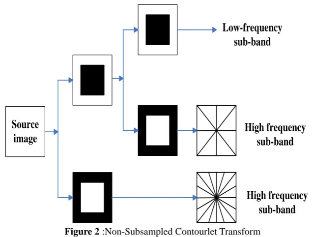
The image fusion method can be divided into image fusion based on spatial domain and image fusion based on transform domain. Image fusion based on spatial domain is the fusion of images directly in the gray space of image pixels, such as weighted average fusion method, IHS spatial fusion method and principal component analysis fusion method etc.. The method of spatial domain fusion is simple and the method has low complexity, but the method is difficult to achieve satisfactory fusion effect in most applications. At present, the fusion method based on transform domain is the focus of the research. Discrete wavelet transform (DWT) as an image multi-scale geometric analysis tool, which has good time-frequency local analysis, but the orthogonal wavelet transform based on the Mallat method can only decompose the finite direction information, and does not have translation invariance, resulting in ringing effect in the reconstructed image[5]. At present, scholars have proposed many multi-scale geometric analysis methods base on the wavelet transform theory: Candes and Donoho proposed the Ridgelet Transform[6], Curvelet transform[7], Do and Vetterli proposed Contourlets transform (CT)[8], Contourlets transform is a "true" two-dimensional image representation. Compared with the traditional DWT transform, Contourlets transform not only has the characteristics of multiresolution and time-frequency local analysis, but also has multi-directional and anisotropy, which can effectively capture the smooth contours in the image, and effectively describe the directional texture of the image Information. However, due to the up and down sampling operations in decomposition and reconstruction, Contourlets transform does not have the translation invariance, prone to spectral aliasing, resulting in the emergence of pseudo-Gibbs phenomenon. In order to compensate the pseudo-Gibbs phenomenon, Cunha proposed Nonsubsampled contourlet (NSCT)[9], This method inherits the Contourlet transform, avoids the lower sampling operation, has translational invariance, can better express the contour information and edge information of the image, and overcomes the phenomenon of pseudo Gibbs. So, NSCT for image fusion can achieve better fusion effect. After multi-scale decomposition, the corresponding fusion rules should be developed for different types of images. In view of the multi focus images, Song Ruixia[10] proposed to use the weighted average of spatial frequency, variance, and improved Laplasse energy to fuse the low-frequency sub-bands in the NSCT domain, using local texture features to fuse high frequency sub-bands. For brain medical images, Dai Wenzhan proposed to use regional energy and mean gradient to fuse the low frequency sub-bands in the NSCT domain, the high frequency sub-band is fused by Region Laplasse energy, directional contrast, and PCNN. Yang Licai[12] proposed the medical image is decomposed by wavelet packet, and the wavelet coefficients and the decomposed sub-images are processed by adaptive operator. The medical fusion image is obtained by wavelet packet reconstruction., Li Xine[13] proposed to use regional energy to fuse the low frequency sub-bands in the NSCT domain, using the band-pass direction sub-band coefficient as the external input excitation of the PCNN to obtain the ignition map to fuse the high frequency sub-band.
II.
REGISTRATION
In this paper, an image registration method based on accelerated robust feature (SURF) [14] feature points is proposed. The fluorescence image is selected as the reference image, and the color fundus image is the floating image to be registered. Firstly, extracting feature points in different scale spaces. Then, the feature vectors are computed by feature description, and the initial matching of feature points is achieved by using the ratio of the nearest neighbor to the next nearest neighbor. Then, using the RANSIC method to remove false match pairs. Finally, the transformation parameters between the images are calculated, and the spatial transformation of the color images is completed. The experiments show that this method can realize automatic registration of images under different pathological conditions.2.1 Feature extraction
The detection of SURF feature points is based on Hessian matrix and scale space theory, using the Hessian matrix to detect the extreme points in the image scale space as candidate feature points. In image f , given
anyp
x y,
, in positionp
and scale , the second order Hessian matrixH p
,
can be defined as:
)
,
(
)
,
(
)
,
(
)
,
(
)
,
(
p
I
p
I
p
I
p
I
p
H
yy xy xy xx (1)Where:Ixx,Ixy,IyxandIyyexpress the convolution of higher-tow-order partial derivative in spot
p
withimage f .
In order to detect the feature points in different scale spaces, using Gauss difference function to construct the scale space Pyramid to guarantee method has scale invariance. In order to improve the calculation speed of convolution, using box filter to instead Gauss two derivative by adjusting the size of the block filter. Then, the scale space D p
,
is constructed by convolution of the original image with block filters of different sizes. Finally, in the three-dimensional space
x y, ,
, each feature point is subjected to non maximal suppression operations in 3*3*3neighborhood. By comparing with the surrounding 26 points, the point with the largest response value is selected as the feature point.III.
FEATURE DESCRIPTION
PIIFD(Partial Intensity Invariant Feature Descriptor)is a local feature descriptor proposed by Chen Jian [15] in multi-modal fundus image registration. PIIFD has image rotation invariance, partial intensity, affine transformation, and angle invariance. The PIIFD presentation is based on such image features: (1) The structure area of an image corresponds to a similar contour in the corresponding region of the other image. In this paper, the image contour extraction is simplified to extract the image gradient. (2) the gradient direction of the corresponding position of the two multimodal images points in the same direction or the opposite direction. In order to extract PIIFD, firstly, the gradient size and direction are sampled in the neighborhood of the feature point. In the scale space of the SURF feature point, taking the point as the center of 40×40 pixel size of the neighborhood. Then, this neighborhood is divided into 4×4 sub neighborhood, calculating the pixel gradient in each sub neighborhood and accumulating in the corresponding histogram, a direction histogram covering 0 to 360 degrees, dividing the histogram into uniform 16 directions (0o,22.5o,45o,...,337.5o). Then, Standardizing the size of the gradient, thereby reducing the impact of changes in the size of the gradient. In the neighborhood of the feature point, the strongest 20% of the gradient is represented by 1, Sub strong is represented by 0.75, and so on, the weakest 20% is represented by 0. The second step is to reduce the 16 directions in 8 directions by means of summing in the opposite direction, (0o,22.5o,45o,...,157.5o). All histograms can be represented as:
44 43 42 41 34 33 32 31 24 23 22 21 14 13 12 11 P P P P P P P P P P P P P P P P P (2)
represents a histogram containing 8 directions, the PIIFD feature descriptor is expressed as:
WhereP1is the direction histogram of the first row, P2, P3, P4, and so on. rot P( , )
p represents the direction of the histogram rotated 180o, where:
,
3
,
4
)
,
(
max
)
,
(
max
i
P
rot
P
P
rot
P
i i
i i
(4)So,the PIIFD feature descriptor is 4*4*8=128 dimensional feature vector, normalized to a unit length, and has the features of no rotation distortion, partial intensity invariance and view invariance.
Feature Point Matching:The feature matching is to search for the most similar eigenvectors in the vector space. After the feature description vectors are generated, the nearest neighbor and sub nearest neighbor ratio method is used to realize the initial matching of feature points. RANSIC method is used to realize the precise matching of feature points, when the correct matching points are obtained, the least squares method is used to estimate the parameters of the transform model. Then, the color fundus images are processed by geometric transformation through using the calculated parameters. The two source images and the color fundus images after registration a.
(a) (b) (c)
Figure (A) Color Fundus Image (B) Fluorescein Fundus Imag (C) Color Fundus Image Afte Registration Image Fusion
Source
image
Low-frequency
sub-band
High frequency
sub-band
High frequency
sub-band
Figure 2 :Non-Subsampled Contourlet Transform
Low frequency sub-band coefficient fusion: After the image is transformed by NSCT, the low frequency part concentrates most of the energy of the image. The purpose of low frequency fusion is to effectively preserve the information of color fundus images, and to fuse the feature information of fluorescein angiography fundus images. The regional energy can reflect the distribution of the luminance information, and the area energy is the weight of the fusion coefficient to effectively preserve the brightness information of the source image. The energy of the low frequency sub-band centered on
i,j is calculated, define as follows:2
( ) ( )
( , ) [ ( , )]
M N M N
L L
M S N T
E i j C i m j n
(5)( )
M N L
C Represents the low frequency coefficient of point
i j, , the area size isS T .When the region energy of image M are greater than the image N, it is shown that the information of the image M is significant at this time, and the coefficient of image after fusion selects low-frequency coefficient of image M. Conversely, Selecting the low frequency coefficients of image N as the coefficient of image after fusion. In addition to the above two cases, regional energy is used to weighted fusion can preserve most of the information of color fundus images.
Define the region energy duty ratio of image M and N:
( , ) ( , ) ( , ) ( , ) ( , ) ( , ) M L
M M N
L L
N L
N M N
L L
E i j K
E i j E i j E i j K
E i j E i j
(6)
Finally, the low-frequency sub-band coefficients of the fused image are determined:
(7)
Where:ELM
i,j ,E
i jN
L , respectively represents the regional energy of the low-frequency subband M and N at the point
i j, .3.3 High frequency sub-band coefficient fusion 3.3.1 PCNN principle
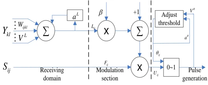
PCNN is widely used in image processing due to its global coupling and dynamic pulse propagation characteristics[18]. A large number of nonlinear modulation mechanisms and multiple leakage integrators exist in the classical PCNN model, which of parameter setting is complex and inconvenient for practical application. So, in this paper, a simplified PCNN model is used, its mathematical equation is described below:
ij ijF n S (8) ij exp L ij 1 L ijkl kl 1
kl
L n L n V
W Y n(9)
1
ij ij ij
U n F n L n
(10)
exp
ij 1
ij
1
ij n n V Y n
(11)
1,
0,
ij ij
ij
U n n
Y n else (12)
Where:Sijis an external input stimulus;
i j, represents the coordinates of the pixel or the coordinates of the neurons; Fijis the feedback input of neurons; Lijis the link input of the neuron; Uijis an internal activityitem;
ijis a dynamic threshold; Yijrepresents the pulse output of PCNN; nis the number of iterations; Wijklisthe matrix of the connection weights between neurons;
L,
is the time constant that connects the input andvariable threshold functions; VL,Vis the amplitude coefficient of the connection input and variable threshold
function;
is the link strength factor. IfUij
n ij
n , the neurons produce a pulse, called an ignition. PCNN model structure shown in Figure 3.∑
x
x
∑
0~1
Adjust
threshold
+1
Receiving
domain
Modulation
section
Pulse
generation
La
klY
W
ijklL
V
ijS
ij L ij F a V ij ij UFigure 3: Simplified model of PCNN
Adaptive PCNN:
In traditional PCNN model, all neuronal link strength
is a constant value, but the visual characteristics of the human eye are not consistent with all characteristics of the image, and the neuron connection strength
is not fixed but varies according to the characteristics of the image at any time. Since the pixel values of image pixels are constantly changing, each neuron should have its own link strength according to the regional characteristics of the image. In addition, since the link strength
is in the PCNN model Therefore, the link strength
also reflects the regional characteristics of the image. In order to obtain a better fusion effect by using the PCNN model, this paper chooses to improve the Laplacian energy and IEOL as neuronal link strength
, improve Laplacian energy and can effectively represent the image edge, direction and other details, suitable for high-frequency subband fusion, to improve the Laplacian energy and is defined as follows:) 1 , ( ) 1 , ( ) , ( 2 ) , 1 ( ) , 1 ( ) , ( 2 ) , ( , , , , , ,
, i j d i j d i j d i j d i j d i j d i j
MLkl kl kl kl kl kl kl (13)
where,dk,l
i,j denotes the coefficient of the high frequency subband in layer K, direction L, and theweighted Laplacian energy in the neighborhood centered at
i,j is:) , ( ) 1 , 1 ( ) , ( 1 1 1 1
, i j wi j ML x i y i
IEOL i i j j l
k
(14)
Where w is the weight matrix,
3.3.3 High frequency sub-band fusion rule
The source image is decomposed by NSCT to obtain three layers of high frequency sub-bands. The PCNN method is used for each layer to obtain the firing times of each pixel point of each layer of high frequency sub-bands. According to the number of ignition times to fuse, the fusion rule is as follows:
, , ,
, , , ,
, , , ,
( , ), ( , ) ( , ),
( ( , ) ( , )) / 2,
M M N
k l k l k l
F N M N
k l k l k l k l
M N M N
k l k l k l k l
d i j R R
d i j d i j R R
d i j d i j R R
(16)
Where: F,( , )
k l
d i j Is the high frequency subband fusion coefficient, M, ( , )
k l
d i j , N,( , )
k l
d i j respectively represents the coefficients of the high frequency subband M and N in layer K, direction L.
4 Experiment
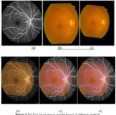
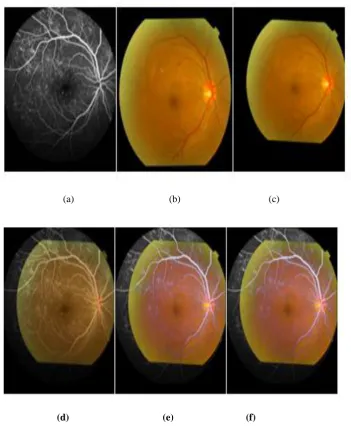
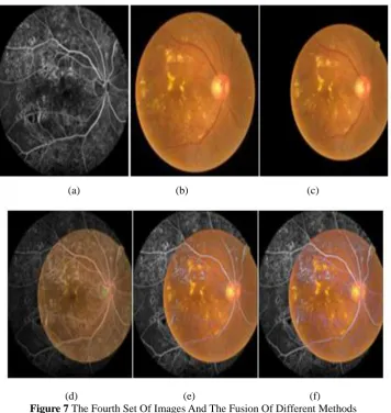
In order to verify the effectiveness of this method, simulation experiments are carried out on the Matlab2012a platform. 30 groups of color fundus images and fluorescein fundus images collected from Tianjin Medical University of Second Hospital were used as experimental subjects. Each group of images are the same person with the same eye at the same time by different types of sensors shooting, there are differences issues in spatial location and resolution. So, before fusion, we need to unify the resolution of two images and register them. In this paper, we select 4 groups images of lesions of varying degrees to experiment. The fluorescence fundus images and the color fundus images are shown in Figure 4 to 7 (a),(b),(c). (a)is the fluorescence fundus image; (b)is the color fundus image before registration;(c)is the color fundus image after registration. because the fundus image are different from other medical fusion images, the general fusion method is not suitable for fundus image fusion. Therefore, two representative methods for fundus image fusion are selected for comparison.(d)is the fusion result of the literature [11] method, (e)is the fusion result of the literature [12] method,(f)is the fusion result of this paper.
(a) (b) (c)
(d) (e) (f)
(d) (e) (f) Figure 6 : The third set of images and the fusion of different methods
(a) (b) (c)
(d) (e) (f)
Figure 7 The Fourth Set Of Images And The Fusion Of Different Methods
paper, the fusion of the images is superior to other representative methods in detail performance, such as the edge of lesion and microvascular.
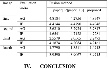
In order to further analyze the fusion results, we select average gradient (AG) and information entropy (IE) to evaluate the above three representative methods, as shown in Table 1. As can be seen from the table, this method of paper is higher than other representative methods in the evaluation index, more information is obtained from the source image, and more clearly on the lesion display.
Table 1Comparison of Objective Evaluation Indexes of Different Fusion Methods
Image Evaluation index
Fusion method
paper[12]paper [13] proposed first AG 4.8184 4.2756 4.8347
IE 4.4144 4.4790 4.4948 second AG 3.6210 3.2924 3.7847 IE 4.6541 4.7128 4.7281 third AG 2.5579 2.0565 2.2493 IE 4.1874 4.2084 4.2161 fourth AG 1.7790 1.3511 1.4713 IE 3.9590 3.9067 3.9715
IV.
CONCLUSION
The fusion of fundus images requires that the fused image as accurately as possible can reflect the lesion, blood vessels and other information. In this paper, a method for registration and fusion of fundus images based on NSCT and adaptive PCNN is proposed: The color fundus image and the fluorescence contrast fundus image were NSCT decomposed in each channel of R, G, B to obtain the high-frequency sub-bands and low-frequency sub-band of the two images .and the fusion of the low-frequency sub-band with the regional energy Which can effectively preserve the complementary information between the two kinds of fundus images. For the high-frequency part of the two images, a simplified PCNN model is used for processing. Considering that the link strength parameter of the PCNN model has a great influence on the fusion result, the Laplacian energy as PCNN adaptive link strength, By comparing the number of ignition times to be fused, finally the NSCT inverse transformation outputs the fusion result of the channel, and finally the three-channel fusion result is synthesized into the total fusion result. The experimental results show that the method of this paper is superior to other methods in both subjective and objective evaluation.
REFERENCE
[1]. Goshtasby A A,Nikolov S. Image fusion:advances in the state of the art [J].Information Fusion(S1566-2535),2007,8(2):114–118.
[2]. Ju J, Loew M, Ku B, et al. Erratum: Hybrid Retinal Image Registration Using Mutual Information and Salient Features [IEICE Transactions on Information and Systems Vol.E99.D (2016), No.6 pp.1729-1732][J]. Ieice Transactions on Information & Systems, 2016, E99.D(6):1729-1732.
[3]. Fan Guo, Xin Zhao, Beiji Zou, et al. Automatic retinal image registration using blood vessel segmentation and SIFT feature[J]. International Journal of Pattern Recognition & Artificial Intelligence, 2017.
[4]. Miri M S, Abràmoff M D, Kwon Y H, et al. Multimodal registration of SD-OCT volumes and fundus photographs using histograms of oriented gradients[J]. Biomedical Optics Express, 2016, 7(12):5252-5267.
[5]. Sun Y, Jiang L. Color multi-focus image fusion algorithm based on fuzzy theory and dual-tree comple wavelet transform[J].Journal ofAlgorithms& Computational Technology, 2017, 11(2):164-169.
[6]. Candes E J.Ridgelets:Theory and application[D].USA:Department of statistics,Stanford University,1999. [7]. Nencini F,Garzelli A,Baronti S,et al. Remote sensing image fusion using the curvelet transform [J].
Information Fusion(S1566-2535),2007,8(2):143–156.
[8]. Yang Shu-yuan,WangMin,JIAO Li-cheng,et al. Image fusion based on a new contourlet packet [J]. Information Fusion(S1566-2535),2010,11(2):78–84.
[9]. Cunha A L,Zhou J,Do M N.The nonsubsamples contourlet transform:Theory,design,and application[J].IEEE Trans.Image Proc,2006,15(10):3089-3101.
[10]. Song Ruixia, Wang Meng, Wang Xiaochun. Multifocus Image Fusion Algorithm Based on NSCT and Edge Detection[J]. Journal of Computer - Aided Design and Graphics, 2016, 28(12):2134-2141.
[13]. Li Xine, Ren Jianyue, Lv Zengming,. Multispectral and Panchromatic Image Fusion Methods Based on Improved PCNN and Regional Energy in NSCT Domain[J]. Infrared and Laser Engineering, 2013, 42(11):3096-3102.
[14]. Luo Tianjian, Liu Binghan. Fast SURF registration algorithm for fusion features[J]. Journal of Image and Graphics, 2015, 20(01):95-103.
[15]. Jian Chen, Jie Tian,A Partial Intensity Invariant Feature Descriptor for Multimodal Retinal Image
Registration[J].IEEETRANSACTIONS ONBIOMEDI -CAL ENGINEERING, VOL.
2010,57(7):1707-1718.