Teeth
Ronald J. Billings, DDS, MSD; Robert J. Berkowitz, DDS; and Gene Watson, DDS, PhD
ABSTRACT. Common environmental chemicals,
drugs, or physical agents can adversely affect human teeth during their embryonic development and after their eruption into the oral cavity. One of the more common elemental toxicants is lead. Teeth are known to accumu-late lead during their development. Both animal and human studies have shown that teeth with high lead levels are generally more susceptible to dental caries. Similarly, although inorganic fluorides have long been recognized for their potential to prevent dental caries, exposure to excessive amounts of fluoride when enamel is forming often leads to a type of enamel hypoplasia referred to as dental fluorosis or mottled enamel. Tera-togenic agents, such as tetracyclines, a class of antibiotic drugs commonly administered to infants and children, will often result in the discoloration of tooth enamel when prescribed during tooth development. It has re-cently been suggested that childhood exposure to passive smoking increases the risk for dental caries. Environmen-tal tobacco smoke has previously been linked to peri-odontal disease in adults. However, this is the first report of an association between passive tobacco smoke and increased susceptibility to dental caries. Last, an often-overlooked source of damage to teeth among all age groups after their eruption into the oral cavity is physical trauma from a variety of sources, especially sports-re-lated injuries. Epidemiologic data suggest that up to one third of all dental injuries are sports related.Pediatrics
2004;113:1120 –1127; environmental chemicals, drugs, physical agents, tooth development, teeth, dental caries.
ABBREVIATIONS. ETS, environmental tobacco smoke; PAH, polyhalogenated aromatic hydrocarbon; PCB, polychlorinated bi-phenyl; TCDD, 2,3,7,8-tetrachlorodibenzo-p-dioxin.
H
umans have 2 sets of teeth: the primary, ordeciduous, dentition followed by the perma-nent dentition. Primary tooth formation and development usually begins, on average, between the 13th and 16th weeks of gestation for incisors and between the 14th and 24th weeks for canines and molars. Mineralization of the enamel surface is usu-ally complete by the end of the first year of life and root development by 12 to 18 months after eruption. The permanent dentition begins to form in utero, and mineralization is usually complete by age 4 or 5 and root development by 2 to 3 years after eruption.
Teeth are most susceptible to developmental distur-bances during the mineralization phase of tooth for-mation. In general, the permanent dentition is more susceptible to disturbances in mineralization by en-vironmental toxicants and drugs than is the primary dentition, most likely as a consequence of its later development. As such, this review focuses on some of the more commonly observed developmental dis-turbances of the permanent dentition that result from excess exposure to various environmental chemicals and drugs and touches on other, somewhat contro-versial, toxicants that may adversely affect the child as a whole as well as the dentition. Damage to the teeth from orofacial trauma is also reviewed for its impact on the dentition of both children and adoles-cents.
ENVIRONMENTAL CHEMICALS Lead
It has long been known that lead crosses the pla-centa and has the potential to affect the development of different organ systems,1,2including teeth,3–7and its accumulation in developing teeth has provided a useful record of both fetal8,9 and long-term uptake for the study of neuropsychological effect of uniden-tified childhood exposure to lead.10,11 Although clear, unequivocal evidence of a cause-and-effect re-lationship between embryonic and early childhood exposure to lead and enhanced susceptibility to den-tal caries is lacking, the preponderance of epidemio-logic data, combined with data from animal studies, are suggestive of such a relationship. Bowen12 de-scribed several potential mechanisms on how pre-eruptive exposure to lead could enhance caries risk. However, a dose–response relationship between preeruptive exposure to lead and dental caries has never been determined. Lead has also been shown to accumulate in teeth posteruptively.3,13 Although a number of ecological and cross-sectional studies con-ducted in the 1960s and 1970s implicated posterup-tive exposure to lead as a risk factor for caries, the data are not clear that posteruptive exposure to lead has a direct effect on caries susceptibility.14A more recent study of the association of dental caries and blood lead levels in children 5 to 17 years of age showed that a 5-g/dL change in blood lead level was associated with an increased risk for caries and that ⬎2 million excess cases of dental caries in US children may be attributable to environmental lead exposure or factors that are linked to lead expo-sure.15However, as the data were cross-sectional in nature, no temporal or causal relationship could be established. In a study specifically designed to
exam-From the Eastman Department of Dentistry, University of Rochester, School of Medicine and Dentistry, Rochester, New York.
Received for publication Oct 7, 2003; accepted Oct 20, 2003.
Reprint requests to (R.J.B.) Eastman Department of Dentistry, University of Rochester, School of Medicine and Dentistry, 625 Elmwood Ave, Rochester, NY 14620. E-mail: [email protected]
ine the temporal association between lead exposure and caries, second- and fifth-grade children who were known to have blood levels up to 45 g/dL (mean: 10.7g/dL) as toddlers were not shown to be at greater risk for caries than children who had little or no lead exposure.16As noted by the authors, how-ever, the study had limited statistical power to detect a clinically relevant odds ratio.
Other ways in which lead could have an impact on caries risk revolves around its effect on salivary gland development and function. Work by Watson et al17 reported significantly diminished salivary flow rates and subsequent high caries levels in the off-spring of laboratory rats whose dams were exposed to 34 ppm lead in the drinking water during preg-nancy and during lactation. These observations are currently being followed up in an ongoing clinical study of caries risk resulting from environmental lead exposure in a birth cohort of 245 Cincinnati children who have been participants in the Cincin-nati Lead Study since late 1979. This group of black and white Appalachian children is arguably the most well-described longitudinal cohort ever studied for prenatal and postnatal lead exposure, with peak blood levels ranging from 5 to ⬎809g/dL.18,19 A major goal of this study is to measure potential con-founding factors, including dietary habits, oral hy-giene, and socioeconomic status, and to assess the influence of these on any observed association be-tween dental caries and lead exposure. If a causal relationship between environmental lead and dental caries is supported, then more aggressive measures for caries prevention could be targeted toward chil-dren with high blood lead levels.
Tobacco
Several recent studies have reported on the ad-verse impact of both smoked and smokeless tobacco on the oral health of children, adolescents,20 –22 and adults.23–26Investigators have specifically linked cigarette smoking with periodontal disease in adults,25,27,28 and a relationship between environ-mental tobacco smoke (ETS) and periodontal health in adults has been reported.28Conversely, only a few studies on the effect of active or passive smoking on oral health, including dental caries and periodontal disease, in children have been reported. No studies have been reported on the effect of maternal active or passive smoking on the preeruptive development of teeth. Data from the 1995 UK National Diet and Nutrition Survey, however, have suggested that ma-ternal but not pama-ternal smoking is a significant risk factor for predicting caries in young children.29 Re-cent work by Aligne et al30 based on a secondary analysis of data from the Third National Health and Nutrition Examination Survey (1988 –1994) has pro-vided the strongest evidence yet of an increased risk of dental caries in the deciduous dentition of chil-dren who are 4 to 11 years of age and have been exposed to ETS. That no effect on permanent teeth was observed is somewhat puzzling, as it would be expected that any effect of ETS on the developing dentition would affect both deciduous and perma-nent teeth alike. Similarly, if the main effect of ETS is
more related to posteruptive forces, then a similar pattern of caries susceptibility in the permanent den-tition should be observed. These findings are clearly provocative and beg for aggressive studies that will elucidate the causative role of ETS on the oral health of children and adolescents. Other studies that clearly need to be undertaken revolve around poten-tial mechanisms underlying tobacco’s cariogenic po-tential. For example, 1 in vitro study suggested that tobacco may have an effect on the growth of oral cariogenic streptococci.31 However, the study in-volved only smokeless tobacco products, and, as the authors noted, growth of cariogenic organisms may have been attributable entirely to the manufacturer’s added sugar and not to natural tobacco sugar. No reported studies have investigated a potential rela-tionship between tobacco in other forms, eg, smoked tobacco, whether active or passive, and growth of cariogenic oral flora. Clearly, much work remains to be undertaken in this area.
Mercury
resto-rations coupled with a lesser reliance on amalgam by dentists, large data sets of highly caries-active chil-dren and chilchil-dren either free of caries or with few fillings would be available for study. One question of interest would be the relationship between caries-free children and caries-active children on health-related variables associated with exposure to mer-cury vapor from amalgam fillings. Although dental surveys do not record the type of filling material used, it can reasonably be assumed, at least until very recently, that the filling material of choice would have been amalgam.
Polyhalogenated Aromatic Hydrocarbons
Polyhalogenated aromatic hydrocarbons (PAHs) and related compounds, including polychlorinated
dibenzofurans and polychlorinated biphenyls
(PCBs), and polychlorinated dibenzo-p-dioxins con-tinue to be of great environmental concern. As a group, the polychlorinated dibenzofurans consist of 135 congeners, the PCBs consist of 209 congeners, and the polychlorinated dibenzo-p-dioxins consist of 75 congeners, including 2,3,7,8-tetrachlorodibenzo-p-dioxin (TCDD), which is commonly referred to by the public as simply “dioxin.” The individual conge-ners vary substantially in their toxicity depending on the number and the position of the chlorine groups. TCDD is considered the most toxic of this class of compounds with the other PAHs scaled by using the concept of toxicity equivalence. The primary mecha-nism of action of all of the PAHs is thought to occur when they bind to a cytoplasmic receptor known as the aryl hydrocarbon receptor, resulting in transfor-mation and translocation of the complex to the nu-cleus where induction of dioxin-responsive elements modulate expression of specific genes, including cy-tochromes CYP1A1 and CYP1A2. On the basis of information from either incidents of inadvertent hu-man exposure or controlled animal studies, chlor-acne, teratogenicity, carcinogenicity, immunosup-pression, thymic atrophy, and wasting syndrome all have been associated with exposure to dioxin and dioxin-like compounds. Oral hard and soft tissues may also be susceptible to the adverse effects of dioxins; although epidemiologic studies on acciden-tal exposure to dioxins and related compounds have not produced consistent findings, overall, they seem to suggest that both prenatal and postnatal exposure to these compounds may cause oral soft tissue ab-normalities and mineralization defects in children’s teeth. In 1 study, children who were born to mothers who were exposed to very high levels of PCBs con-tained in contaminated cooking oil experienced in-creased prevalence of natal teeth, gingival hypertro-phy, intraoral hyperpigmentation, tooth chipping, and dental caries.46As a consequence of its fat con-tent, breast milk has been identified as a potentially significant source of postnatal dioxin exposure for an infant.47– 49However, gingival pigmentation, mottled enamel, and dental caries levels in children of moth-ers who were exposed to PCBs in an electric capaci-tor faccapaci-tory, whose reported PCB levels in blood and milk were 10 to 100 times greater than nonexposed mothers, were not found to differ from the children
of nonexposed mothers.48One study reported a sig-nificant association between the duration of breast-feeding and mineralization defects in the first per-manent teeth of offspring.49 In this study, mothers did not have known previous occupational or envi-ronmental exposure to elevated levels of TCDD and other PCBs, thus suggesting that “background” lev-els of these compounds could adversely influence dental development. In a follow-up study, the au-thors went on to suggest that the high frequency of hypomineralized dental defects among the children may be a sign of exposure to dioxin and its conge-ners, and the presence of defects could potentially be used as a biomarker of exposure.50 More recently, developmental defects in enamel of permanent teeth were reported in 71.3% of children who were ex-posed long term to PCBs, compared with 49.5% in the control group (P⫽.0001).51Overall, these studies seem to suggest that developing oral structures, in particular dental enamel, may be especially suscep-tible to minute dioxin exposure.
Fluoride
Inorganic fluorides have long been recognized for their potential to reduce the magnitude and the se-verity of dental caries in children as well as adults.52,53Although fluoride has substantial benefits in the prevention of tooth decay, depending on the level and source of exposure, fluorides also have adverse effects on human tissues.54In North Amer-ica, the major sources of fluoride are from drinking water, beverages, food, and oral hygiene products, including dietary fluoride supplements. The most common adverse effect of excess exposure to fluoride is dental fluorosis, a permanent hypomineralization of enamel, characterized in its mildest form as small, barely visible, white flecks found primarily on cusp tips and on facial surfaces of the permanent denti-tion.55 The moderate to severe forms, infrequently observed in North America, are found on most per-manent tooth surfaces and range between white opaque areas (Fig 1) to darkly stained and pitted enamel.55The critical window of exposure for fluo-rosis to manifest occurs during the early maturation stage of tooth development,56 beginning when the child is approximately 2 years of age and continuing for several years thereafter until later-developing teeth have matured.57 Dental fluorosis is a dose– response condition, and the higher the level of expo-sure during tooth development, the more severe the fluorosis.58 – 60In general, fluoride intake during crit-ical periods of tooth development and maturation from approximately birth to 8 years of age is in the range of 0.03 to 0.1 mg F/kg body weight per day.61,62
catego-ries.72–74 However, unintentional ingestion of fluo-ride-containing dentifrices deserves special mention. As pointed out in many recent reports, fluoride-containing toothpastes, particularly when used by toddlers and young children in an unsupervised manner and often in combination with fluoride tab-lets, can provide another major source of fluoride exposure.75–78 Accordingly, toddlers and preschool-ers should not have access to fluoride-containing toothpaste. Parents should supervise tooth brushing and should place an amount the size of a pea on the toothbrush.
Dental fluorosis has not been identified as a public health problem in North America. However, given the trend toward the use of agents for whitening teeth and the increased demand for cosmetic dentist-ry,78 – 80public rejection of even the mildest form of fluorosis could pose problems for dentistry’s time-tested reliance on this proven and cost-effective car-ies preventive agent.
DRUGS Tetracyclines
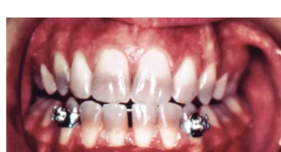
Perhaps the best-known and studied interaction between a therapeutic medication and an adverse interaction with tooth development is tetracycline. It has been known for many years that tetracycline has the potential to cause discoloration of teeth81 and that intensity of discoloration is both time and dose dependent82 (Fig 2). For example, tetracycline ingestion during periods of enamel calcification ei-ther by the moei-ther during gestation83 or by the
child up to approximately age 584,85 will result in discoloration of the enamel ranging from yellow (tet-racycline, demethylchlortet(tet-racycline, and oxytetracy-cline) to gray-brown (chlortetracyoxytetracy-cline). Oxytetracy-cline causes the least staining, and doxycyOxytetracy-cline does not stain at all.86 Permanent teeth are less intensely stained but more diffusely stained than primary teeth. As timing of administration, dosage, and type of tetracycline all are related to the extent and the type of enamel discoloration, tetracycline adminis-tration during pregnancy or periods of enamel calci-fication should be avoided to the extent possible. In general, tetracycline staining in most of North Amer-ica is seen only in adults today, rarely in children, as tetracycline is no longer recommended during preg-nancy and in young children. One way to differen-tiate tetracycline staining from other causes of enamel staining is to apply ultraviolet light to the teeth in question. Tetracycline-stained teeth will usu-ally fluoresce. On a final note, it is interesting to point out that chronic ingestion of tetracyclines may cause tooth discoloration, but the antimicrobial ef-fects prevent dental caries as evidenced by earlier observations in children with cystic fibrosis.
Other Drugs
There have been many reports over the past sev-eral years of prenatal and postnatal administration of anticonvulsants and chemotherapeutic drugs that have an adverse effect on teeth and oral tissues.87–97 Studies on medications used for the treatment of childhood cancer and leukemia have consistently
Fig 1. Moderate fluorosis.
shown that children younger than 5 years at diagno-sis and start of treatment exhibit abnormal dental development.93The severity of dentofacial-develop-mental and tooth-related abnormalities secondary to therapy are related to the age of the child, dosage, and duration of treatment.93 Dental abnormalities include tooth agenesis, arrested root development, microdontia, and enamel disturbances. However, no association between anticancer drugs and increased or decreased risk for dental caries has been reported. In contrast, though, but not part of this review, radi-ation therapy for head and neck cancer often results in partial or complete destruction of salivary gland tissues, and, unless extremely aggressive preventive measures are undertaken as part of the overall treat-ment plan, the onset of acute and rampant dental caries will occur rapidly with devastating conse-quences to the dentition for both children and adults. Drugs that are used for the treatment of asthma, including inhaler medicaments such as corticoste-roids and2 agonists, have not been shown to have adverse effects on tooth development. They have been shown to decrease plaque pH after administra-tion.98 However, their association with increased susceptibility or resistance to dental caries has been equivocal, with some studies reporting an increased susceptibility to caries with use of antiasthma med-ications99 –101and others reporting no increased sus-ceptibility.102
PHYSICAL AGENTS
Few physical agents are as damaging to the den-tition and craniofacial complex as is trauma. Further-more, it has been estimated that as many as one third of all dental injuries and up to 19% of injuries to the head and face are sports related.103–107 The conse-quences of sports-related injuries run the gamut, ranging from chipped or fractured teeth (Fig 3) to loss of 1 or more teeth, facial lacerations, contusions, and bone fractures. The majority of these injuries occur in children and adolescents, although protec-tive equipment including helmets, face shields, and mouth guards are mandatory in many sports today. Although use of appropriate head, face, eye, and mouth protection for children and adolescents who participate in school-sponsored physical activities has been encouraged and adopted as a Healthy Peo-ple 2010 objective,105there is, as yet, insufficient ev-idence that planned interventions have been effective in reducing the prevalence or incidence of sports-related injuries to the mouth and face.108 Clearly, much remains to be discovered regarding attitude and effective use of protective equipment.
CONCLUSION
In this brief review, we have described what is known about the more common environmental chemicals, drugs, and physical agents on teeth
dur-Fig 3. Fractured permanent teeth.
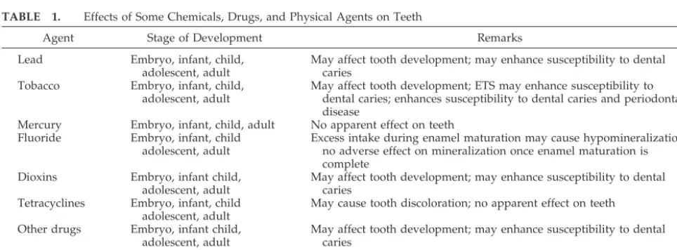
TABLE 1. Effects of Some Chemicals, Drugs, and Physical Agents on Teeth
Agent Stage of Development Remarks
Lead Embryo, infant, child,
adolescent, adult
May affect tooth development; may enhance susceptibility to dental caries
Tobacco Embryo, infant, child, adolescent, adult
May affect tooth development; ETS may enhance susceptibility to dental caries; enhances susceptibility to dental caries and periodontal disease
Mercury Embryo, infant, child, adult No apparent effect on teeth Fluoride Embryo, infant, child
adolescent, adult
Excess intake during enamel maturation may cause hypomineralization; no adverse effect on mineralization once enamel maturation is complete
Dioxins Embryo, infant child, adolescent, adult
May affect tooth development; may enhance susceptibility to dental caries
Tetracyclines Embryo, infant, child adolescent, adult
May cause tooth discoloration; no apparent effect on teeth Other drugs Embryo, infant child,
adolescent, adult
May affect tooth development; may enhance susceptibility to dental caries
Physical agents (trauma)
Embryo, infant, child infant, child, adolescent, adult
ing various stages of development, from the embryo to the adult (Table 1). As a generalization, teeth are most vulnerable and sensitive to the toxic effects of environmental chemicals and drugs during their de-velopment and before eruption into the mouth. However, emerging data suggestive of posteruptive effects of some environmental toxicants on dental health may also be revealing and may help to ex-plain, in part, the disproportionately high level of dental caries in children who are exposed to ETS, for example. Whether ETS also has an adverse effect on the gingival and mucosal tissues of children remains to be elucidated. Clearly, data from adult studies showing a strong relationship between adult-onset periodontal disease and ETS merit investigation for similar effects in children. The risk of physical injury to teeth and supporting structures must also be con-sidered in children and adolescents. Studies of sports-related injuries in adults are especially appli-cable to children. Although many questions remain on the prevention and control of sports-related craniofacial injuries, many of the data on safeguards to protect the teeth and craniofacial complex of adults can be extrapolated to children. However, there is a significant need for continued research on more effective methods to educate parents, coaches, health professionals, and children about the dangers of unprotected teeth and sports-related craniofacial injuries.
REFERENCES
1. Clark ARL. Placental transfer of lead and its effects on the newborn.
Postgrad Med J. 1977;53:674 – 678
2. Goyer RA. Transplacental transport of lead.Environ Health Perspect. 1990;89:101–105
3. Brudevold F, Steadman LT. The distribution of lead in human enamel.
J Dent Res. 1956;35:430 – 437
4. Lawson BF, Stout FW, Ahern DE, Sneed WD. The incidence of enamel hypoplasia associated with chronic pediatric lead poisoning.S C Dent J. 1971;29:5–10
5. Needleman HL, Tuncay OC, Shapiro IM. Lead levels in deciduous teeth of urban and suburban American children.Nature. 1972;235: 111–112
6. Stack MV, Burkitt AJ, Nickless G. Lead in children’s teeth [letter].
Nature. 1975;255:169
7. Pearl M, Roland NM. Delayed primary dentition in a case of congenital lead poisoning.ASDC J Dent Child. 1980;47:269 –271
8. Stack MV, Burkitt AJ, Nickless G. Trace metals in teeth at birth (1957–1963 and 1972–1973). Bull Environ Contam Toxicol. 1976;16: 764 –766
9. Gulson B, Wilson D. History of lead exposure in children revealed from isotopic analyses of teeth.Arch Environ Health. 1994;49:279 –283 10. Needleman HL, Shapiro IM. Dentine lead levels in asymptomatic
Philadelphia school children: subclinical exposure in high and low risk groups.Environ Health Perspect. 1974;7:27–31
11. Needleman HL, Gunnoe C, Leviton A, et al. Deficits in psychologic and classroom performance of children with elevated dentine lead levels.N Engl J Med. 1979;300:689 – 695
12. Bowen WH. Exposure to metal ions and susceptibility to dental caries.
J Dent Educ. 2001;65:1046 –1053
13. Brudevold F, Aasenden R, Srinivasian BN, Bakhos Y. Lead in enamel and saliva, dental caries and the use of enamel biopsies for measuring past exposure to lead.J Dent Res. 1977;56:1165–1171
14. Stack MV. Lead. In: Curzon M, Cutress TW, eds.Trace Elements and Dental Disease. Boston, MA: John Wright; 1983:357–385
15. Moss ME, Lanphear BP, Auinger P. Association of dental caries and blood lead levels.JAMA. 1999;281:2294 –2298
16. Campbell JR, Moss ME, Raubertas RF. The association between caries and childhood lead exposure. Environ Health Perspect. 2000;108: 1099 –1102
17. Watson GE, Davis BA, Raubertas RF, Pearson SK, Bowen WH.
Influ-ence of maternal lead ingestion on caries in rat pups.Nat Med. 1997; 3:1024 –1025
18. Dietrich KN, Berger OG, Succop PA. Lead exposure and the motor developmental status of urban six-year-old children in the Cincinnati Prospective Study.Pediatrics. 1993;91:301–307
19. Dietrich KN, Berger OG, Succop PA, Hammond PB, Bornschein RL. The developmental consequences of low to moderate prenatal and postnatal lead exposure: intellectual attainment in the Cincinnati Lead Study Cohort following school entry. Neurotoxicol Teratol. 1993;15: 37– 44
20. Christen AG, McDonald JL Jr, Olson BL, Christen JA. Smokeless to-bacco addiction: a threat to the oral and systemic health of the child and adolescent.Pediatrician. 1989;16:170 –177
21. Kleinman DV, Swango PA, Pindborg JJ. Epidemiology of oral mucosal lesions in United States schoolchildren: 1986 – 87.Community Dent Oral Epidemiol. 1994;22:243–253
22. Tomar SL, Winn DM, Swango PA, Giovino GA, Kleinman DV. Oral mucosal smokeless tobacco lesions among adolescents in the United States.J Dent Res. 1997;76:1277–1286
23. Tyc VL, Hopkins KP. Smoking interventions delivered by pediatric dentists: special recommendations for pediatric cancer patients.Pediatr Dent. 2000;22:43– 48
24. Tomar SL, Winn DM. Chewing tobacco use and dental caries among U.S. men.J Am Dent Assoc. 1999;130:1601–1610
25. Johnson GK, Slach NA. Impact of tobacco use on periodontal status.J Dent Educ. 2001;65:313–321
26. Winn DM. Tobacco use and oral disease.J Dent Educ. 2001;65:306 –312 27. Machuca G, Rosales I, Lacalle JR, Machuca C, Bullon P. Effect of cigarette smoking on periodontal status of healthy young adults.J Periodontol. 2000;71:73–78
28. Arbes SJ Jr, Agustsdottir H, Slade GD. Environmental tobacco smoke and periodontal disease in the United States. Am J Public Health. 2001;91:253–257
29. Williams SA, Kwan SY, Parsons S. Parental smoking practices and caries experience in pre-school children.Caries Res. 2000;34:117–122 30. Aligne CA, Moss ME, Auinger P, Pearson TA, Weitzman M.
Associa-tion of pediatric dental caries with passive smoking: an analysis of NHANES III.JAMA. 2003;289:1258 –1264
31. Lindemeyer RG, Baum RH, Hsu SC, Going RE. In vitro effect of tobacco on the growth of oral cariogenic streptococci.J Am Dent Assoc. 1981;103:719 –722
32. Mackert JR Jr. Factors affecting estimation of dental amalgam mercury exposure from measurements of mercury vapor levels in intra-oral and expired air.J Dent Res. 1987;66:1775–1780
33. Snapp KR, Boyer DB, Peterson LC, Svare CW. The contribution of dental amalgam to mercury in blood.J Dent Res. 1989;68:780 –785 34. Mackert JR Jr, Leffell MS, Wagner DA, Powell BJ. Lymphocyte levels in
subjects with and without amalgam restorations.J Am Dent Assoc. 1991;122:49 –53
35. Osborne JW. Dental amalgam and mercury vapor release.Adv Dent Res. 1992;6:135–138
36. Lorscheider FL, Vimy MJ, Summers AO. Mercury exposure from “silver” tooth fillings: emerging evidence questions a traditional dental paradigm.FASEB J. 1995;9:504 –508
37. Ingalls TH. Epidemiology, etiology, and prevention of multiple scle-rosis. Hypothesis and fact.Am J Forensic Med Pathol. 1983;4:55– 61 38. MacEntee MI, Mojon P. Issues in the amalgam debate.J Can Dent Assoc.
1991;57:931–936
39. Bjorkman L, Pedersen NL, Lichtenstein P. Physical and mental health related to dental amalgam fillings in Swedish twins.Community Dent Oral Epidemiol. 1996;24:260 –267
40. McGrother CW, Dugmore C, Phillips MJ, Raymond NT, Garrick P, Baird WO. Multiple sclerosis, dental caries and fillings: a case-control study.Br Dent J. 1999;187:261–264
41. Langworth S, Bjorkman L, Elinder CG, Jarup L, Savlin P. Multidisci-plinary examination of patients with illness attributed to dental fill-ings.J Oral Rehabil. 2002;29:705–713
42. Council on Dental Materials, Instruments, and Equipment, Council on Dental Therapeutics. Safety of dental amalgam. J Am Dent Assoc. 1983;106:519 –520
43. Council on Dental Materials, Instruments and Equipment. Recommen-dations in dental mercury hygiene.J Am Dent Assoc. 1984;109:617– 619 44. Langan DC, Fan PL, Hoos AA. The use of mercury in dentistry: a critical review of the recent literature. J Am Dent Assoc. 1987;115: 867– 880
US Department of Health and Human Services, Committee to Coor-dinate Environmental Health and related Programs, US Public Health Service; 1993
46. Rogan WJ, Gladen BC, Hung KL, et al. Congenital poisoning by polychlorinated biphenyls and their contaminants in Taiwan.Science. 1988;241:334 –336
47. Holtta P, Kiviranta H, Leppaniemi A, Vartiainen T, Lukinmaa PL, Alaluusua S. Developmental dental defects in children who reside by a river polluted by dioxins and furans.Arch Environ Health. 2001;56: 522–528
48. Hara I. Health status and PCBs in blood of workers exposed to PCBs and of their children.Environ Health Perspect. 1985;59:85–90 49. Alaluusua S, Lukinmaa PL, Koskimies M, et al. Developmental dental
defects associated with long breast feeding.Eur J Oral Sci. 1996;104: 493– 497
50. Alaluusua S, Lukinmaa PL, Torppa J, Tuomisto J, Vartiainen T. Devel-oping teeth as biomarker of dioxin exposure.Lancet. 1999;353:206 51. Jan J, Vrbic V. Polychlorinated biphenyls cause developmental enamel
defects in children.Caries Res. 2000;34:469 – 473
52. McClure FJ.Water Fluoridation: The Search and the Victory. Bethesda, MD: US Department of Health, Education and Welfare, National In-stitutes of Health; 1970
53. Klein H. Dental caries inhibition by fluorine—the historical perspec-tive.J Ir Dent Assoc. 1972;18:9 –21
54.Review of Fluoride: Benefits and Risks. Report of the Ad Hoc Subcommittee on Fluoride of the Committee to Coordinate Environmental Health and Related Programs. Washington, DC: US Department of Health and Human Services, US Public Health Service; 1991
55. Dean HT. Classification of mottled enamel diagnosis.J Am Dent Assoc. 1934;21:1421–1426
56. Den Besten PK. Mechanism and timing of fluoride effects on develop-ing enamel.J Public Health Dent. 1999;59:247–251
57. Evans RW, Stamm JW. Dental fluorosis following downward adjust-ment of fluoride in drinking water.J Public Health Dent. 1991;51:91–98 58. Dean HT. The investigation of physiological effects by the epidemio-logical method. In: Moulton FR, ed.Fluorine and Dental Health. Wash-ington, DC: American Association for the Advancement of Science; 1942:23–31
59. Eklund SA, Burt BA, Ismail AI, Calderone JJ. High-fluoride drinking water, fluorosis, and dental caries in adults.J Am Dent Assoc. 1987;114: 324 –328
60. Fejerskov O, Manji F, Baelum V. The nature and mechanisms of dental fluorosis in man.J Dent Res.1990;69:692–700
61. Grobler SR, van Wyk CW, Kotze D. Relationship between enamel fluoride levels, degree of fluorosis and caries experience in communi-ties with a nearly optimal and a high fluoride level in the drinking water.Caries Res. 1986;20:284 –288
62. Fejerskov O, Stephen KW, Richards A, Speirs R. Combined effect of systemic and topical fluoride treatments on human deciduous teeth— case studies.Caries Res. 1987;21:452– 459
63. Pendrys DG, Katz RV. Risk of enamel fluorosis associated with fluo-ride supplementation, infant formula, and fluofluo-ride dentifrice use.Am J Epidemiol. 1989;130:1199 –1208
64. Bohaty BS, Parker WA, Seale NS, Zimmerman ER. The prevalence of fluorosis-like lesions associated with topical and systemic fluoride usage in an area of optimal water fluoridation.Pediatr Dent. 1989;11: 125–128
65. Bagramian RA, Narendran S, Ward M. Relationship of dental caries and fluorosis to fluoride supplement history in a non-fluoridated sample of schoolchildren.Adv Dent Res. 1989;3:161–167
66. Larsen MJ, Kirkegaard E, Poulsen S, Fejerskov O. Dental fluorosis among participants in a non-supervised fluoride tablet program. Com-munity Dent Oral Epidemiol. 1989;17:204 –206
67. Woolfolk MW, Faja BW, Bagramian RA. Relation of sources of sys-temic fluoride to prevalence of dental fluorosis.J Public Health Dent. 1989;49:78 – 82
68. Szpunar SM, Burt BA. Evaluation of appropriate use of dietary fluo-ride supplements in the US.Community Dent Oral Epidemiol. 1992;20: 148 –154
69. Kalsbeek H, Verrips GH, Backer Dirks O. Use of fluoride tablets and effect on prevalence of dental caries and dental fluorosis.Community Dent Oral Epidemiol. 1992;20:241–245
70. McKnight-Hanes MC, Leverett DH, Adair SM, Shields CP. Fluoride content of infant formulas: soy-based formulas as a potential factor in dental fluorosis.Pediatr Dent.1988;10:189 –194
71. Ripa LW. Topical fluorides: a discussion of risks and benefits.J Dent Res. 1987;66:1079 –1083
72. Horowitz HS. Fluoride and enamel defects. Adv Dent Res. 1989;3: 143–146
73. Horowitz HS. Proper use of fluoride products in fluoridated commu-nities.Lancet. 1999;353:1462
74. Bowen WH. Fluorosis: is it really a problem?J Am Dent Assoc. 2002; 133:1405–1407
75. Osuji OO, Leake JL, Chipman ML, Nikiforuk G, Locker D, Levine N. Risk factors for dental fluorosis in a fluoridated community.J Dent Res. 1988;67:1488 –1492
76. Woltgens JHM, Etty EJ, Nieuwland WMD, Lyaruu DM. Use of fluoride by young children and prevalence of mottled enamel.Adv Dent Res. 1989;3:177–182
77. Kaminsky LS, Mahoney MC, Leach J, Melius J, Miller M. Fluoride: benefits and risks of exposure.Crit Rev Oral Biol Med. 1990;1:261–281 78. Riordan PJ. Perceptions of dental fluorosis. J Dent Res. 1993;72:
1268 –1274
79. Clark DC. Evaluation of aesthetics for the different classifications of the Tooth Surface Index of Fluorosis.Community Dent Oral Epidemiol. 1995;23:80 – 83
80. Hawley GM, Ellwood RP, Davies RM. Dental caries, fluorosis and the cosmetic implications of different TF scores in 14-year-old adolescents.
Community Dent Health. 1996;13:189 –192
81. Kutscher AH, Zegarelli EV, Tovell HM, Hochberg B, Hauptman J. Discoloration of deciduous teeth induced by administration of tetra-cycline antepartum.Am J Obstet Gynecol. 1966;96:291–292
82. Grossman ER, Walchek A, Freedman H. Tetracyclines and permanent teeth: the relation between dose and tooth color.Pediatrics. 1971;47: 567–570
83. Genot MT, Golan HP, Porter PJ, Kass EH. Effect of administration of tetracycline in pregnancy on the primary dentition of the offspring.
J Oral Med. 1970;25:75–79
84. Zegarelli EV, Denning CR, Kutscher AH, Tuoti F, DiSant’Agnese PA. Tooth discoloration in cystic fibrosis.Pediatrics.1960;26:1050 –1051 85. Harcourt JK, Johnson NW, Storey E. In vivo incorporation of
tetracy-cline in teeth of man.Arch Oral Biol. 1962;7:431– 437
86. Stewart RE, Witkop CJ, Bixler D. The dentition. In: Stewart RE, Barber TK, Troutman KC, Wei SHY, eds.Pediatric Dentistry. St. Louis, MO: CV Mosby Co; 1962:104 –105
87. Jaffe N, Toth BB, Hoar RE, Ried HL, Sullivan MP, McNeese MD. Dental and maxillofacial abnormalities in long-term survivors of child-hood cancer: effects of treatment with chemotherapy and radiation to the head and neck.Pediatrics. 1984;73:816 – 823
88. Durr DP, Adair SM, Novak EV. Dental abnormalities associated with the treatment of Hodgkin’s disease in a young patient.J Pedod. 1987; 12:98 –104
89. Rosenberg SW, Kolodney H, Wong GY, Murphy ML. Altered dental root development in long-term survivors of pediatric acute lympho-blastic leukemia. A review of 17 cases.Cancer. 1987;59:1640 –1648 90. Pajari U, Lanning M, Larmas M. Prevalence and location of enamel
opacities in children after antineoplastic therapy.Community Dent Oral Epidemiol. 1988;16:222–226
91. Sonis AL, Tarbell N, Valachovic RW, Gelber R, Schwenn M, Sallan S. Dentofacial development in long-term survivors of acute lymphoblas-tic leukemia. A comparison of three treatment modalities. Cancer. 1990;66:2645–2652
92. Nunn JH, Welbury RR, Gordon PH, Kernahan J, Craft AW. Dental caries and dental anomalies in children treated by chemotherapy for malignant disease: a study in the north of England.Int J Paediatr Dent. 1991;1:131–135
93. Dahllof G, Rozell B, Forsberg CM, Borgstrom B. Histologic changes in dental morphology induced by high dose chemotherapy and total body irradiation.Oral Surg Oral Med Oral Pathol. 1994;77:56 – 60 94. Kaste SC, Hopkins KP, Jenkins JJ III. Abnormal odontogenesis in
children treated with radiation and chemotherapy: imaging findings.
AJR Am J Roentgenol.1994;162:1407–1411
95. Kaste SC, Hopkins KP, Jones D, Crom D, Greenwald CA, Santana VM. Dental abnormalities in children treated for acute lymphoblastic leu-kemia.Leukemia. 1997;11:792–796
96. Orup HI Jr, Keith DA, Holmes LB. Prenatal anticonvulsant drug exposure: teratogenic effect on the dentition.J Craniofac Genet Dev Biol. 1998;18:129 –137
97. Alpaslan G, Alpaslan C, Gogen H, Oguz A, Cetiner S, Karadeniz C. Disturbances in oral and dental structures in patients with pediatric lymphoma after chemotherapy: a preliminary report.Oral Surg Oral Med Oral Pathol Oral Radiol Endod. 1999;87:317–321
medica-ment effects on saliva and plaque pH in asthmatic children.J Clin Pediatr Dent. 1998;22:137–140
99. Ryberg M, Moller C, Ericson T. Saliva composition and caries devel-opment in asthmatic patients treated with beta 2-adrenoceptor agonists: a 4-year follow-up study.Scand J Dent Res. 1991;99:212–218 100. McDerra EJ, Pollard MA, Curzon ME. The dental status of asthmatic
British school children.Pediatr Dent. 1998;20:281–287
101. Milano M. Increased risk for dental caries in asthmatic children.Tex Dent J. 1999;116:35– 42
102. Shulman JD, Taylor SE, Nunn ME. The association between asthma and dental caries in children and adolescents: a population-based case-control study.Caries Res. 2001;35:240 –246
103. Meadow D, Lindner G, Needleman H. Oral trauma in children.Pediatr Dent. 1984;6:248 –251
104. Lephart SM, Fu FH. Emergency treatment of athletic injuries.Dent Clin North Am. 1991;35:707–717
105.Understanding and Improving Health and Objectives for Improving Health. 2nd ed. Washington, DC: US Department of Health and Human Ser-vices, Healthy People 2010; 2000
106.Oral Health in America: A Report of the Surgeon General. Rockville, MD: US Department of Health and Human Services, National Institutes of Health, National Institute of Dental and Craniofacial Research; 2000
107. Burt CW, Overpeck MD. Emergency visits for sports-related injuries.
Ann Emerg Med. 2001;37:301–308
2004;113;1120
Pediatrics
Ronald J. Billings, Robert J. Berkowitz and Gene Watson
Teeth
Services
Updated Information &
http://pediatrics.aappublications.org/content/113/Supplement_3/1120 including high resolution figures, can be found at:
References
#BIBL
http://pediatrics.aappublications.org/content/113/Supplement_3/1120 This article cites 100 articles, 14 of which you can access for free at:
Subspecialty Collections
sub
http://www.aappublications.org/cgi/collection/environmental_health_
Environmental Health
ub
http://www.aappublications.org/cgi/collection/dentistry:oral_health_s
Dentistry/Oral Health
following collection(s):
This article, along with others on similar topics, appears in the
Permissions & Licensing
http://www.aappublications.org/site/misc/Permissions.xhtml in its entirety can be found online at:
Information about reproducing this article in parts (figures, tables) or
Reprints
2004;113;1120
Pediatrics
Ronald J. Billings, Robert J. Berkowitz and Gene Watson
Teeth
http://pediatrics.aappublications.org/content/113/Supplement_3/1120
located on the World Wide Web at:
The online version of this article, along with updated information and services, is
by the American Academy of Pediatrics. All rights reserved. Print ISSN: 1073-0397.