review article
Mechanisms of Disease
Bone Quality — The Material and Structural
Basis of Bone Strength and Fragility
Ego Seeman, M.D., M.B., B.S., and Pierre D. Delmas, M.D., Ph.D.
From the Department of Endocrinology, Austin Health, University of Melbourne, Melbourne, Australia (E.S.); and the De-partment of Rheumatology, Université Claude Bernard Lyon 1, and INSERM Re-search Unit 403 — both in Lyon, France (P.D.D.). Address reprint requests to Dr. Seeman at the Department of Endocri-nology, Austin Health, Heidelberg 3084, Melbourne, Australia, or at egos@unimelb. edu.au.
N Engl J Med 2006;354:2250-61.
Copyright © 2006 Massachusetts Medical Society.
P
rogress in understanding the pathogenesis of bone fr agility is hampered by the inaccessibility of bone for investigation. Bone densitom-etry is an effective, noninvasive, and quantitative method for the assessment of the risk of fracture, but structures such as the vertebral body are depicted as a two-dimensional image — the areal bone mineral density cast by the attenuation of photons by mineral during their passage through bone. Just as the shadow of the earth, cast on the moon, reveals nothing of the topology of the earth’s mountain ranges, the densitometric image tells us little about the two properties that deter-mine bone strength: its material composition and its structural design.1,2In this review, we define how the composition and structure of bone determine its strength, describe bone modeling and remodeling — the cellular machinery re-sponsible for constructing bone during growth and reconstructing it during adult-hood, demonstrate how age-related abnormalities in these processes compromise the composition and structure of bone, and show how the mechanisms underlying the structural decay of bone offer rational approaches to the use of drugs that in-hibit bone resorption and stimulate bone formation.
F a b r i c a n d S t r u c t u r e o f B o n e — L e v e r s a n d S p r i n g s
The strength of bone is determined by its material composition and structure.2 Bone must be stiff and able to resist deformation, thereby making loading possible. Bone must also be flexible: it must be able to absorb energy by deforming, to short-en and widshort-en whshort-en compressed, and to lshort-engthshort-en and narrow in tshort-ension without crack-ing. If bone is brittle (i.e., too stiff and unable to deform a little), the energy imposed during loading will be released by structural failure — initially by the development of microcracks and then by complete fracture. If bone is too flexible and deforms beyond its peak strain, it will also crack. Bone must also be light to facilitate move-ment. A unique feature of bone is that it can serve these contradictory needs of stiff-ness yet flexibility and lightstiff-ness yet strength.3
Composition of Bone
Bone is composed of type I collagen stiffened by crystals of calcium hydroxyapa-tite. An increase in tissue mineral density increases the stiffness of the fabric but sacrifices flexibility.2,4
Variations in tissue mineral density affect function. Audi-tory ossicles are 90 percent mineral, conferring the stiffness essential for the fidelity of sound transmission (like tuning forks). Animal antlers are 40 percent mineral, conferring the flexibility needed to absorb energy during head butting to defeat suitors in mating season. Human bone is about 60 percent mineralized. The com-position and degree of collagen cross-linking also influence function.5-8
helix of type I collagen confers strength in ten-sion. The cross-links in collagen keep its helixes fastened. If there are too few cross-links, the he-lixes may separate; if there are too many, the abil-ity to absorb energy diminishes.
Microstructure and Macrostructure of bone Bone fabric is woven at submicroscopic, micro-scopic, and macroscopic levels into an architec-tural masterpiece of biomechanical engineering — with an optimal mass adapted in size, shape, and architecture for structural strength (i.e., the ability to resist cracking).9
Just as a wall is con-structed with overlapping bricks, cortical bone consists of overlapping parallel osteons, the ana-tomical remnants of a completed remodeling event (Fig. 1).10
A large number of osteons per unit of bone volume limits the propagation of cracking because they obstruct the passage of a crack as it navigates between the many osteons.9 The entry of cracks into the osteon is blocked by the cement line delimiting each osteon and by concentric lamellae of mineralized collagen fi-bers that are packed in an alternating loose and dense pattern and are orientated in various direc-tions. In addition, uncracked bone tissue within a crack forms a bridge that carries the load that otherwise would be used to drive the crack for-ward.11
As a result, cracks are largely confined to the older, more densely mineralized interstitial
bone between osteons.12
Although small, con-fined cracks are undesirable, they are a final means of dissipating energy as a defense against the alter-native means of energy release imposed by the stress on bone — fracture.2
Figure 1. The Hierarchical Structure of Cortical Bone.
Within a cortical bone shaft, shown in cross-section (Panel A) are osteons surrounded by interstitial bone and many osteocytic lacunae distributed around the central haversian canal (Panel B). Panel C shows a mi-crocrack that is largely confined to interstitial bone. Panel D shows the haversian canal system in cortical bone (microcomputed tomography courtesy of M.A. Knackstedt, Australian National University). In Panel E, alternating high-density and low-density concentric lamellae of an osteon produce a composite structure that is resistant to cracking, with an osteocytic lacuna at a higher resolution showing collagen fibers (Panel F)
(scanning electron microscopy reprinted from Marotti10
with the permission of the publisher). In Panel G, osteo-cytes connect with lining cells and with one another through a network of canaliculi (scanning electron mi-croscopy of an acid-etched resin-embedded murine bone section, courtesy of Drs. Lynda F. Bonewald and Jian Q. Feng, University of Missouri–Kansas City). Panel H shows the detail of an osteoblast lining cell connected to an osteocyte (transmission electron microscopy
reprint-ed from Marotti10
with the permission of the publisher).
A B E G H F C B E E G H F C D
Lever Action of Long Bones
Cortical bone is used to build long bones. Long bones are levers needed for loading and move-ment, with rigidity favored over flexibility. Struc-tural stiffness and lightness are achieved by the construction of a marrow cavity. Long bones grow in length by endochondral apposition on the in-ner, or endosteal, surface and in width by the de-position of bone on the outer, or periosteal, sur-face. Resorptive excavation of a marrow cavity during fetal and postnatal growth shifts the thick-ening cortex away from the neutral axis, thereby increasing resistance to bending.13
Sex and racial differences in the extent of periosteal apposition and endocortical resorption during growth and aging establish variations in the diameter and cortical thickness of bone and in the distance of the cortical mass from the neutral axis — and thus differences in bone strength.14-17
Long bones are not like drinking straws, which have the same diameter and thickness through-out. The conical metaphyses are fashioned by the resorption and formation of bone on the periosteal surface, whereas endochondral bone forms the trabecular network. External and internal contours differ at each point along and around the shaft. For example, the femoral neck adjacent to the shaft is elliptical, with the longest diameter in the superior–inferior direction and greater cor-tical thickness inferiorly; these features minimize bending.18
Near the femoral head, where stresses are mainly compressive, the femoral neck is more circular and largely trabecular, with a cortex of similar thickness around its perimeter. These structural adaptations to loading are not seen in quadrupedal primates.19
Spring Action of Vertebral Bodies
Bone that will become vertebral bodies is assem-bled as an open-celled, porous structure that func-tions more like a spring than a lever in that the sponge-like structural design can absorb more energy by deforming more before cracking than can long bones. However, this structure sacrifices the ability to tolerate the peak loads that can be borne by long bones. The interconnecting trabecu-lar plates achieve lightness and favor structural flexibility over stiffness.20
The greater loads that are better tolerated in men than in women and in some races better than in others are largely due to differences in bone dimensions.14,15
Men and women generally have
similar vertebral trabecular volumetric density (number plus thickness) and similar vertebral heights; the larger vertebral cross-sectional area in men contributes to sex-based differ-ences in bone strength.14 Black people tend to have wider but shorter vertebral bodies and higher measures of trabecular volumetric density than do white people owing to thicker trabecu-lae, a feature that may protect against the ef-fects of age-related bone loss.21-23
The structure of bone is contained in the ge-netic blueprint — fetal lower limb buds grown in vitro have the shape of the proximal femur.24 Although structure determines the loads a bone will tolerate, the reverse also applies: loads de-termine structure. Bone can adapt its composi-tion and structure to prevailing loads.25 Adap-tation in size and shape in the playing arm of tennis players is well documented.2 The Mov13 mouse, a model of the mild form of osteogenesis imperfecta, compensates for defects in bone col-lagen by a structural adaptation that entails peri-osteal apposition; the Brittle IV mouse, another model of osteogenesis imperfecta, makes adap-tations in the mineral:collagen ratio.26,27 How-ever, such adaptations may be unsuccessful. In the osteogenesis imperfecta (oim/oim) mouse, a com-pensatory increase in bone formation with de-fective collagen does not correct bone fragility.28 Thus, bone fragility can be the result of failed mate-rial or structural adaptations or both, not just low bone mass.
M o d e l i n g a n d R e m o d e l i n g o f B o n e
The cellular mechanisms responsible for the ad-aptation of bone are modeling (construction) and remodeling (reconstruction). Bone modeling pro-duces a change in the size and shape of bone when new bone is deposited without previous bone re-sorption. During bone remodeling, resorption by osteoclasts precedes bone formation by os-teoblasts. Osteoblasts and osteoclasts form the bone multicellular unit that reconstructs bone in distinct locations on the three components (en-docortical, intracortical, and trabecular) of its endosteal envelope and, to a lesser extent, on the periosteal envelope.29 Bone modeling and remod-eling modify the external size and contours of bone and its internal architecture by the deposi-tion or removal of bone from the surface of bone,
causing cortical and trabecular thickening dur-ing growth and thinndur-ing durdur-ing agdur-ing (Fig. 2).
The purpose of modeling and remodeling dur-ing growth is to establish the skeleton’s peak bone strength; its purpose in adulthood is to main-tain bone strength. In bone (as in roads, build-ings, and bridges), damage due to fatigue devel-ops during repeated loading, but only bone has the mechanism to detect the location and mag-nitude of the damage, remove it, replace it with new bone, and then reconstruct the material com-position, microarchitecture, and macroarchitec-ture.30,31
The role of remodeling in calcium homeo-stasis is outside the scope of this article.
Bone resorption is not necessarily bad. Dur-ing growth it is essential for the excavation of a marrow cavity and the fashioning of cortical and trabecular bone. In adults, the resorptive phase of the remodeling cycle removes damaged bone, and the formation phase restores the structure. The restitution of structure requires balanced re-modeling; the volume of damaged bone removed must be replaced by the same volume of nor-mal bone.
The most likely reason that a given point on a quiescent bone surface becomes a remodeling site is removal of damage.32-36
But how does bone
know the location of damage, as well as how much damage to remove and how much bone needs to be replaced? This process depends on the normal production, work, and life span of osteoclasts and osteoblasts, but another cell — the osteocyte — is also a likely participant.
Osteocytes are the most numerous, longest-lived, and least studied cells of bone. They are osteoblasts that have become entombed in la-cunae in the bone matrix (osteoid) that they synthesize. These osteoblasts undergo a morpho-logic change and become osteocytes with cyto-plasmic processes that connect them with other osteocytes and flattened lining cells (Fig. 1G and 1H).33
The dense, lace-like communicating net-work of osteocytes ensures that no part of bone is more than several microns from a lacuna con-taining its osteocyte. This arrangement suggests that osteocytes are part of the machinery guard-ing the integrity of the material and structural strength of bone.30-37
Osteocytes probably sense bone deformation, thereby signaling the need for adaptive remodel-ing of bone size, shape, and distribution to ac-commodate prevailing loads.33
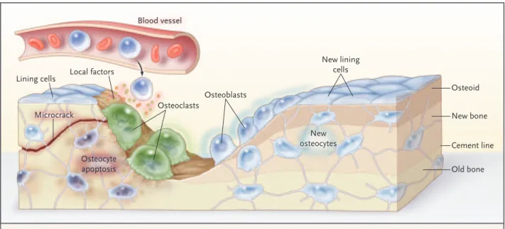
The death of os-teocytes by apoptosis — in estrogen deficiency, during corticosteroid therapy, in advancing age, Microcrack Osteoid Lining cells New lining cells New osteocytes Local factors Osteoclasts Osteoblasts Blood vessel Osteocyte apoptosis New bone Old bone Cement line
Figure 2. The Remodeling Cycle on a Trabecula.
A microcrack severs canaliculi, which causes osteocytic apoptosis, with the location and extent of the damage defined by signals to lin-ing cells. Linlin-ing cells and osteocytes release local factors that attract cells from blood and marrow into the remodellin-ing compartment in which osteoclastogenesis occurs. Osteoclasts resorb matrix and the microcrack, then successive teams of osteoblasts deposit new la-mellar bone. Osteoblasts that are trapped in the matrix become osteocytes; others die or form new, flattened osteoblast lining cells.
or after damage to bone — is associated with a loss of bone strength before any bone loss.35-37 The death of these cells probably heralds (through biochemical and chemotactic signals) the pres-ence of damage and its location and the initia-tion of targeted remodeling. Regions of micro-damage contain apoptotic osteocytes, whereas quiescent zones do not.37 The number of osteo-cytes that undergo apoptosis may provide the topo-graphic information needed to target removal of damage by osteoclasts.
Hence, the first step in remodeling is unlikely to be bone resorption. Osteoclasts must first be formed and be told where to go and how much bone to resorb. These instructions are likely to arise from signals produced by the deformation or death of osteocytes, which define the location and amount of resorption needed. The signals are probably relayed, in part, by the cytoplasmic pro-cesses connected to flattened osteoblast lining cells.31 These lining cells partly form the canopy of a bone-remodeling compartment.38 Within this microenvironment, local factors, including vas-cular growth factors, provide the vasvas-cular supply, osteoclast precursors, macrophages, and activated T cells that participate with osteoblast precursors in osteoclastogenesis. Osteoblast precursors go on to become mature osteoid-forming osteoblasts. Bone formation may also be coupled with bone resorption by products from the resorbed matrix and from osteoclasts themselves.39,40 Thus, the osteocyte is involved in initiating remodeling, and subsequent local regulation is bidirectional, with osteoblast precursors directing osteoclastogenesis and products of the osteoclast and of the resorbed matrix modulating bone formation.
N e g a t i v e B a l a n c e i n t h e B o n e M u l t i c e l l u l a r U n i t
A negative balance in the bone multicellular unit — which causes bone loss, an increased remod-eling rate, or both — compromises the strength of bone. During growth, the balance between the volume of bone that is resorbed and the volume that is formed in the bone multicellular unit is positive on a trabecular surface, so that each re-modeling event adds a small moiety of bone. As skeletal size reaches its programmed dimensions, the need for rapid remodeling and a positive bal-ance between the volume of bone removed and the volume of bone deposited in each bone
multi-cellular unit lessens. The remodeling rate decreas-es as longitudinal growth ceasdecreas-es with epiphyseal closure.41 The volume of bone formed in each bone multicellular unit may also decline, shifting the balance between the volume of bone that is re-moved and the volume that is formed in each bone multicellular unit from positive to equal (i.e., zero).
In adults, one of the first changes in the remod-eling machinery that leads to bone loss is likely to be a decline in bone formation within the bone multicellular unit. There is evidence of a reduc-tion in bone formareduc-tion in midlife,42,43 but this may begin in young adults when the need to build the skeleton (and thus the need for bone forma-tion) declines.44-46 When bone formation is less than prior bone resorption, each remodeling event removes a small moiety of bone from the skeleton, resulting in bone loss and structural damage.
The positive balance in the bone multicellular unit (net bone formation) during growth and the negative balance (net bone loss) during aging are small. For these reasons, the rate of gain in bone during growth and loss during aging is driven more by a high remodeling rate than by the mag-nitude of the positive or negative balance in the bone multicellular unit. This consideration is im-portant, given the effect of antiresorptive agents such as the bisphosphonates on the rate of remod-eling. Largely on the basis of cross-sectional data, bone loss appears to begin between the ages of 18 and 30 years, but the process is slow because remodeling is slow.47
Rapid remodeling (independent of an imbal-ance in the bone multicellular unit) is associated with an increased risk of fracture for several rea-sons. First, more densely mineralized bone is re-moved and replaced with younger, less densely mineralized bone, reducing material stiffness.48-50 As a result, bone may become too flexible, bend excessively, and crack under usual loading condi-tions. Second, excavated resorption sites remain temporarily unfilled (because of the delay in the initiation and slower completion of bone forma-tion that is coupled with resorpforma-tion), creating stress concentrators that predispose bone to micro-damage (as a small cut on the surface of a glass cylinder makes the tube easy to snap).2 Third, increased remodeling impairs isomerization and maturation of collagen, which increases the fragil-ity of bone,5,6 probably by altering the cross-linking between adjacent collagen fibers.
in-creases the rate of remodeling and the volume of bone that is resorbed by prolonging the life span of osteoclasts. It also decreases the volume of bone that is formed by reducing the life span of osteo-blasts, thereby aggravating the negative bone bal-ance in the bone multicellular unit.51 The combi-nation of a rapid rate of remodeling and increased imbalance in the bone multicellular unit acceler-ates bone loss and structural decay after meno-pause.
Trabecular Thinning and Loss of Connectivit y
Remodeling occurs on bone surfaces. Trabecular bone is fashioned with more surface than corti-cal bone. Since there are more remodeling sites per unit volume in trabecular bone than in corti-cal bone, a greater proportion of trabecular bone is turned over and lost as each remodeling event removes more bone than it puts back.52 The high remodeling rate and deep resorption cavities pro-duce a loss of trabecular plates and of their con-nection (connectivity), which in turn produce a greater deficit in bone strength than does tra-becular thinning.53
Bone fragility is more common in women than in men partly because the production of sex hor-mones in males does not decrease rapidly and there is no increase in the bone-remodeling rate in midlife. Although perforation and loss of con-nectivity between trabeculae occur in men, bone loss in men proceeds more by trabecular thinning (due to reduced bone formation within each bone multicellular unit) than by trabecular per-foration (due to increased bone resorption with-in each bone multicellular unit).54-57
Cortical Thinning and Porosit y
Rapid remodeling does not slow down with age; it continues because of persistent hypogonadism in women, emerging hypogonadism in some men, and secondary hyperparathyroidism in both sexes. With continued remodeling, trabeculae perforate and some disappear completely, and remodeling is more active on the endocortical surface than on remaining trabecular surfaces. Active endocorti-cal and intracortiendocorti-cal remodeling “trabecularizes” cortical bone (i.e., creates cortical bone with more surface area), so bone loss becomes mainly cortical in origin.52,58,59
Structural decay accelerates as each remodel-ing event removes bone from an ever-decreasremodel-ing
total volume of bone. Older, more densely min-eralized interstitial bone, distant from surface re-modeling, has a reduced number of osteocytes and accumulates microdamage.12 Cortical thinning and porosity reduces the resistance of bone to the propagation of cracks. Pores coalesce, and the re-duced bone mass cannot absorb the energy im-parted by a fall.
Periosteal Apposition
Concurrent periosteal apposition, by depositing new bone on the external surface, partly offsets the loss of compressive and bending strength pro-duced by cortical thinning and porosity, so that cortical thickness is better maintained than would occur without periosteal apposition.54-62 However, details of the magnitude of changes in periosteal apposition during advancing age — as well as the effects of site, sex, and race — are difficult to evaluate prospectively, given the small changes in periosteal apposition (a few millimeters) throughout adult life. In addition, periosteal ap-position is difficult to interpret in cross-sectional studies, given secular trends in bone mass and dimensions.63 The findings in one study62 that periosteal apposition occurs in the years after menopause to compensate for accelerated bone loss needs confirmation. Even the notion that periosteal apposition is greater in men than in women remains controversial1,64,65; recent stud-ies suggest that sex-based differences occur at some, but not all, sites.14,15 Moreover, these sex-based differences may vary according to race.
Albright and his colleagues suggested that osteoporosis is a disorder of reduced bone for-mation,66 but they did not specify its morpho-logic basis. We now believe that in addition to the decreased volume of bone that is formed in each bone multicellular unit during aging,42 the re-duced formation of periosteal bone during growth, aging, or both partly explains the smaller size of the vertebral body and smaller bone mineral mass in women and men with vertebral fractures.67,68
Bone Fragilit y in Patients with Fractures Not all fractures have the same pathogenesis or structural abnormalities that cause bone fragil-ity. Some fractures are associated with reduced tissue mineral density69
; in others, there is a re-duced density of osteocytes.70
Women with frac-tures may have high, normal, or low rates of remod-eling. Some women with fractures have a negative
balance in the bone multicellular unit owing to reduced bone formation, increased resorption, or both; other women with fractures have no nega-tive balance in the bone multicellular unit bal-ance71-73 (Fig. 3). The heterogeneity of mecha-nisms suggests that all patients with fragility fractures should not be treated in the same way. Effects of Antiresorptive and Bone-forming Agents
In most postmenopausal women, the remodel-ing rate is high — a large number of bone mul-ticellular units excavate cavities while other units are at various stages involved in the completion of remodeling. When an antiresorptive agent is given, this steady state is perturbed.74-76 The birth rate of new bone multicellular units decreases quickly when treatment is started, whereas the many bone multicellular units at various stages in the remodeling cycle complete the remodeling process by depositing a volume of new bone that reduces the depth of the excavated site (Fig. 4). The newly deposited bone undergoes primary min-eralization and then slower secondary mineral-ization. (Primary mineralization is the rapid lay-ing down of mineral durlay-ing the deposition of osteoid in the formation period of the remodel-ing cycle. Secondary mineralization is the slow enlargement of the crystals occurring thereafter.) The deposition of bone reduces cortical porosity and focal stress, thereby preventing microdamage.
These early material and structural changes part-ly reverse fragility by helping to restore bone strength, which may account for the early reduc-tion in fracture risk during treatment.
The increased tissue mineral density and re-duced porosity slightly improve bone strength. During treatment, the slow remodeling rate and the reduced depth of a decreased number of exca-vated sites produce bone loss and structural decay but more slowly than before, and bone fragility reemerges. Fractures continue but are less fre-quent than in untreated controls in whom rapid remodeling and a negative balance in bone multi-cellular units exponentially increase bone fragil-ity. Thus, early in treatment, antiresorptive agents partly restore bone strength by reducing the rate of bone remodeling and by promoting the com-pletion of remodeling by bone formation in the many excavated sites that were present before treatment. The drugs then slow the progression of fragility by suppressing the rate of remodeling and reducing the depth of resorption in each of the reduced number of bone multicellular units engaged in remodeling bone.
Given that the purpose of remodeling is to maintain bone strength by repairing microdam-age, could such suppression of remodeling be harmful? Heterogeneity in the distribution of tis-sue mineral density — with younger, less densely mineralized regions adjacent to older, more dense-ly mineralized regions — obstructs the
progres-Rate of Bone Formation (%/yr)
20 0 100 80 60 40
Bone Multicellular Unit Balance (µm)
¡60 20
0
¡20
¡40
Rate of Bone Resorption (%/yr)
20 0 100 80 60 40
Figure 3. Heterogeneity in the Pathogenesis of Bone Fragility in Women with Fractures.
Women with fractures may have rates of bone formation and resorption that are high, normal, or low and a normal or negative bone multicellular unit balance. The shaded area in each graph depicts the normal range (the 10th to
sion of microcracking. Since remodeling is slow during treatment with antiresorptive agents, more time is available for secondary mineralization of the new bone deposited in the many sites of re-sorption that were present before treatment and in bone that has not undergone remodeling be-cause it is distant from the endosteal surface. The slower remodeling allows tissue mineral density to increase so that there is a loss of heterogeneity in the distribution of tissue mineral density be-tween adjacent regions. The former increases tis-sue stiffness, thereby predisposing the bone to microdamage. The greater homogeneity in tissue density offers less resistance to the propagation of cracking.2
Reduced remodeling may also reduce removal of microdamage in bone.49,50,76-81
Microdamage and increased brittleness of bone occur in animals given high doses of bisphospho-nates. Although convincing evidence in humans is lacking, case reports suggest that research is needed to determine whether prolonged suppres-sion of remodeling is deleterious82,83
and wheth-er drugs that greatly suppress remodeling are more appropriate in persons with high remodel-ing rates and low tissue mineral density but less appropriate in persons with lower remodeling rates and normal tissue mineral density, in whom further suppression may predispose bone to micro-damage.
Anabolic agents such as parathyroid hormone
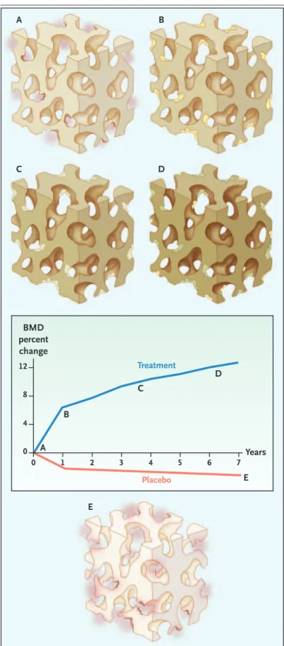
Figure 4. Effects of Antiresorptive Treatment on Bone Remodeling.
Before treatment, rapid remodeling and resorption in each remodeling unit produce numerous deep cavities on trabecular surfaces (colored pink) (Panel A and point A in graph). Treatment with antiresorptive agents suppresses the birth of new remodeling units, and re-modeling continues with the deposition of new bone (colored yellow) (Panel B and points A to B in the graph). The newly deposited bone partly maintains bone structure. Slow remodeling during treatment per-mits more complete secondary mineralization of the newly deposited bone and the rest of the bone (darker color) (Panels C and D), which slowly increases bone mineral density (BMD) for years (points B to D in the graph). Remodeling continues, but fewer and more shallow resorption cavities (colored yellow) remove less bone (Panel D). In the placebo group, rapid re-modeling reduces tissue mineral density (colored light gray), and deep resorption in each cavity produces tra-becular thinning, perforation, and a loss of connectivi-ty (colored pink), with a decrease in BMD (Panel E and points A to E in the graph).
BMD percent change Treatment Placebo Years 0 0 1 2 3 4 5 6 7 4 8 12 A A B C D B E E C D
stimulate periosteal apposition in growing ani-mals (Fig. 5). Evidence in adult subjects is limit-ed.84
Most of the increase in cortical and trabecu-lar thickness, as well as the improved trabecutrabecu-lar
structure induced by parathyroid hormone, is caused by bone formation on the inner surfaces adjacent to marrow. Although seemingly more ap-propriate in patients with a reduced rate of re-modeling and bone formation, the anabolic action may require bone resorption85
; efficacy in the pre-vention of fracture appears to be similar in per-sons with low rates and those with high rates of remodeling.86
There is no evidence that fracture rates are reduced more by combined therapy with antiresorptive agents and parathyroid hormone than by either therapy alone. Previous use of anti-resorptive agents does not seem to influence the eventual response to the hormone.
Advances in noninvasive techniques are likely to provide insights into the effects of these thera-peutic agents on bone structure and increasingly accurate information concerning the structural heterogeneity of bone fragility from patient to patient and so may improve the sensitivity of the prediction of fracture risk.87
Evidence of this is suggested by the finding that patients with low bone mass, high remodeling rates, and a preva-lent fracture have an increased fracture risk. When the absolute risk of fracture is high, more patients who are exposed to treatment actually benefit from it because a greater proportion of these high-risk patients have fractures.88,89
C o n c l u s i o n s
The purpose of bone modeling and remodeling throughout life is to adapt the material composi-tion and structure of bone to prevailing loads. During growth, these processes fashion a struc-ture able to accommodate Herculean loads and
Baseline Final
Parathyroid hormone Risedronate No treatment
Figure 5. Effects of Treatment with Anabolic or Anti-resorptive Agents.
Microcomputed tomography of biopsy specimens of the iliac crest at baseline and after three years in an untreated control subject shows cortical and trabecular thinning and a loss of connectivity (Panel A). In Panel B, treatment with the antiresorptive agent risedronate maintains structure. (microcomputed tomography in Panels A and B courtesy of B. Borah, Procter & Gamble Pharmaceuticals.) In Panel C, treatment with parathy-roid hormone, an anabolic agent, promotes the depo-sition of bone and cortical and trabecular thickening, as shown on microcomputed tomography of biopsy specimens of the iliac crest at baseline and after 18
months of treatment. (Reprinted from Jiang et al.8 4
maintain its strength by adapting one trait to compensate for a defect in another. Advancing age is accompanied by accumulating abnormalities in this cellular machinery, hormonal deficiency and excess, deficiency and excess of local growth factors, declining muscle mass and mobility, nutri-tional deficiencies, and other factors that over-whelm the declining ability of the remodeling machinery to adapt bone to prevailing loads. Ab-normalities in the balance and rate of remodel-ing and limits to periosteal apposition compromise the material composition and structural design
of bone so that it is no longer “just right” for the loads it must endure. Bone fragility is the conse-quence of failed adaptation. Why bones become fragile is a problem of cell biology. How and when bones fail is a problem of biomechanical engineer-ing. The solution to the problem of structural fail-ure requires a study of the qualities of bone and the cellular mechanisms maintaining these qual-ities from region to region in the body.
No potential conflict of interest relevant to this article was reported.
We are indebted to T.J. Martin of the University of Melbourne for his advice and critical comments.
Refe renc e s
Seeman E. From density to structure: growing up and growing old on the sur-faces of bone. J Bone Miner Res 1997;12: 509-21.
Currey JD. Bones: structure and me-chanics. Princeton, N.J.: Princeton Uni-versity Press, 2002:1-380.
Wainwright SA, Biggs WD, Currey JD, Gosline JM. Mechanical design in organ-isms. Princeton, N.J.: Princeton University Press, 1982:1-436.
Currey JD. The mechanical conse-quences of variation in the mineral con-tent of bone. J Biomech 1969;2:1-11.
Viguet-Carrin S, Garnero P, Delmas PD. The role of collagen in bone strength. Osteoporos Int 2006;17:319-36.
Garnero P, Cloos P, Sornay-Rendu E, Qvist P, Delmas PD. Type I collagen racemi-zation and isomeriracemi-zation and the risk of fracture in postmenopausal women: the OFELY prospective study. J Bone Miner Res 2002;17:826-33.
Bailey AJ, Sims TJ, Ebbesen EN, Man-sell JP, Thomsen JS, Mosekilde L. Age-related changes in the biochemical prop-erties of human cancellous bone collagen: relationship to bone strength. Calcif Tis-sue Int 1999;65:203-10.
Banse X, Sims TJ, Bailey AJ. Mechani-cal properties of adult vertebral cancellous bone: correlation with collagen intermo-lecular cross-links. J Bone Miner Res 2002; 17:1621-8.
Yeni YN, Brown CU, Wang Z, Norman TL. The influence of bone morphology on fracture toughness of the human femur and tibia. Bone 1997;21:453-9.
Marotti G. The structure of bone tis-sues and the cellular control of their depo-sition. Ital J Anat Embryol 1996;101:25-79.
Nalla RK, Kruzic JJ, Kinney JH, Ritchie RO. Effect of aging on the toughness of human cortical bone: evaluation by R-curves. Bone 2004;35:1240-6.
Qiu S, Rao DS, Fyhrie DP, Palnitkar S, Parfitt AM. The morphological association between microcracks and osteocyte lacu-nae in human cortical bone. Bone 2005;37: 10-5. 1. 2. 3. 4. 5. 6. 7. 8. 9. 10. 11. 12.
Ruff CB, Hayes WC. Sex differences in age-related remodeling of the femur and tibia. J Orthop Res 1988;6:886-96.
Duan Y, Wang XF, Evans A, Seeman E. Structural and biomechanical basis of ra-cial and sex differences in vertebral fragil-ity in Chinese and Caucasians. Bone 2005; 36:987-98.
Wang XF, Duan Y, Beck T, Seeman ER. Varying contributions of growth and age-ing to racial and sex differences in femoral neck structure and strength in old age. Bone 2005;36:978-86. [Erratum, Bone 2005; 37:599.]
Theobald TM, Cauley JA, Gluer CC, Bunker CH, Ukoli FAM, Genant HK. Black-white differences in hip geometry. Osteo-poros Int 1998;8:61-7.
Nieves JW, Formica C, Ruffing J, et al. Males have a larger skeletal size and bone mass than females, despite comparable body size. J Bone Miner Res 2005;20:529-35.
Zebaze RMD, Jones A, Welsh F, Knack-stedt M, Seeman E. Femoral neck shape and the spatial distribution of its mineral mass varies with its size: clinical and bio-mechanical implications. Bone 2005;37: 243-52.
Ohman JC, Krochta TJ, Lovejoy CO, Mensforth RP, Latimer B. Cortical bone distribution in the femoral neck of homi-noids: implications for the locomotion of Australopithecus afarensis. Am J Phys An-thropol 1997;104:117-31.
Turner CH. Biomechanics of bone: de-terminants of skeletal fragility and bone quality. Osteoporos Int 2002;13:97-104.
Gilsanz V, Roe TF, Mora S, Costin G, Goodman WG. Changes in vertebral bone density in black girls and white girls dur-ing childhood and puberty. N Engl J Med 1991;325:1597-600.
Gilsanz V, Gibbens DT, Roe TF, et al. Vertebral bone density in children: effect of puberty. Radiology 1988;166:847-50.
Han Z-H, Palnitkar S, Rao DS, Nelson D, Parfitt AM. Effect of ethnicity and age or menopause on the structure and geom-etry of iliac bone. J Bone Miner Res 1996;
13. 14. 15. 16. 17. 18. 19. 20. 21. 22. 23.
11:1967-75. [Erratum, J Bone Miner Res 1997;12:867.]
Murray PDF, Huxley JS. Self-differen-tiation in the grafted limb bud of the chick. J Anat 1925;59:379-84.
Frost HM. The laws of bone structure. Springfield, Ill.: Charles C Thomas, 1964.
Bonadio J, Jepsen KJ, Mansoura MK, Jaenisch R, Kuhn JL, Goldstein SA. A mu-rine skeletal adaptation that significantly increases cortical bone mechanical prop-erties: implications for human skeletal fragility. J Clin Invest 1993;92:1697-705.
Kozloff KM, Carden A, Bergwitz C, et al. Brittle IV mouse model for osteogen-esis imperfecta IV demonstrates postpu-bertal adaptations to improve whole bone strength. J Bone Miner Res 2004;19:614-22.
McBride DJ Jr, Shapiro JR, Dunn MG. Bone geometry and strength measurements in aging mice with the oim mutation. Cal-cif Tissue Int 1998;62:172-6.
Orwoll ES. Toward an expanded under-standing of the role of the periosteum in skeletal health. J Bone Miner Res 2003; 18:949-54.
Parfitt AM. Skeletal heterogeneity and the purposes of bone remodelling: impli-cations for the understanding of osteopo-rosis. In: Marcus R, Feldman D, Kelsey J, eds. Osteoporosis. San Diego, Calif.: Aca-demic Press, 1996:315-39.
Idem. Targeted and non-targeted bone remodeling: relationship to basic multi-cellular unit origination and progression. Bone 2002;30:5-7.
Verborgt O, Gibson GJ, Schaffler MB. Loss of osteocyte integrity in association with microdamage and bone remodeling after fatigue damage in vivo. J Bone Miner Res 2000;15:60-7.
Han Y, Cowin SC, Schaffler MB, Wein-baum S. Mechanotransduction and strain amplification in osteocyte cell processes. Proc Natl Acad Sci U S A 2004;101:16689-94.
Hazenberg JG, Taylor D, Lee TC. Crack opening and shear displacements cause damage to cell processes: is this the cel-lular transducer? In: Proceedings book of
24. 25. 26. 27. 28. 29. 30. 31. 32. 33. 34.
the 14th Conference of the European Soci-ety of Biomechanics, ’s Hertogenbosch, the Netherlands, July 4–7, 2004. abstract.
Taylor D. Bone maintenance and re-modeling: a control system based on fatigue damage. J Orthop Res 1997;15:601-6.
O’Brien CA, Jia D, Plotkin LI, et al. Glu-cocorticoids act directly on osteoblasts and osteocytes to induce their apoptosis and reduce bone formation and strength. En-docrinology 2004;145:1835-41.
Schaffler MB, Majeska RJ. Role of the osteocyte in mechanotransduction and skeletal fragility. In: Proceedings of the NIAMS–ASBMR scientific meeting, Bone quality: what is it and can we measure it? Bethesda, Md., May 2–3, 2005. abstract.
Hauge EM, Qvesel D, Eriksen EF, Mosekilde L, Melsen F. Cancellous bone remodeling occurs in specialized compart-ments lined by cells expressing osteoblas-tic markers. J Bone Miner Res 2001;16:1575- 82.
Martin TJ, Sims NA. Osteoclast-derived activity in the coupling of bone formation to resorption. Trends Mol Med 2005;11:76-81.
Lorenzo J. Interactions between im-mune and bone cells: new insights with many remaining questions. J Clin Invest 2000;106:749-52.
Parfitt AM, Travers R, Rauch F, Glorieux FH. Structural and cellular changes during bone growth in healthy children. Bone 2000;27:487-94.
Lips P, Courpron P, Meunier PJ. Mean wall thickness of trabecular bone packets in the human iliac crest: changes with age. Calcif Tissue Res 1978;26:13-7.
Vedi S, Compston JE, Webb A, Tighe JR. Histomorphometric analysis of dynam-ic parameters of trabecular bone forma-tion in the iliac crest of normal British sub-jects. Metab Bone Dis Relat Res 1983-84; 5:69-74.
Nishida S, Endo N, Yamagiwa H, Tani-zawa T, Takahashi HE. Number of osteo-progenitor cells in human bone marrow markedly decreases after skeletal matu-ration. J Bone Miner Metab 1999;17:171-7. Stenderup K, Justesen J, Eriksen EF, Rattan SI, Kassem M. Number and prolif-erative capacity of osteogenic stem cells are maintained during aging and in patients with osteoporosis. J Bone Miner Res 2001; 16:1120-9.
Oreffo RO, Bord S, Triffitt JT. Skeletal progenitor cells and ageing human popu-lations. Clin Sci (Lond) 1998;94:549-55.
Gilsanz V, Gibbens DT, Carlson M, Boe-chat I, Cann CE, Schulz EE. Peak trabecu-lar bone density: a comparison of adoles-cent and adult females. Calcif Tissue Int 1988;43:260-2.
Delmas PD. The use of biochemical markers in the evaluation of fracture risk and treatment response. Osteoporos Int 2000;11:Suppl 1:S5-S6. 35. 36. 37. 38. 39. 40. 41. 42. 43. 44. 45. 46. 47. 48.
Boivin G, Lips P, Ott SM, et al. Contri-bution of raloxifene and calcium and vita-min D3 supplementation to the increase of the degree of mineralization of bone in postmenopausal women. J Clin Endocri-nol Metab 2003;88:4199-205.
Boivin G, Meunier PJ. Changes in bone remodeling rate influence the degree of mineralization of bone. Connect Tissue Res 2002;43:535-7.
Manolagas SC. Birth and death of bone cells: basic regulatory mechanisms and im-plications for the pathogenesis and treat-ment of osteoporosis. Endocr Rev 2000;21: 115-37.
Parfitt AM, Mathews CHE, Villanueva AR, Kleerehoper M, Frame B, Rao DS. Rela-tionship between surface, volume, and thick-ness of iliac trabecular bone in aging and in osteoporosis: implications for the micro-anatomic and cellular mechanism of bone loss. J Clin Invest 1983;72:1396-409.
van der Linden JC, Homminga J, Ver-haar JAN, Weinans H. Mechanical conse-quences of bone loss in cancellous bone. J Bone Miner Res 2001;16:457-65.
Aaron JE, Makins NB, Sagreiya K. The microanatomy of trabecular bone loss in normal aging men and women. Clin Or-thop Relat Res 1987;215:260-71.
Khosla S, Melton LJ III, Atkinson EJ, O’Fallon WM. Relationship of serum sex steroid levels to longitudinal changes in bone density in young versus elderly men. J Clin Endocrinol Metab 2001;86:3555-61.
Legrand E, Chappard D, Pascaretti C, et al. Trabecular bone microarchitecture, bone mineral density, and vertebral frac-tures in male osteoporosis. J Bone Miner Res 2000;15:13-9.
Aaron JE, Shore PA, Shore RC, Bene-ton M, Kanis JA. Trabecular architecture in women and men of similar bone mass with and without vertebral fracture: II. Three-dimensional histology. Bone 2000; 27:277-82.
Brown JP, Delmas PD, Arlot M, Meunier PJ. Active bone turnover of the cortico-endosteal envelope in postmenopausal os-teoporosis. J Clin Endocrinol Metab 1987; 64:954-9.
Foldes J, Parfitt AM, Shih M-S, Rao DS, Kleerekoper M. Structural and geometric changes in iliac bone: relationship to nor-mal aging and osteoporosis. J Bone Miner Res 1991;6:759-66.
Balena R, Shih M-S, Parfitt AM. Bone resorption and formation on the perios-teal envelope of the ilium: a histomorpho-metric study in healthy women. J Bone Miner Res 1992;7:1475-82.
Seeman E. Periosteal bone formation — a neglected determinant of bone strength. N Engl J Med 2003;349:320-3.
Ahlborg HG, Johnell O, Turner CH, Rannevik G, Karlsson MK. Bone loss and bone size after menopause. N Engl J Med 2003;349:327-34. 49. 50. 51. 52. 53. 54. 55. 56. 57. 58. 59. 60. 61. 62.
Ruff CB, Trinkaus E, Walker A, Lar-sen CS. Postcranial robusticity in Homo. I: Temporal trends and mechanical inter-pretation. Am J Phys Anthropol 1993;91: 21-53.
Duan Y, Beck TJ, Wang X-F, Seeman E. Structural and biomechanical basis of sex-ual dimorphism in femoral neck fragility has its origins in growth and aging. J Bone Miner Res 2003;18:1766-74.
Riggs BL, Melton LJ III, Robb RA, et al. A population-based study of age and sex differences in bone volumetric density, size, geometry and structure at different skeletal sites. J Bone Miner Res 2004;19:1945-54.
Albright F, Smith PH, Richardson AM. Postmenopausal osteoporosis. JAMA 1941; 116:2465-74.
Seeman E, Hopper JL, Bach LA, et al. Reduced bone mass in daughters of wom-en with osteoporosis. N Engl J Med 1989; 320:554-8.
Duan Y, Turner CH, Kim BT, Seeman E. Sexual dimorphism in vertebral fragil-ity is more the result of gender differences in age-related bone gain than bone loss. J Bone Miner Res 2001;16:2267-75.
Ciarelli TE, Fyhrie DP, Parfitt AM. Ef-fects of vertebral bone fragility and bone formation rate on the mineralization lev-els of cancellous bone from white females. Bone 2003;32:311-5.
Qiu S, Rao RD, Palnitkar S, Parfitt AM. Reduced iliac cancellous osteocyte densi-ty in patients with osteoporotic vertebral fracture. J Bone Miner Res 2003;18:1657-63.
Eriksen EF, Hodgson SF, Eastell R, Cedel SL, O’Fallon WM, Riggs BL. Can-cellous bone remodeling in type I (post-menopausal) osteoporosis: quantitative assessment of rates of formation, resorp-tion, and bone loss at tissue and cellular levels. J Bone Miner Res 1990;5:311-9.
Arlot ME, Delmas PD, Chappard D, Meunier PJ. Trabecular and endocortical bone remodeling in postmenopausal osteo-porosis: comparison with normal post-menopausal women. Osteoporos Int 1990;1: 41-9.
Brown JP, Delmas PD, Malaval L, Ed-ouard C, Chapuy MC, Meunier PJ. Serum bone Gla-protein: a specific marker for bone formation in postmenopausal osteoporo-sis. Lancet 1984;1:1091-3.
Delmas PD. How does antiresorptive therapy decrease the risk of fracture in women with osteoporosis? Bone 2000;27: 1-3.
Parfitt AM. Morphologic basis of bone mineral measurements: transient and steady-state effects of treatment in os-teoporosis. Miner Electrolyte Metab 1980;4: 273-87.
Borah B, Dufresne TE, Chmielewski PA, Johnson TD, Chines A, Manhart MD. Risedronate preserves bone architecture in postmenopausal women with
osteopo-63. 64. 65. 66. 67. 68. 69. 70. 71. 72. 73. 74. 75. 76.
rosis as measured by three-dimensional microcomputed tomography. Bone 2004;34: 736-46.
Meunier PJ, Boivin GY. Bone mineral density reflects bone mass but also the de-gree of mineralization of bone: therapeu-tic implications. Bone 1997;21:373-7.
Boivin GY, Chavassieux PM, Santora AC, Yates J, Meunier PJ. Alendronate increas-es bone strength by increasing the mean degree of mineralization of bone tissue in osteoporotic women. Bone 2000;27:687-94.
Mashiba T, Hirano T, Turner CH, For-wood MR, Johnston CC, Burr DB. Suppressed bone turnover by bisphosphonates increas-es microdamage accumulation and reducincreas-es some biomechanical properties in dog rib. J Bone Miner Res 2000;15:613-20.
Li J, Mashiba T, Burr DB. Bisphospho-nate treatment suppresses not only sto-chastic remodeling but also the targeted repair of microdamage. Calcif Tissue Int 2001;69:281-6.
77.
78.
79.
80.
Komatsubara S, Mori S, Mashiba T, et al. Long-term treatment of incadronate disodium accumulates microdamage but improves the trabecular bone microarchi-tecture in dog vertebra. J Bone Miner Res 2003;18:512-20.
Whyte MP, Wenkert D, Clements KL, McAlister WH, Mumm S. Bisphosphonate-induced osteopetrosis. N Engl J Med 2003; 349:457-63.
Odvina CV, Zerwekh JE, Rao DS, Maalouf N, Gottschalk FA, Pak CY. Severely suppressed bone turnover: a potential com-plication of alendronate therapy. J Clin Endocrinol Metab 2005;90:1294-301.
Jiang Y, Zhao JJ, Mitlak BH, Wang O, Genant HK, Eriksen EF. Recombinant hu-man parathyroid hormone (1-34) [teripa-ratide] improves both cortical and cancel-lous bone structure. J Bone Miner Res 2003; 18:1932-41.
Martin TJ. Does bone resorption inhi-bition affect the anabolic response to
81.
82.
83.
84.
85.
parathyroid hormone? Trends Endocrinol Metab 2004;15:49-50.
Delmas PD, Licata AA, Reginster JY, et al. Fracture risk reduction during treat-ment with teriparatide is independent of pretreatment bone turnover. Bone (in press).
Boutroy S, Bouxsein ML, Munoz F, Delmas PD. In vivo assessment of trabec-ular bone microarchitecture by high-reso-lution peripheral quantitative computed tomography. J Clin Endocrinol Metab 2005; 90:6508-15.
Sornay-Rendu E, Munoz F, Garnero P, Duboeuf F, Delmas PD. Identification of osteopenic women at high risk of frac-ture: the OFELY study. J Bone Miner Res 2005;20:1813-9.
Laupacis A, Sackett DL, Roberts RS. An assessment of clinically useful mea-sures of the consequences of treatment. N Engl J Med 1988;318:1728-33. Copyright © 2006 Massachusetts Medical Society. 86.
87.
88.
89.
VIEWCURRENTJOBPOSTINGSATTHENEJMCAREERCENTER
Visit our online CareerCenter for physicians at www.nejmjobs.org to see the expanded features and services available. Physicians can conduct a quick search
of the public data base by specialty and view hundreds of current openings that are updated daily online