DISSERTATION ON
A STUDY ON THYROID FUNCTION TESTS IN
CHRONIC KIDNEY DISEASE PATIENTS
Dissertation Submitted To
THE TAMILNADU Dr. M.G.R MEDICAL UNIVERSITY,
In partial fulfillment of therules and regulations, for the award of the
M.D. DEGREE IN GENERAL MEDICINE
BRANCH – I
THANJAVUR MEDICAL COLLEGE
THANJAVUR – 613004
THE TAMILNADU Dr. M.G.R. MEDICAL UNIVERSITY
CHENNAI – 600032
CERTIFICATE
This is to certify that this dissertation entitled “
A STUDY ON
THYROID FUNCTION TESTS IN CHRONIC KIDNEY DISEASE
PATIENTS”
is the bonafide original work of
Dr. LOGESH M.R
in partial
fulfillment of the requirements for M.D Branch 1 (General Medicine )
examination of The Tamilnadu Dr M.G.R Medical University to be held in
April 2017. The period of study was from 2016 January to 2016 June.
Prof.Dr.C.Paranthakan.M.D
Prof.Dr.C.Ganesan.M.D
Unit Chief Head of the Department
Department of Internal Medicine
Department of Internal Medicine
Thanjavur Medical College Thanjavur Medical College
Thanjavur – 613004
Thanjavur – 613004
Prof.Dr.M.Vanithamani .M.S,Mch
Dean
Thanjavur Medical College
Thanjavur- 613004
CERTIFICATE BY THE GUIDE
Certified that the thesis entitled “
A STUDY ON THYROID
FUNCTION TESTS IN CHRONIC KIDNEY DISEASE PATIENTS”
has been carried out by
Dr. LOGESH M.R
, under my direct supervision
and guidance. All the observations and conclusions have been made by the
candidate himself and have been checked by me periodically.
Place: Thanjavur
Date :
Prof.Dr.C.Paranthakan.M.D
Professor and Unit Chief
Department Of Internal Medicine
Thanjavur Medical College
DECLARATION BY THE CANDIDATE
I ,
Dr. LOGESH M.R
, solemnly declare that the dissertation titled “
A
STUDY ON THYROID FUNCTION TESTS IN CHRONIC KIDNEY
DISEASE PATIENTS”
is a bonafide work done by me at Thanjavur
Medical College , Thanjavur during
2016 JANUARY TO 2016 JUNE
under the guidance and supervision of
Prof.Dr.C.Paranthakan.M.D.
, Unit
Chief, Department Of Internal Medicine, Thanjavur Medical College,
Thanjavur . This dissertation is submitted to Dr. M.G.R Medical University,
Tamilnadu towards the partial fulfilment of requirement for the award of
M.D. Degree (Branch -1) in General Medicine
Place :Thanjavur
Date :
Dr. LOGESH, M.R
Post graduate in General Medicine
Thanjavur Medical College
Submission author: Assignment title: Submission title: File name: File size: Page count: Word count: Character count: Submission date: Submission ID:
Digital Receipt
This receipt acknowledges that Turnitin received your paper. Below you will f ind the receipt inf ormation regarding your submission.
The f irst page of your submissions is displayed below.
201411207 Md Genmed Logesh.M.R 2015-2015 plagiarism
A study on thyroid f unction tests in… f inal.docx 1.12M 78 7,338 42,054 16-Sep-2016 04:15PM 705356133
ACKNOWLEDGEMENT
I would like to express my gratitude to the Dean,
PROF Dr. M.VANITHAMANI, M.S.,M.Ch., Thanjavur Medical College, Thanjavur for giving me permission to do the dissertation and utilize the institutional facilities .
I acknowledge my heartfelt thanks to PROF. Dr. C GANESAN, M.D.,
Head Of The Department, Department Of Internal Medicine, Thanjavur Medical College, for his generous help and guidance throughout my study and post graduate period.
I profusely thank PROF Dr.C.PARANTHAKAN M.D., my Professor and Unit Chief, who is my guide for this dissertation, for his valuable criticism, suggestions and fully fledged support during the preparation of this dissertation.
I profusely thank PROF Dr.K.NAGARAJAN, M.D., who is also my guide for this dissertation.
I also express my sincere thanks to Dr. V.P.KANNAN. M.D, (Registrar) for his guidance and support which helped me finish this dissertation .
I am deeply indebted to the Assistant Professor
Dr. A.GUNASEKARAN, M.D.,D.M., for motivating and encouraging me.
I would like to gratefully acknowledge the assistance rendered by Nephrology Assistant Professor Dr.V.RAJAKUMAR M.D., D.M., and who helped me perform this study.
Last but not the least, I also thank all my patients for their cooperation and patience without whom this study would not have been completed. A special mention to my family and friends for their unfailing support.
ABBREVIATIONS
CKD - CHRONIC KIDNEY DISEASE
ESRD - END STAGE RENAL DISEASE
eGFR - ESTIMATED GLOMERULA FLTRATION RATE
TSH - THIROD STIMULATING HORMONE
T3 - TRIIODOTHYRONINE
T4 - THYROXINE
FT4 - FREE THYROXINE
MIT - MONOIODOTYROSINE
CONTENTS
NUMBER
CHAPTER
PAGE
NUMBER
1
INTRODUCTION
1
2
AIM AND OBJECTIVES
5
3
REVIEW OF LITERATURE
6
4
MATERIALS AND METHODS
50
5
STATISTICAL ANALYSIS
54
6
RESULTS
55
7
DISCUSSION
73
8
LIMITATIONS OF THE STUDY
76
9
STRENGTH OF THE STUDY
77
10
CONCLUSION
78
11
BIBLIOGRAPHY
79
12
ANNEXURE
1.PROFORMA
2. CONSENT FORM
3. INFORMATION SHEET
4. MASTER CHART
INTRODUCTION
Maintenance of a balance or homeostasis of metabolic functions in all body organs is critical in the humans . This balance is achieved by actions of hormones. Thyroid hormones affects growth and differentiation of cells and modulate important functions in virtually all cells, tissues and organs.
kidney is involved in metabolic waste excretion, maintenance of fluid and acid base balance by regulating the concentration of hydrogen, sodium, potassium, phosphate and other ions in the extracellular fluid, secretion and metabolism of hormones which are important in haemodynamic control, erythrocyte production and metabolism of various minerals.
Chronic Kidney Disease (CKD) is a worldwide public health and health care problem with an increasing prevalence, poor outcomes, mortality and high cost.
Chronic Kidney disease (CKD) includes a spectrum of different pathologic and physiologic processes which are associated with progressive decrease in GFR.
It is a disease process that is associated with early death of patients and reduced quality of life. A trend towards an raise in its occurance and prevalence has been reported worldwide, as epidemics in some countries and regions.
Adverse effects of CKD such as renal failure, cardiovascular disease and mortality can be prevented or delayed. Early stages of CKD can be screened through laboratory testing and if treated effectively, it would slowdown progression towards kidney failure and cardiovascular disease.
Improving the future of CKD patients would require a co-ordinated approach for prevention of adverse effects by identifying the disease at early stages, determine its burden on the community, identify risk factors, treatment of at risk populations.
Chronic kidney disease is a clinical syndrome characterized by irreversible dysfunction of kidneys that ultimately results in excretory, metabolic and synthetic failure that leads to death without renal replacement therapy.
The complex interaction of the kidney and thyroid gland is well studied. Kidney metabolizes and eliminates thyroid hormones in humans. The growth and development of kidney, water and electrolyte homeostasis requires action of thyroid hormones.
The knowledge of metabolic and hormonal abnormalities in milder degrees of renal dysfunction is growing but only little is known about thyroid dysfunction in CKD patients. The literature available regarding thyroid dysfunction in chronic disease on conservative management is still low.
Patients with advanced CKD has symptoms like easyfatigueability, lethargy, cognitive decline and sexual dysfunction, that considerably overlaps with hypothyroidism but only a few studies are done so far about the prevalence or severity of thyroid dysfunction in patients with CKD.
Thyroid hormones are important for growth and differentiation, and modulation of physiological functions in all the tissues including the kidney. They also play a vital role in water and electrolyte homeostasis.
Either hypo or hyperthyroidism is accompanied by modulations in water and electrolytes metabolism , as well as cardiovascular function. kidney is a target organ for thyroid hormone and for the metabolism and elimination of hormones.
Decline in kidney function is associated with abnormalities in the thyroid hormone functions. CKD affects both HPT axis and thyroid hormone’s catabolism. These effects of reduced renal function may lead to hypothyroidism, hyperthyroidism and non-thyroidal illness which are associated with cardiovascular dysfunction which will have adverse effects on the prognosis of CKD.
The importance of knowing the prevalence of thyroid dysfunction in CKD patients lies in the fact that it adds to already high cardiovascular mortality risk in this patients group.
This original research was under taken to study the thyroid dysfunction that occurs in CKD patients not on maintenance dialysis.
AIMS AND OBJECTIVES
To study thyroid function tests in chronic kidney disease patients on conservative management.
To study the proportion of thyroid dysfunction in chronic kidney disease patients on conservative management.
To study the correlation between the severity of renal dysfunction with thyroid abnormality.
REVIEW OF LITERAURE
REVIEW ANATOMY & PHYSIOLOGY: GROSS ANATOMY:
Kidneys are two in number located in the retroperitoneal space. The Kidneys extended vertically from T12 to L3 vertebra.
DIMENSIONS:
Each adult Human Kidney weighs about 130 to 180 grams in males and 110 to 160 grams in females. It is 10 to 13 cm in length, 5 to 8 cm in breadth and 2 to 3.0 cm thick.
MICROSCOPIC ANATOMY:
The complex structure of mammalian kidney can be simplified as a unipapillary model. The unipapillary kidney consists of a cortex and medulla. The kidney of humans contains about 1 million nephrons.
NEPHRONS:
Main segments of nephron include glomerulus, renal tebule and collecting duct.
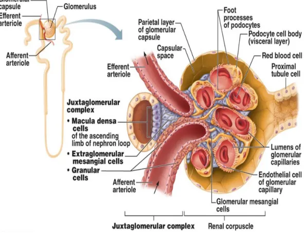
GLOMERULUS:
The glomerulus has special capillaries to mesangium, which is enclosed in the glomerular (Bowman) capsule. The epithelial cells (podocytes), covers the mesangium and capillaries forming the visceral epithelium of bowman capsule.
Between glomerular capillaries, mesangium and the podocytes layer, the glomerular basement membrane (GBM) develops. On entering the glomerular tuft, the afferent arteriole divides and forms an anatomizing capillary network representing a glomerular lobule.
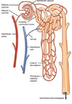
RENAL TUBULE:
The real tubule comprises of convoluted tubules proximal and distal , henle’s loop, the collecting duct. The henle’ loop comprises of thin descending and ascending limbs and thick ascending limb. Two different pathways exist. A transcelluar pathway across the luminal, basolateral memberanes, and cytoplasm and paracelluar pathway through the junctional and intercellular spaces.
COLLECTING DUCT:
The collecting duct includes cortical, outer & inner medullary collecting duct. Principal (light) cells and intercalated (dark) cells are the two types of cells in collecting duct. The collecting duct plays crucial role in handling Na+, Cl- and K+ and acid in response to vasopressin can or dilute urine.
MICROVASCULATURE:
The renal artery divides into interlobar arteries after entering the renal sinus. At cortico medullary junction, the interlobar arteries divide into the arcuate arteries, which branch into cortical radial arteries.
Afferent arterioles supply the glomerular tufts and generally arise from cortical radial arteries. The blood supply of the peritubular capillaries is mainly postglomerular.
The efferent arterioles drain the glomerulus. Cortical efferent arterioles, from superficial and mid cortical glomeruli, supplies cortex. The efferent arteriole supplies the renal medulla. In outer stripe of the medulla, these vessels divide into the descending vasa recta.
CHRONIC KIDNEY DISEASE
Chronic kidney disease (CKD) entails the presence of kidney damage or decreased level of function for 3 months or more. While no local prevalence data exist, the risk factors such as post-streptococcal glomerulonephritis, hypertension, diabetes and lately HIV associated nephropathy (HIVAN) are on the rise. Recent studies show that early diagnosis allows for institution of therapy to either arrest or reverse progression of the global challenge that CKD has become.
In 2000, the National Kidney Foundation (NKF), and Kidney Disease Outcome Quality Initiative (KDOQI) Advisory Board approved clinical practise guidelines to define CKD and to classify stages in the progression of CKD based on eGFR.
Glomerular diseases such as post-streptococcal glomerulonephritis contribute to a large proportion of early CKD. Chronic pyelonephritis and tuberculosis are notable infectious risk factors of which HIV-associated nephropathy is routinely encountered. Congenital anomaliese.g. polycystic kidney disease and obstructive processes such as calculi are also culpable causative factors. Collagen disease e.g. SLE and vascular diseases such as renal nephrosclerosis may also lead to CKD. Nephrotoxic agents e.g.
aminoglycoside therapy are occasionally implicated. Chronic kidney disease may be a progression from acute renal failure.
GFR is widely accepted as the best measure for renal function in health and disease. Providers and patients are familiar with the concept that “the kidney is like a filter.” The ‘gold standard’ of measuring GFR is by the renal clearance of exogenous markers Inulin, radio-labelled EDTA (51Cr-EDTA) and technecium-labelled diethylene-triaminepentacetate( 99mTc-DTPA) and iohexol.
These tests are however time-consuming, labour intensive,invasive, costly and require specialized equipment restricting their use in routine individual cases monitoring or in large epidemiological studies. While an ideal endogenous marker should meet 3 criteria of complete filtration at the glomerulus, absent tubular secretion and no tubular reabsorption, serum creatinine is widely accepted as an endogenous marker for assessing renal function.
Currently, a spot serum creatinine level is favoured with creatinine-based equations for estimating GFR being employed. They include; the Cockcroft-Gault (CG), the 4-variable Modifications of Diet in Renal Disease (4v-MDRD) and the Mayo Clinic Quadratic formulae.
These equations are used interchangeably to suit various study populations. Most CKD patients tend to progress over time and worsen. The risk of adverse outcomes in CKD can be further stratified by the severity of disease and rate of progression.
The outcomes of CKD include loss of kidney function leading to kidney failure and development of cardiovascular disease.
The evaluation and management of CKD as recommended by the work group includes treatment of co-morbid illness, preventing the loss of kidney failure and delay disease process, preventing and early treatment of cardiovascular disease, preventing and early treatment of complications of decreased renal function and early initiation of dialysis to replace renal function and plan for renal transplantation in possible patients.
Thus CKD is a disease process in which early diagnosis and proper follow up to control risk factors will reduce the progression of renal dysfunction.
ESTIMATION OF KIDNEY FUNCTION:
The current CKD classification is based on eGFR. The Modification of Diet in Renal Disease (MDRD) study equation and the more recently developed Chronic Kidney Disease Epidemiology Collaboration (CKD-EPI) equation are both commonly used equations to calculate eGFR; however, both are relatively inaccurate in reflecting measured glomerular filtration rate (mGFR) above 60 ml/ min/1.73 m2. This underestimation by eGFR of true mGFR can lead to misclassification of a large number of individuals as having CKD stage 3a, whereas in reality their true GFR is above 60 ml/min. Also, the inaccuracy of eGFR to reflect mGFR above 60 ml/min/1.73 m2 makes the distinction between CKD stages 1 and 2 difficult and artificial. The limitations of using creatinine-based estimations have spurred the search for alternative filtration biomarkers, such as β- trace protein (βTP), cystatin C, and β2-microglobulin (β2M)
GLOMERULAR FILTRATION RATE:
MDRD (Modification of Diet in Renal Disease Equation)
GFR (mL / min / 1.73 m3) = (Scr)-1.154 x (Age)-0.203 x (0.742 if African America)
The results of this equation are reported normalized to 1.73 m2 body surface area, which is an accepted adult surface area which eliminates the need for knowing accurate weight and height variables.
RISK FACTOR:
• Age (older)
• Race of ethnicity (Non – Caucasian)
• Genetics
• Birth weight (low)
• Systemic hypertension
• Diabetes mellitus
• Cardiovascular disease
• Albuminuria
• Obesity or metabaolic syndrome
• Dyslipidemia
• Hyperuricemia
• Smoking
• Low socioeconomic status
• Nephrotoxin exposure: non steroidal anti-inflammatory drugs (NSAIDs), lead, traditional herbal use.
PATHOPHYSIOLOGY:
Kidney scarring or fibrosis is a complex, overlapping, multistage phenomenon that could be characterized by a number of processes:
■ An inflammatory response with infiltration of damaged kidneys by extrinsic inflammatory cells (blood borne and bone marrow derived)
■ Activation, proliferation, and loss of intrinsic renal cells (through apoptosis or necrosis and including mesangiolysis and podocytopenia)
■ Activation and proliferation of extracellular matrix (ECM)–producing cells, including myofibroblasts and fibroblasts
■ Deposition of ECM replacing the normal renal architecture
INITIATION OF INJURY:
The underlying initiates the renal damage by specific mechanism (eg., genetically determined abnormalities, immune complex deposition, or toxin exposure) which leads to adaptive changes.
PROGRESSION OF DAMAGE:
The hyperfiltration and hypertrophy of the remaining functioning nephrons occurs in response to injury which in long-term leads to reduction of renal mass, irrespective of underlying aetiology. These maladaptive leads to sclerosis and dropout of the remaining nephrons. This eventually leads to progressive decline in renal function over many years.
RECOMMENDED KIDNEY PROTECTIVE THERAPIES:
LEVEL 1 RECOMMENDATIONS
1. Control blood pressure (BP).
2. Administer ACE inhibitor, ARB , or renin inhibitor.
3. Avoid dihydropyridine calcium channel blockers (DHCCBs) unless needed for BP control
4. Control protein intake.
LEVEL 2 RECOMMENDATIONS
1. Restrict NaCl intake and use diuretics.
3. Control each characteristics of the metabolic syndrome.
4. Administer aldosterone antagonist therapy.
5. Administer allopurinol therapy
6. Control serum phosphorous.
7. Instigate smoking cessation.
8. Perform alkali therapy.
9. Administer β-blocker therapy.
DEFINITION OF CHRONIC KIDNEY DISEASE:
Criteria for CKD (either of he following present for > 3 months) I. Markers of kidney damage (one or more)
• Albuminuria (AER ≥ 30 mg/24 hours; ACR ≥ 30 mg/g)
• Urine sediment abnormalities
• Electrolyte and other abnormalities due to tubular disorders
• Abnormalities detected by histology
• Structural abnormalities detected by imaging
• History of kidney transplantation
II. Decreased GFR – GFR < 60 ml/min/1.73 m2
Abbreviations:
ACR – Albumin to Creatinine Ratio, AER – Albumin Excretion Ratio
STAGING KIDNEY DISEASE: TABLE 1 GFR CATEGORY eGFR (ml/min/1.73 m2) Terminology G1 < 90 Normal to High G2 60 – 89 Mildly decreased
G3a 45 – 59 Mild to moderately decreased
G3b 30 – 44 Moderately to severely decreased
G4 15 – 29 Severely decreased
COMPLICATION OF CKD:
The complication of chronic kidney disease increases with progression of disease. The prevalence of complications in stage 1 CKD is 0.28, and it rises to an average of 1.71in stage 4.
The well know complications of CKD are as follows,
• Anemia
• Bone-mineral disorder
• Metabolic acidosis
• Cardio – vascular risk
• Dyslipidaemia
The thyroid is unique among the endocrine glands by virtue of the large store of hormone it contains the low rate at which the hormones turnover (1 % per day). Iodine is ingested in both inorganic and organic bound forms. Iodine is rapidly and efficiently absorbed from the gastrointestinal tract.
The thyroid gland produces two related hormones, thyroxine (T4) and Triiodothyrone (T3).Acts through nuclear receptors, these hormones play a vital role in cell differentiation and help maintain homeostasis in the adult.
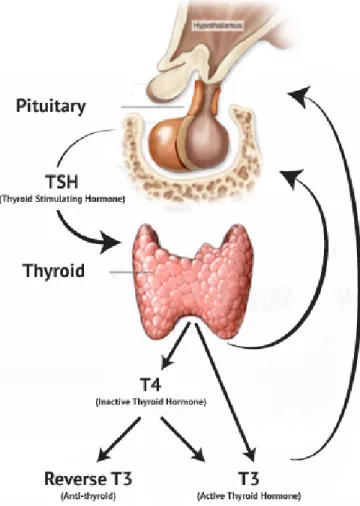
Thyriod stimulating hormone (TSH), secreted by the anterior pituitary is responsible for control of the thyroid axis. Thyroid hormones are derived from thyroglobulin, a glycoprotein, which is iodinated on tyrosine residues.
T4 is secreted from the thyroid gland in about 20 times more than T3. Both hormones bind to proteins in the plasma like thyroid-binding globulin (TBG); transthyretin (TTR, also known as thyroxine-binding prealbumin, or TBPA); and albumin. These proteins increase the levels of circulating hormones, reduce its clearance, and modulate delivery of hormones to various tissue sites. When the effects of various binding proteins are combined, app 99.98% of T4 and 99.7% of T3 are bound to proteins. T4 is converted to T3 by the de-iodinase enzymes.
T4 to T3 conversion is reduced by fasting, systemic illness anda variety of medications including propyl thiouracil, propranolol, amiodarone, glucocorticoids. Type III de-iodinase inactivates T4 and T3 and is the most important source of reverse T3 (rT3)
IODINE TRAPPING:
It is then actively transformed into the thyroid cell which is accomplished by a membrane protein called sodium-iodine symporter (NIS).
ORGANIFICATION:
Oxidation of iodine and incorporation of the resulting intermediate, covert iodine into the hormonally inactive iodotyrosines {Mono iodotyrosines and diiodotyrosine}.
COUPLING:
MIT & DIT are coupled to form T3 whereas two DIT are coupled to form T4. Oxidation, iodination and coupling reactions are mediated by heme-containing protein thyroid peroxidase (TPO).
SECRETION:
The hormone produced binds with thyroglobulin till secreted. In the blood, it is transported in bound and free from. Most common binding proteins are thyroid binding globulin, prealbumin and albumin. T4 is predominantly bound to thyroid binding globulin whereas T3 is predominantly bound to albumin. The other form is free T3 & T4. These free forms are in equilibrium with bound form.
PHERIPHERAL METABOLISM:
In the periphery one third of T4 is converted to T3 by 5’ Deiodenase
and 45% to rT3 by 5 Deiodenase. They are further metabolized to Diiodothyronines. Only about 13% of T3 is produced from thyroid gland and remaining 87% is formed from T4.
FIGURE 3 – HYPOTHALAMIC PITUITARY AXIS
Two major factors control synthesis and release of TSH:
1. T3 level within thyrotropic cells, which regulates mRNA expression, TSH translation.
OTHER FACTORS:
TSH synthesis and release are inhibited by high serum levels of T4 and T3 (hyperthyroidism) and stimulated by low levels of thyroid hormone (hypothyroidism). Somatostatin, dopamine, dopamine agonists (bromocriptine) and glucocortocoids and inhibit TSH secretion. Acute illness can inhibit TSH secretion followed by rebound rise in TSH as the patient recovers.
From a clinical practice viewpoint, both hypothyroidism and hyperthyroidism are accompanied by remarkable alterations in the water and electrolyte metabolism, as well as in cardiovascular function..
The major pathway of thyroid hormone metabolism is by de-iodination and only 25% is conjugated, deaminated or decarboxylated. Although the liver is the major site of metabolism, the kidney also participates to a lesser degree by deamination and decarboxylation. Renal conversion of T4 to T3 and reverse T3 (rT3) also occurs. The kidney also is the most important route for iodine excretion and in renal dysfunction the plasma levels of inorganic iodine are raised.
On the other hand, the different treatments used in management of renal disease and thyroid diseases may be accompanied by adverse events that alter thyroid and kidney function respectively.
The decrease in the activity of Thyroid hormone results in inability to excrete water results in water overload, this effect is due to reduction in the GFR. Thyroid hormones enhance tubular transport of Na+, via their actions on Na/K ATPase and K+ ions in the membrane of proximal tubules.
TH moderate renin release from the juxta glomerular cells through a mechanism that does not involve the ouabain-sensitive sodium pump effects kidney angiotensinase activity.
KIDNEY AND THYROID:
The growth of the kidneys also depends upon adequate thyroid hormones. The kidneys also metabolize and excrete thyroid hormones. Thyroid abnormalities can significantly alter kidney function and fluid & water balance.
EFFECT OF THYROID HORMONES IN RENAL PHYSIOLOGY:
Thyroid hormones influences directly as well as indirectly 1.The direct effect of thyroid hormones on
a.glomerular filtration rate.
b.hormonal influences on renal tubular physiology. c. tubular secretion and re-absorptive processes
2.Indirect effects are through influence of thyroid hormones on renal blood flow and cardiovascular system3
Thyroid hormones affect renal clearance of water,increases the activity of the Na/K ATPase18.
Thyroid hormones also regulates the renin-angiotensin-aldosterone axis40.
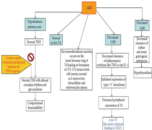
CHRONIC KIDNEY DISEASE AND THYROID:
CRD affects peripheral metabolism of thyroid hormones and hypothalamus-pituitary-thyroid axis19.
URAEMIA AND THYROID:20,23
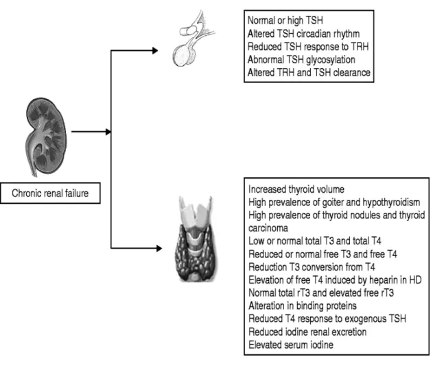
Uraemia causes blunted and delayed response of TSH receptors to TRH and altered circadian rhythm which leads to normal or elevated levels of TSH, indicating pituitary disturbances. The TSH glycosylation is altered in CKD, which compromises its activity23.
Uraemia affects the volume and functions of the thyroid. Women who has uraemia had higher volume of thyroid and increased prevalence of goitre when compared to women with normal renal function. Thyroid carcinoma and thyroid nodules is found to be more common in uremic patients27.
Effects of chronic kidney disease on thyroid hormone function
Chronic kidney disease causes multiple abnormalities in the thyroid physiology. These include the state of chronic illness, malnutrition and negative nitrogen balance, the presence of circulating inhibitors of hormone metabolism and a multitude of hormone alterations. CKD interferes with H-P-T axis and also in TH metabolism.
Uraemia causes dysfunction and alters volume of the thyroid. ckd patients have thyromegaly compared with subjects. There is higher prevalence of goitre, more in women. Nodules and thyroid malignancy are more common in CKD patients when compared to normal population.
THYROXINE
Total T4 levels are either normal or reduced in CKD pts, this is due to reduced binding to the carrier proteins. concentrations of major carrier proteins appears to be normal in ckd, and it is postulated that accumulations of many Uraemic toxins reduce T4 binding to carriers which causes low T4 levels but free t4 levels remains unaffected.
The available test estimates low values of free T4. fT4 measurement uses equilibrium dialysis, dialyzable circulating inhibitors are removed. This inturn raises T4 binding to carriers, resulting in low free T4 levels.
fT4 and total T4 either be normal, or reduced, or sometimes the free T4 be high due to heparin which used during haemodialysis, which inhibits T4 binding.
TRIIODOTHYRONINE (T3)
Total and fT3 levels are reduced in CKD. The low T3 syndrome is the most observed thyroid alteration in ckd patients. This low T3 is caused by decreased peripheral conversion of T4.
Chronic metabolic acidosis is common in CKD. This inturn may further contribute to this reduced conversion. Acumulation of uraemic toxins reduces binding of T3 to serum carrier proteins which leads to reduced T3 levels.
Low fT3 levels in uraemia is interpreted as a response to reduce the energy expenditure of cells and also for minimizing metabolism of proteins.
Also Low T3 is indicator of mal-adaptation contributing to worsening of the ckd.
Many studies have now shown that reduced T3 levels are correlated to inflammatory markers and now an important predictor of death in ckd patients on maintaenance dialysis.
THYROID STIMULATING HORMONE (TSH)
Serum TSH is ether normal or elevated in CKD,
TSH response to its releasing hormone (TRH) is low. These findings suggest some intra thyroidal and pituitary disturbances are associated with uraemia.
Circadian rhythm of TSH secretion and glycosylation of TSH are affected in CKD. Alteration In glycosylation compromise TSH biological action. Both normal nuclear T3 levels and also normal thyroid receptor action in pituitary explains the normal TSH levels.
This Abnormal glycosylation of TSH , altered rhythm of secretion and pulsatile secretion of TSH, and blunted TSH response to TRH are reported in Uraemic patients points to a disordered function at the hypothalamics level and pituitary levels.
Interestingly, in Uraemic patients with primary non-immune hypothyroidism, TSH may increase appropriately.
Free T3 Assay(fT3)
The fT3 test is a solid phase ELISA. Patient samples, standard samples, and T3 -Conjugated Working Reagent are added to wells coated with monoclonal T3 Ab.
fT3 in the patient sample and conjugated T3 competes for the binding sites of the Ab. After 60min period of incubation at 37 degree, the wells are washed thoroughly with water to remove the unbound excess T3 conjugate.
Then H2O2/TMB solution is added and again incubated for another 20 min, till appearance of blue color. The color developed can be stopped by adding 3N HCl, and the sample now is taken for spectro photometry and viewed at 450 nm. The intensity of the color is proportional directly to level of enzymes present and inversely to the level of unlabeled fT3.
Free T4 Assay
The fT4 test is a solid phase ELISA. Patient samples, standard samples, and T4-Conjugated Working Reagent are added to wells coated with monoclonal T4 Ab.
fT4 in the patient sample and conjugated T4 competes for the binding sites of the Ab. After 60min period of incubation at 37 degree, the wells are washed thoroughly with water to remove the unbound excess T4 conjugate.
Then H2O2/TMB solution is added to the wells and again incubated for another 20 min, till appearance of blue color. The color developed can be stopped by adding 3N HCl, and the sample now is taken for spectro-photometry and viewed at 450 nm. The intensity of the color is proportional directly to level of enzymes present and inversely to the level of unlabeled fT4.
.
TSH Assay
TSH assay is a solid phase ELISA.
The assay systems uses the monoclonal Abs directed against the antigenic sites on the intact TSH molecule.
Mouse monoclonal anti-TSH Ab is used for the solid phase of ELISA (microtiter wells), and goat anti-TSH Ab is used in Ab-enzyme (horse-radish peroxidase enzyme) conjugate solution.
The test sample is allowed to react with the Abs, thus the TSH in the sample gets sandwiched between the two, the solid phase and enzyme-Ab complex.
After a incubating at 37 degree for 2 hour, then after shaking well, the solid phase then washed thoroughly with distilled water. This removes all the unbound labeled antibodies and leaves behind the bound TSH.
A solution of tetramethylbenzidine added to the wells and once again incubated for another 20 min, which results in a blue color. This color development can be stopped by adding 1N HCl,
Final yellow color produced after addition of HCL, is now measured for adsorbance spectro-photometrically at 450 nm. The amount of TSH is proportional directly to the intensity of the colour obtained in test sample
CKD AND LOW T3 SYNDROME:
Low T3 syndrome is the most common thyroid dysfunction noted in CKD patients42. Following reasons are said to be responsible for the low T3
syndrome:
1. The conversion of T4 to T3 in peripheral tissues is inhibited By Chronic metabolic acidosis which affects deodination process.
2. (TNF)-a and interleukin (IL)-1 which are abundant in CKD inhibits type 15’-deiodinase thereby preventing the conversion of T4 to T3.
3. Wolff-Chaikoff effect due to impaired renal regulation of iodine which increases serum iodine levels2.
FIGURE 6 - CKD AND THYROID DISORDERS
CLINICAL EFFECTS OF LOW T3 SYNDROME:
The low T3 levels in CKD patients is associated with higher all-cause as well as cardiovascular mortality.
But the clinical importance of T3 syndrome remains controversial and this association is not invariable as demonstrated by recent studies8.
CKD EUTHYROID SICK SYNDROME VS NON-CKD
EUTHYROID SICK SYNDROME:15
In NON-CKD ESS there is increase in rT3 which is characteristically normal in CKD ESS. This can be explained by the fact redistribution of rT3 across vascular space in CKD patients leads to normal rT3 levels. But free rT3 concentrations may be high because of reduced renal clearance20.
HYPOTHYROIDISIM IN RENAL FAILURE:3,34
High prevalence of overt, subclinical hypothyroidism and not hyperthyroidism is found in CKD patients.1,14
The Renal blood flow is decreased in hypothyroidism by decreased CO , raised PVR, increased intrarenal vaso constriction, blunted renal response to vasodilators such as VEGF an IGF-1
Pathologic changes seen in the glomerulus in hypothyroidism are thickening of GBM and mesangial expansion. this may also contribute to reduced RBF in hypothyroidism.
The GFR is reversibly decreased by 40% in >55% of adults with hypothyroidism.
There is reduced sensitivity to β-adrenergic stimulus, there is reduced renin angiotensin II release from kidney which in turn impairs RAAS action, reduction of GFR.
There is also a structural constraint because of limited glomerular filtration surface due to impaired renal parenchymal growth in hypothyroidism.
There is a loss of proximal tubular re-absorption of sodium, chloride, and water. Basolateral Cl channel expression is reduced.
Impaired Cl reabsorption increases the distal Cl delivery, triggering the macula densa mediated feedback mechanism which inturn reduces the RAAS action.
The tubular transport capacity is impaired and the activity of Na/K ATPase is lost in the proximal tubules predominantly and later virtually in almost all segments of the nephron.
Net reduction in sodium and bicarbonate reabsorption, leads to increased loss in urine results in reduced urinary acidification. There is also an inability to maintain the medullary hypertonicity.
Loss of medullary hypertonicity in hypothyroidism results isothenuria.
However, hypothyroidism raise vasopressin sensitivity of CD favouring free water reabsorption in collecting ducts. The reduced GFR, impaired Na reabsorption, and excess ADH secretion and ADH mediated reduced free water clearance, contribute to Hyponatremia.
Hypothyroidism leads to reversible increase in Creatinine and reduction of cystatin c levels.
Thus Hypothyroidism reduces the renal blood flow by variety of mechanisms including negative chronotropic and inotropic effects and deceased response to vasodilators. It also produces pathological changes in renal microanatomy like mesangial expansion and thickening of basement membrane. In 55% of hypothyroid patients GFR is reversibly reduced.
Hypothyroidism also interferes with renal functions including decreased absorption of sodium, chloride and water in proximal convoluted tubule which ultimately leads to fall in GFR by reducing RASS activity.
Low thyroid activity also interferes with urinary acidification and urinary concentration mechanisms by decreased sodium and bicarbonate excretion and abolishing the modularly hyper tonicity respectively. Hypothyroidism leading to hyponatremia is well known fact, the reason for which is reversible increase in ADH sensitivity of collecting ducts.
COMPLICATIONS OF CKD WORSENED BY COEXISTING HYPOTHYROIDISM ARE,
I. Cardiovascular Complications
• Secondary hypertension
• LV failure and pulmonary edema • Accelerated atherosclerosis • Myocardial infarction • Pericarditis
• Uremic cardiomyopathy
Hypothyroidism is associated with raised LDL-c levels due to a reduced LDL receptor formation in liver and impaired clearance of LDL.
II. CNS and neuromuscular complications
• Dementia
• Uremic encephalopathy • Peripheral neuropathy • Proximal muscle weakness
III. Hematological complications like anemia
Hypothyroidism can cause normocytic normochromic, macrocytic anemia or iron deficiency anemia due to menorrhagia.
IV. Electrolyte imbalance like hyponatremia
Hypothyroidism can cause euvolumic hypoosmolar hyponatremia. The hyponatremia in hypothyroidism suggests that the disease is severe- myxedema coma.
V. Fluid overload – edema.
Myxedema may worsen the volume overload state of CRF
CHRONIC KIDNEY DISEASE AND THYROID DYSFUNCTION
Hyperthyroidism accelerate CKD by several mechanisms. hyperthyroidism increase intra-glomerular pressure that leads to glomerular hyperfiltration.
Hyperthyroidism produces proteinuria, which cause direct renal toxicity. Hyperthyroidism cause increased mitochondrial energy metabolism along with down-regulation of superoxide dismutase that leads to the free radical production and kidney damage. Oxidative stress leads to hypertension further to CKD worsening.
RAAS activity is increased that further can induce fibrosis and progression of CKD . anemia exacerbated by hyperthyrodism in CKD patients further adds to the resistance to EPO(recombinant)
Hypothyroidism by the mild to moderate reduction in GFR it cause worsening in CKD. Hypothyroidism if treated appropriately it will cause improvement of GFR.
Primary hypothyroidism is commonly observed in CKD patients. Esthe prevalence of subclinical hypothyroidism increases consistently with decline in GFR.
Most common thyroid abnormality in CKD is “low T3 syndrome” Fasting, acidosis and malnutrition reduces deiodination of iodothyronine, and reduces conversion of T4 to T3. TNF-a and IL-1 inhibit type 1 5′ -deiodinase, affects conversion of T4 to T3.
A prolonged Wolff – Chaikoff effect due to improper handling of iodine by kidneys.
TREATMENT OF THYROID DYSFUNCTION IN CKD:
At present, there are no recommendations available regarding the treatment of thyroid hormone abnormalities in CRF patients.
Low thyroid state (low TT3, TT4, FT3) in uremia acts defence against the protein wasting and if improper attempts to replete the thyroid hormone stores may further worsen malnutrition in CRF patients.
But at the same time, subclinical and clinical hypothyroidism are associated with increased risk of cardiovascular disease and these conditions need treatment with thyroid hormone.
THYROID DYSFUNCTION IN DIALYSIS AND KIDNEY TRANSPLANTATION
Patients on HD in CKD have decreased T3 T4 and raised TSH. This small rise in TSH (6 – 15 mU/l), seen in 30% of CKD patients, are usually not considered to be hypothyroidism.
Total T4 is less, heparin inhibits T4 protein binding, hence freeT4 is raised in CKD patients on heparin dialysis. In CKD patients on HD, compensatory cellular transport of thyroid hormones, maintains the euthyroid state.
Because of these reasons, inspite of serum thyroid hormone levels beng less, supplemental thyroxine should not be startes without marked elevation in TSH level.
Among patients on PD, there is increased prevalence of subclinical hypothyroidism and low T3 syndrome. TBG, T4, and T3 are lost in the PD effluent. There is continuous and marked protein loss during PD, even then TBG is normal.
T4 and T3 losses are minimal(12% and 2%,) and thus are compensated without producing any effects. hence T4 replacement is not needed in PD.
Kidney transplantation reverses the CKD effects on thyroid. The low T3 and T4 reverse after transplant, slowly over 4–5months. In initial months after transplant, there is decrease in T4 to levels lesser than the pre-transplant level, later It rises back to normal.
Post-transplant thyroid volume and fT3 levels correlate well with graft function. low T3 before transplant is associated with risk of graft loss.
MATERIALS AND METHODS
This is a prospective, Cross-sectional study conducted in teaching general hospital, Thanjavur Medical college, Thanjavur from 100 consecutive Chronic kidney disease patients in medical wards and nephron out patient department fulfilling our criteria for inclusion and exclusion and those people with willingness to participate in the study were included. Those who were not willing to give consent for the study & too ill to participate are excluded in our study.
The approval for conducting the study was taken from the institutional ethics committee. After explaining details about the study involved, an informed consent is taken from the concerned participant or representative in case the patient is not able to provide consent.
INCLUSION CRITERIA:
Adult patients who fulfill the criteria for chronic kidney disease and who are on conservative management not undergoing renal replacement therapy.
CRITERIA FOR CHRONIC KIDNEY DISEASE:21
(either of the following present for >3 months ) i) Markers of kidney damage (one or more )
Albuminuria (AER ≥ 30mg/24 hours; ACR ≥ 30 mg/g )
Using sediment abnormalities
Electrolyte and other abnormalities due to tubular disorders
Abnormalities detected by histology
Structural abnormalities detected by imaging
History of kidney transplantation
ii) Decreased GFR - GFR <60ml/min/1.73m2
EXCLUSION CRITERIA :
1. Previously diagnosed cases of hypothyroidism and hyperthyroidism 2. Patients on Hem dialysis or Peritoneal dialysis
3. Other condition like
i) Patient on thyroxin supplementation, iodine containing medications ( amiodarone ) , anti-thyroid drugs.
ii) Acute illness & ICU admission. iii) Recent Surgery or trauma.
Detailed history and examination undertaken with special focus on thyroid & renal system.
The following investigation were done :
1. Urine routine and microscopic examination 2. Peripheral smear
3. Blood urea, serum creatinine and creatinine clearance (MDRD formula )
4. Serum electrolytes including calcium and phosphorous 5. Ultrasound abdomen
6. FNAC in patients presenting with thyroid swelling
7. After selecting patients fulfilling the above criteria blood sample is collected in non heparinised serum bottle and quantitative assessment of serum total T3, Free T4 and serum TSH were done by Electro chemiluminescence immunoassay method
NORMAL VALUES :
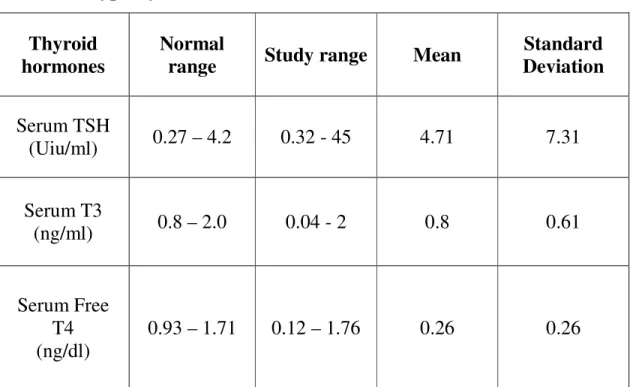
Serum TSH = 0.27 – 4.2 uIU / ml Serum Total T3 = 0.8 – 2.0 ng / ml Serum Free T4 – 0.932 – 1.7 ng / dl
CATEGORISATION OF THYROID ABNORMALITY :11,26,30
LOW T3 SYNDROME-Patients with low serum T3 levels and normal TSH and FT4.
SUBCLINICAL HYPOTHYROIDISM : Patients with Serum TSH >
4.20 and normal FT4 levels.
HYPOTHYRODISM : Patients with Serum TSH > 4.20 and FT4 < 0.93
SUBCLINICAL HYPERTHYROIDISM : Patients with Serum TSH
<0.20 and normal FT4 levels.
STATISTICAL ANALYSIS
The data was collected using a Performa. Data was analyzed for proportion of thyroid dysfunction in chronic disease patients and correlation of thyroid dysfunction with creatinine clearance.
Statistical analysis were performed by “ Statistical Package for Social Sciences (SPSS) 17 software” (SPSS Inc., Chicago, IL ,USA). Pearson’s chi-square test and fisher test used for analysis of qualitative data.
RESULTS
100 CKD patients on conservative management who fulfilled the criteria were studied and the results analyzed.
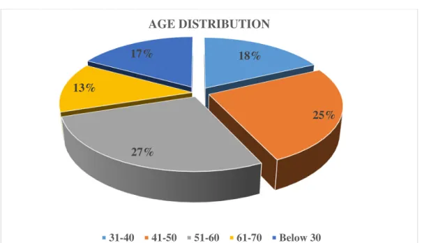
Of 100 patients 66 (66%) were males and 34 (34%) were females. 17 patients (17%) were 30 years and younger and 18 (18%) patients were 60 years and above. 30-60 years age group constituted 65 (65%) patients.
TABLE 2 GENDER DISTRIBUTION Gender Percentage Male 66% Female 34% TABLE 3 AGE DISTRIBUTION
Age in yrs Percentage
31-40 18%
41-50 25%
51-60 27%
61-70 13%
FIGURE 6 : Mean age = 46.86 ± 13.49 3 18% 25% 27% 13% 17% AGE DISTRIBUTION 31-40 41-50 51-60 61-70 Below 30
FIGURE 7:
Male, 66% Female, 34%
GENDER DISTRIBUTION
TABLE 4 CKD STAGES Stages of CKD Percentage Stage 3 18% Stage 4 43% Stage 5 39%
In our study 18 patients (18%) were stage 3 CKD, 43 patients (43%) were stage 4 CKD and 39 patients (39%) belonged to stage 5 CKD. Creatinine clearance varied from 5.5 ti 60mi/min/1.73m2
FIGURE 8 :
18%
43%
39%
CKD STAGES
SERUM CONCENTRATION OF THYROID HORMONES (EXCLUDING HYPOTHYROIDISM) :
Serum TSH varied from 0.32 – 45 with mean of 4.71 and S.D. of 7.31 Serum Free varied from 0.12 – 1.76 with mean of 1.31 and S.D. of 0.26
Serum Total T3 ranged from 0.04 to 2 with mean of 0.8 and S.D. of 0.61
Table 5 : Serum Concentration Of Thyroid Hormones (Excluding Hypothyroidism )
Thyroid hormones
Normal
range Study range Mean
Standard Deviation Serum TSH (Uiu/ml) 0.27 – 4.2 0.32 - 45 4.71 7.31 Serum T3 (ng/ml) 0.8 – 2.0 0.04 - 2 0.8 0.61 Serum Free T4 (ng/dl) 0.93 – 1.71 0.12 – 1.76 0.26 0.26
FIGURE 9 :
Mean and Standard Deviation of Thyroid Hormones
0 2 4 6 8 10 12
Serum TSH Serum T3 Serum free T4
M
ea
n
In present study overall 66 patients (66%) had thyroid dysfunction, 34 patients (34%) had normal thyroid function tests
Figure 10 :Analysis of Thyroid Dysfunction
34 45 16 5 0 5 10 15 20 25 30 35 40 45 50 Normal Low T3 Syndrome Sub Clinical Hypothyroidism Hypothyroidism N o. of P at ie nt s
ANALYSIS OF SUBTYPES OFTHYROID DYSFUNCTION :
Low T3 syndrome was the commonest thyroid dysfunction observed in 45 patients (45%). 3 patients (16.7%) of stage 3 CKD ,19 patients (44.2%) of stage 4 CKD and 23 patients (59%) of stage 5 CKD had low T3 syndrome.
Subclinical hypothyroidism was the second common thyroid dysfunction. It was detected in 16 patients (16%). 1 patient(5.6%) of stage 3, 3 patients (7.0%) of stage 4 and 12 patients (30.8%) of stage 5 had subclinical hypothyroidism.
5 patients had frank hypothyroidism in our study. 3 patients (7.7%) were stage 5 CKD. 1 patient (2.3%) in stage 4 and 1 patient (5.6%)in stage 3 CKD.
CORRELATION OF THYROID DYSFUNCTION WITH CREATININE CLEARANCE :
T3 LEVELS AND CREATININE CLEARANCE :
Number of patients with low serum T3 levels increased proportionately with decreasing creatinine clearances with p = 0.023(S)
Table 6: Serum T3 Correlation with Creatinine Clearance
SERUM T3 (0.80-2.0) Total LOW NORMAL CREATININE CLEARANCE Stage 3 5 27.8% 9.6% 13 72.2% 27.1% 18 100.0% 18.0% Stage 4 21 48.6% 40.4% 22 51.2% 45.8% 43 100.0% 43.0% Stage 5 26 66.7% 50.05% 13 33.3% 27.1% 39 100.0% 39.0% Total 52 52.0% 100.0% 48 48.0% 100.0% 100 100.0% 100.0% X2 = 7.67 p= 0.023 significant
FT4 LEVELS AND CREATININE CLEARANCE :
Serum FT4 levels was not significantly related to worsening renal function p = 0.227 (NS)
TABLE 7: SERUM FREE T4 CORRELATION WITH CREATININECLEARANCE :
FREE T4 (0.932-1.71)
Total Low Normal High
CRETININIE CLEARANCE STAGE 3 2 11.1% 33.3% 15 83.3% 16.3% 1 5.6% 50.0% 18 100.0% 18.0% STAGE 4 1 2.3 16.7% 41 95.3% 44.6% 1 2.3 50.0% 43 100.0% 43.0% STAGE 5 3 7.7% 50.0% 36 92.3% 39.1% 0 0% 0% 39 100.0% 39.0% TOTAL 6 6.0% 100.0% 92 92.0% 100.0% 2 2.0% 100.0% 100 100.0% 100.0%
TSH LEVELS AND CREATININE CLEARANCE :
Higher Serum TSH levels were found in more number of patients as the creatinine clearance decreased with a p = 0.004(HS)
TABLE 8 : Serum TSH Correlation with Creatinine Clearance
SERUM TSH (0.27.4.2) Total Normal High CREATININE CLEARANCE STAGE 3 16 88.9% 20.3% 2 11.1% 9.5% 18 100.0% 18.0% STAGE 4 39 90.7% 49.4% 4 9.3% 19.0% 43 100.0% 43.0% STAGE 5 24 61.5% 30.4% 15 38.5% 71.4% 39 100.0% 39.0% TOTAL 79 79.0% 100.0% 21 21.0% 100.0% 100 100.0% 100.0% X2 =11.77, p=0.004, HS
The proportion of CKD patients with SCH was 5.6% in stage 3, 3.7% in stage 4 and 30.8 in stage 5 CKD.
The Low T3 syndrome was found in 16.7% in CKD stage 3, 44.2% in stage 4 CKD and 59%in stage 5 CKD
Hypothyroidism was found in 5.6% of stage 3CKD, 2.3%of stage 4 and 7.7% of stage 5 CKD
Table 9 : Thyroid Dysfunction correlation with Creatinine Clearance
Types
Normal Low T3 Sub clinical
Hypo Hypo Total
CREATININE CLERANCE STAGE 3 13 72.2% 38.2% 3 16.7% 6.7% 1 5.6% 6.3% 1 5.6% 20.0% 18 100.0% 18.0% STAGE 4 20 46.5% 58.5% 19 44.2% 42.2% 3 7.0% 18.8% 12.3% 20.0% 43 100.0% 43.0% STAGE 5 1 2.6% 2.9% 23 59.0% 51.1% 12 30.8% 75.0% 3 7.7% 60.0% 39 100.0% 39.0% Total 34 34.0% 100.0% 45 45.0% 100.0% 16 16.0% 100.0% 5 5.0% 100.0% 100 100.0% 100.0%
Low T3 syndrome & Creatinine Clearance : x2 = 14.933, p = 0.001 (HS)
Subclinical hypothyroidism & Creatinine Clearance : x2 = 12.875, p = 0.002 (HS)
Figure 11 : Thyroid Dysfunction In Stage 3 CKD 18 13 3 1 1 0 5 10 15 20 25 30 35 40 45 50 N o. of P at ie n ts STAGE 3 CKD No of Patients Normal
Low T3 Syndrome 2 Subclinical Hypothyroidism Hypothyroidism
Figure 12 : Thyroid Dysfunction In Stage 4 CKD 43 20 19 3 1 0 5 10 15 20 25 30 35 40 45 50 N o. of P at ie n ts STAGE 4 CKD No of Patients Normal
Low T3 Syndrome 2 Subclinical Hypothyroidism Hypothyroidism
FIGURE 13 : Thyroid Dysfunction In Stage 5 CKD 39 1 23 12 3 0 10 20 30 40 50 N o. of P at ie n ts STAGE 5 CKD No of Patients Normal
Low T3 Syndrome 2 Subclinical Hypothyroidism Hypothyroidism
Figure 14 : Thyroid Dysfunction And Correlation with Creatinine Clearance 18 43 39 13 20 1 3 19 23 1 3 12 1 1 3 0 5 10 15 20 25 30 35 40 45 50
STAGE 3 STAGE 4 STAGE 5
T ot al P at ie nt CKD No of Patients Normal
Low T3 Syndrome Subclinical Hypothyroidism
OTHER FINDINGS
ANEMIA : 76 patients (76%) were anemic with peripheral smear showing normocytic normochromic picture in 64 patients (84%) and microcytic hypochromic anemia seen in 12 patients (16%).
CALCIUM &PHOSPHOROUS : Hypocalcaemia was noted in 62
patients (62%) and hyperphosphatemia was noted in 43 patients(43%).
USG abdomen revealed bilateral shrunken kidneys in 80 patients (80%), Hydrouretronephrosis in15 patients (15%) and normal sized kidneys in 5 patients (5%)
Thyroid swelling was found in 5 patients. FNAC revealed multi nodular goiter in all 5 patients.
DISCUSSION
The complex interplay of thyroid and renal function is highlighted by recent studies showing that subclinical hypothyroidism is common in patients with CKD22. The GFR is reduced in more than 55% of adults with
hypothyroidism and this is reversible with treatment3. In CKD and ESRD
patients, cardiovascular disease is the leading cause of death, accounting for 50% of all deaths 43.
The fact that hypothyroidism is associated with raised cardiovascular complications and death, underscores the need for knowing the prevalence of thyroid dysfunction in CKD patients5.
In present study we have consciously excluded patients with CKD undergoing dialysis because dialysis in CKD patients independently alters thyroid hormones as demonstrated by Rhee et al36, Ramirez et al32 .
We found that 66% of CKD patients had thyroid dysfunction and 34% had normal thyroid status. Low T3 syndrome was the commonest dysfunction that occurred in CKD patients on conservative management which is consistent with Singh et al37, Ramirez et al32, P Iglesias et
In our study 16% of CKD patients had subclinical hypothyroidism. A recent study by Choncholet al6 estimated 18% of CKD patients on conservative management had subclinical hypothyroidism. Lo et al25 found
23% of patients with eGFR< 30 had hypothyroidism and 56% of overall hypothyroids were in subclinical hypothyroidism category.
The present study showed 5% of patients had frank hypothyroidism with clinically detectable thyroid swelling. FNAC of thyroid swelling showed Multi nodular goiter in all 5 patients.
Detailed study by Kaptein et al estimated the prevalence of primary hypothyroidism to be about 2.5 times much frequent in CKD patients. A study by Quvionverdeet al31 estimated about 5% ESRD patients had
hypothyroidism.
Ramirez et al33 showed high prevalence of goiter in patients with
CKD especially those on chronic dialysis. Hegeduset al13 also showed
increased thyroid gland volume in CKD patients. None of the study subjects had hyperthyroidism
THYROID DYSFUNCTION CORRELATION WITH eGFR :
Choncholet al6, Lo et al25, in their study demonstrated an inverse relationship between the stage of CKD and the prevalence of Subclinical hypothyroidism. Chonchol et al 6 study described that prevalence of
Subclinical hypothyroidism raised from 7% to 17.9% when eGFR was above 90 and below 60 respectively.
Lo et al25 also showed similar results. In their study the Subclinical hypothyroidism prevalence was 5.4% in patients with eGFR> 90 ml/min/1.73m2 and it increased to 23% in patients with eGFR< 30 ml/min/1.73m2
In the present study the proportion of CKD patients with Subclinical hypothyroidism increased from 5.6% in stage 3 to 30.8% in stage 5 with p value of 0.002 which is highly significant.
The patients with Low T3 syndrome increased from 16.7% in CKD stage 3 to 59 % in stage 5 CKD with p value of 0.001 which is highly significant . Hypothyroidism did not have any correlation with creatinine clearance.
LIMITATIONS OF THE STUDY
This is an cross sectional study and hence it has its limitations in reliably establishing an causal relationship between Subclinical hypothyroidism and Chronic kidney disease.
We did not identify the etiology of CKD patients so the effect of individual disease process on thyroid dysfunction could not be studied.
STRENGTH OF THE STUDY
1. The study was done in CKD patients who were not on dialysis which eliminated the dialysis induced changes in thyroid physiology.
2. Free thyroxin hormone levels were used in the study as total thyroxin levels may be deranged in various non thyroidal diseases due to alteration in binding protein levels.
CONCLUSION
1. Thyroid dysfunction occurred in 66% of Chronic Kidney Disease patients.
2. Low T3 syndrome was the commonest thyroid abnormality detected. This can be viewed as protective mechanism to conserve protein in Chronic Kidney Disease patients.
3. Subclinical hypothyroidism was the second most common thyroid abnormality detected. It occurred in 16% patients indicating significant alteration of thyroid hormone physiology in Chronic Kidney Disease patients.
4. Frank hypothyroidism was the least commonly detected thyroid abnormality.
5. Number of patient with Low T3 syndrome and subclinical hypothyroidism progressively increased with increasing severity of Chronic Kidney Disease.
BIBLIOGRAPHY
1. Avasthi G et al, Study of Thyroid function in patients of chronic renal
failure. Indian Journal of Nephrology, 2001 ; 11 : 165-170
2. Bando Y , Ushiogi Y , Okafuji K , Toya D, Tanaka N, Miura S,
Non-autoimmune primary hypothyroidism in diabetic and non-diabetic chronic renal dysfunction. ExpClinEndocrinol Diabetes 2002 ; 110 : 408 -415
3. Basu, G., &Mohapatra A. Interactions between thyroid disorder and kidney
disease. Indian Journal of Endocrinology and Metabolism. 2012 ; 16 (2) , 204.
4. Beckett, GJ, Henderson, CJ, Elwes, R, Seth,J&Lambie, ‘Thyroid status in
patients with chronic-renal-failure’ Clinical Nephrology, 1983 ; vol 19, no.4,pg. 172-178
5. Biondi, B., & Cooper, D. S. The clinical significance of subclinical thyroid
dysfunction. Endocrine Reviews, 2008 ; 29(1), 76-131
6. Chonchol, M., Lippi, G., Salvagno, G., Zoppini, G., Muggeo, M.,
&Targher, G. Prevalence of subclinical hypothyroidism in patients with chronic kidney disease. Clinical Journal of the American Society of Nephrology : CJASN, 2008 ; 3(5), 1296-1300
7. David S. Cooper, Paul W. Ladenson, The Thyroid Gland. Greenspan’s
Basic and Clinical Endocrinology, 9th Edison; David Gardner Dolores
8. Fernandez-Reyes MJ, Diez JJ, Collado A, Iglesias P , Bajo MA, Estrada p, et al. Are low concentrations of serum triiodothyronine a good marker for long-term mortality in hemodialysis patients, ClinNephrol 2010 ; 73 : 238-40
9. Ganong WF, Renal functions and micturition, Review of Medical
Physiology 21st edition ; Mc Graw Hill : 2001, pg 702-7
10.Ganong WF, Thyroid gland, Review of Medical physiology, 21st edition ;
Mc Graw Hill : 2001, pg 317-324
11.Garber, J.R., Cobin, R.H., Gharon ,H., Hennessey, J.V., Klein, I.,
Mechanick, J. I., Woeber, K. Clinical Practice Guidelines for
Hypothyroidism in Adults; American Association of Clinical
Endocrinologists and the American Thyroid Association. (2012)
12.Harrison’s principles of internal medicine 17th edition, 2008; vol 2: pg
1761-1762.
13.Hegedus L et al.Thyroid gland volume and serum concentrations of
thyroid hormone in chronic renal failure. Nephron, 1985 ; 40 : 171-4.
14.Iglesias, P., &Diez, J.J. Thyroid dysfunction and kidney disease. European
Journal of Endocrinology, 2009; 160 (4), 503-515.
15.John T. Nicoloff et al. Non thyroidal illness. Degroot’s Endocrinology,3’
Edition. Volume 1.
16.KapteinEM . The Thyroid hormone metabolism and thyroid diseases in
17.Kaptein E et al. The Thyroid in end stage renal diseases, Medicine, 1988 ; 67: 189-97
18.Katz AI, Lindheimer MD. Renal sodium and potassium-activated
adenosine triphosphatase and sodium reabsorption in the hypothyroid rat.JClin Invest 1973;52:796-804
19.Keptein EM. Thyroid hormone metabolism and thyroid diseases in chronic
renal failure. Endocrine review 1996; 17 45-63
20.Kaptein EM, Feinstein EI, Nicoloff JT 7 Massry SG. Serum reverse
triiodothyronine and thyroxine kinetics in patients with chronic renal failure. Journal of Clinical Endocrinology and Metabolism, 1983; 57 181-189
21.Kidney Disease : Improving Global Outcomes ( KDIGO) CKD Work
Group. KDIGO 2012 Clinical Practice Guideline for the Evaluation and Management of Chronic Kidney Disease. Kidney International
Supplements, 2013 ; 3(1).4-4.
22. Kim, E.O., Lee, I.S., Choi, Y. a, Lee,S. J., Chang, Y. K., Hwnag, H. S.
Unresolved subclinical hypothyroidism is independently associated with progression of chronic kidney disease. International Journal of Medical Sciences, 2014 ; 11(1), 52-9.
23. Lim VS et al. Thyroid dysfunction in chronic renal failure. A study of the
Pituitary thyroid axis an peripheral turnover kinetics of thyroxine and triiodothyroxine. J Kin Invest, 1977 ; 60 : 522-34.