HISTOLOGY
AND
HISTOPATHOLOGY
(non-edited
manuscript)
ONLINE FIRSTThis is a provisional PDF only. Copyedited and fully formatted versión will be made available at final publication
This article has been peer reviewed and published immdediately upon acceptance. Articles in “Histology and Histopathology” are listed in Pubmed.
Pre-print author´s version
ISSN: 0213-3911 e-ISSN: 1699-5848
Submit your article to this Journal (http://www.hh.um.es/Instructions.htm)
Two updates on oesophagogastric junction adenocarcinoma from the fifth WHO classification: alteration of definition and emphasis on HER2 test
Authors: Yunzhu Li, Jiayu Li and Jiman Li
DOI: 10.14670/HH-18-296 Article type: ORIGINAL ARTICLE Accepted: 2020-12-30
HISTOLOGY
AND
HISTOPATHOLOGY
(non-edited
manuscript)
Two updates on oesophagogastric junction adenocarcinoma from the fifth WHO classification: alteration of definition and emphasis on HER2 test.
Author Names: Yunzhu Li1, Jiayu Li1, Jiman Li1*
Authors’ Affiliations: 1. Department of Pathology, Sichuan Cancer Hospital & Institute, Sichuan Cancer Center, School of Medicine, University of Electronic Science and Technology of China, Chengdu, China.
Corresponding author:
* Jiman Li, M.D., Department of Pathology, Sichuan Cancer Hospital & Institute, Sichuan Cancer Center, School of Medicine, University of Electronic Science and Technology of China, No.55, section 4, Renmin South Road, 610041, Chengdu, China; E-mail: [email protected].
Running title: Updates on oesophagogastric junction adenocarcinoma
Funding: This work was supported by Foundation of Health Department of Sichuan Province (No.120044).
Disclosure: The authors declared no potential conflicts of interest.
Keywords: Oesophagogastric junction adenocarcinoma, Siewert’s classification, Nishi’s classification, the fifth WHO classification, epidermal growth factor receptor 2 (HER2)
HISTOLOGY
AND
HISTOPATHOLOGY
(non-edited
manuscript)
AbstractIntruduction: The incidence of oesophagogastric junction adenocarcinoma has increased rapidly but remains controversial over the last decades. There are two crucial updates of the fifth World Health Organization (WHO) classification, including the alteration of its definition and the emphasis on the human epidermal growth factor receptor 2 (HER2) test.
Methods: A total of 566 clinicopathological samples from patients who were diagnosed with gastric adenocarcinoma were retrospectively analyzed. We comprehensively compared the clinicopathological features of oesophagogastric junction adenocarcinoma between the fourth (V4.0) and fifth (V5.0) WHO versions. The clinicalpathological features among oesophagogastric junction, proximal and distal gastric tumors with fourth and fifth edition were also compared, respectively. Also, we discuss the correlation of HER2-expression with clinicopathological features according to the V5.0.
Results: The results showed that the difference was mainly between oesophagogastric junction and distal adenocarcinoma in V4.0, while some were found between proximal and distal adenocarcinoma in V5.0. Tumors invading the oesophagus more than 3cm were still mainly oesophagogastric junction tumors. The expression of HER2 in oesophagogastric junction and proximal gastric adenocarcinoma was still higher than that in gastric body and distal sites.
Conclusions: The clinicopathological parameters of the oesophagogastric junction tumors changed to some extent in the updated WHO version. The proximal gastric tumors tended to be more invasive, more than those located in oesophagogastric junction. But the latter with oesophageal invasion required additional management. The HER2-expression of oesophagogastric junction adenocarcinoma is the highest. The classification of V5.0 is reasonable and worth recommendation.
List of abbreviations
WHO: World Health Organization; HER2: human epidermal growth factor receptor 2; V4.0: fourth version; V4.0: fourth version; CT: computed tomography; HE:
HISTOLOGY
AND
HISTOPATHOLOGY
(non-edited
manuscript)
hematoxylin and eosin-stained; MMR: mismatch repair protein; MSS: microsatellite stability; MSI-H: microsatellite instability of high frequency; FISH: Fluorescence in situ hybridization
Introduction
Gastric cancer is the fifth most common cancer worldwide and adenocarcinoma of the oesophagogastric junction has drawn considerable attention because of its remarkable increasing incidence (Cowan et al., 2016). Compared with oesophageal and gastric carcinoma, oesophagogastric junction adenocarcinoma requires different surgical procedures, as well as lymph node dissection. The location of pathological anatomy was considered the most accurate after operation, but the definition of oesophagogastric junction adenocarcinoma remains controversial in the last decades. Siewert (Siewert et al., 1998) has classified oesophagogastric junction adenocarcinoma into three types, including type I with its epicentre 1-5cm above the oesophagogastric junction, type II with its epicentre between 1cm above and 2cm below the junction, and type III with its epicentre 2-5 cm distal from the junction. Meanwhile, Nishi (Nishi et al., 1978) proposed that oesophagogastric junction tumor is located 2 cm above and 2 cm below the oesophagogastric junction, regardless of its different histological subtype. Except in Japan, Siewert’s classification has been widely used to distinguish the oesophagogastric junction adenocarcinoma and recommended in the fourth World Health Organization (WHO) classification (V4.0) of tumors of the digestive system. In 2019, the fifth WHO classification (V5.0) of tumors of the digestive system redefined the oesophagogastric junction adenocarcinoma as having its epicentre from 5 cm to 2cm, showing almost no difference with Nishi’s classification (Nagtegaal et al., 2020). The new definition of oesophagogastric junction adenocarcinoma is similar to Siewert II. Efforts have been made to discuss the difference of clinicopathological features among the three Siewert subtypes, but with no consistent conclusion (Feith et al., 2006; Suh et al., 2012). Moreover, when compared with distal gastric tumors, oesophagogastric junction adenocarcinoma was associated with worse outcome (Costa
HISTOLOGY
AND
HISTOPATHOLOGY
(non-edited
manuscript)
et al., 2016). Therefore, a detailed and comprehensive analysis among the clinicopathological features is necessary, not only between the V4.0 and the V5.0 for oesophagogastric junction adenocarcinoma, but also between the gastric and oesophagogastric junction adenocarcinoma.
Besides, it is now increasingly clear that human epidermal growth factor receptor 2 (HER2) should be detected routinely to identify patients who may benefit from the target therapy of trastuzumab, which has been proposed in the V5.0 (Nagtegaal et al., 2020). HER2 is a pro-oncogene encoded by erbB2 on chromosome 17 and its amplification may result in angiogenesis, tumorigenesis and excessive cell growth in several tissues (Roskoski, 2014). The HER2-amplication rate exhibits a great discrepancy, which varies with an extremely wide range, from 4% to 53% (median, 20.2%) (Maresch et al., 2012; Abrahao-Machado et al., 2016). Previous studies showed that HER2-expression was heterogeneous in gastric or oesophagogastric junction adenocarcinoma (Oono et al., 2018; Roy et al., 2019), thus it is important to review HER2 status and its correlating clinicopathological features according to V5.0.
In this study, we compared the clinicopathological features between gastric and oesophagogastric junction adenocarcinoma with V4.0 and V5.0, respectively. With that, we sought to assess the relationship between clinicopathological features and HER2-expression with V5.0, aiming to make an in-depth and comprehensive understanding about the V5.0 for oesophagogastric junction adenocarcinoma.
Material and methods Case Selection
A retrospective analysis was conducted. A total of 566 patients were included, among which, 464 were gastric adenocarcinoma and the remainder were oesophagogastric junction adenocarcinoma (V4.0). All patients received radical resection of the tumor at Sichuan Cancer Hospital & Institute between 2016 and 2019. Patients who received neoadjuvant therapies were excluded. The study was approved by the ethics committee of Sichuan Cancer Hospital.
HISTOLOGY
AND
HISTOPATHOLOGY
(non-edited
manuscript)
Histological evaluationUsing a multi-headed microscope, hematoxylin and eosin-stained (HE) sections from surgical excisions of specimens in all cases were reviewed by two pathologists. The histologic features were assessed as following: T classification (depth of tumor invasion), N classification (nodal involvement), degree of tumor differentiation, lymphovascular invasion, nerve invasion and histologic type (Lauren’s classification). The TNM classification was consistent with the AJCC eighth edition (Agnes et al., 2020). Moreover, the sites of lymph node metastasis included four groups: lower mediastinal/periesophageal (No110-No.112), perigastric (No.1-No.6), suprapancreatic (No.7-No.11), para-aortic (No.16) (Yamashita et al., 2017). The sites of lymph node metastasis were divided into three classifications: none (0), periesophageal (1), more than periesophageal and perigastric (≥2).
Immunohistochemistry
The rabbit monoclonal antibodies included anti-CDX-2 (RMA-0631, Maxim, Fuzhou, China), anti-CK7 (Kit-0021, Maxim), anti-CK20 (Kit-0025, Maxim) anti-KI67 (MIB-1, Maxim), anti-C-erbB-2 (EP3, Maxim) and antibodies for mismatch repair protein (MMR: MLH1, PMS2, MSH2 and MSH6). All procedures were performed in the EnVision System by a Benchmark-ULTRA automatic immunohistochemical staining instrument (Asia-core, China). HER2 scoring system proposed by Hoffman (Hofmann et al., 2008) was set as the criteria. HER2 status was considered negative (HER2-) with scores of 0 and 1 (No membranous reactivity in <10% or faint or barely perceptible reactivity in ≥10% of tumor cells). HER2 with a score of 3 (strong and complete basolateral membranous reactivity in ≥10 of tumor cells) was considered HER2-amplification (Figure 1). HER2 status with score 2 was considered positive unless tested for gene amplification. The status of microsatellites was evaluated by four markers of mismatch repair protein (MMR), including microsatellite stability (MSS) with four positive markers and microsatellite instability of high frequency (MSI-H) with deficiency of more than two markers (Chaves et al., 2000).
HISTOLOGY
AND
HISTOPATHOLOGY
(non-edited
manuscript)
Fluorescence in situ hybridization (FISH)
The levels of HER2 amplification were tested with the cases whose immunohistochemistry score was two. FISH test of HER2-amplification was performed with PanthVysion kit (GSP, LBP, Guangzhou, China). The evaluation towards HER2-amplification was based on the ratio of HER2 to centromere 17 copy number, according to the guidelines of 2007 ASCO/CAP (Wolff et al., 2007). Cases were considered gene amplified for the HER2/CEP17 ratio of more than 2.2 (Figure 2), equivocal with ratio less than 2.2 but more than 1.8 and negative with the ratio less than 1.8. The equivocal cases were not selected in our cohort.
Statistical analysis
Data was analyzed with SPSS software for Windows, Version 20. The clinicopathological parameters were collected according to a standardized protocol. The Mann-Whitney U test and Kruskal-Wallis test were performed to assess the difference of clinicopathological features among oesophagogastric junction adenocarcinoma, proximal and distal gastric tumors. The difference of clinicopathological features between oesophagogastric junction adenocarcinoma of V4.0 and V5.0, and the correlation between clinicopathological features and HER2-expression were evaluated with the Mann-Whitney U test and Fisher’s exact test. P-value <0.05 was considered significantly different.
Results
Comparison of the clinicopathological features of gastric (proximal gastric, gastric body and distal gastric) and oesophagogastric junction adenocarcinoma between V4.0 and V5.0
All 566 patients were divided into different groups according to the location of the disease. The Clinicopathological features of gastric (proximal gastric, gastric body and distal gastric) and oesophagogastric junction adenocarcinoma (V4.0 and V5.0) are summarized in Table1. In V4.0, there was no significant difference in patient age, histological type, degree of differentiation, M-classification and HER2-status between
HISTOLOGY
AND
HISTOPATHOLOGY
(non-edited
manuscript)
gastric and oesophagogastric junction adenocarcinoma. Compared with distal gastric cancer, oesophagogastric junction adenocarcinoma was associated with larger tumor (P<0.001), higher T classification (P<0.001), and more frequent nodal metastases (P=0.011). In addition, oesophagogastric junction adenocarcinoma had more lymph node metastasis than gastric body (P=0.015) and distal gastric tumors (P=0.006). There was no difference in other parameters among oesophagogastric junction, proximal gastric and gastric body adenocarcinoma. HER2 status was merely different between proximal and distal gastric (P=0.001).
According to the new version (V5.0), there was no significant difference in patient age, histological type, N classification, M classification, sites of lymph node metastasis and HER2 status between gastric and oesophagogastric junction adenocarcinoma. Compared with proximal gastric, oesophagogastric junction adenocarcinoma had a higher degree of differentiation (P=0.029). Besides, although oesophagogastric junction tumors were smaller than proximal gastric (P=0.041) and gastric body (P=0.032), it had more advanced T classification than distal gastric (P=0.012) tumors. The comparison of HER2 status was consistent with that in V4.0, suggesting significant difference was only detected between proximal and distal gastric (P=0.021).
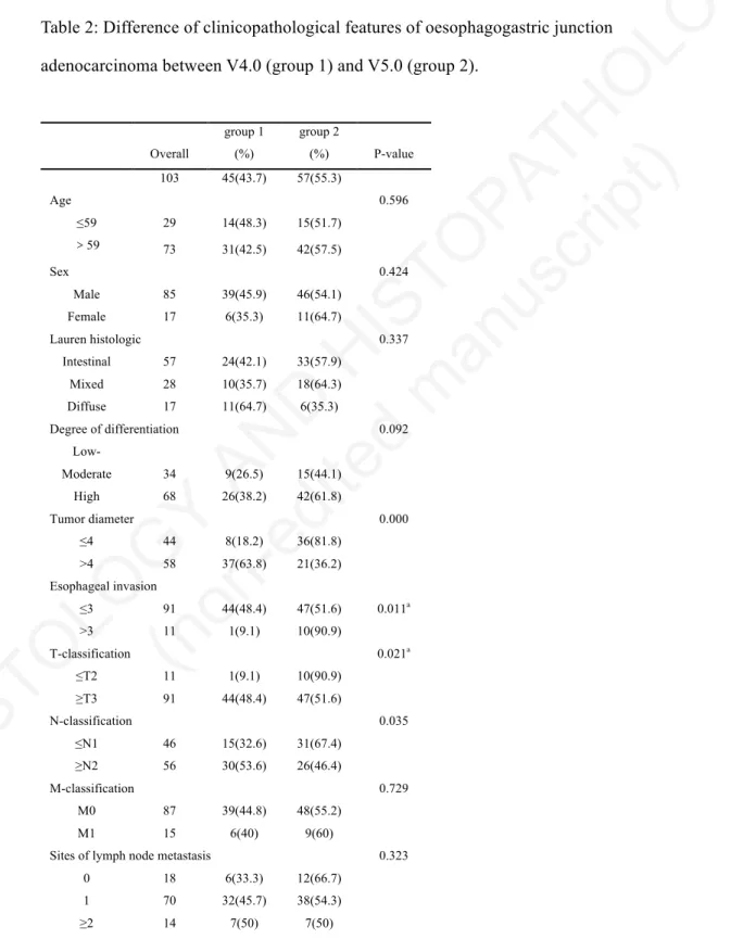
Difference of clinicopathological features of oesophagogastric junction adenocarcinoma between V4.0 (group 1) and V5.0 (group 2)
In order to have a more comprehensive understanding of the impact of the change in the definition of oesophagogastric junction adenocarcinoma, we selected 103 patients with oesophagogastric junction adenocarcinoma and compared the clinicopathological features between the fourth and fifth edition of oesophagogastric junction adenocarcinoma (Table 2). All cases of oesophagogastric junction adenocarcinoma were divided into two groups: group 1was diagnosed with V4.0 criteria, within 2cm to 5cm from oesophagogastric junction, while group 2 was diagnosed with V5.0 criteria, within 2cm from oesophagogastric junction. There were 45 (44%) cases of group 1 and 57 (56%) cases of group 2. Candidates in group 2 showed smaller tumor (P<0.001), earlier T classification (P=0.021) and N classification (P=0.035). There was no
HISTOLOGY
AND
HISTOPATHOLOGY
(non-edited
manuscript)
significant difference in other parameters between the two groups.
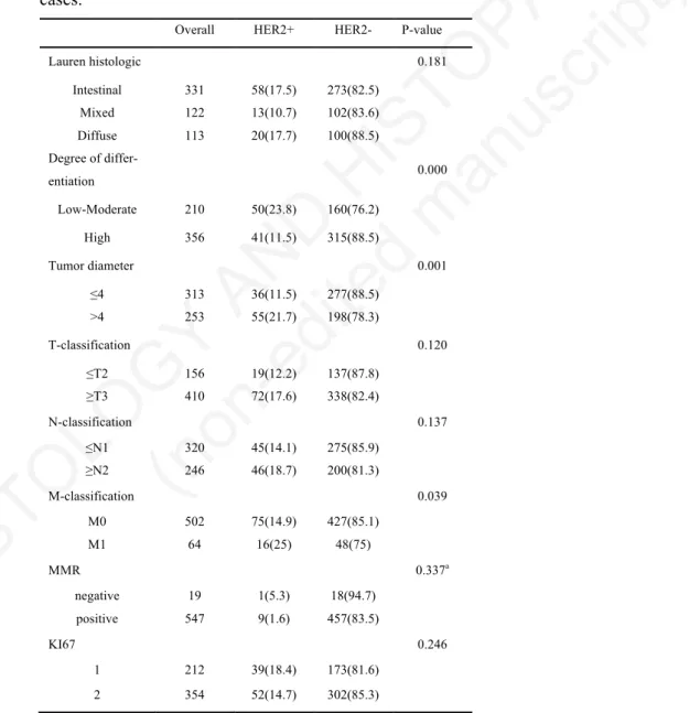
Correlation between HER2 status and clinicopathological characteristics of all 566 cases
We evaluated the association between HER2 status and clinicopathological features among all 566 cases and the cases were divided according to the criteria of V5.0 (Table 3). The incidence of HER2-expression in oesophagogastric junction (25%) and proximal gastric (22%) were dramatically higher than gastric body (13%) and distal gastric (11%) tumors (P=0.001). HER2-positivity was more common in low-moderate differentiation cases than the higher ones (P<0.001). Moreover, tumors of HER2-expression were distinctly associated with larger tumor (P<0.001) and more advanced M classification (P=0.039). Interestingly, there was no statistically difference in T classification and N classification. Although no difference in MMR status, there was merely one case of MSI-H in HER2-expression tumors.
Discussion
In V5.0, the definition of oesophagogastric junction adenocarcinoma was the same as Nishi’s classification in addition to emphasis on adenocarcinoma. The change in the definition of oesophagogastric junction tumors is very important for clinicopathological assessment and clinical management, such as the surgical dissection procedure.
In this study, we compared the clinicopathological features between gastric and oesophagogastric junction adenocarcinoma with V4.0 and V5.0, respectively. The oesophagogastric junction adenocarcinoma had more advanced T-classification than distal gastric adenocarcinoma with the criteria of both the V4.0 and V5.0. This was concurred by previous studies, that proximal (oesophagogastric junction and cardia) tumors were associated with poor outcomes (Kattan et al., 2003; Talamonti et al 2003). However, there were still some changes between the clinicopathological features of V4.0 and V 5.0. When compared with V4.0, the differences of N-classification and the sites of lymph node metastasis were mainly focused on oesophagogastric junction and distal gastric adenocarcinoma. But these differences were mainly found on proximal
HISTOLOGY
AND
HISTOPATHOLOGY
(non-edited
manuscript)
gastric and distal gastric adenocarcinoma with the V5.0. This was perhaps because the oesophagogastric junction tumors of 2cm-5cm from the oesophagogastric junction with V4.0 were classified to the proximal gastric tumors in the V5.0. The majority of patients were found to have more lymph node involvement in the Siewert III (Barbour et al., 2007), which was consistent with our results. Based on these findings, the Siewert III tumors were no longer included in oesophagogastric junction adenocarcinoma and the treatment for oesophagogastric junction tumors needs to be updated. Total gastrectomy or more extensive distal gastric lymph node dissection may not be considered. On the other hand, the scope of proximal gastric tumors also changed, which statistically exhibited larger tumor, more advanced T-classification and N-classification than distal gastric adenocarcinoma. Thus, proximal gastric tumors, rather than oesophagogastric junction adenocarcinoma, may have worse survival and need more aggressive treatment. Furthermore, we also compared the clinicopathological features between the V4.0 (group 1) and V5.0 (group 2) of oesophagogastric junction adenocarcinoma. The data showed the group 2 had smaller tumor, and earlier T-classification and N-classification than group 1. It seemed that group 1 was less invasive. Notably, the extent of lymph node dissection in the mediastinum and the choice of distal esophagectomy were of great importance in the treatment for oesophagogastric junction adenocarcinoma. A multicenter retrospective study indicated only the distance from the oesophagogastric junction was significantly related to metastasis. The longer the distance is, the higher rate of lymph node metastasis is (Hosokawa et al., 2012). In the newer version, the distance of oesophagogastric junction adenocarcinoma was smaller, which supported that oesophagogastric junction adenocarcinoma was less likely to have lymph node involvement. Besides, some studies suggested the extent of upper or middle mediastinal lymphadenectomy was for oesophageal invasion of ≥ 3 cm (Koyanagi et al., 2018; Kumamoto et al., 2019). Group 2 had a higher rate than older ones in terms of the extent of oesophageal invasion of ≥ 3 cm, which showed that oesophagogastric junction adenocarcinoma need upper or middle mediastinal lymphadenectomy. With that, we compared the difference of mediastinal lymph node involvement between the two groups. The proportion of group 2 (57%) were slightly higher than group 1, but with no
HISTOLOGY
AND
HISTOPATHOLOGY
(non-edited
manuscript)
statistical difference. This may be due to the lack of an accurate assessment of the extent of oesophageal invasion before operation and the incomplete extent of lymph node dissection.
HER2 test is another significant point which was formally recommended in the V5.0. Its expression and relevant clinicopathological features have been well studied in previous research with Siewert’s classification (Madani et al., 2015; Grillo et al., 2016). A Japanese study indicated that HER2-overexpression was not associated with tumor location with Siewert’s classification (Oono et al., 2018). An Italian study also showed HER2 amplification was not correlated with tumor location and prevailed in intestinal-type and low-grade tumors (Rocco Cappellesso et al., 2015). On the contrary, we re-evaluated the clinicopathological features of a total of 566 cases with their HER2 status, using V5.0. The HER2-expression in oesophagogastric junction and proximal tumors were statistically higher than that in body and distal tumors, which was consistent with some studies (Shan et al., 2013; Madani et al., 2015). This was perhaps because we used the latest classification to compare the HER2 status of different sites and chose a small sample size. Our results demonstrated oesophagogastric junction tumors had a higher expression of HER2 than body and distal tumors even if the scope of oesophagogastric junction adenocarcinoma was narrowed with the new criterion. This should be critical to emphasize the HER2 test in oesophagogastric junction adenocarcinoma. In addition, our analyses showed a statistically significant association between HER2-expression and pathological grade, tumor diameter and M-classification for gastric tumors. HER2-expression tumors had poor differentiation, larger diameter and more metastasis than HER2-negative ones, which indicated that HER2-expression tumors were more aggressive. These results were consistent with previous studies (Rajagopal et al., 2015). Therefore, the relevant clinicopathological features of HER2-expressing oesophagogastric junction adenocarcinoma remained unchanged in the V5.0. The status of microsatellites was another significant molecular test in gastric carcinoma, which was related to the contraction or expansion or of microsatellite sequences owing to the replication errors caused by mutations in the mismatch repair (MMR) in most cases (Shokal et al., 2012). Patients with deficiency
HISTOLOGY
AND
HISTOPATHOLOGY
(non-edited
manuscript)
of more than two markers of mismatch repair protein were considered microsatellite instability of high frequency (MSI-H). More than 30% patients with MSI-H were likely to develop Lynch syndrome. Even though there was no statistically significant difference between microsatellite status and HER2-expression, only one of the 19 MSI-H cases showed positive for MSI-HER2, while another 18 cases were all negative for MSI-HER2. This demonstrated that the HER2-expression cases probably did not suffer from MSI-H, but further verification is required.
Conclusions
The clinicopathological parameters of the oesophagogastric junction adenocarcinoma changed in the updated WHO classification. The analyses showed that the difference was mainly between proximal and distal adenocarcinoma and the proximal gastric tumors seem to be more invasive than oesophagogastric junction in the newer version. Although the treatment tends to be more unified and standardized, the oesophagogastric junction tumors with extent of oesophageal invasion more than 3cm required additional management. The HER2-expression of oesophagogastric junction adenocarcinoma is still higher than that of other sites of gastric adenocarcinoma in the updated version. Therefore, the emphasis on the detection of HER2 in oesophagogastric junction tumors is of great significance in clinical practice. The overall analyses showed it is reasonable to recommend the updated V5.0 in pathological diagnosis, as well as clinical practice.
Acknowledgements
The authors thank Sichuan Cancer Hospital & Institute for clinical data supported.
Funding
This work was supported by Foundation of Health Department of Sichuan Province (No.120044).
Competing interests
HISTOLOGY
AND
HISTOPATHOLOGY
(non-edited
manuscript)
ReferencesAbrahao-Machado L.F. and Scapulatempo-Neto C. (2016). HER2 testing in gastric cancer: An update. World J. Gastroenterol. 22, 4619.
Agnes A., Biondi A., Laurino A., Persiani R. and D'Ugo D. (2020). Global updates in the treatment of gastric cancer: a systematic review. Part 1: staging, classification and surgical treatment. Updates Surg. 1-13.
Barbour A P, Rizk N P, Gonen M and Tang L.H., Bains M.S., Rusch V.W., Coit D.G. and Brennan M.F. (2007). Adenocarcinoma of the gastroesophageal junction: influence of esophageal resection margin and operative approach on outcome. Ann. Surg. 246, 1.
Cappellesso, R., Fassan, M., Hanspeter, E., Bornschein, J., d'Amore, E.S., Cuorvo, L.V., and Malfertheiner P. (2015). HER2 status in gastroesophageal cancer: a tissue microarray study of 1040 cases. Human Pathology, 46, 665-672.
Chaves P., Cruz C., Lage P., Claro I., Cravo M., Leitao C.N. and Soares J. (2000). Immunohistochemical detection of mismatch repair gene proteins as a useful tool for the identification of colorectal carcinoma with the mutator phenotype. J. Pathol. 191, 355-360.
COSTA L.B., Toneto M.G. and Moreira L.F. (2016). Do proximal and distal gastric tumours behave differently? Arquivos Brasilros De Cirurgia Digestiva Abcd. 29, 232-235.
Cowan A.J., Allen C., Barac A., Basaleem H., Bensenor I. and Curado M.P. (2018). Global burden of multiple myeloma: a systematic analysis for the global burden of disease study 2016. JAMA Oncol. 4, 1221-1227.
Feith M., Stein H.J. and Siewert J.R. (2006). Adenocarcinoma of the esophagogastric junction: surgical therapy based on 1602 consecutive resected patients. Surg. Oncol. Clin. N. Am. 15, 751-764.
Grillo F., Fassan M., Sarocchi F., Fiocca R. and Mastracci L. (2016). HER2 heterogeneity in gastric/gastroesophageal cancers: from benchside to practice. World J. Gastroenterol. 22, 5879.
HISTOLOGY
AND
HISTOPATHOLOGY
(non-edited
manuscript)
Hofmann M., Stoss O., Shi D., Buttner R., Vijver M.J.V.D., Kim W., Ochiai A., Ruschoff J. and Henkel T. (2008). Assessment of a HER2 scoring system for gastric cancer: results from a validation study. Histopathology 52, 797-805. Hosokawa Y., Kinoshita T., Konishi M., Takahashi S., Gotohda N., Kato Y., Daiko H.,
Nishimura M., Katsumata K., Sugiyama Y. and Kinoshita T. (2012). Clinicopathological features and prognostic factors of adenocarcinoma of the esophagogastric junction according to Siewert classification: experiences at a single institution in Japan. Ann. Surg. Oncol. 19, 677-683.
Kattan M.W., Karpeh M.S., Mazumdar M. and Brennan M.F. (2003). Postoperative nomogram for disease-specific survival after an R0 resection for gastric carcinoma. J. Clin. Oncol.21, 3647-3650.
Koyanagi K., Kato F., Kanamori J., Daiko H., Ozawa S. and Tachimori Y. (2018). Clinical significance of esophageal invasion length for the prediction of mediastinal lymph node metastasis in Siewert type II adenocarcinoma: A retrospective single institution study. Ann. Gastroenterol. Surg. 2, 187-196. Kumamoto T., Kurahashi Y., Niwa H., Nakanishi Y. Kumamoto T., Kurahashi Y., Niwa
H., Nakanishi Y., Okumura K., Ozawa R., Ishida Y. and Shinohara H. (2019). True esophagogastric junction adenocarcinoma: background of its definition and current surgical trends. Surg. Today. 1-6.
Madani S.H, Rahmati A., Sadeghi E., Khazaei S., Sadeghi M., Payandeh M. and Amirifard N. (2015). Survey of Her2-neu expression and its correlation with histology of gastric carcinoma and gastroesophageal junction adenocarcinoma. Asian Pac. J. Cancer Prev. 16, 7755-7758.
Maresch J., Schoppmann S.F., Thallinger C.M.R., Zielinski C.C. and Hejna M. (2012). Her-2/neu gene amplification and over-expression in stomach and esophageal adenocarcinoma: from pathology to treatment. Crit. Rev. Oncol. Hematol. 82, 310-322.
Nagtegaal I.D., Odze R.D., Klimstra D., Paradis V., Rugge M., Schirmacher P., Washington K.M., Carneiro F. and Cree I.A. (2020). The 2019 WHO classification of tumours of the digestive system. Histopathology 76, 182-188.
HISTOLOGY
AND
HISTOPATHOLOGY
(non-edited
manuscript)
Nishi M., Nomura H. and Kajisa T. (1978). Surgical problem of carcinoma in the esophagogastric junction. Stomach and Intestine 13, 1497-1507.
Oono Y., Kuwata T., Takashima K., Yoda Y., Ikematsu H., Shitara K., Kinoshita T. and Yano T. (2018). Clinicopathological features and endoscopic findings of HER2-positive gastric cancer. Surg. Endosc. 32, 3964-3971.
Rajagopal I., Niveditha S.R., Sahadev R., Nagappa P.K. and Rajendra S.G. (2015). HER 2 expression in gastric and gastro-esophageal junction (GEJ) adenocarcinomas. J. Clin. Diagn. Res. 9, EC06.
Roskoski J.R. (2014). The ErbB/HER family of protein-tyrosine kinases and cancer. Pharmacol. Res. 79, 34-74.
Roy P.S., Nyodu T., Hazarika M., Saikia B.J., Bhuyan C., Inamdar A., Nyuthe C.W., Borthakur C.W. and Sharma J.D. (2019). Prevalence of HER2 Expression and Its Correlation with Clinicopathological Parameters in Gastric or Gastroesophageal Junction Adenocarcinoma in North-East Indian Population. Asian Pac. J. Cancer Prev. 20, 1139-1145.
Shan L., Ying J. and Lu N. (2013). HER2 expression and relevant clinicopathological features in gastric and gastroesophageal junction adenocarcinoma in a Chinese population. Diagn Pathol. 8, 76.
Shokal U. and Sharma P.C. (2012). Implication of microsatellite instability in human gastric cancers. Indian J. Med. Res. 35, 599.
Siewert J.R. and Stein H.J. (1998). Classification of adenocarcinoma of the oesophagogastric junction. Br. J. Surg. 85, 1457-1459.
Suh Y.S., Han D.S. and Kong S.H. (2012). Should adenocarcinoma of the esophagogastric junction be classified as esophageal cancer? A comparative analysis according to the seventh AJCC TNM classification. Ann. Surg. 255, 908-915.
Talamonti M.S., Kim S.P., Yao K.A., Wayne J.D., Feinglass J.M., Bennett C.L. and Rao S. (2003). Surgical outcomes of patients with gastric carcinoma: the importance of primary tumor location and microvessel invasion. Surgery 134, 720-727. Wolff A.C., Hammond M.E.H., Schwartz J.N., Hagerty K.L., Allred D.C., Cote R.J.,
HISTOLOGY
AND
HISTOPATHOLOGY
(non-edited
manuscript)
Dowsett M., Fitzgibbons P.L., Hanna W. and Langer A.S. (2007). American Society of Clinical Oncology/College of American Pathologists guideline recommendations for human epidermal growth factor receptor 2 testing in breast cancer. J. Clin. Oncol. 25,118-145.
Yamashita H., Seto Y., Sano T., Makuuchi H., Ando N. and Sasako M. (2017). Results of a nation-wide retrospective study of lymphadenectomy for esophagogastric junction carcinoma. Gastric Cancer 20, 69-83.
HISTOLOGY
AND
HISTOPATHOLOGY
(non-edited
manuscript)
Table1: Comparison of the clinicopathological features of gastric (proximal gastric, gastric body and distal gastric) and oesophagogastric junction adenocarcinoma between V4.0 and V5.0.
Location P value EGJ (V4.0/V5.0) Proximal (V4.0/V5.0) Body Distal EGJ vs Proximal (V4.0/V5.0) EGJ vs Body (V4.0/V5.0) EGJ vs Dis-tal (V4.0/V5.0) Proximal vs Distal (V4.0/V5.0) Age ≤59 29/15 29/43 57 103 1.000 /1.000 0.411 /1.000 0.082 /0.693 0.052 /0.012 >59 73/42 82/113 81 112 Sex Male 85/11 93/132 101 152 1.000 /1.000 0.258 /0.304 0.005 /0.018 0.001 /0.000 Female 17/46 18/24 37 63 Lauren histologic Intestinal 57/33 73/97 84 117 0.181 /0.900 0.737 /0.826 0.489 /0.264 0.183 /0.102 Mixed 28/18 22/32 25 47 Diffuse 17/6 16/27 29 51 Degree of differenti-ation Low-Moderate 34/15 55/82 43 78 0.087 /0.029 1.000 /1.000 1.000 /1.000 0.113 /0.169 High 68/42 56/82 95 137 Tumor diameter ≤4 44/36 58/64 57 154 1.000 /0.041 1.000 /0.032 0.000 /1.000 0.005 /0.000 >4 58/21 53/90 81 61 T-classification ≤T2 11/10 27/28 36 82 0.164 /1.000 0.053 /1.000 0.000 /0.012 0.049 /0.000 ≥T3 91/47 84/128 102 133 N-classification ≤N1 46/31 65/80 72 137 0.288 /0.690 1.000 /0.779 0.011 /0.198 1.000 /0.017 ≥N2 56/26 46/76 66 78 M-classification M0 87/9 12/138 121 195 0.394 /0.410 0.592 /0.518 0.152 /0.159 0.665 /0.484 M1 15/48 99/18 17 20
Sites of lymph node metastasis
None 18/12 37/42 47 94 1.000 /1.000 0.015 /0.250 0.006 /0.188 0.125 /0.009 Periesophageal 70/38 47/79 86 86
HISTOLOGY
AND
HISTOPATHOLOGY
(non-edited
manuscript)
HER2 Negative 84/43 81/123 120 24 0.575 /1.000 1.000 /0.280 0.548 /0.087 0.001 /0.021 Positive 19/14 30/35 18 24Table 2: Difference of clinicopathological features of oesophagogastric junction adenocarcinoma between V4.0 (group 1) and V5.0 (group 2).
Overall group 1 (%) group 2 (%) P-value 103 45(43.7) 57(55.3) Age 0.596 ≤59 29 14(48.3) 15(51.7) >59 73 31(42.5) 42(57.5) Sex 0.424 Male 85 39(45.9) 46(54.1) Female 17 6(35.3) 11(64.7) Lauren histologic 0.337 Intestinal 57 24(42.1) 33(57.9) Mixed 28 10(35.7) 18(64.3) Diffuse 17 11(64.7) 6(35.3) Degree of differentiation 0.092 Low-Moderate 34 9(26.5) 15(44.1) High 68 26(38.2) 42(61.8) Tumor diameter 0.000 ≤4 44 8(18.2) 36(81.8) >4 58 37(63.8) 21(36.2) Esophageal invasion ≤3 91 44(48.4) 47(51.6) 0.011a >3 11 1(9.1) 10(90.9) T-classification 0.021a ≤T2 11 1(9.1) 10(90.9) ≥T3 91 44(48.4) 47(51.6) N-classification 0.035 ≤N1 46 15(32.6) 31(67.4) ≥N2 56 30(53.6) 26(46.4) M-classification 0.729 M0 87 39(44.8) 48(55.2) M1 15 6(40) 9(60)
Sites of lymph node metastasis 0.323 0 18 6(33.3) 12(66.7)
1 70 32(45.7) 38(54.3)
HISTOLOGY
AND
HISTOPATHOLOGY
(non-edited
manuscript)
Periesophageal lymph node metastasis 0.897Negative 81 36(44.4) 45(55.6) Positive 21 9(42.9) 12(57.1) Lymphovascular invasion 0.989 Negative 25 11(44) 14(56) Positive 77 34(44.2) 43(55.8) HER2 status 0.085 Negative 83 40(48.2) 43(51.8) Positive 19 5(26.3) 14(73.7) a Fisher’s exact test
Table 3: Correlation between HER2 status and clinicopathological characteristics of all 566 cases.
Overall HER2+ HER2- P-value
Lauren histologic 0.181 Intestinal 331 58(17.5) 273(82.5) Mixed 122 13(10.7) 102(83.6) Diffuse 113 20(17.7) 100(88.5) Degree of differ-entiation 0.000 Low-Moderate 210 50(23.8) 160(76.2) High 356 41(11.5) 315(88.5) Tumor diameter 0.001 ≤4 313 36(11.5) 277(88.5) >4 253 55(21.7) 198(78.3) T-classification 0.120 ≤T2 156 19(12.2) 137(87.8) ≥T3 410 72(17.6) 338(82.4) N-classification 0.137 ≤N1 320 45(14.1) 275(85.9) ≥N2 246 46(18.7) 200(81.3) M-classification 0.039 M0 502 75(14.9) 427(85.1) M1 64 16(25) 48(75) MMR 0.337a negative 19 1(5.3) 18(94.7) positive 547 9(1.6) 457(83.5) KI67 0.246 1 212 39(18.4) 173(81.6) 2 354 52(14.7) 302(85.3)
HISTOLOGY
AND
HISTOPATHOLOGY
(non-edited
manuscript)
Figure legendsFigure 1: HER2-immunohistochemistry of score 3 showed strong and complete basolateral membranous reactivity in 90% adenocarcinoma cells (≥10 of tumor cells), which was considered as HER2-amplification. (×100)
Figure 2: FISH result showed case with HER2/CEP17 ratio of more than 2.2 was considered HER2 gene amplified. (×1000). (The red signal represented HER2, the blue signal represented CEP17)