The Ligament Anatomy of the Deltoid Complex
of the Ankle: A Qualitative and Quantitative
Anatomical Study
Kevin J. Campbell, BS, Max P. Michalski, MSc, Katharine J. Wilson, MSc, Mary T. Goldsmith, MS, Coen A. Wijdicks, PhD, Robert F. LaPrade, PhD, MD, and Thomas O. Clanton, MD Investigation performed at the Department of Biomedical Engineering, Steadman Philippon Research Institute,
and the Steadman Clinic, Vail, Colorado
Background: The deltoid ligament has both superficial and deep layers and consists of up to six ligamentous bands. The prevalence of the individual bands is variable, and no consensus as to which bands are constant or variable exists. Although other studies have looked at the variance in the deltoid anatomy, none have quantified the distance to relevant osseous landmarks.
Methods: The deltoid ligaments from fourteen non-paired, fresh-frozen cadaveric specimens were isolated and the ligamentous bands were identified. The lengths, footprint areas, orientations, and distances from relevant osseous landmarks were measured with a three-dimensional coordinate measurement device.
Results: In all specimens, the tibionavicular, tibiospring, and deep posterior tibiotalar ligaments were identified. Three additional bands were variable in our specimen cohort: the tibiocalcaneal, superficial posterior tibiotalar, and deep anterior tibiotalar ligaments. The deep posterior tibiotalar ligament was the largest band of the deltoid ligament. The origins from the distal center of the intercollicular groove were 16.1 mm (95% confidence interval, 14.7 to 17.5 mm) for the tibionavicular ligament, 13.1 mm (95% confidence interval, 11.1 to 15.1 mm) for the tibiospring ligament, and 7.6 mm (95% confidence interval, 6.7 to 8.5 mm) for the deep posterior tibiotalar ligament. Relevant to other pertinent osseous landmarks, the tibionavicular ligament inserted at 9.7 mm (95% confidence interval, 8.4 to 11.0 mm) from the tuberosity of the navicular, the tibiospring inserted at 35% (95% confidence interval, 33.4% to 36.6%) of the spring ligament’s posteroanterior distance, and the deep posterior tibiotalar ligament inserted at 17.8 mm (95% confidence interval, 16.3 to 19.3 mm) from the posteromedial talar tubercle.
Conclusions: The tibionavicular, tibiospring, and deep posterior tibiotalar ligament bands were constant components of the deltoid ligament. The deep posterior tibiotalar ligament was the largest band of the deltoid ligament.
Clinical Relevance: The anatomical data regarding the deltoid ligament bands in this study will help to guide anatomical placement of repairs and reconstructions for deltoid ligament injury or instability.
E
arly descriptions of the deltoid ligament anatomy pre-sented general anatomical information regarding its origin on the medial malleolus and insertion on the navicular, talus, and calcaneus1. Later qualitative observations introduced the fact that the deltoid ligament was composed ofsuperficial and deep layers, often described as being separated by a fat pad2-5
. These initial qualitative observations were later expanded further when reports were published highlighting the presence of multiple ligamentous bands identified as part of the superficial and deep layers6-8.
Disclosure:None of the authors received payments or services, either directly or indirectly (i.e., via his or her institution), from a third party in support of any aspect of this work. One or more of the authors, or his or her institution, has had a financial relationship, in the thirty-six months prior to submission of this work, with an entity in the biomedical arena that could be perceived to influence or have the potential to influence what is written in this work. No author has had any other relationships, or has engaged in any other activities, that could be perceived to influence or have the potential to influence what is written in this work. The completeDisclosures of Potential Conflicts of Interestsubmitted by authors are always provided with the online version of the article.
Peer Review:This article was reviewed by the Editor-in-Chief and one Deputy Editor, and it underwent blinded review by two or more outside experts. The Deputy Editor
reviewed each revision of the article, and it underwent a final review by the Editor-in-Chief prior to publication. Final corrections and clarifications occurred during one or more exchanges between the author(s) and copyeditors.
The deltoid ligament is a complex structure that spans from the medial malleolus to the navicular, talus, and calca-neus. It is primarily responsible for stabilizing the medial side of the ankle and its functions are to limit anterior, posterior, and lateral translation of the talus and to restrain talar ab-duction at the talocrural joint9-14. Specifically, the superficial deltoid resists eversion of the hindfoot and the deep deltoid is the primary restraint to external rotation of the talus9.
Deltoid ligament pathology has a number of different causes. Medial ankle sprains accounted for 5.1% of the ankle sprains reported in a two-year study of West Point cadets by Waterman et al.15. The highest prevalence reported was a two-year study of soccer players in the Greek Amateur Association. Of the 139 ankle sprains reported in that study, twenty-two (15.8%) were to the medial ligaments16
. Deltoid ligament injury can also occur with concomitant lateral ankle sprains17,18, during fractures of the lateral malleolus13, and in association with pos-terior tibial tendon pathology19. When injury to the deltoid ligament occurs, there is frequently associated injury to other
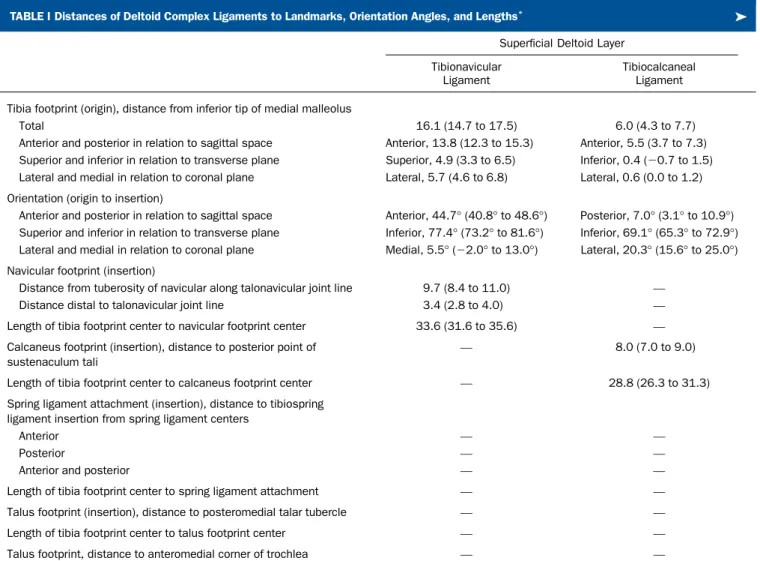
anatomical structures of the ankle including the fibula, syndes-mosis, and articular cartilage surfaces of the tibiotalar joint20. The majority of deltoid ligament injuries can be treated non-operatively. However, in patients who develop chronic medial ankle instability, a surgical procedure to repair or to reconstruct the deltoid ligament may be necessary17. A complete under-standing of the origins, insertions, and orientations of the in-dividual ligamentous bands of the deltoid ligament is critical to achieve a successful anatomical surgical repair or reconstruction. The superficial and deep layers of the deltoid ligament can consist of up to six individual ligamentous bands6,21-26. The prev-alence of the individual ligamentous bands is variable among in-dividuals, and there is no consensus as to which bands are constant or variable. Further, there is no agreement as to the size and ori-entation of the ligamentous bands, their spatial relationship to each other, or their distance from osseous landmarks. This anatomical information would help to develop a knowledge base for multiple applications, including biomechanical analyses of the ligamentous bands’ functional roles, clinical diagnosis of injured structures on TABLE I Distances of Deltoid Complex Ligaments to Landmarks, Orientation Angles, and Lengths* !
Superficial Deltoid Layer Tibionavicular
Ligament TibiocalcanealLigament
Tibia footprint (origin), distance from inferior tip of medial malleolus
Total 16.1 (14.7 to 17.5) 6.0 (4.3 to 7.7)
Anterior and posterior in relation to sagittal space Anterior, 13.8 (12.3 to 15.3) Anterior, 5.5 (3.7 to 7.3) Superior and inferior in relation to transverse plane Superior, 4.9 (3.3 to 6.5) Inferior, 0.4 (20.7 to 1.5) Lateral and medial in relation to coronal plane Lateral, 5.7 (4.6 to 6.8) Lateral, 0.6 (0.0 to 1.2) Orientation (origin to insertion)
Anterior and posterior in relation to sagittal space Anterior, 44.7"(40.8"to 48.6") Posterior, 7.0"(3.1"to 10.9") Superior and inferior in relation to transverse plane Inferior, 77.4"(73.2"to 81.6") Inferior, 69.1"(65.3"to 72.9") Lateral and medial in relation to coronal plane Medial, 5.5"(22.0"to 13.0") Lateral, 20.3"(15.6"to 25.0") Navicular footprint (insertion)
Distance from tuberosity of navicular along talonavicular joint line 9.7 (8.4 to 11.0) —
Distance distal to talonavicular joint line 3.4 (2.8 to 4.0) —
Length of tibia footprint center to navicular footprint center 33.6 (31.6 to 35.6) —
Calcaneus footprint (insertion), distance to posterior point of sustenaculum tali
— 8.0 (7.0 to 9.0)
Length of tibia footprint center to calcaneus footprint center — 28.8 (26.3 to 31.3)
Spring ligament attachment (insertion), distance to tibiospring ligament insertion from spring ligament centers
Anterior — —
Posterior — —
Anterior and posterior — —
Length of tibia footprint center to spring ligament attachment — —
Talus footprint (insertion), distance to posteromedial talar tubercle — —
Length of tibia footprint center to talus footprint center — —
Talus footprint, distance to anteromedial corner of trochlea — —
*The values are given as the mean of fourteen specimens with the parametric 95% CI in parentheses; negative 95% CI values correspond to the opposite anterior and posterior, superior and inferior, and lateral and medial direction.
magnetic resonance imaging (MRI) scans, and development of anatomical surgical repairs and reconstruction. Although ana-tomical repairs and reconstruction are becoming the standard of care and are improving outcomes in other joints, they have not been defined in detail for the deltoid ligament of the ankle.
The purpose of this study was to provide qualitative and quantitative information regarding the origins and insertions of the ligamentous bands of the deltoid ligament and to quantify the prevalence of individual bands. It was hypothesized that for each of the ligamentous bands of the superficial and deep layers of the deltoid ligament, their origins and insertions in relation to pertinent osseous landmarks, spatial relationships to each other, footprint areas, and ligament lengths could be defined. A com-prehensive understanding of the anatomical attachment site of the ligamentous bands of the deltoid will assist in guiding medial ankle surgical repair and reconstruction techniques.
Materials and Methods Specimen Preparation
F
ourteen non-paired, fresh-frozen cadaveric specimens (six female and eight male; eight left and six right), with a mean age of 50.4 years (range,twenty-seven to sixty years), a mean foot length (and standard deviation) of 25.2±2.3 cm (the average adult foot length [and standard deviation] is 24.7±1.1 cm)27, and no history of injury were used in the study. The dissections consisted of identification of the medial ligament structures originating from the anterior and posterior colliculi of the medial malleolus and inserting onto the navicular, talus, and calcaneus. The ligamentous bands of the deltoid ligament were identified on the basis of their distinct origins and insertions and were tagged with 2-0 nylon sutures. Specimens were then loaded into a custom jig, where a bicortical bone screw through the calcaneus and a lag screw through the cal-caneus and talus were placed. The phalanges were secured to a plate to ensure rigid fixation of the ankle and to prevent movement during testing. The tibia and fibula were then secured to a stabilizing tower with rigid screws. Neutral alignment in the sagittal plane at zero degrees of dorsiflexion was achieved with a goniometer. Three local coordinate frames were secured to the calcaneus, tibia, and fibula to allow for anatomical positioning of the bones during analysis. The tibia and fibula were then disarticulated from the foot to allow for accurate measurements of the ligamentous centers on the tibia, fibula, and foot.
Anatomical Measurements
Following secure fixation of the calcaneus, tibia, and distal metatarsals, three-dimensional positional data of the ligament attachments and osseous land-marks on the disarticulated tibia, fibula, and foot were collected using a coordinate measuring device (MicroScribe MX; GoMeasure3D, Amherst, Virginia). For accurate measurement of the ligament footprints, the individual
Superficial Deltoid Layer Deep Deltoid Layer
Tibiospring
Ligament Superficial PosteriorTibiotalar Ligament Deep Anterior TibiotalarLigament Deep Posterior TibiotalarLigament
13.1 (11.1 to 15.1) 3.5 (3.0 to 4.0) 11.1 (9.6 to 12.6) 7.6 (6.7 to 8.5)
Anterior, 10.8 (8.9 to 12.7) Posterior, 0.2 (21.2 to 1.6) Anterior, 9.0 (7.4 to 10.6) Posterior, 0.7 (20.9 to 2.3) Superior, 6.9 (5.8 to 8.1) Inferior, 1.1 (0.4 to 1.8) Inferior, 3.6 (2.8 to 4.4) Inferior, 2.2 (1.3 to 3.1) Lateral, 1.8 (1.1 to 2.5) Lateral, 1.5 (1.0 to 2.0) Lateral, 4.9 (3.9 to 5.9) Lateral, 6.4 (5.5 to 7.3) Anterior, 8.6"(3.5"to 13.7") Posterior, 12.2"(6.1"to 18.3") Anterior, 22.5"(11.9"to 33.1") Anterior, 8.5"(25.4"to 22.4") Inferior, 75.6"(71.4"to 79.8") Inferior, 58.3"(54.1"to 62.5") Inferior, 59.9"(53.8"to 66.0") Inferior, 56.0"(48.4"to 63.6") Lateral, 11.2"(4.8"to 17.6") Lateral, 31.7"(27.5"to 35.9") Lateral, 23.8"(12.0"to 35.6") Lateral, 31.9"(22.0"to 41.8")
— — — — — — — — — — — — — — — — — — — — 22.1 (20.3 to 23.9) — — — 12.0 (11.1 to 12.9) — — — 35.0 (33.4 to 36.6) — — — 25.0 (23.2 to 26.8) — — — — 10.4 (8.9 to 11.9) — 17.8 (16.3 to 19.3) — 21.0 (20.0 to 22.1) 12.0 (10.5 to 13.5) 10.3 (9.0 to 11.6) — — 12.2 (11.1 to 13.4) — TABLE I (continued)
Fig. 1
Dissection photographs presenting the ligamentous bands (tagged with suture) of the superficial layer of the deltoid ligament in a left ankle. Four ligamentous bands composed the superficial layer of the deltoid ligament in this specimen and included the tibionavicular, tibiospring, tibiocalcaneal, and superficial posterior tibiotalar ligaments. Fig.1-AAnteromedial view of the deltoid ligament with the tibionavicular ligament grasped.Fig.1-BMedial view of the deltoid ligament with the tibiospring ligament grasped. Fig.1-CMedial view of the deltoid ligament with the tibiocalcaneal ligament grasped.Fig. 1-D Posteromedial view of the deltoid ligament with the superficial posterior tibiotalar ligament in view.
Fig. 2
Dissection photograph showing the deep layer of the deltoid ligament in a left ankle. Two ligamentous bands composed the deep layer in this specimen and included the deep anterior and posterior tibiotalar ligaments.
ligamentous bands were tagged with sutures, were cut at their midsubstance, and were reflected to clearly identify their attachments. The periphery of each ligament attachment site was measured with points collected in approximately 1-mm increments. Data points of pertinent osseous landmarks were also col-lected. All points were transformed from their respective local coordinate system on the tibia, fibula, and foot to the anatomical ankle joint coordinate system, as described below. From these points, the ligament attachment centers, distances to osseous prominences, ligament footprint areas, and ligament lengths were calculated. Footprint areas were calculated using the Heron for-mula28,29. All reported measurements were performed by the same individual (K.J.C.) with the senior author’s (T.O.C.) guidance to eliminate interobserver variability.
The anatomical ankle joint coordinate system was based on recom-mendations by the International Society of Biomechanics (ISB) for the calca-neal coordinate system in neutral stance30. The center of the ankle joint coordinate system was positioned midway between the tip of the lateral and medial malleoli and was orientated with the positive directions pointing su-periorly, anteriorly, and laterally. The superior and inferior axis was aligned with a line coincident with the long axis of the tibia and fibula. The ISB recommendations use the tibial plateau to calculate the long axis of the tibia and fibula; however, this plateau was not present in the ankle specimens and therefore circumferential points around the tibial diaphysis were used to cal-culate the long axis. The anterior and posterior axis was created perpendicular to the superior and inferior axis and a line connecting the medial and lateral malleoli. The lateral and medial axis was then defined perpendicular to the superior and inferior axis and the anterior and posterior axis. All measurements reported in this study were made in the ankle joint coordinate frame.
System Validation
The manufacturer (GoMeasure3D) performed a single-point articulation performance test (SPAT) based on the B89.4.22 ASME (American Society of Mechanical Engineers) standard as an assessment of accuracy for the coordinate measuring device used in this study. The SPAT result reflected twice the stan-dard deviation for repeatability of measurements using this device. The average volumetric SPAT result was reported to be±0.01 mm3.
Statistical Methods
Data were assessed for symmetry and normality and no evidence was found for deviations from normality. Continuous variables were reported as the mean and the parametric 95% confidence interval (95% CI).
Source of Funding
No external sources of funding were received for this study.
Results
I
n all fourteen specimens, the deltoid ligament was on the medial ankle deep to the tendons of the posterior tibialis, flexor digitorum longus, and flexor hallucis longus muscles. At initial inspection, the individual bands of the deltoid ligament were contiguous with each other and were almost completely covered by the ligamentous sheaths of the posterior tibialis and flexor digitorum tendons. Fine dissection of the ligamentous sheath layer beneath the posterior tibialis and flexor digitorum tendons allowed for clear visualization of between three and six Fig. 3Illustration of the medial view of a left ankle showing the anatomical attachment sites of the ligamentous bands of the deltoid ligament and their spatial relationships to surgically relevant osseous landmarks: (A) distal center of the intercollicular groove, (B) posteromedial talar tubercle, (C) posterior point of the sustenaculum tali, (D) tuberosity of the navicular, and (E) anteromedial corner of the trochlea.
individual ligamentous bands of the deltoid, with some inter-specimen variability (Figs. 1 and 2) (see Appendix). These bands were consistently separated into superficial and deep layers separated by a thin layer of adipose tissue. The superficial layer crossed the tibiotalar and subtalar joints (Fig. 3), whereas the deep layer only crossed the tibiotalar joint (see Appendix). Figure 4 displays the anatomical attachment sites of each lig-ament band and their associated footprint area. Qualitative and quantitative descriptions of each ligamentous band of the deltoid are detailed below. Ligament band length, footprint area, and orientation are presented in Table I as mean values and the 95% CIs. We used an anatomical naming system originally proposed by Milner and Soames21, which named ligamentous bands of the deltoid ligament on the basis of their attachment sites.
Superficial Layer of the Deltoid Ligament
Tibionavicular Ligament
The tibionavicular ligament, identified in all fourteen speci-mens, was a thin and broad band of tissue whose fibers com-prised the most anterior aspect of the superficial deltoid layer. The ligament originated on the anterior colliculus of the medial malleolus and inserted in an expansive manner onto the dor-somedial surface of the navicular immediately anterior to the talonavicular articular cartilage border (see Appendix).
The tibia footprint was 16.1 mm (95% CI, 14.7 to 17.5 mm) from the distal center of the intercollicular groove, with a superior aspect of 4.9 mm (95% CI, 3.3 to 6.5 mm) and an anterior aspect of 13.8 mm (95% CI, 12.3 to 15.3 mm). The navicular footprint was 9.7 mm (95% CI, 8.4 to 11.0 mm) from the tuberosity of the navicular along the talonavicular joint line and 3.4 mm (95% CI, 2.8 to 4.0 mm) distal to the talonavicular joint line (Table I).
Tibiospring Ligament
The tibiospring ligament was identified in all fourteen speci-mens and was a flat and broad consolidation of ligament fibers. Its tibial attachment was slightly proximal and posterior to the tibionavicular ligament, and it was consistently the most su-perficial band of the deltoid ligament. It originated on the anterior tibial colliculus and inserted onto the plantar calca-neonavicular (spring) ligament at 35% (95% CI, 33.4% to 36.6%) of its posteroanterior distance (see Appendix).
Its insertion width at the plantar calcaneonavicular lig-ament junction was 5.9 mm (95% CI, 5.1 to 6.7 mm). The tibia footprint was 13.1 mm (95% CI, 11.1 to 15.1 mm) from the distal center of the intercollicular groove, with a superior aspect of 6.9 mm (95% CI, 5.8 to 8.1 mm) and an anterior aspect of 10.8 mm (95% CI, 8.9 to 12.7 mm) (Table I).
Fig. 4
Illustration of the medial view of a left ankle demonstrating the anatomical footprints of the deep and superficial bands of the deltoid ligament and their respective measured footprint areas. The values are given as the mean, with the 95% CI in parentheses.
Tibiocalcaneal Ligament
The tibiocalcaneal ligament, identified in eleven (79%) of fourteen specimens, attached near the intercollicular groove of the medial malleolus and attached at the most posterior aspect of the sustenaculum tali on the calcaneus, immediately pos-terior to the spring ligament’s insertion on the sustenaculum tali (Fig. 5).
The tibia footprint was 6.0 mm (95% CI, 4.3 to 7.7 mm) from the distal center of the intercollicular groove, with an inferior aspect of 0.4 mm (95% CI,20.7 to 1.5 mm) and an anterior aspect of 5.5 mm (95% CI, 3.7 to 7.3 mm). On the calcaneus, it attached on the posterior border of the suste-naculum tali at 8.0 mm (95% CI, 7.0 to 9.0 mm) from the posterior point of the sustenaculum tali (Table I).
Superficial Posterior Tibiotalar Ligament
The superficial posterior tibiotalar ligament was identified in eleven (79%) of fourteen specimens and was the most pos-terior ligament of the superficial layer of the deltoid liga-ment. The ligament center originated near the distal center of the intercollicular groove and attached onto the inferior medial talar body, anterosuperior to the postero-medial talar tubercle (Fig. 5).
The tibia footprint was 3.5 mm (95% CI, 3.0 to 4.0 mm) from the distal center of the intercollicular groove, with an inferior aspect of 1.1 mm (95% CI, 0.4 to 1.8 mm) and a posterior aspect of 0.2 mm (95% CI, 21.2 to 1.6 mm). The talus footprint was 10.4 mm (95% CI, 8.9 to 11.9 mm) an-terosuperior to the posteromedial talar tubercle (Table I).
Deep Layer of the Deltoid Ligament
Deep Anterior Tibiotalar Ligament
The deep anterior tibiotalar ligament was identified in thir-teen (93%) of fourthir-teen specimens and was located immedi-ately deep to the tibionavicular and tibiospring ligaments of the superficial deltoid layer. This short ligament originated at the most inferior and anterior areas of the medial malleolus and inserted onto the anterosuperior portions of the medial talus body immediately inferior to the articular cartilage of the trochlea (Fig. 6).
The tibia origin was 11.1 mm (95% CI, 9.6 to 12.6 mm) from the distal center of the intercollicular groove, with an inferior aspect of 3.6 mm (95% CI, 2.8 to 4.4 mm) and an anterior aspect of 9.0 mm (95% CI, 7.4 to 10.6 mm). Its talus insertion was 12.2 mm (95% CI, 11.1 to 13.4 mm) postero-inferior to the anteromedial corner of the trochlea (Table I). Fig. 5
Illustration of the medial view of a left ankle showing the attachment sites of the tibiocalcaneal and superficial posterior tibiotalar ligaments. The tibiocalcaneal ligament originated 6.0 mm from the distal center of the intercollicular groove and inserted 8.0 mm from the posterior point of the sustenaculum tali. The superficial posterior tibiotalar ligament originated 3.5 mm from the distal center of the intercollicular groove (no distance labeled) and inserted onto the talus 10.4 mm from the posteromedial talar tubercle. Ligament attachment centers are marked with an asterisk.
Deep Posterior Tibiotalar Ligament
The deep posterior tibiotalar ligament was identified in all fourteen specimens and was located deep to the tibiocalcaneal and superficial posterior tibiotalar ligaments. It was consis-tently the largest and thickest band of the superficial and deep layers. The deep posterior tibiotalar ligament originated near the distal center of the intercollicular groove and inserted on the posterosuperior aspect of the medial talar body inferior to the articular cartilage of the trochlea (Fig. 6).
The tibia footprint was 7.6 mm (95% CI, 6.7 to 8.5 mm) from the distal center of the intercollicular groove, with an inferior aspect of 2.2 mm (95% CI, 1.3 to 3.1 mm) and a lateral aspect of 6.4 mm (95% CI, 5.5 to 7.3 mm). Its talar attachment center was 17.8 mm (95% CI, 16.3 to 19.3 mm) anterosuperior to the posteromedial talar tubercle (Table I).
Discussion
W
e provide a detailed quantitative and qualitative as-sessment of the deltoid ligament. Through meticulous dissection and measurement, we confirmed our hypothesis and presented an anatomical description of the ligamentousbands of the superficial and deep ligamentous layers of the deltoid ligament, their origins and insertions in relation to pertinent osseous landmarks, their spatial relationships to each other, their footprint areas, and their ligament lengths. The most salient findings in this study were that the deep posterior tibiotalar ligament was the largest band of the deltoid ligament and that three constant ligamentous bands were found in all specimens and included the tibionavicular, tibiospring, and deep posterior tibiotalar ligaments. These ligament band data advance our understanding of the deltoid ligament and should aid in the development of anatomical repair and reconstruction guidelines, biomechanical studies that investigate their indi-vidual functional roles, and diagnosis of injury on MRI scans. Surgery is often performed on the injured deltoid liga-ment. Patients with large or complete tears that fail to heal primarily and who develop chronic medial ankle instability will often undergo a surgical repair or reconstruction of the deltoid ligament17,19. Concurrent injury to the deltoid ligament occurs in up to 83% of lateral ankle ligament injuries18 and may occasionally require surgical repair. In patients with posterior tibial tendon rupture or pathology, insufficiency of this dynamic Fig. 6
Illustration of the medial view of the deep deltoid ligament in a left ankle showing the anatomical attachment sites of the deep anterior and posterior tibiotalar ligaments. The deep anterior tibiotalar ligament originated 11.1 mm (9.0 mm anterior and 3.6 mm inferior) from the distal center of the intercollicular groove and inserted 12.2 mm from the anteromedial corner of the trochlea. The deep posterior tibiotalar ligament originated 6.6 mm lateral (no distance labeled) to the distal center of the intercollicular groove and inserted 17.8 mm anterosuperior to the posteromedial talar tubercle. Ligament attachment centers are marked with an asterisk.
stabilizer can lead to failure of the deltoid ligament requiring repair19
. Similarly, posterior tibial tendon reconstruction often requires contemporaneous surgical correction of the deltoid and spring ligament complexes19,31-34. Finally, in patients undergoing total ankle arthroplasty, attention must be paid to the integrity of the deltoid ligament to achieve proper ligamentous balance35.
The most important ligamentous bands of the deltoid ligament to repair or to reconstruct are not currently known. Haddad et al. reported on reconstruction of the tibiocalcaneal and deep posterior tibiotalar ligaments19. Various other au-tograft and allograft reconstruction techniques have been reported with little reference to the anatomical bands31-34,36. The paucity of data regarding the exact origins and insertions of the ligamentous bands of the deltoid ligament may have contributed to the less reliable results of secondary repairs or reconstructions for the deltoid ligament complex compared with lateral ankle ligament procedures. This study provides further information for repair and reconstruction procedures by showing the relative size of the ligamentous bands and their spatial relationships to each other and to osseous landmarks.
Several publications in recent years have described the prevalence of each of the bands of the deltoid ligament complex and, to a limited extent, some quantitative information including the ligament band lengths and orientations21,23,37
. The results of these studies in comparison with the findings in the current study are presented in the Appendix21,23,37. A major limitation of these previous studies was that the physical locations of the ligament insertion sites were not described or were not referenced from surgically relevant landmarks. This information is imperative for incorporating the findings into surgical practice for anatomically based treatment. This limitation was overcome in the present study by presenting quantitative data in reference to the distal center of the intercollicular groove, tuberosity of the navicular, posteromedial talar tubercle, and posterior point of the suste-naculum tali that will enable accurate re-creation of the normal anatomy during repair and reconstructive procedures.
A major strength of this study is that it shows the qual-itative and quantqual-itative morphology of the highly complex deltoid ligament and provides anatomical illustrations with distances from ligament band attachment centers to surgically relevant osseous landmarks. In addition, our measurement tech-nique used a highly precise measuring device that captured the three-dimensional distance relationships among the ligamentous and osseous structures of interest. Lastly, the average foot length (and standard deviation) of the specimens in this cohort was 25.2±2.3 cm, which is matched to the average adult foot length (and standard deviation) of 24.7±1.1 cm27.
We acknowledge some limitations of this study. First, four-teen ankles were included and our results may not have been wholly inclusive of the population variability. Our cohort was smaller than those used in the previous investigations by Milner and Soames21
, whose study included forty specimens, and Siegler et al.37, whose
study included twenty specimens. Second, the inherent contiguous nature of the ligamentous bands of the deltoid ligament may have introduced some variability during identification of the individual bands. We tried to avoid this limitation by meticulously dissecting the ligamentous joint capsule layer to clearly visualize the fiber direction and attachment sites. Lastly, our study cohort average age was 50.4 years. Although each ankle was screened for major degenerative changes or deformities and specimens were excluded if they had a history of ankle instability or surgical procedures, the anatomical findings in this group may not be matched to the findings present in a younger or older population.
In conclusion, in all specimens, the tibionavicular, tibiospring, and deep posterior tibiotalar ligaments were identified. Three additional ligamentous bands were a variable finding in our specimen cohort: the tibiocalcaneal, superficial posterior tibiotalar, and deep anterior tibiotalar ligaments. The detailed information presented in the current study will help guide biomechanical testing, diagnosis of injuries on MRI scans, and development of anatomical repair and reconstruction procedures of the deltoid ligament.
Appendix
Figures showing medial views of a left ankle with regard to the attachment positions and sites of the superficial layer of the deltoid ligament, the deep layer of the deltoid lig-ament and the anatomical attachment sites of the deep anterior and posterior tibiotalar ligaments, and the anatomical attach-ment sites of the tibiospring ligaattach-ment on the tibia and spring ligament as well as tables showing comparisons between the literature and the current study with regard to the prevalence of individual bands of the deltoid ligament and the deltoid liga-ment individual band lengths are available with the online version of this article as a data supplement at jbjs.org.n
NOTE: We would like to acknowledge Kyle Jansson for his mechanical expertise in developing the rigid fixture for this study.
Kevin J. Campbell, BS Max P. Michalski, MSc Katharine J. Wilson, MSc Mary T. Goldsmith, MS Coen A. Wijdicks, PhD
Department of Biomedical Engineering, Steadman Philippon Research Institute, 181 West Meadow Drive,
Suite 1000, Vail, CO 81657 Robert F. LaPrade, PhD, MD Thomas O. Clanton, MD The Steadman Clinic, 181 West Meadow Drive, Suite 400, Vail, CO 81657.
E-mail address for T.O. Clanton: [email protected]
References
1.Toldt C. Anatomischer Atlas f¨ur Studierende und ¨Arzte unter mitwirkung von Alois Dalla Rosa. 11th ed. Berlin: Urban & Schwarzenberg; 1921. German.
2. Bonnin JG. Injuries to the ankle. London: William Heinemann Medical Books Ltd.; 1950.
3.Dziob JM. Ligamentous injuries about the ankle joint. Am J Surg. 1956 Apr;91(4):692-7; discussion 697–8.
4.Close JR. Some applications of the functional anatomy of the ankle joint. J Bone Joint Surg Am. 1956;38(4):761-81.
5.Grath GB. Widening of the ankle mortise. A clinical and experimental study. Acta Chir Scand Suppl. 1960;Suppl 263:1-88.
6.Pankovich AM, Shivaram MS. Anatomical basis of variability in injuries of the medial malleolus and the deltoid ligament. I. Anatomical studies. Acta Orthop Scand. 1979 Apr;50(2):217-23.
7.Wilson FD. Fractures and dislocations of the ankle. In: Rockwood CA Jr, Green DD, editors. Fractures. Philadelphia: J.B. Lippincott; 1975.
8.Gray H. Anatomy of the human body. 29th ed. Goss CM, editor. Philadelphia: Lea and Febiger; 1973.
9.Earll M, Wayne J, Brodrick C, Vokshoor A, Adelaar R. Contribution of the deltoid ligament to ankle joint contact characteristics: a cadaver study. Foot Ankle Int. 1996 Jun;17(6):317-24.
10. Harper MC. An anatomic study of the short oblique fracture of the distal fibula and ankle stability. Foot Ankle. 1983 Jul-Aug;4(1):23-9.
11. Harper MC. Deltoid ligament: an anatomical evaluation of function. Foot Ankle. 1987 Aug;8(1):19-22.
12. Harper MC. The deltoid ligament. An evaluation of need for surgical repair. Clin Orthop Relat Res. 1988 Jan;(226):156-68.
13. Lauge-Hansen N. Ligamentous ankle fractures; diagnosis and treatment. Acta Chir Scand. 1949 Mar 23;97(6):544-50.
14. Lee SH, Jacobson J, Trudell D, Resnick D. Ligaments of the ankle: normal anatomy with MR arthrography. J Comput Assist Tomogr. 1998 Sep-Oct;22(5): 807-13.
15. Waterman BR, Belmont PJ Jr, Cameron KL, Deberardino TM, Owens BD. Epi-demiology of ankle sprain at the United States Military Academy. Am J Sports Med. 2010 Apr;38(4):797-803. Epub 2010 Feb 9.
16. Kofotolis ND, Kellis E, Vlachopoulos SP. Ankle sprain injuries and risk factors in amateur soccer players during a 2-year period. Am J Sports Med. 2007
Mar;35(3):458-66. Epub 2007 Jan 11.
17. Hintermann B, Valderrabano V, Boss A, Trouillier HH, Dick W. Medial ankle instability: an exploratory, prospective study of fifty-two cases. Am J Sports Med. 2004 Jan-Feb;32(1):183-90.
18. Buchhorn T, Sabeti-Aschraf M, Dlaska CE, Wenzel F, Graf A, Ziai P. Combined medial and lateral anatomic ligament reconstruction for chronic rotational instability of the ankle. Foot Ankle Int. 2011 Dec;32(12):1122-6.
19. Haddad SL, Dedhia S, Ren Y, Rotstein J, Zhang LQ. Deltoid ligament recon-struction: a novel technique with biomechanical analysis. Foot Ankle Int. 2010 Jul;31(7):639-51.
20. Savage-Elliott I, Murawski CD, Smyth NA, Golano P, Kennedy JG. The deltoid ligament: an in-depth review of anatomy, function, and treatment strategies. Knee Surg Sports Traumatol Arthrosc. 2013 Jun;21(6):1316-27. Epub 2012 Aug 10.
21. Milner CE, Soames RW. The medial collateral ligaments of the human ankle joint: anatomical variations. Foot Ankle Int. 1998 May;19(5):289-92.
22. Sarrafian SK, Kelikian AS, editors. Sarrafian’s Anatomy of the foot and ankle. Descriptive, topographic, functional. 3rd ed. Philadelphia: Lippincott Williams & Wilkins; 2011.
23. Boss AP, Hintermann B. Anatomical study of the medial ankle ligament com-plex. Foot Ankle Int. 2002 Jun;23(6):547-53.
24. Stufkens SA, van den Bekerom MP, Knupp M, Hintermann B, van Dijk CN. The diagnosis and treatment of deltoid ligament lesions in supination-external rotation ankle fractures: a review. Strategies Trauma Limb Reconstr. 2012 Aug;7(2):73-85. Epub 2012 Jul 6.
25. Pankovich AM, Shivaram MS. Anatomical basis of variability in injuries of the medial malleolus and the deltoid ligament. II. Clinical studies. Acta Orthop Scand. 1979 Apr;50(2):225-36.
26. Mengiardi B, Pfirrmann CW, Vienne P, Hodler J, Zanetti M. Medial collateral ligament complex of the ankle: MR appearance in asymptomatic subjects. Radiol-ogy. 2007 Mar;242(3):817-24. Epub 2007 Jan 5.
27. Agnihotri AK, Purwar B, Googoolye K, Agnihotri S, Jeebun N. Estimation of stature by foot length. J Forensic Leg Med. 2007 Jul;14(5):279-83. Epub 2007 Jan 18.
28. Johannsen AM, Civitarese DM, Padalecki JR, Goldsmith MT, Wijdicks CA, LaPrade RF. Qualitative and quantitative anatomic analysis of the posterior root attachments of the medial and lateral menisci. Am J Sports Med. 2012 Oct;40(10):2342-7. Epub 2012 Sep 7.
29. Nelsen RB. Heron’s formula via proofs without words. Coll Math J. 2001;32(4):290-2.
30. Wu G, Siegler S, Allard P, Kirtley C, Leardini A, Rosenbaum D, Whittle M, D’Lima DD, Cristofolini L, Witte H, Schmid O, Stokes I; Standardization and Terminology Committee of the International Society of Biomechanics; International Society of Biomechanics. ISB recommendation on definitions of joint coordinate system of various joints for the reporting of human joint motion—part I: ankle, hip, and spine. J Biomech. 2002 Apr;35(4):543-8.
31. Deland JT, de Asla RJ, Sung IH, Ernberg LA, Potter HG. Posterior tibial tendon insufficiency: which ligaments are involved? Foot Ankle Int. 2005 Jun;26(6):427-35.
32. Ellis SJ, Williams BR, Wagshul AD, Pavlov H, Deland JT. Deltoid ligament re-construction with peroneus longus autograft in flatfoot deformity. Foot Ankle Int. 2010 Sep;31(9):781-9.
33. Kitaoka HB, Luo ZP, An KN. Reconstruction operations for acquired flatfoot: biomechanical evaluation. Foot Ankle Int. 1998 Apr;19(4):203-7.
34. Bluman EM, Myerson MS. Stage IV posterior tibial tendon rupture. Foot Ankle Clin. 2007 Jun;12(2):341-62, viii.
35. Haddad SL, Coetzee JC, Estok R, Fahrbach K, Banel D, Nalysnyk L. Intermediate and long-term outcomes of total ankle arthroplasty and ankle arthrodesis. A sys-tematic review of the literature. J Bone Joint Surg Am. 2007 Sep;89(9):1899-905.
36. Xu C, Zhang MY, Lei GH, Zhang C, Gao SG, Ting W, Li KH. Biomechanical evaluation of tenodesis reconstruction in ankle with deltoid ligament deficiency: a finite element analysis. Knee Surg Sports Traumatol Arthrosc. 2012
Sep;20(9):1854-62. Epub 2011 Nov 11.
37. Siegler S, Block J, Schneck CD. The mechanical characteristics of the collateral ligaments of the human ankle joint. Foot Ankle. 1988 Apr;8(5):234-42.