Thermodynamic mapping of effector protein interfaces with RalA and RalB†
Louise J. Campbell, Maria Peppa, Michael D. Crabtree, Arooj Shafiq, Nicholas F. McGough, Helen R. Mott* and Darerca Owen*
Department of Biochemistry, University of Cambridge, 80 Tennis Court Road, Cambridge. CB2 1GA. U.K.
Author Information Corresponding Authors
*Correspondence should be addressed to DO, [email protected] or HRM, [email protected] Funding
This research was supported by a BBSRC Studentship to MC, a Pakistan HEC and Cambridge Overseas Trust Studentship to AS, a CR-UK project grant C9467/A4658 (to DO and HRM) and MRC project grants G0700057 and MR/J007803/1 (to DO and HRM).
Notes
The authors declare no competing financial interest.
Abbreviations
RBD, Ral binding domain; IPTG, isopropyl--D-1-thiogalactopyranoside; GST, glutathione-S-transferase; GTP, guanosine-5'-triphosphate; GDP, guanosine-5'-diphosphate; DTT, dithiothreitol; SPA, scintillation proximity assay.
Abstract
RalA and RalB are members of the Ras family of small G proteins and are activated downstream of Ras via RalGEFs. The RalGEF-Ral axis represents one of the major effector pathways
controlled by Ras and as such is an important pharmacological target. RalA and RalB are approximately 80% identical at the amino acid level; despite this they have distinct roles both in normal cells and in the disease state. We have used our structure of RalB-RLIP76 to guide an analysis of Ral-effector interaction interfaces, creating panels of mutant proteins to probe the energetics of these interactions. The data provide a physical mechanism that underpins the effector selective mutations commonly employed to dissect Ral G protein function. Comparing the
energetic landscape of the RalB-RLIP76 and RalB-Sec5 complexes reveals mutations in RalB that differentially bind the two effector proteins. A panel of RLIP76 mutants was used to probe the interaction between RLIP76 and RalA/B. Despite 100% sequence identity in the RalA/B contact residues with RLIP76, differences still exist in the energetic profiles of the two complexes. Therefore we have revealed properties that may account for some of the functional separation observed with RalA and RalB at a cellular level. Our mutations, in both the Ral isoforms and RLIP76, provide new tools that can be employed to parse the complex biology of Ral G protein signalling networks. The combination of this thermodynamic and structural data can also guide efforts to ablate RalA/B activity with small molecules and peptides.
The Ral proteins are small G proteins that are activated downstream of Ras. There are two human Ral proteins, RalA and RalB, which despite having 82% sequence identity at the amino acid level have distinct cellular functions. Both proteins have roles in the regulation of cytokinesis. RalA acts first to secure the exocyst complex to the cytokinetic furrow; this is followed by RalB activity, which engages the exocyst at the midbody of the cytoplasmic bridge to drive abscission1. RalB has been demonstrated to promote autophagocytosis2, while RalA controls mitochondrial fission at mitosis3. Both Ral proteins also have roles in targeted exocytosis, receptor mediated endocytosis and the regulation of the actin cytoskeleton and while individual roles have not been assigned to RalA and RalB in these processes, it seems likely they will emerge. In fact some specific roles are already known, for example, RalA drives polarized exocytosis in epithelial cells4, whereas RalB controls exocytosis during polarized cell migration5. Distinctive roles have also been assigned to RalA and RalB in the disease state: RalA is required for anchorage-independent proliferation in cancer cell lines, while RalB is necessary for tumour cells to avoid apoptosis6.
In common with most other small G proteins, the Ral proteins are found in two forms. When bound to GDP, they are inactive but when bound to GTP, they adopt their active conformation and can engage with downstream effector proteins, initiate signalling cascades and control cellular outcomes. The functional individuality of RalA and RalB is even more surprising considering they interact with the same set of downstream effector proteins and therefore share control of the same collection of signalling pathways7. Proposals for the mechanism underlying the specific actions of the Ral proteins in the absence of differential sets of effector proteins include distinctive cellular localization and divergent activation. The active conformation of small G proteins is based around
two regions of the protein, known as switch 1 and switch 2, which are sensitive to the presence of the terminal phosphate in GTP. These switch regions mediate the majority of contacts with the effector proteins. RalA and RalB are identical in the switch regions, and most of the variation between the two proteins lies at their C-termini, beyond the structured G domain, in the residues that comprise the ‘hypervariable region’8. In most small G proteins, this region controls membrane localization9. Both proteins have been observed localized at the plasma membrane and also at endomembranes. RalA and RalB are geranylgeranylated at Cys203, which constitutes the primary membrane attachment cue and both contain multiple positively charged sidechains preceding this that could act as secondary membrane localization signals. Both proteins also carry
phosphorylation sites in this region. RalA is phosphorylated at Ser183 and Ser194: pSer194 is a consequence of Aurora A activity and results in translocation of RalA to mitochondria3. RalB is phosphorylated by PKC on Ser198; the outcome of this is relocation to endomembranes10. Specific membrane localization could result in the Ral proteins encountering subsets of effector proteins and therefore activating specific signalling pathways. Likewise, specificity could also come via distinct actuation signals and activators. Ral proteins are activated by RalGEFs, some of which provide the direct link to Ras signalling. However there are currently six GEFs that have been identified for the Ral proteins7. In the case of cytokinesis, the distinct roles for RalA and RalB are designated by individual pairs of RalGEFs, which coordinate several input signals1.
Several effector proteins have been identified for RalA and RalB. The first one to be identified was RLIP76 (also known as RalBP1 and RIP111-13). RLIP76 appears to play multiple, disparate roles in Ral signalling7. Alongside RLIP76, two components of the exocyst complex, Sec5 and Exo84 are
the best-characterized effector proteins for the Ral proteins. Through these effectors the Ral proteins control polarized exocytosis14 but also non-exocyst functions including activation of TBK115 and autophagosome assembly2. Ral proteins are also known to interact with ZONAB, a transcription regulator16, and filamin, the actin crosslinking protein17. Interestingly, the Ral proteins also seem to interact with phospholipase D and phospholipase C-1 but in a nucleotide-independent manner18, 19. The latter four interactions are not as well characterized. There is some evidence that RalA and RalB do have differential affinity for their effector proteins4 and this would certainly contribute to conferring specific cellular roles to the proteins.
We have previously solved the structure of the complex that forms between RalB and the RLIP76 RBD20. We have now used this structure to design mutants of both RalB and RLIP76 to elucidate the thermodynamics of the binding interface produced by the two proteins interacting. In addition, we have used the panel of RalB mutants that we generated to probe the interaction between RalB and a second effector protein, Sec5. Comparing the energetic landscape of the two complexes has revealed mutations in RalB that differentially bind the two effector proteins. We have also used the RLIP76 mutants to probe the interaction between RLIP76 and RalA to compare the energetics of the RalA and RalB complexes. Despite 100% sequence identity in the RalA/B contact residues for RLIP76, differences still exist in the energetic profiles of the two complexes.
Methods
Protein Expression Constructs
Simian RalA C (residues 1-184) was amplified by PCR and cloned into pMAT10 (DO,
unpublished) using BamHI and EcoRI sites that had been incorporated into the PCR primers. The resulting construct expresses RalA as an N-terminal His-MBP fusion protein with a thrombin cleavable tag. Full-length Simian RalA (residues 1-206) was amplified by PCR and cloned into pGEX-6P (GE Healthcare) using BamHI and EcoRI sites that had been incorporated into the PCR primers. RalB C (residues 1-185) was cloned into pET16b using NdeI and BamHI sites that had been incorporated into the PCR primers. Full-length Human RalB (residues 1-206) was amplified by PCR and cloned into pMAT10P (DO, unpublished) using BamHI and EcoRI sites that had been incorporated into the PCR primers. The resulting construct expresses RalB as an N-terminal His-MBP fusion protein with the tag cleavable using PreScission protease. All Ral expression constructs incorporate the activating mutation Q72L and were expressed in E. coli BL21(DE3) (Invitrogen).
The RBD of human RLIP76 (393-446) was cloned into a modified version of pGEX-His-221. A thrombin cleavage site was engineered into pGEX-His-2, 5' to the BamHI cloning site. RLIP76 (393-446) was amplified by PCR and cloned into modified pGEX-His-2 using BamHI and XhoI restriction sites that had been incorporated into the PCR primers. The resulting construct expressed GST-RLIP76 RBD with a C-terminal His tag. The C411S mutation was introduced as described below. The construct was expressed in E. coli BL21 (Invitrogen). The Sec5 RBD expression construct has been described elsewhere22.
Recombinant Protein Production
A stationary culture containing pMAT10-RalA C was diluted 1 in 10 into 2TY and grown to an A600 of ~0.8 at 37 °C, induced with 1mM IPTG and grown for a further 16 h at 20°C. Cells were lysed and the fusion protein purified using Ni-NTA resin (Qiagen) following manufacturer’s instructions. The fusion protein was cleaved with thrombin to remove the His-MBP tag. A stationary culture containing pGEX-6P-full-length RalA was diluted 1 in 10 into 2TY and grown to an A600 of ~0.8 at 37 °C, induced with 0.1mM IPTG and grown for a further 5 h at 37°C. Cells were lysed and the fusion protein purified using glutathione-agarose resin (Sigma-Aldrich)
following manufacturer’s instructions. The fusion protein was cleaved with PreScission protease to remove the GST tag. A stationary culture containing pET16b-RalB was diluted 1 in 10 into 2TY and grown to an A600 of ~0.8 at 37 °C, induced with 1mM IPTG and grown for a further 3h at 37°C. Cells were lysed and the fusion protein purified using Ni-NTA resin (Qiagen) as above. The fusion protein was cleaved with Factor Xa (Roche) to remove the His tag. A stationary culture containing pMAT10P-full-length RalB was diluted 1 in 10 into 2TY and grown to an A600 of ~0.8 at 37 °C, induced with 1mM IPTG and grown for a further 16 h at 20°C. Cells were lysed and the fusion protein purified using Ni-NTA resin (Qiagen) as above. The fusion protein was cleaved with PreScission protease to remove the His-MBP tag. All Ral proteins were further purified by gel filtration (S75 16/60, GE Healthcare).
A stationary culture of each RLIP76 RBD construct was diluted 1 in 10 into 2TY, grown to an A600 of ~0.8 at 37 °C, induced with 0.1mM IPTG and grown for a further 5h at 37 °C. Cells were lysed
and the fusion protein purified using glutathione agarose (Sigma-Aldrich) following
manufacturer’s instructions. The fusion protein was cleaved with thrombin to remove the GST tag and further purified by gel filtration (S30 16/60, GE Healthcare). An accurate concentration of each protein was determined using amino acid analysis by the Protein and Nucleic Acid Chemistry Facility, Dept. Biochemistry, University of Cambridge. This protein was then used directly in SPAs. Purification of the Sec5 RBD has been published previously22.
Mutagenesis of the RLIP76 RBD
Mutations were introduced, as specified, into the coding region of RLIP76 RBD using the QuikChange Lightning Multi Site Directed Mutagenesis Kit (Agilent) following manufacturer’s instructions. The sequences of the coding regions of all mutants were verified by the DNA Sequencing Facility, Department of Biochemistry, University of Cambridge.
Nucleotide Exchange
Ral proteins were labelled with [3H]GTP for use in binding assays as described previously22.
Scintillation Proximity Assays (SPA)
Affinities of Ral proteins for the RLIP76 RBD-His domain and its variants were measured using SPA. 80 nM of RLIP76 RBD-His variants were immobilised on Protein A SPA
fluoromicrospheres via an anti-His antibody (Sigma-Aldrich). 20 nM GST-Sec5 was immobilized via an anti-GST antibody as described previously22. The equilibrium binding constants (Kd) of the effector-G protein interaction were determined by monitoring the SPA signal in the presence of
varying concentrations of [3H]GTP·Ral, as described previously23. Binding of Ral to the effector protein brings the radiolabelled nucleotide close enough to the scintillant to obtain a signal. For each Ral protein, an experiment was performed in the absence of effector, which resulted in a linear increase in background SPA counts. This data set was then subtracted from the data points obtained in the presence of effector and plotted as a function of increasing concentration of Ral protein. For each affinity determination, data points were obtained for at least 10 different G protein concentrations. Binding curves were fitted using a direct binding isotherm 23 to obtain Kd values and their standard errors for the G protein-effector interactions.
Results
Binding affinity of RLIP76 RBD for Ral isoforms
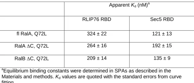
Previously we had measured the affinity of the RLIP76 RBD for both RalA and RalB and found that the two Ral isoforms interacted with similar affinities (our unpublished data and20). We described a similar situation with another Ral effector, Sec522. For practical reasons, these measurements had been undertaken with RalA 1-206 (full-length) and RalB 1-185 (C-terminal truncation). This prompted us to investigate the binding affinities of full-length and truncated RalA and truncated RalB for the RLIP76 RBD, along with the RBD of a second Ral effector, Sec5 for comparison. The apparent Kd values for the interaction between the Ral variants and the RLIP76 RBD were determined by SPA. The binding isotherms are shown in Figure 1 and the affinities are summarized in Table 1. Full-length RalA binds RLIP76 RBD with a Kd of 324 ± 22nM, while RalA C binds with a Kd of 264 ± 16 nM. RalB C binds with a Kd of 209 ± 14 nM. Despite repeated attempts, we were unable to purify sufficiently high quality full-length RalB to perform equivalent experiments with this variant. Full-length RalA binds Sec5 RBD with a Kd of 121 ± 13nM, while RalA C binds with a Kd of 192 ± 15 nM. RalB C binds with a Kd of 135 ± 9 nM. The C-terminal tail of the Ral proteins, as with most small G proteins, is thought to direct
membrane localization in the cell. As such it is usually unavailable for effector binding, although we have previously identified a role for the C-terminal polybasic region from one small G protein (Rac1) in effector binding24. These data indicate that full-length RalA and RalA C bind with equivalent affinity to RLIP 76 and Sec5. RalA C and RalB C also bind with similar affinities to each effector protein. We decided to proceed with a comparative study of RalA C and RalB C
binding to RLIP76 to look for thermodynamic differences in the two Ral-RLIP76 complexes.
Thermodynamic mapping of the RLIP76 and Sec5 binding surfaces on RalB
The structures of Ral proteins in complex with the RLIP76 and Sec5 Ral binding domains25, 26 provided the starting points for mapping the energetics of the interface between RalB and these effectors. We analysed the interfaces of the complexes (PDB codes 1UAD and 2KWI) to identify residues on the small G protein that were within 4Å of an effector residue and mutated these to alanine in RalB. There are two sidechains in RalB that interact with these effectors that are already alanine: Ala48 and Ala77. Ala48 contacts both RLIP76 and Sec5 and was changed to Gly, so that the requirement for the alanine methyl group could be probed. Ala48 lies within switch 1, which is unstructured and highly flexible in active RalB, so substitution with a glycine is unlikely to affect the secondary structure. Ala77 is within a helix in switch 2 in RalB and interacts with His413 and various hydrophobic residues in RLIP76. We therefore mutated it to a larger, charged residue (Arg). Other mutations were also made as follows. Tyr36 forms a hydrogen bond in both Sec5 and RLIP76 complexes, so we mutated it to Phe, reasoning that this would remove the hydroxyl group, while maintaining the bulky aromatic ring. Glu38 forms a salt bridge with Arg27 in the Sec5 complex, so we mutated it to Gln, hence removing the charge but maintaining the size of the sidechain. Two mutations were made to Thr46, although this residue does not interact directly with either effector. Thr46 is equivalent to Ras Thr35, whose mutation to Ser prevented Ras binding to RalGDS and PI3K but not to Raf27, 28. The T46S mutant is the direct equivalent and was tested to investigate whether this mutation would discriminate between RLIP76 and Sec5. The T46A mutation was also generated, to study the effect of removing the hydroxyl group completely.
Finally, the D49E mutation was generated because this is mutant is known to prevent binding to Sec5 but not to RLIP76 in yeast two-hybrid experiments29. This therefore allowed an assessment of whether the in vitro affinity measurements correlate with the effects of this mutation in a (yeast) cell.
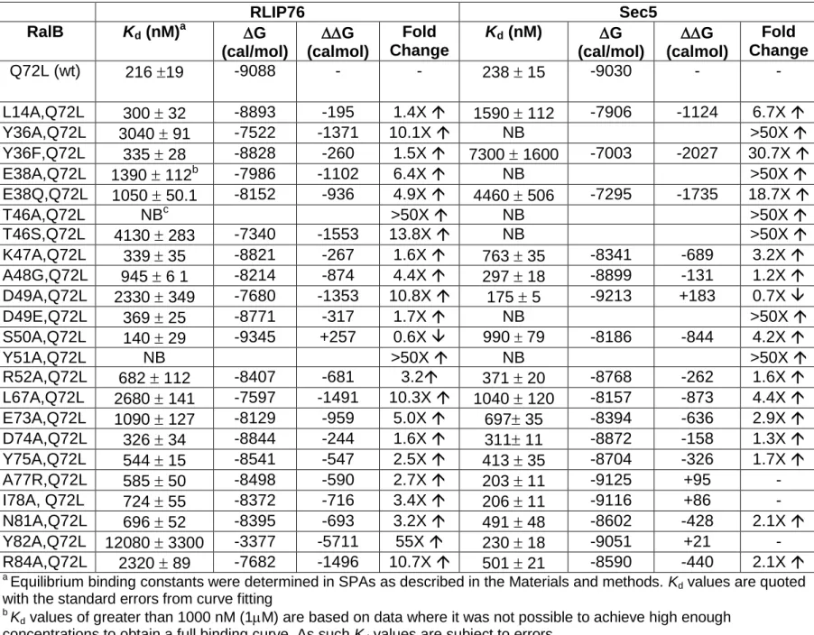
The apparent Kd values for the interaction between the RalB mutants and the RLIP76 and Sec5 RBDs were determined by SPA. Selected binding isotherms are shown in Figure 2A and 2B and the affinities are summarized in Table 2.
Mutations that reduce the binding between RLIP76 RBD and RalB include Y36A, T46A, T46S, D49A, Y51A, L67A, Y82A and R84A, which decrease the affinity more than 10-fold, mutations E38A and E73A, which decrease the affinity between 5 and 10-fold and mutations E38Q, A48G, R52A, I78A and N81A, which all decrease the affinity between 3 and 5-fold. Interestingly one mutation, S50A, shows a small increase in binding affinity for RLIP76. Thus Tyr36, Thr46, Asp49, Tyr51, Leu67, Glu73, Tyr82 and Arg84 all contribute at least 1.2kcal/mol to the interface, with Tyr51 and Tyr82 making the largest individual contributions.
Mutations Y36A/F, E38A/Q, T46A/S, D49E and Y51A all effectively abrogate binding to Sec5, mutations L14A, K47A and L67A all decrease the affinity for Sec5 between 3 and 7-fold.
Mutations at other residues show only a minor effect or no change in the binding affinity. Mutation D49A shows a small increase in binding affinity for Sec5.
Thermodynamic mapping of the RalA and RalB binding surfaces on the RLIP76 RBD Having investigated the differences in thermodynamics between a Ral protein and two of its effector proteins, we next wanted to extend our studies to see if there were any differences in the energetic contributions of an effector protein for the two Ral proteins. RalA and RalB have 82% sequence identity but are 100% identical across the residues that contact RLIP7620 so we next investigated the energetic contributions of sidechains on the RLIP76 RBD to binding to the Ral small G proteins. In a similar manner to our dissection of the RalB interaction surface, we identified sidechains of RLIP76 residues that were within 4Å of a RalB sidechain in our RalB-RLIP76 RBD structure and mutated them to alanine. The apparent Kd values for the interaction between the RLIP76 RBD mutants with RalB were determined by SPA. Selected binding isotherms are shown in Figure 3A and the affinities are summarized in Table 3.
Substitution of RLIP76 residues His413, Trp430 and Thr437 with alanine ablated the binding to RalB, alanine substitutions of Leu409, Leu429 and Lys440 reduced the affinity significantly (16.5, 22.8 and 8.8-fold respectively) and substitution of Leu416 with alanine reduced the affinity 4.7-fold. Mutations at the remaining residues tested had deleterious effects on RalB binding but to a lesser extent and were thus considered to be individually insignificant. Exceptionally, introducing the Q417A mutation into the RLIP76 RBD enhanced binding to RalB by more than 2-fold.
We then tested the same panel of RLIP76 RBD mutants for their ability to bind RalA. Selected binding isotherms are shown in Figure 3B and the affinities are summarized in Table 3. Similarly to RalB, mutation to alanine at residues Leu409, His413, Trp430, Thr437 and Lys440 all decrease
binding by ~9-fold or more. Mutation at residue Leu416 decreased the affinity 9-fold, which is larger than the reduction in affinity for RalB for this mutation, while mutation at Leu429 reduced the affinity by 5.6-fold, significantly less than the effect on RalB affinity. Mutating Arg434 to alanine abrogated binding to RalA but had very little effect (2-3-fold reduction) on RalB. The remaining residues, when mutated, all had a small detrimental affect on binding to RalA and were not considered to be significant, except Q433A, which had no affect on RalA binding. There were no mutations that increased the binding of RalA.
Residues Leu409, His413, Leu429, Trp430, Thr437 and Lys440 all contribute >1.2 kcal/mol to the RLIP76-RalB interface. Residues Leu409, His413, Leu416, Tyr430, Arg434, Thr437 and Lys440 of RLIP76 all contribute >1.2 kcal/mol to the RLIP76-RalA interface.
Discussion
Using our structure of RalB bound to the RLIP76 RBD20 and the structure of RalA bound to Sec526, we have designed mutations to probe the thermodynamics of the binding interfaces and dissect the energetic contributions of specific RalB residues for these two effector proteins. Figures 4A and 4B show heat maps for the RLIP76 and Sec5 binding surfaces on RalB. It is immediately striking that the energetically important binding surface for RLIP76 is much more extensive than that for Sec5. This is not surprising, since a comparison of the Ral complex structures reveals that the effectors themselves are strikingly different, as are the regions that they contact on the Ral proteins (Figures 4C and 4D). The Sec5 RBD has an all sheet, Ig-like fold, which forms an intermolecular antiparallel sheet with the 2 strand of RalA, interacting exclusively with residues
in and around switch 1 and burying ~1000 Å2 in the interface26. The RLIP76 RBD-RalB structure20 shows that the RLIP76 RBD forms a simple coiled-coil, which interacts with both switch 1 and switch 2 in RalB and buries ~1700 Å2. It is logical, therefore that we find residues with significant energetic input into RalB-RLIP76 complex formation across RalB switches 1 and 2. For Sec5 the highest energetic contributions are exclusively found from residues in switch 1 of RalB. These thermodynamic differences highlight certain RalB residues that if mutated would differentially discriminate between effectors. For example, RalB Y82A or D49A mutants would no longer bind to RLIP76 but would retain the ability to bind to Sec5. Conversely RalB Y36F or D49E should retain the ability to bind RLIP76 but no longer be competent to bind Sec5. Such mutations should be useful tools to dissect Ral effector pathways in vivo. In fact the affinities of the D49E mutant that we have quantified in vitro are in agreement with the results found for this mutation by yeast two-hybrid and by co-immunoprecipitation in HEK293T cells29. Thus all of the mutants that we describe here are likely to have the same effects in vivo as we have seen in vitro.
Some of the residues whose mutation affects binding are not involved in directly contacting the effector but play a supporting role in maintaining the structure of the RalB interacting residues. For example, in both complexes Thr46 makes no direct contact with the effectors (Figure 4C, 4D) but its mutation to Ser or Ala reduces the binding to both significantly. As this Thr residue contacts the Mg2+ ion in the RalB protein it is not surprising that the integrity of the switch regions is
compromised. The T46A/S mutants can still, however be loaded with the GTP-analogue and T46S shows some affinity for RLIP76. Similarly, Leu67, which is close to the interface in both
effect on binding, presumably because it supports the RalB 2 strand, which is involved in an intermolecular -sheet with Sec5 (Figure 4D). This is also the case for Leu14, which is within 1 and packs next to Leu67. Leu14 reduces Sec5 binding but not RLIP76 binding, highlighting the importance of the intermolecular -sheet in the Sec5 interaction. There are also supporting residues in the RalB-RLIP76 interaction whose mutations affect binding. The E38A mutant knocks out Sec5 binding and reduces RLIP76 binding. In the Sec5 complex Glu38 forms a salt bridge with Sec5 residue Arg27 but Glu38 does not contact RLIP76 directly. Instead, it is likely to be involved in maintaining the position of the neighbouring residue, Tyr36, which does contact RLIP76. It is possible that replacing the charged Glu38 with the smaller, hydrophobic Ala allows the 1 helix to be extended to residue 38, altering its conformation and that of switch 1. This is consistent with the observation that the E38Q mutation has a lesser effect on binding of RLIP76, since Gln38 is not charged but is more polar than Ala. A similar argument may explain the effect of the E73A mutant, which reduces the binding of RLIP76 5-fold. Glu73 is next to the switch 2 helix, 2, and its
mutation to Ala is likely to alter 2 and therefore the ability of residues in this helix to contact RLIP76.
Some of the mutations that we have tested here have previously been generated in RalA and their effects on Exo84 and Sec5 measured30. The effects on Sec5 binding were broadly consistent with our observations on the RalB-Sec5 interaction, for example, the E38A mutation did not affect Exo84 binding but reduced the affinity for Sec5 ~45-fold just as in RalB (Table 1). The R52A mutant of RalA bound to Sec5, with a similar affinity to wild-type, similar to its effects in RalB,
but reduced the affinity of Exo84 18-fold. Arg52 forms a hydrogen bond in the RalA-Exo84 complex so the effects of this mutation can be explained.
We next resolved the contribution of RLIP76 residues to complex formation with RalA and RalB. Figures 5A and 5B show the energetically important residues on RLIP76 for RalA and RalB binding. It is striking that although the residues in RalB that contact RLIP76 are 100% conserved in RalA, the binding hotspots on RLIP76 are not identical (Table 3): removal of the Leu412 sidechain has a small affect on RalA binding but does not change RalB binding; the L429A mutation reduces RalA binding less than 6-fold but has a more significant effect on RalB binding (23-fold); and R434A in the RLIP76 RBD abrogates binding to RalA completely, while binding to RalB is not significantly affected (less than 3-fold).
Leu412 does not directly contact RalB and instead is packed within the RLIP76 coiled-coil directly behind Leu429. It is therefore likely that the effects of the L412A mutation are mediated via small changes in the orientation of the helices of the coiled-coil, which in turn lead to subtle
rearrangements of the sidechains that do interact with the RalA molecule. Replacement of Leu412 with the smaller Ala sidechain would create a cavity inside the coiled coil unless the two helices shift closer together. Such a shift would move one of the helices further away from the Ral molecule.
Leu429 contacts three residues in RalB switch 2, Ala77, Asn81 and Tyr82, all of which are conserved in RalA. Arg434 makes contacts with Lys16 and Asp65, which again are conserved in
RalA. Indeed a comparison of all of the differences in sequence between the truncated versions of RalA and RalB that were used in this study shows that all of the changes are away from the switch regions and the RLIP76 binding site (Figure 5C). The most dramatic difference between RalA and RalB lies in the insertion of Ala116 in the loop between helix 3 and strand 5. The same loop is also modified by the substitution of the neutral Asn119 in RalA with Lys120 in RalB. These changes lead to a different conformation in this loop when RalA and RalB are compared. In both Ral proteins, this loop is mobile, exhibiting high temperature factors in the X-ray structures of RalA and dynamics on a psec-nsec timescale in RalB22. The insertion in the loop is likely to cause a subtle shift in the position of the C-terminus of helix 3, which is in direct contact with the 2 helix at the C-terminus of switch 2 (Figure 5C). Any changes in switch 2 would also be readily transmitted to the 1 and 2 strands, which lie underneath switch 2. Hence, the changes in the 3-5 loop sequence and length may be responsible for the differences in contribution of residues in switch 2 (Ala77, Asn81, Tyr82) and the 1/2 strands (Lys16, Asp65) to interactions with RLIP76 Leu429 and Arg434. For the purposes of this discussion, we have assumed that Leu429 and
Arg434 contact broadly the same residues in RalA as in RalB, which is likely although not certain in the absence of a RalA-RLIP76 structure.
Interestingly substitution of one residue, Gln417, with Ala had little affect on RalA binding but slightly increased (2.2-fold) the affinity of the RalB complex. This residue also contacts RalB switch 2, via the sidechain of Tyr75. The effects of this mutation therefore also supports the idea that switch 2 is subtly different in the RalA and RalB isoforms. This is in agreement with our
NMR analysis, which suggests that switch 2 has some differences in msec timescale dynamics in the two Ral proteins (manuscript in preparation).
A number of the mutations that we have identified are likely to mediate their effects on binding via medium range structural rearrangements. Allostery in GTPases has been observed in several situations and its exploitation is becoming increasingly important for targetting these proteins. Binding of calcium acetate to helix3-loop7 of Ha-Ras, in a crystallized form, results in structural rearrangements that ultimately order the N-terminal region of switch II and position Glu61 in the active site31. Identification of a unique pocket in the Ki-Ras G12C oncogenic variant prompted screening for binding compounds. This pocket is adjacent to the nucleotide binding site and
binding compounds have been identified that change the nucleotide affinity leading to a preference for GDP over GTP and simultaneously block GEF activation using an allosteric mechanism32. A region adjacent to but distinct from the nucleotide binding site has also been utilized to target inhibitors to the Ral GTPases. Again binding to this allosteric site is sufficient to modulate GTPase activity in vitro and in vivo33. Dynamic exchange between different conformations is also well documented in small G proteins. NMR has shown that Ha-Ras bound to GTP or GTP analogues exhibits dynamics on a msec timescale34, 35. Furthermore, 31P NMR experiments suggested the presence of multiple conformations in active forms of small GTPases22, 36. Taken together these studies demonstrate the importance of conformational and structure flexibility to small G protein function and also establish the utility of this feature as a means of attacking these proteins therapeutically.
In summary, our hotspot analysis of the binding of RLIP76 to RalA and RalB suggests that at least some of the differences in the Ral proteins lie in the structure and dynamics of switch 2 and the consequences for effector binding that ensue. We have identified some residues of RLIP76 that can be mutated to prevent its binding to both RalA and RalB e.g. His413 and Trp430. Furthermore, we have identified two residues whose mutation will allow discrimination between the Ral isoforms: the L429A mutation will reduce RalB binding significantly more than RalA binding, while the R434A mutation abrogates RalA binding but has little effect on RalB binding. These mutants represent essential tools for dissecting the roles of RalA and RalB in vivo.
Acknowledgments
We are grateful to the Captain Stephanos Foundation for support to MP during her undergraduate training.
References
[1] Cascone, I., Selimoglu, R., Ozdemir, C., Del Nery, E., Yeaman, C., White, M., and Camonis, J. (2008) Distinct roles of RalA and RalB in the progression of cytokinesis are supported by distinct RalGEFs, EMBO Journal 27, 2375-2387.
[2] Bodemann, B. O., Orvedahl, A., Cheng, T., Ram, R. R., Ou, Y.-H., Formstecher, E., Maiti, M., Hazelett, C. C., Wauson, E. M., Balakireva, M., Camonis, J. H., Yeaman, C., Levine, B., and White, M. A. (2011) RalB and the Exocyst Mediate the Cellular Starvation Response by Direct Activation of Autophagosome Assembly, Cell 144, 253-267.
[3] Kashatus, D. F., Lim, K.-H., Brady, D. C., Pershing, N. L. K., Cox, A. D., and Counter, C. M. (2011) RalA and RalBP1 regulate mitochondrial fission at mitosis, Nature Cell Biology 13, 1108-U1138.
[4] Shipitsin, M., and Feig, L. A. (2004) RalA but not RalB enhances polarized delivery of membrane proteins to the basolateral surface of epithelial cells, Molecular and Cellular Biology 24, 5746-5756.
[5] Rosse, C., Hatzoglou, A., Parrini, M. C., White, M. A., Chavrier, P., and Camonis, J. (2006) RalB mobilizes the exocyst to drive cell migration, Molecular and Cellular Biology 26, 727.
[6] Chien, Y. C., and White, M. A. (2003) Ral GTPases are linchpin modulators of human tumour-cell proliferation and survival, EMBO Reports 4, 800.
[7] Kashatus, D. F. (2013) Ral GTPases in tumorigenesis: Emerging from the shadows, Experimental Cell Research 319, 2337-2342.
[8] Chardin, P., and Tavitian, A. (1989) Coding Sequences of Human RalA and RalB cDNAs, Nucleic Acids Research 17, 4380-4380.
[9] Takai, Y., Sasaki, T., and Matozaki, T. (2001) Small GTP-binding proteins, Physiological Reviews 81, 153-208.
[10] Martin, T. D., Mitin, N., Cox, A. D., Yeh, J. J., and Der, C. J. (2012) Phosphorylation by Protein Kinase C Regulates RalB Small GTPase Protein Activation, Subcellular Localization, and Effector Utilization, Journal of Biological Chemistry 287, 14827-14836. [11] Jullien-Flores, V., Dorseuil, O., Romero, F., Letourneur, F., Saragosti, S., Berger, R.,
Tavitian, A., Gacon, G., and Camonis, J. H. (1995) Bridging Ral GTPase to Rho-Pathways - RLIP76, a Ral Effector With Cdc42/Rac GTPase-Activating Protein Activity, Journal of Biological Chemistry 270, 22473-22477.
[12] Cantor, S. B., Urano, T., and Feig, L. A. (1995) Identification and Characterization of Ral-Binding Protein-1, a Potential Downstream Target of Ral GTPases, Molecular and Cellular Biology 15, 4578.
[13] Park, S. H., and Weinberg, R. A. (1995) A putative effector of Ral has homology to Rho/Rac GTPase activating proteins, Oncogene 11, 2349.
[14] Moskalenko, S., Tong, C., Rosse, C., Mirey, G., Formstecher, E., Daviet, L., Camonis, J., and White, M. A. (2003) Ral GTPases regulate exocyst assembly through dual subunit interactions, Journal of Biological Chemistry 278, 51743-51748.
[15] Chien, Y. C., Kim, S., Bumeister, R., Loo, Y. M., Kwon, S. W., Johnson, C. L., Balakireva, M. G., Romeo, Y., Kopelovich, L., Gale, M., Yeaman, C., Camonis, J. H., Zhao, Y. M.,
and White, M. A. (2006) RalB GTPase-mediated activation of the I kappa B family kinase TBK1 couples innate immune signaling to tumor cell survival, Cell 127, 157-170.
[16] Frankel, P., Aronheim, A., Kavanagh, E., Balda, M. S., Matter, K., Bunney, T. D., and Marshall, C. J. (2005) RalA interacts with ZONAB in a cell density-dependent manner and regulates its transcriptional activity, EMBO Journal 24, 54.
[17] Ohta, Y., Suzuki, N., Nakamura, S., Hartwig, J. H., and Stossel, T. P. (1999) The small GTPase RalA targets filamin to induce filopodia, Proceedings of the National Academy of Sciences of the United States of America 96, 2122-2128.
[18] Sidhu, R. S., Clough, R. R., and Bhullar, R. P. (2005) Regulation of phospholipase C-1 through direct interactions with the small GTPase Ral and calmodulin, Journal of Biological Chemistry 280, 21933.
[19] Jiang, H., Luo, J. Q., Urano, T., Frankel, P., Lu, Z. M., Foster, D. A., and Feig, L. A. (1995) Involvement of Ral GTPase in v-Src-Induced Phospholipase-D Activation, Nature 378, 409.
[20] Fenwick, R. B., Campbell, L. J., Rajasekar, K., Prasannan, S., Nietlispach, D., Camonis, J., Owen, D., and Mott, H. R. (2010) The RaIB-RLIP76 Complex Reveals a Novel Mode of Ral-Effector Interaction, Structure 18, 985-995.
[21] Strugnell, S. A., Wiefling, B. A., and DeLuca, H. F. (1997) A modified pGEX vector with a C-terminal histidine tag: Recombinant double-tagged protein obtained in greater yield and purity, Analytical Biochemistry 254, 147-149.
[22] Fenwick, R. B., Prasannan, S., Campbell, L. J., Nietlispach, D., Evetts, K. A., Camonis, J., Mott, H. R., and Owen, D. (2009) Solution Structure and Dynamics of the Small GTPase RalB in Its Active Conformation: Significance for Effector Protein Binding, Biochemistry 48, 2192-2206.
[23] Graham, D. L., Eccleston, J. F., and Lowe, P. N. (1999) The conserved arginine in Rho-GTPase-activating protein is essential for efficient catalysis but not for complex formation with Rho GDP and aluminum fluoride, Biochemistry 38, 985-991.
[24] Modha, R., Campbell, L. J., Nietlispach, D., Buhecha, H. R., Owen, D., and Mott, H. R. (2008) The Rac1 polybasic region is required for interaction with its effector PRK1, Journal of Biological Chemistry 283, 1492-1500.
[25] Fenwick, R., Prasannan, S., Campbell, L. J., Evetts, K. A., Nietlispach, D., Owen, D., and Mott, H. R. (2008) (1)H, (13)C and (15)N resonance assignments for the active conformation of the small G protein RalB in complex with its effector RLIP76, Biomolecular NMR Assignments 2, 179-182.
[26] Fukai, S., Matern, H. T., Jagath, J. R., Scheller, R. H., and Brunger, A. T. (2003) Structural basis of the interaction between RalA and Sec5, a subunit of the sec6/8 complex, EMBO Journal 22, 3267.
[27] White, M. A., Nicolette, C., Minden, A., Polverino, A., Vanaelst, L., Karin, M., and Wigler, M. H. (1995) Multiple Ras Functions Can Contribute to Mammalian-Cell Transformation, Cell 80, 533-541.
[28] RodriguezViciana, P., Warne, P. H., Khwaja, A., Marte, B. M., Pappin, D., Das, P., Waterfield, M. D., Ridley, A., and Downward, J. (1997) Role of phosphoinositide 3-OH kinase in cell transformation and control of the actin cytoskeleton by Ras, Cell 89, 457-467.
[29] Moskalenko, S., Henry, D. O., Rosse, C., Mirey, G., Camonis, J. H., and White, M. A. (2002) The exocyst is a Ral effector complex, Nature Cell Biology 4, 66-72.
[30] Jin, R. S., Junutula, J. R., Matern, H. T., Ervin, K. E., Scheller, R. H., and Brunger, A. T. (2005) Exo84 and Sec5 are competitive regulatory Sec6/8 effectors to the RalA GTPase, EMBO Journal 24, 2064.
[31] Buhrman, G., Holzapfel, G., Fetics, S., and Mattos, C. (2010) Allosteric modulation of Ras positions Q61 for a direct role in catalysis, Proceedings of the National Academy of Sciences of the United States of America 107, 4931-4936.
[32] Ostrem, J. M., Peters, U., Sos, M. L., Wells, J. A., and Shokat, K. M. (2013) K-Ras(G12C) inhibitors allosterically control GTP affinity and effector interactions, Nature 503, 548-551. [33] Yan, C., Liu, D., Li, L., Wempe, M. F., Guin, S., Khanna, M., Meier, J., Hoffman, B., Owens, C., Wysoczynski, C. L., Nitz, M. D., Knabe, W. E., Ahmed, M., Brautigan, D. L., Paschal, B. M., Schwartz, M. A., Jones, D. N. M., Ross, D., Meroueh, S. O., and Theodorescu, D. (2014) Discovery and characterization of small molecules that target the GTPase Ral, Nature 515, 443-447.
[34] Ito, Y., Yamasaki, K., Iwahara, J., Terada, T., Kamiya, A., Shirouzu, M., Muto, Y., Kawai, G., Yokoyama, S., Laue, E. D., Walchli, M., Shibata, T., Nishimura, S., and Miyazawa, T. (1997) Regional polysterism in the GTP-bound form of the human c-Ha-Ras protein, Biochemistry 36, 9109-9119.
[35] O'Connor, C., and Kovrigin, E. L. (2008) Global conformational dynamics in Ras, Biochemistry 47, 10244-10246.
[36] Geyer, M., Schweins, T., Herrmann, C., Prisner, T., Wittinghofer, A., and Kalbitzer, H. R. (1996) Conformational transitions in p21(ras) and in its complexes with the effector protein Raf-RBD and the GTPase activating protein GAP, Biochemistry 35, 10308-10320.
[37] Nicely, N. I., Kosak, J., de Serrano, V., and Mattos, C. (2004) Crystal structures of Ral-GppNHp and Ral-GDP reveal two binding sites that are also present in Ras and Rap, Structure 12, 2025-2036.
[38] Eswar, N., Webb, B., Marti-Renom, M. A., Madhusudhan, M. S., Eramian, D., Shen, M.-Y., Pieper, U., and Sali, A. (2006) Comparative protein structure modeling using Modeller, Current protocols in bioinformatics Chapter 5, Unit 5.6.
Table 1: The affinities of Ral variants for RLIP76 and Sec5 RBDs Apparent Kd (nM)a RLIP76 RBD Sec5 RBD fl RalA, Q72L 324 ± 22 121 ± 13 RalA C, Q72L 264 ± 16 192 ± 15 RalB C, Q72L 209 ± 14 135 ± 9
aEquilibrium binding constants were determined in SPAs as described in the
Materials and methods. Kd values are quoted with the standard errors from curve
Table 2: The affinities of RalB mutants for RLI76 RBD and Sec5 RBD RLIP76 Sec5 RalB Kd (nM)a G (cal/mol) G (calmol) Fold Change Kd (nM) G (cal/mol) G (calmol) Fold Change Q72L (wt) 216 19 -9088 - - 238 15 -9030 - - L14A,Q72L 300 32 -8893 -195 1.4X 1590 112 -7906 -1124 6.7X Y36A,Q72L 3040 91 -7522 -1371 10.1X NB >50X Y36F,Q72L 335 28 -8828 -260 1.5X 7300 1600 -7003 -2027 30.7X E38A,Q72L 1390 112b -7986 -1102 6.4X NB >50X E38Q,Q72L 1050 50.1 -8152 -936 4.9X 4460 506 -7295 -1735 18.7X T46A,Q72L NBc >50X NB >50X T46S,Q72L 4130 283 -7340 -1553 13.8X NB >50X K47A,Q72L 339 35 -8821 -267 1.6X 763 35 -8341 -689 3.2X A48G,Q72L 945 6 1 -8214 -874 4.4X 297 18 -8899 -131 1.2X D49A,Q72L 2330 349 -7680 -1353 10.8X 175 5 -9213 +183 0.7X D49E,Q72L 369 25 -8771 -317 1.7X NB >50X S50A,Q72L 140 29 -9345 +257 0.6X 990 79 -8186 -844 4.2X Y51A,Q72L NB >50X NB >50X R52A,Q72L 682 112 -8407 -681 3.2 371 20 -8768 -262 1.6X L67A,Q72L 2680 141 -7597 -1491 10.3X 1040 120 -8157 -873 4.4X E73A,Q72L 1090 127 -8129 -959 5.0X 697 35 -8394 -636 2.9X D74A,Q72L 326 34 -8844 -244 1.6X 311 11 -8872 -158 1.3X Y75A,Q72L 544 15 -8541 -547 2.5X 413 35 -8704 -326 1.7X A77R,Q72L 585 50 -8498 -590 2.7X 203 11 -9125 +95 - I78A, Q72L 724 55 -8372 -716 3.4X 206 11 -9116 +86 - N81A,Q72L 696 52 -8395 -693 3.2X 491 48 -8602 -428 2.1X Y82A,Q72L 12080 3300 -3377 -5711 55X 230 18 -9051 +21 - R84A,Q72L 2320 89 -7682 -1496 10.7X 501 21 -8590 -440 2.1X
a Equilibrium binding constants were determined in SPAs as described in the Materials and methods. K
d values are quoted
with the standard errors from curve fitting
b K
d values of greater than 1000 nM (1M) are based on data where it was not possible to achieve high enough
concentrations to obtain a full binding curve. As such Kd values are subject to errors. c NB (no binding) denotes data that could not be fitted to the binding isotherm
Table 3: The affinities of RLIP76 RBD mutants for Ral isoformsa
RalA RalB
RLIP76 RBD Kd (nM) G (cal/mol) G (cal/mol) Fold
Change
Kd (nM) G (cal/mol) G (cal/mol) Fold
Change C411S (wt) 185 ± 5 -9180 - - 261 ± 17 -8976 - - L409A,C411S 1880 ± 297b -7807 -1373 10.0X 4300 ± 1400 -7317 -1659 16.5X L412A,C411S 774 ± 123 -8332 -848 4.2X 228 ± 24 -9056 - - H413A,C411S NBc >69X NB > 16X L416A,C411S 1700 ± 309 -7866 -1314 9.2X 1240 ± 147 -8053 -923 4.7X Q417A,C411S 126 ± 27 -9407 - - 99 ± 22 -9550 +574 2.2X K421A,C411S 433 ± 37 -8676 -504 2.3X 441 ± 30 -8665 -311 0.6X E426A,C411S 437 ± 131 -8671 -509 2.4X 670 ± 33 -8418 -558 2.6X E427A,C411S 452 ± 45 -8651 -529 2.4X 618 ± 39 -8465 -511 2.4X L429A,C411S 1030 ± 308 -8163 -1017 5.6X 5940 ± 928 -3588 -5388 22.8X W430A,C411S NB >69X NB > 16X Q433A,C411S 216 ± 43 -9088 - - 197 ± 20 -9142 - - R434A,C411S NB >69X 678 ± 77 -8411 -565 2.6X T437A,C411S NB >69X NB > 16X K440A,C411S 1590 ± 410 -7906 -1274 8.6X 2300 ± 450 -7687 -1289 8.8X
a Equilibrium binding constants were determined in SPAs as described in the Materials and methods. K
d values are quoted with the standard errors from curve
fitting
b K
d values of greater than 1000 nM (1M) are based on data where it was not possible to achieve high enough concentrations to obtain a full binding curve. As
such Kd values are subject to errors.
Figure 1: SPA binding data for full-length RalA, truncated RalA and truncated RalB with the RLIP76 RBD and the Sec5 RBD. The indicated concentration of [3H]GTP-labelled G protein was incubated with either His-tagged RLIP76 RBD or GST-tagged Sec5 RBD, as appropriate, in each SPA. The SPA signal was corrected by subtraction of the background signal from parallel measurements in which the effector protein was omitted. The effect of the concentration of G protein on this corrected SPA signal was fitted to a binding isotherm to give an apparent Kd value and the signal at saturating G protein concentrations. The data and curve fits are displayed as a percentage of this maximal signal: (A) binding isotherms of full-length and truncated RalA and truncated RalB with RLIP76 RBD, (B) binding isotherms of full-length and truncated RalA and truncated RalB with Sec5 RBD.
Figure 2: SPA binding data for truncated RalB and mutant variants with the RLIP76 RBD and the Sec5 RBD. The indicated concentration of [3H]GTP-labelled G protein was incubated with either His-tagged RLIP76 RBD or GST-tagged Sec5 RBD, as appropriate, in each SPA. The SPA signal was corrected by subtraction of the background signal from parallel measurements in which the effector protein was omitted. The effect of the concentration of G protein on this
corrected SPA signal was fitted to a binding isotherm to give an apparent Kd value and the signal at saturating G protein concentrations. The data and curve fits are displayed as a percentage of this maximal signal: (A) binding isotherms of truncated RalB and mutants with the RLIP76 RBD, (B) binding isotherms of truncated RalB and mutants with the Sec5 RBD.
Figure 3: SPA binding data for the RLIP RBD and mutant variants with truncated RalA and RalB. The indicated concentration of [3H]GTP-labelled G protein was incubated with the
appropriate His-tagged RLIP76 RBD variant in each SPA. The SPA signal was corrected by subtraction of the background signal from parallel measurements in which the effector protein was omitted. The effect of the concentration of G protein on this corrected SPA signal was fitted to a binding isotherm to give an apparent Kd value and the signal at saturating G protein concentrations. The data and curve fits are displayed as a percentage of this maximal signal: (A) binding isotherms of the RLIP76 RBD variants with truncated RalB, (B) binding isotherms of the RLIP76 RBD variants with truncated RalA.
Figure 4: Structural details of the RalB-RLIP76 and RalB-Sec5 interfaces.
A. Residues whose mutation to Ala affects binding to RLIP76. RalB is shown in a blue ribbon representation, overlaid with a semi-transparent blue surface. Relevant residues are coloured as follows: red, more than 10-fold weaker affinity; orange, 5 to 10-fold weaker affinity; yellow, 3 to 5-fold weaker affinity. Switch 1 encompasses residues 40-50 and switch 2 encompasses residues 70-84, as assigned by comparing the structures in PDB entries 1U8Y and 1U9037.
B. Residues whose mutation to Ala affects binding to Sec5. The colours are the as the same as in A.
C. The structure of the RalB-RLIP76 complex (PDB 2KWI) is shown with the residues whose mutation affects RLIP binding shown as sticks. RalB is blue, RLIP76 is dark pink. The mutated residues are shown in the same colour scheme as in A.
D. A model of RalB-Sec5 is shown, constructed using Modeller38 based on PDB 1UAD, with the residues that affect Sec5 binding shown as sticks. RalB is blue and Sec5 is pale pink. The mutated residues are shown in the same colour scheme as in A.
Figure 5: Hotspots on RLIP76 for binding to Ral proteins
A. Residues whose mutation to Ala disrupts binding to RalA. Relevant residues are coloured as follows: red, more than 10-fold weaker affinity; orange, 5 to 10-fold weaker affinity; yellow, 3 to 5-fold weaker affinity.
B. Residues whose mutation to Ala disrupts binding to RalB. The colour scheme for residues is the same as in A.
C. The two residues whose mutation has drastically different effects on RalA and RalB binding to RLIP76. RalB is blue and RLIP76 is dark pink. Leu429 and Arg434 are shown in green and the residues that they contact in RalB are shown in yellow. The positions of conservative changes between RalA and RalB are shown as cyan spheres: these include Asp/Glu, Lys/Arg, Val/Ile/Leu or Asn/Gln exchanges only. The positions of less conservative changes between the two proteins are shown as orange spheres. Finally, the position of a single amino acid insertion (Ala116) in RalB is shown as a red sphere.