Stem Cell Reports
Article
Deterministic
HOX
Patterning in Human Pluripotent Stem Cell-Derived
Neuroectoderm
Ethan S. Lippmann,1,2Clay E. Williams,3,4David A. Ruhl,5,6Maria C. Estevez-Silva,1,2Edwin R. Chapman,5,6 Joshua J. Coon,3,4,7and Randolph S. Ashton1,2,*
1Department of Biomedical Engineering 2Wisconsin Institute for Discovery 3Genome Center of Wisconsin 4Department of Biomolecular Chemistry 5Department of Neuroscience
6Howard Hughes Medical Institute 7Department of Chemistry
University of Wisconsin, Madison, WI 53706, USA *Correspondence:[email protected]
http://dx.doi.org/10.1016/j.stemcr.2015.02.018
This is an open access article under the CC BY-NC-ND license (http://creativecommons.org/licenses/by-nc-nd/4.0/). SUMMARY
ColinearHOXexpression during hindbrain and spinal cord development diversifies and assigns regional neural phenotypes to discrete rhombomeric and vertebral domains. Despite the precision ofHOXpatterning in vivo, in vitro approaches for differentiating human pluripotent stem cells (hPSCs) to posterior neural fates coarsely patternHOXexpression thereby generating cultures broadly specified to hindbrain or spinal cord regions. Here, we demonstrate that successive activation of fibroblast growth factor, Wnt/b-catenin, and growth differentiation factor signaling during hPSC differentiation generates stable, homogenous SOX2+/Brachyury+neuromesoderm that exhibits progressive, full colinearHOXactivation over 7 days. Switching to retinoic acid treatment at any point during this process halts colinearHOXactivation and transitions the neuromesoderm into SOX2+/PAX6+neuroectoderm with predictable, discreteHOX gene/protein profiles that can be further differentiated into region-specific cells, e.g., motor neurons. This fully defined approach signif-icantly expands capabilities to derive regional neural phenotypes from diverse hindbrain and spinal cord domains.
INTRODUCTION
The human genome contains 39 HOX genes divided among four clusters and classified into 13 paralogous groups based on sequence homology (Figure 1A). During embryonic stages of body axis elongation, newly formed tissues express HOX genes in sequential, contiguous domains consistent with their order in each cluster, i.e., colinearly. This phenomenon is evolutionarily conserved in bilaterian species and spatially assigns body segment-specific differentiation trajectories to axial progenitors of all three germ layers (Lewis, 1978). During formation of the posterior CNS, progenitors proximal to the node progressively transition from a 30 to 50 HOX expression profile as the primitive streak regresses (Iimura and Pour-quie´, 2006). This process produces nested and overlapping axial domains ofHOXexpression within the neuroepithe-lium of hindbrain rhombomeric (HOX1-5) and spinal cord cervical (HOX5-9), thoracic (HOX9-10), and lumbosa-cral (HOX10-13) vertebral segments (Figure 1A) ( Philippi-dou and Dasen, 2013). The spatial variation of HOX expression along the rostrocaudal (R/C) axis of the poste-rior CNS diversifies the fates of neuroepithelial progeny and precisely restricts the development of specific neural subtypes to discrete axial positions (Philippidou and Dasen, 2013).
Retinoic acid (RA), wingless-type MMTV integration site protein family (Wnt), fibroblast growth factor (FGF), and growth differentiation factor (GDF) signaling intricately regulate HOX expression during posterior CNS develop-ment (Liu et al., 2001; Nordstro¨m et al., 2006). Yet, it re-mains controversial how these factors should be applied to human pluripotent stem cells (hPSCs) to recapitulate full colinear HOX activation and enable deterministic patterning of diverse R/CHOXprofiles, i.e., regional hind-brain or spinal cord phenotypes, during neural differentia-tion. RA is used ubiquitously to caudalize hPSC-derived neuroectoderm because its secretion by paraxial somitic mesoderm is believed to convert 30HOXgenes in hindbrain and spinal cord neural tissues from an epigenetically repressed to a transcriptionally active state (Gould et al., 1998; Mazzoni et al., 2013; Bel-Vialar et al., 2002). How-ever, RA activatesHOX1-5chromatin domains in a salta-tory (Mazzoni et al., 2013) versus colinear manner, which patterns mouse (mPSC) and human PSC-derived neuro-ectoderm with a broad caudal hindbrain thru cervical spinal cord identity. This is evidenced by demonstra-tions that RA-mediated caudalization generates HOXB4+ (Li et al., 2008), HOXC4+/HOXA5+/HOXC6/HOXC8 (Mazzoni et al., 2013), HOXC5+/HOXC6+ (Peljto et al., 2010; Wichterle et al., 2002), or HOXC6+/HOXC8+ (Lee et al., 2007; Li et al., 2005) motor neurons (MNs), or
HOXB4+ astrocytes (Krencik et al., 2011). Alternatively, Wnt and FGF treatments in the absence of RA can caudalize neurally differentiating mPSCs by inducing saltatory activation ofHOX1-9paralogs to coarsely pattern a hetero-geneous mixture of HOXC6+ cervical and HOXD9+ thoracic spinal tissues, which can be further caudalized to also contain HOXD10+lumbar tissues upon GDF11 sup-plementation (Mazzoni et al., 2013; Peljto et al., 2010). Also, a recent report demonstrated that manipulation of Wnt and RA concentrations could yield MNs possessing hindbrain or rostral spinal phenotypes but more caudal and further partitioning of regional HOX identity was not achieved (Maury et al., 2014). Overall, an approach
for hPSC neural differentiation that recapitulates the preci-sion, extent, and predictability of colinearHOXpatterning observed in vivo remains elusive. Its development would enable unprecedented access to diverse regional pheno-types that populate posterior CNS tissues and generation of human disease models containing regional neural phenotypes with differential susceptibility to neurodegen-erative disorders (Brockington et al., 2013; Kaplan et al., 2014; Sandoe and Eggan, 2013).
Recent progress in understanding how the posterior CNS develops led us to consider other methods for in vitroHOXpatterning. New evidence suggest that bipo-tent SOX2+/Brachyury(T)+neuromesodermal progenitors Figure 1. Wnt/b-Catenin and FGF Signaling Synergistically CoordinateHOXActivation during hPSC Differentiation
(A)HOXparalog expression in the hindbrain and spinal cord color-coded to the region where its expression is first detected (modified from
a previous report;Philippidou and Dasen, 2013).
(B) Timeline of H9 hESC differentiation corresponding to (C)–(E). (C) RT-PCR analysis of cultures at days 2–5.
(D) qPCR analysis at day 5. *p < 0.005; **p < 0.001.
(E) OTX2 and SOX2 expression at day 5 after RA or CHIR treatment. Scale bars, 100mm. Adjacent images are the same field.
(F) qPCR analysis at day 2 using the H9 ishcat2 line with or without doxycycline treatment. *p < 0.02; **p < 0.005. (G) qPCR analysis at day 5 using the H9 ishcat2 line with or without doxycycline treatment. *p < 0.02; **p < 0.0001.
All qPCR data are presented as mean±SD calculated from independent biological triplicates. H9 data are normalized to the condition
yielding maximum expression and ishcat2 data are normalized to each doxycycline-free condition. Statistical significance was calculated using the Student’s unpaired t test.
(NMPs) residing in a stem zone proximal to the primitive streak’s regressing node are the cell source for both the posterior neural tube and flanking paraxial mesoderm (Delfino-Machı´n et al., 2005; Takemoto et al., 2011; Tzoua-nacou et al., 2009). These NMPs are maintained by syner-gistic Wnt/b-catenin and FGF signaling from the node and primitive streak that induces SOX2 expression via activation of enhancer N1 (Takemoto et al., 2006, 2011). FGF signaling from the node also helps maintain the NMP state by repressing the expression of the neuroecto-derm fate determinant PAX6 (Bertrand et al., 2000; Mathis et al., 2001). As the node regresses, NMPs rostral to the stem zone experience a decline in FGF signaling, which initiates paraxial mesoderm differentiation (i.e., somite formation). Then, the somites secrete RA back to the presumptive neural tube, which induces PAX6 expression to generate definitive neuroectoderm of the posterior CNS (Diez del Corral et al., 2003; Molotkova et al., 2005). The discovery that Wnt/FGF signaling induces NMPs that progressively caudalizeHOXexpression and undergo RA-induced neuro-ectoderm differentiation to form the hindbrain and spinal cord suggests an alternative approach for patterningHOX expression in vitro. Guoti et al. recently had partial success recapitulating theHOX-expressing NMP state from hPSCs in vitro, but the NMP phenotype could only be maintained for 3 days preventing acquisition of more caudal HOX genes/proteins before exhibiting a mesodermal shift (Gouti et al., 2014). Also, they were unable to partition HOX patterning within hindbrain and spinal cord regions. Therefore, we proceeded to test whether (1) Wnt/b-catenin and FGF signaling could differentiate hPSCs to stable NMPs to permit full, colinear activation of HOX genes; (2) RA exposure could differentiate NMPs to neuroectoderm; (3) this approach to deriving human neuroectoderm could enable enhanced control ofHOXpatterning.
Using a chemically defined, monolayer neural differen-tiation system (Lippmann et al., 2014), we determined that by temporally modulating Wnt/b-catenin and FGF signaling, hPSCs could be differentiated into pure SOX2+/ Brachyury+ NMPs that were stable for 7 days. Over this period, the NMPs exhibited colinear activation of hind-brain, cervical, and thoracicHOXgenes plus lumbosacral
HOX genes with GDF11 supplementation. At any point during NMP propagation, a transition to RA was necessary and sufficient to induce PAX6+/SOX2+ neuroectodermal differentiation, precisely arrest progressiveHOXactivation, and thereby generate discrete HOX protein profiles indi-cative of distinct R/C domains. Overall, we demonstrate the ability to deterministically pattern neuroectoderm to diverse R/C domains by simply varying the amount of time NMPs spend under Wnt/b-catenin, FGF, and GDF signaling prior to RA exposure. Furthermore, we demonstrate that neuroectoderm of differing HOX
expres-sion profiles produce region-specific hindbrain, cervical, thoracic, and lumbar MN populations. Therefore, this temporal biphasic approach could be used to produce regional neural phenotypes from diverse hindbrain and spinal cord domains.
RESULTS
Regulation ofHOXandCDXExpression by RA, Wnt/b-Catenin, and FGF Signaling
We used the chemically defined ‘‘E6’’ method to investigate the effects of Wnt, FGF, RA, and GDF11 during hPSC neural differentiation because it lacks the use of small molecule inhibitors that could influence fate choices and does not form non-neural germ layers that could initiate unknown signaling crosstalk (Lippmann et al., 2014). We first differ-entiated H9 human embryonic stem cells (hESCs) in E6 medium for 24 hr and then added RA, CHIR99021 (CHIR; a small molecule Wnt/b-catenin agonist), FGF8b, or their relevant combinations and monitored HOX
expression by RT- and quantitative PCR (qPCR) over 4 days (Figures 1B–1D). The addition of RA or CHIR, but not FGF8b, induced temporal, progressive expression of
HOXA1-HOXC5 (Figure 1C). However, CHIR/FGF8b, but not RA/FGF8b, accelerated the progression ofHOX activa-tion inducing colinear activaactiva-tion ofHOXC6,HOXC8, and
HOXC9 at levels ranging from 39- to 3,400-fold higher than the other treatments (Figures 1C and 1D). Moreover, the caudalizing effect of CHIR/FGF8b was not impeded by the addition of HX531, an RXR inhibitor, and CHIR but not RA treatment was able to reduce gene and protein expression of the midbrain/forebrain marker OTX2 (Figures 1C–1E; Figure S1). These results indicate that while RA can induce some degree of posterior identity indicated by HOX1-5 expression, CHIR/FGF8b induces rapid patterning of differentiating hESCs to an OTX2 posterior phenotype with caudal HOX1-9 expression in an RA-independent manner. This is in agreement with other neural differentiation studies (Gouti et al., 2014; Mazzoni et al., 2013)
Additionally, CHIR/FGF8b treatment yielded substan-tially elevated expression of CDX genes, which are the upstream regulators of HOX genes (Nordstro¨m et al., 2006): compared to RA, CHIR/FGF8b upregulatedCDX1 by >6-fold,CDX2by >1,800-fold, andCDX4by >330-fold (Figure 1D). To confirm that CHIR-mediatedHOX induc-tion requiresb-catenin signaling, we utilized the H9 ishcat2 line, which harbors a doxycycline-inducible b-catenin small hairpin RNA (shRNA) cassette (Lian et al., 2012). Addition of doxycycline prior to CHIR treatment reduced
b-catenin (CTNNB1) expression by 3-fold and decreased
respectively (Figure 1F). In contrast, while doxycycline treatment also reducedb-catenin by 3-fold in RA-treated cells, it did not reduceHOXA1orHOXB1expression, sug-gesting RA-mediated induction of HOX transcription is
b-catenin independent (Figure 1F). To verify sustained
b-catenin signaling promotes progressiveHOXactivation, we inducedHOXexpression with CHIR/FGF8b for 1 day and continued the treatment for 3 days with and without doxycycline. Doxycycline treatment reducedCTNNB1by 4-fold,HOXC8by 2-fold, andHOXC9by 2-fold, indicating that sustainedb-catenin signaling contributes to progres-siveHOXactivation (Figure 1G).
Derivation of Stable NMPs Exhibiting Full Colinear HOXActivation
Gouti et al. recently demonstrated that simultaneous induction of Wnt/b-catenin and FGF signaling can
differ-entiate hPSCs to SOX2+/Brachyury(T)+NMPs, but NMPs persisted for only 3 days before shifting to a mesodermal fate under their treatment regimen (Gouti et al., 2014). We observed a similar trend with simultaneous CHIR and FGF8b treatment inducing uniform Brachyury but also causing a sharp decrease in SOX2 expression (23%±0% SOX2+), indicating a mesodermal fate shift (Figure 2A, Route 1). Conversely, pre-treatment with FGF8b prior to CHIR (pre-FGF8b/CHIR) yielded uniform expression of both Brachyury and SOX2 that could be maintained (75%–100% SOX2+/Brachyury+) for 7 days (Figure 2A, Route 2, and Figure 2B). Thus, FGF signaling upstream of Wnt/b-catenin signaling effectively induces a stable NMP identity during hPSC differentiation.
Next, we investigated whether colinearHOXexpression was evident within our NMP cultures. Analogous to their in vivo counterparts, these cultures exhibited colinear
Figure 2. ColinearHOXExpression in hPSC-Derived NMPs
(A) Analysis of the NMP phenotype by flow cytometry and immunocytochemistry. Gray histogram, immunoglobulin G (IgG) control; red
histogram, label of interest. Scale bars, 100mm.
(B) Optimized NMP propagation scheme. The addition of CHIR is denoted as t = 0 hr in all panels. Purity of NMPs was assessed by flow
cytometry in the presence or absence of GDF11 (data are presented as mean±SD). qPCR analysis ofHOXD10andHOXD11was conducted in
the presence or absence of GDF11 (expression normalized to the time point of maximum expression).
(C) Schematic forHOXinduction in NMPs. ‘‘t’’ indicates time under Wnt, FGF, and GDF signaling.
(D) qPCR analysis of colinearHOXexpression normalized to the time point of maximum expression. GDF11 was included according to (B).
qPCR data in all panels are presented as mean±SD calculated from technical duplicates. Full profiles for all analyzed genes can be found in
activation ofHOXgenes over the 7-day period (Figures 2B– 2D;Figure S2). In our system, 30HOXtranscripts remained expressed even after activation of caudal paralogs and fluc-tuated within an5-fold range (Figure S2A), also agreeing with Gouti et al. (2014). Supplementation with GDF11, a transforming growth factor b (TGF-b) family member expressed at later stages of NMP propagation in vivo (Liu, 2006; Liu et al., 2001), was necessary for robust activation of lumbosacral HOX paralogs (Philippidou and Dasen, 2013) (Figures 2B–2D;Figure S2A) but did not significantly repress transcription of rostralHOXgenes (Figure S2B) or disrupt the NMP state (Figure 2B). Moreover, premature GDF11 treatment (initiated after 1 day of FGF8b/CHIR) could induceHOXD10but at >100-fold lower expression compared to GDF11 treatment after 4 days of FGF8b/ CHIR, indicating robust lumbosacral patterning requires prior activation of rostral HOX genes (Figure S2C). We were able to detect colinear transcription of 33 of the 39
HOX genes in hPSC-derived NMP cultures, excluding
HOXB3, HOXC12, and theHOX13 paralogs, over a time span consistent with posterior neural tube formation according to the Carnegie stages of human development (Figure S2A). Moreover, temporal HOX activation was also observed in NMPs derived from IMR90-4 iPSCs (Figure S2D), indicating these mechanisms translate to other hPSC lines. While we believe the lack of HOXB3 detection was due to experimental or qPCR reagent error, our extensive literature search was unable to find definitive documentation ofHOXC12orHOX13paralog expression in the posterior CNS (Dasen et al., 2005).
RA Converts NMPs to Neuroectoderm
In vivo, regression of the stem zone along the primitive streak yields a rostral decline in FGF signaling that coin-cides with RA secretion from newly formed somites and PAX6 expression in the newly formed neural tube (Diez del Corral et al., 2003). This suggests that RA could induce a NMP-to-neuroectoderm fate switch. To test this in vitro, we exposed hPSC-derived NMPs to E6 medium containing RA, CHIR, or neither for 2 days. Cultures exposed to only E6 medium maintained SOX2 and Brachyury expression but gained minimal PAX6 (96%±4% SOX2+, 15%±8% PAX6+, 78% ±16% Brachyury+), while exposure to only CHIR induced a mesodermal fate shift as evidenced by SOX2 downregulation, no PAX6 induction, and uniform maintenance of Brachyury expression (50%±8% SOX2+, 96% ± 1% Brachyury+) (Figure 3A). However, when exposed to RA, the cells gained PAX6 expression along with SOX2 maintenance (98%± 4% SOX2+, 97% ± 3% PAX6+) and Brachyury downregulation (43%± 4%) indi-cating a neuroectodermal fate shift (Figure 3A). Moreover, transitioning to RA at any point during NMP propagation generated a highly pure (>83%) PAX6+/SOX2+
neuroepi-thelial culture, i.e., polarized N-cadherin, within 4 days (Figures 3B–3D). If GDF11 was added to facilitate lumbosa-cral patterning, PAX6 expression in response to RA was decreased in a dose-dependent manner (Figures S3A– S3C). The ability of GDF11 to control R/C patterning is mediated by signaling via ALK5 (Andersson et al., 2006) and activation of the SMAD2/3 complex (Liu, 2006). GDF11 has also been shown to activate SMAD1/5/8 in vitro (Liu, 2006), which contributes to dorsal patterning (Tozer et al., 2013). Since dorsomorphin selectively inhi-bits ALK2, ALK3, and ALK6, which blocks SMAD1/5/8 signaling (Yu et al., 2008), we hypothesized the addition of dorsomorphin should prevent acquisition of a dorsal phenotype without affecting R/C patterning. Indeed, the addition of dorsomorphin with GDF11 and throughout RA treatment was sufficient to recover PAX6 expression (83%±4% PAX6+;Figure S3D). Thus, highly pure neuro-ectoderm cultures could be obtained from NMPs at any point during colinearHOXactivation.
Deterministic Hox Patterning Can Be Achieved Using a Temporal Transition between Wnt/FGF/GDF and RA Treatment
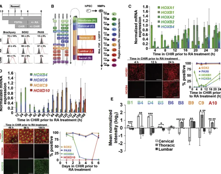
Because RA production in vivo and treatment in vitro coin-cides with neural differentiation, we and others (Diez del Corral and Storey, 2004) posited that it might also serve as a signal to arrest colinearHOXactivation. We therefore tested the ability of RA to generate neuroectoderm with discrete, region-specificHOXexpression profiles in accor-dance with the status of colinearHOXactivation observed in the NMP state (Figure 3B). Also, we assessed the expres-sion of HOX transcription factors (TFs) that would demar-cate distinct hindbrain and spinal cord domains (Dasen et al., 2003, 2005). First, we evaluated our ability to pattern neuroectoderm to hindbrain domains using a 0- to 30-hr FGF8b/CHIR NMP propagation period prior to 4 days of RA treatment (Figures 3B and 3C). qPCR analysis of the re-sulting neuroectoderm cultures revealed elevated expres-sion of HOXA1andHOXA2prior toHOXB1,HOXB2, and
HOXB4. Immunocytochemical analysis revealed that early cultures were HOXB1+/HOXB4(2 hr FGF8b/CHIR, 4 days RA; 63%±6% HOXB1+) but rapidly lost HOXB1 expression if derived after only an additional 4 hr of NMP propagation (6 hr FGF8b/CHIR, 4 days RA; 5% ± 2% HOXB1+) ( Fig-ure 3C). If neuroectoderm was derived after 24 hr of NMP propagation, it reacquired HOXB1 and now also expressed HOXB4 (24 hr FGF8b/CHIR, 4 days RA; 63%±5% HOXB1+ and 37%±8% HOXB4+) (Figure 3C). This mirrors HOX TF expression during development of the fourth, fifth, and sixth through eighth rhombomeres, respectively, indi-cating that transitioning from Wnt/FGF to RA at the 2- to 4-hr time point generates HOXB1+ neuroectoderm that could potentially produce facial nerve MNs whereas
Figure 3. RA Induces a Neuroectodermal Fate and Halts ColinearHOXActivation
(A) NMPs exposed to RA or CHIR acquire a neuroectodermal or mesodermal fate as assessed by flow cytometry at day 6 (gray histogram, IgG control; red histogram, antigen of interest).
(B) Schematic for transition from NMPs to neuroectoderm by RA treatment. ‘‘t’’ indicates time under Wnt, FGF, and GDF signaling prior to
RA treatment, which haltsHOXprogression to yield a defined rostrocaudal identity.
(C and D) Representative hindbrain (C) and spinal cord (D) cultures assessed by qPCR, immunocytochemistry, and flow cytometry. For all
plots, flow cytometry data are presented as mean±SD from biological duplicates, and qPCR data are mean±SD from technical duplicates
normalized to the time point of maximum expression for each gene. For hindbrain cultures, HOXB1 and HOXB4 were quantified by
immunocytochemistry relative to DAPI+nuclei (technical triplicates per time point, minimum 10,000 cells counted, and relative HOX
expression patterns were qualitatively verified across biological duplicates), while SOX2 and PAX6 were quantified by flow cytometry. For
spinal cord cultures, HOXD10 was quantified by immunocytochemistry relative to DAPI+ nuclei (technical duplicates, >2,000 cells
counted), while SOX2, PAX6, and HOXB4 were quantified by flow cytometry. Scale bars, 100mm. For (D), dorsomorphin was included with
GDF11 and throughout RA treatment (further details inFigure S3).
(E) Mass spectrometry comparison of HOX profiles in cervical, thoracic, and lumbar neuroectoderm cultures. Cervical differentiation: 1 day FGF8b, 2 days FGF8b/CHIR, 4 days RA; thoracic differentiation: 1 day FGF8b, 6 days FGF8b/CHIR, 4 days RA; lumbar differentiation: 1 day FGF8b, 4 days FGF8b/CHIR, 2 days FGF8b/CHIR/GDF11, 4 days RA. Biological triplicates (neuroepithelium isolated individually from three separate culture wells) were used to calculate statistical significance between cervical, thoracic, and lumbar samples.
Data are presented as mean±SD and statistical significance was calculated using the Student’s unpaired t test. *p < 0.01; **p < 0.002;
transitioning at 24 hr generates HOXB4+neuroectoderm of the caudal hindbrain (Bell et al., 1999; Philippidou and Dasen, 2013; Bel-Vialar et al., 2002). Interestingly, the high percentage of HOXB1+cells at the 2 hr mark coincided with low HOXB1 expression (10% of the maximum observed expression at 20 hr) suggesting that while tran-script levels are useful for identifying relative HOXgene expression, they may not be directly instructive for pre-dicting HOX TF profiles that arise due to cross-repressive interactions (Bell et al., 1999; Dasen et al., 2003, 2005). Also, the observed abrupt transitions in neuroectoderm HOX TF expression suggest fairly uniform progressive
HOXactivation within the NMPs (Figure 3C).
Next, we performed a similar Wnt/FGF/GDF-to-RA transition time-course analysis focusing on later time points that would generate neuroectoderm patterned to spinal cord domains (Figures 3B and 3D). qPCR analysis again demonstrated that RA treatment could halt the in-duction of 50paralogs that would otherwise have become expressed in continuous Wnt/FGF/GDF11 treatment. Using both immunocytochemistry and flow cytometry, we observed uniform HOXB4 expression in neuroectoder-mal cultures derived from NMPs transitioned to RA after 24–48 hr of propagation, correlating to a rostral cervical spinal cord domain (Figures 3B and 3D). HOXB4 expres-sion dropped sharply (from 84%± 10% down to 12%± 4% HOXB4+ cells) when the transition to RA occurred at 72 hr (Figure 3D), which coincided with the initiation of thoracic HOXC9 expression, a known repressor of
HOX4 paralog expression (Jung et al., 2010). Further-more, HOXD10 was only detected in neuroectodermal cultures when RA was added after144 hr of NMP pro-pagation (Figures 3B and 3D;Figure S3E), while HOXB4 remained repressed. Additional insights into the diversity of HOX expression in neuroectoderm patterned to cervi-cal, thoracic, and lumbar spinal cord domains were ob-tained using quantitative mass spectrometry (Figure 3E; Table S1). As expected, HOXC9 was expressed in thoracic neuroectoderm but repressed in the HOXD10+ lumbar culture, and many (HOXB1, HOXB4, HOXD4, HOXB5, HOXB8, HOXC9, and HOXA10) but not all (HOXB9) detected HOX factors exhibited expression patterns indic-ative of similar cross-repressive interactions (Dasen et al., 2005) (Figure 3E). Thus, these results demonstrate re-gion-specific patterning of hPSC-derived neuroectoderm in the spinal cord, and similar to the hindbrain neuro-ectoderm cultures, repression of HOX proteins occurred even though their HOX transcripts remained expressed, possibly indicating cross-repressive interactions at this stage are occurring at the post-transcriptional stage (Yekta et al., 2004). Hence, our temporal biphasic approach for differentiating hPSCs to neuroectoderm through a stable NMP intermediate enables deterministicHOXpatterning
to impart diverse and discrete hindbrain and spinal cord regional identities (Figure 3B).
Neuroectoderm with Region-SpecificHOXProfiles Generate Regional MN Populations
We used MN differentiation to evaluate whether the regional specification imparted by our HOX patterning approach yielded characteristic differences in the neuroec-toderm progeny. First, we verified that MNs derived from neuroectoderm of discrete hindbrain domains likewise exhibited distinct HOX signatures. Hindbrain neuromeso-derm was propagated for 4, 12, and 24 hr before conversion to ventralized neuroectoderm using RA, sonic hedgehog (SHH) and purmorphamine (PM, a sonic hedgehog agonist) exposure and further differentiation to MNs using media containing RA and neurotrophic factors (NTFs). MN differentiation was verified by the presence of cells co-expressing bIII-tubulin/ISL1 and SMI-32 reactive non-phosphorylated neurofilament heavy chain with ISL1 or synapsin (Amoroso et al., 2013; Li et al., 2005) (Figure 4A). Immunocytochemical analysis of HB9+ MNs generated from 4 hr neuromesoderm revealed no co-expression of HOXB1 or HOXB4. However, HB9+ MNs generated from 12 hr neuromesoderm predominantly co-expressed HOXB1 but not HOXB4, and those generated from 24 hr neuromesoderm predominantly co-expressed both HOXB1 and HOXB4 (Figure 4A). Also, within all cultures, many ISL1+cells co-expressed PHOX2B indicative of hind-brain identity (Maury et al., 2014; Pattyn et al., 2000). Thus, MNs derived from hindbrain neuroectoderm with discrete Hox gene and protein profiles (Figure 3C) likewise express distinct HOX profiles, although the precise regional identity of these MNs based on their HOX signa-tures is currently unclear.
In the developed spinal cord, MNs reside in distinct mo-tor columns: the lateral (LMC) and medial momo-tor columns (MMC) at cervical and lumbar levels, and the preganglionic (PGC), MMC, and hypaxial motor column (HMC) at thoracic levels (Philippidou and Dasen, 2013). MMC MNs exclusively express LHX3 along with HB9 and ISL1, while LMC MNs express LHX1 and FOXP1 with either HB9 or ISL1 depending on their further subdivision into medial and lateral domains (Thaler et al., 1999; Tsuchida et al., 1994). PGC MNs are FOXP1+/ISL1+/HB9, while HMC MNs are LHX3/FOXP1but express both HB9 and ISL1 (Dasen et al., 2008; William et al., 2003). Thus, quantitative immunocytochemistry can be used to observe whether cervical, thoracic, and lumbar neuroectoderm could be differentiated into MN precursors populations exhibiting relative FOXP1/HB9 co-expression patterns characteristic of their R/C domain in vivo, i.e., the presence of FOXP1+/ HB9+cells in cervical and lumbar but not thoracic cultures (Dasen et al., 2008).
Regional spinal MN precursors were derived by exposing appropriately patterned NMPs to RA and PM for 7 days prior to an additional week of differentiation in neuro-trophic factors (Li et al., 2005) (Figure 4B). We observed
no significant differences in the numbers of FOXP1+/ ISL1+MN precursors (37%±9%, 27%±13%, and 26%± 8%, respectively, of the total ISL1+population). However, FOXP1+/HB9+ MN precursors were prevalent in cervical Figure 4. Derivation of Region-Specific MNs
(A) Neuronal maturation from various hindbrain locales. NMPs were propagated for 4, 12, or 24 hr before conversion to ventralized
neuroectoderm and differentiation to neurons. DAPI (blue) is overlaid in most images. Scale bars, 20mm. Quantified data are presented as
mean±SD (four technical replicates, >100 HB9+cells counted per sample).
(B) Evaluation of FOXP1 columnar identity in ISL1+and HB9+MNs. Cervical differentiation (C): 1 day FGF8b, 2 days FGF8b/CHIR; thoracic
differentiation (T): 1 day FGF8b, 5 days FGF8b/CHIR; lumbar differentiation (L): 1 day FGF8b, 4 days FGF8b/CHIR, 2 days FGF8b/CHIR/
GDF11; prior to en bloc passage and further differentiation as indicated. NTFs, neurotrophic factors (described inExperimental
Pro-cedures). For plotted data, open circles are percentages from individual fields and bars indicate the mean. Statistical significance was calculated using the Student’s unpaired t test (three to five fields were counted across three biological replicates for each condition,
minimum 2,500 cells counted). n.s., p > 0.02; **p < 0.000001. Scale bars, 100mm.
(C) Neuronal maturation from cervical, thoracic, and lumbar patterned neuroectoderm corresponding to the NMP state described in (B).
and lumbar cultures (23% ± 6% and 18% ± 4% of the total HB9+ population) but scarce in thoracic cultures (8%±4%; p < 0.000001,Figure 4B). We initially expected that no FOXP1+/HB9+ MN precursors would be present in the thoracic cultures, but PGC MNs proceed through an HB9+state before its expression is rapidly lost (William et al., 2003). Therefore, the small subset of FOXP1+/HB9+ cells in our thoracic cultures likely represents immature PGC MNs. The significant difference in the propensity of cervical and lumbar versus thoracic neuroectoderm to generate FOXP1+/HB9+ MN precursors characteristic of the LMC demonstrates proper regional specification as a result of our deterministicHOXpatterning approach.
Furthermore, we investigated whether accelerated MN differentiation induced by treating ventralized cervi-cal (HOXC6+/HOXC9/HOXD10), thoracic (HOXC6+/
HOXC9+/HOXD10), and lumbar (HOXC6+/HOXC9+/
HOXD10+) (Figure 3D) neuroectoderm with the Notch signaling inhibitor DAPT (Maury et al., 2014), which yielded >92% NeuN+neuronal cultures within 7 days ( Fig-ures 4C and S4A), altered the regional specification imposed at the neuroectoderm stage. Each differentiated culture contained MNs co-expressing bIII-tubulin with ISL1 or HB9 and SMI-32 with ISL1 or synapsin (Figure 4C). RT-PCR analysis revealed that each culture’s regional spinal cord identity patterned at the neuroectodermal state was preserved throughout accelerated neuronal differentiation (Figure 4C). Also, electrophysiological analysis of these cervical, thoracic, and lumbar cultures after additional weeks of maturation determined that each region-specific culture possessed neurons exhibiting mature action potentials (Figures S4B and S4C). Hence, our approach to deterministic patterning ofHOX expres-sion should be broadly applicable to deriving functional and region-specific hindbrain and spinal cord tissues from hPSCs.
DISCUSSION
Regulation ofHOXExpression during Posterior Neural Differentiation of hPSCs
Our results provide unique evidence thatHOXexpression during posterior neural differentiation of hPSCs can be precisely regulated by temporal control of two opposing mechanisms. Wnt/b-catenin, FGF, and GDF signaling synergistically control colinear HOX activation while maintaining an NMP state, and RA arrestsHOXactivation to yield a fixed R/C position and transitions the cultures to definitive neuroectoderm. This approach should enable the derivation of neuroectoderm from any R/C position spanning the hindbrain to lumbosacral spinal cord in a predictable manner.
Specifically, we have demonstrated the ability to generate neuroectoderm with HOX profiles corresponding to spe-cific rhombomeric segments and cervical, thoracic, and lumbosacral vertebral segments. Our findings are distinct from other PSC neural differentiation approaches that un-predictably pattern cells to primarily caudal hindbrain thru cervicalHOXprofiles (Amoroso et al., 2013; Lee et al., 2007; Li et al., 2005) or to heterogeneous mixtures of cervical, thoracic, and lumbar tissues (Patani et al., 2011; Peljto et al., 2010). Given the data presented in our manuscript, it is perhaps surprising that RA-only patterning approaches can yieldHOX6-8expression, but this may be inherent to the choice of differentiation methods. Whereas we utilized a chemically defined monolayer system to screen HOX patterning cues, most other studies have used embryoid body (EB) cultures for posterior neural differentiation. EB differentiation has been shown to activate endogenous Wnt signaling (Li et al., 2009), but cells in outer layers of EBs are exposed to different concentrations of soluble media cues than those on the inside of EBs due to diffu-sional limitations (Van Winkle et al., 2012). Therefore, we suspect in these EB-based systems that competition be-tween endogenous Wnt (activatingHOXprogression) and exogenously added RA (inducing HOX arrest and neural differentiation) could cause heterogeneous R/C patterning, especially when considering relative heterogeneity in EB sizes and shapes. In contrast, exposure to soluble factors in our monolayer differentiation system should be more uniform, possibly enabling our enhancedHOXpatterning precision.
We also provide evidence that Wnt/b-catenin and FGF signaling alone can induce substantial expression ofCDX genes, which are core regulators ofHOXexpression in vivo, similar to results from tissue explant and other PSC differ-entiation studies (Gouti et al., 2014; Maury et al., 2014; Mazzoni et al., 2013; Nordstro¨m et al., 2006). Thus, RA and Wnt/FGF signaling may regulate different epigenetic states, which could potentially explain discrepancies between reports of coarse, saltatory HOX activation in vitro by RA alone (Mazzoni et al., 2013) versus the induc-tion of organized, colinearHOXactivation in vivo by the temporal integration of multiple signaling pathways. Moreover, though previous studies have suggested RA serves as a ‘‘rostralizing’’ factor (Philippidou and Dasen, 2013), our treatment of thoracic or lumbosacral NMPs with RA did not generate rostralized neuroectoderm ( Fig-ure 3D); as such, we postulate these previous studies were actually observing premature termination ofHOX progres-sion. From our results, we posit thatHOXpatterning may involve a temporal, biphasic mechanism where Wnt/FGF primarily controls colinearHOXactivation and RA controls its termination. This is different than the widely cited gradient mechanism that supposes RA controls 30 HOX
patterning and Wnt/FGF controls 50HOXpatterning ( Phil-ippidou and Dasen, 2013). Our patterning mechanism resonates with a progress/stem zone model that has been heavily scrutinized for limb development (Towers and Tickle, 2009). However, our in vitro approach does not allow us to draw direct conclusions about the role each of these factors plays in HOX regulation in vivo. We are instead hopeful that our patterning approach will enable detailed studies ofHOX regulation in vitro, which could then be used to conduct more informative studies of
HOXpatterning in vivo. In addition, since much of the ‘‘Hoxasome’’ is still poorly understood (Mann et al., 2009), the scalability of our chemically defined, deter-ministic HOX patterning protocol will be invaluable to elucidating the molecular mechanisms underlying Hox regulation of downstream neural cell fates.
Implications for Regenerative Therapies and Disease Modeling
Recent studies of cell replacement therapy in the anterior CNS using hPSC-derived progenitors have demonstrated that the implanted cells must possess a regional phenotype that mimics endogenous tissues in order to effectively engraft and correct neural deficits (Kriks et al., 2011; Ma et al., 2012). SinceHOXexpression patterns are crucial de-terminants of cellular phenotype, organization, and neural circuit integration in the developing hindbrain and spinal cord (Philippidou and Dasen, 2013), our patterning approach could serve as the basis for generating a spectrum of posterior neural progeny with highly specific regional identities that may aid regenerative therapy efforts. Also, regional MN phenotypes exhibit differential susceptibility to several neurodegenerative disorders, including amyotro-phic lateral sclerosis (ALS) and spinal muscular atrophy (SMA) (Brockington et al., 2013; Kaplan et al., 2014). Our deterministic HOX patterning approach will facilitate elucidation of the molecular basis underlying differential susceptibility of iPSC-derived MNs with the same disease-causing mutations but different regional phenotypes, including previously inaccessible hindbrain phenotypes. The ability to execute such experimental paradigms should significantly enhance the utility of iPSC-derived MN disease models to identify relevant pathways of neurode-generative disease induction and progression (Chen et al., 2014; Kiskinis et al., 2014).
EXPERIMENTAL PROCEDURES
Induction and Propagation of NMPs and Differentiation to Neural Fates
H9 hESCs (Thomson et al., 1998) (passage 25–45), H9 ishcat2 hESCs (Lian et al., 2012) (passage 33–43), and IMR90-4 iPSCs (Yu et al., 2007) (passage 31–40) were maintained in E8 medium
on Matrigel (BD Biosciences) as previously described (Lippmann et al., 2014). 2mg/ml doxycycline (Sigma) was used forb-catenin knockdown in the ishcat2 line. To initiate differentiation, hPSCs were passaged with accutase (Life Technologies) onto vitronectin (VTN-NC)-coated (Chen et al., 2011) plates at a density of 13 105cells/cm2with 10mM ROCK inhibitor (Y27632; R&D Systems) as previously described (Lippmann et al., 2014). The following day, cells were changed to E6 medium and then changed to E6 medium containing 200 ng/ml FGF8b 24 hr later (PeproTech). After another day, the cells were washed once with 2 ml PBS, accutased for 2 min, and removed from the surface by gentle pipetting. After collection by centrifugation, cells were gently resuspended in E6 medium containing 200 ng/ml FGF8b and CHIR99021 (CHIR, 3mM for H9 hESCs and 2mM for IMR90-4 iPSCs; R&D Systems) and re-seeded on VTN-NC-coated plates at a density of 1.5 3 105 cells/cm2. 10mM Y27632 was included during the re-seeding process, and the medium was not changed again until 48 hr after passaging. If extended NMP propagation was required, this passaging process was repeated on day 3 of CHIR treatment (re-seed density of 1.23 105cells/cm2), and 50 ng/ml GDF11 (PeproTech) was added on day 4 of CHIR treatment to initiate lumbosacral patterning. For some experiments, 1mM dorsomorphin (R&D Systems) was included with GDF11. At any point during CHIR treatment, cells were changed to E6 medium containing 1mM retinoic acid (RA; Sigma) to elicit neural induction and arrestHOXprogression. For assessment of FOXP1 expression in MN precursors, NMPs were passaged en bloc by scraping and re-seeded in VTN-NC-coated chamber slides at a 1:200 ratio in E6 medium containing 1mM RA and 100 nM purmorphamine (PM; Cayman Chemicals) for 7 days, followed by an additional 7-day treatment with 10 ng/ml brain-derived neurotrophic factor (BDNF; PeproTech), 10 ng/ml glial-derived neurotrophic factor (GDNF; PeproTech), and cAMP (1mM; Sigma). For assessment of HOX identity in MNs, NMPs were neuralized and ventralized with 1mM RA, 2mM PM, and 1 mg/ml sonic hedgehog (SHH; R&D Systems) for 2 days and then passaged en bloc or with accutase and re-seeded in VTN-NC-coated chamber slides at 23104cells/cm2in E6 medium con-taining RA, 10mM Y27632, 10 ng/ml BDNF, 10 ng/ml GDNF, and 1mM cAMP for 7–14 days (Y27632 was removed after the first 3 days). 10 nM RA was used for cultures with hindbrainHOX
signatures, and 1mM RA was used for cultures with spinal cord
HOXsignatures. 1mM DAPT (Cayman Chemicals) was included for cultures with spinal cord identity. For extended neuronal differentiation to conduct electrophysiology studies, NMPs were neuralized and ventralized with 1mM RA, 2mM PM, and 1mg/ml SHH, passaged 1:100 onto Matrigel-coated glass coverslips, and cultured for 7 days in E6 medium containing 1mM RA, 100 nM PM, 100 ng/ml SHH, and 1mM DAPT. The cultures were then matured in E6 medium containing 10 ng/ml BDNF, 10 ng/ml GDNF, and 1mM cAMP for at least 2 months prior to electrophys-iology measurements. The cells were supplemented with 1mg/ml mouse laminin (Invitrogen) once per week to help maintain attachment.
SUPPLEMENTAL INFORMATION
Supplemental Information includes Supplemental Experimental Procedures, four figures, and four tables and can be found
with this article online at http://dx.doi.org/10.1016/j.stemcr. 2015.02.018.
AUTHOR CONTRIBUTIONS
E.S.L. and R.S.A. conceived the study. E.S.L. and M.C.E.-S. per-formed all cell-culture experiments and related analyses. C.E.W. and J.J.C. designed the mass spectrometry proteomic analysis and C.E.W. conducted these experiments. D.A.R. and E.R.C. con-ducted and supervised the electrophysiology experiments, respec-tively. All authors contributed to manuscript preparation.
ACKNOWLEDGMENTS
We would like to thank Dr. James Thomson and Nicholas Propson for providing the vitronectin peptide, Dr. Sean Palecek for providing the H9 ishcat2 line, and Dr. John Yin for use of the Biorad CFX96 qPCR machine. The PAX6, HOXB4, HB9, and ISL1 antibodies used in this study were obtained from the Develop-mental Studies Hybridoma Bank developed under the auspices of the NICHD and maintained by the University of Iowa. The HOXD10 antibody was obtained as a pre-release reagent from R&D Systems. This work was supported by funding from the Wisconsin Institutes for Discovery (R.S.A.), a Draper Technology Innovation Fund Award from the Wisconsin Alumni Research Foundation (R.S.A.), and NIH grants R21 NS082618 (R.S.A.), R01 GM080148 (J.J.C.) and R01 MH061876 (E.R.C.). E.S.L. was supported by a postdoctoral fellowship from the University of Wisconsin Stem Cell and Regenerative Medicine Center (SCRMC) and an Individual Postdoctoral National Research Service Award (NRSA) from the NIH (1F32NS083291-01A1). D.A.R. was sup-ported by a National Science Foundation (NSF) GRSP fellowship (DGE-1256259) and the Neuroscience Training Program (T32-GM007507). E.R.C. is an Investigator of the Howard Hughes Med-ical Institute. R.S.A. holds an Innovation in Regulatory Science Award for the Burroughs Wellcome Fund.
Received: January 16, 2015 Revised: February 23, 2015 Accepted: February 24, 2015 Published: April 2, 2015
REFERENCES
Amoroso, M.W., Croft, G.F., Williams, D.J., O’Keeffe, S., Carrasco, M.A., Davis, A.R., Roybon, L., Oakley, D.H., Maniatis, T., Hender-son, C.E., and Wichterle, H. (2013). Accelerated high-yield genera-tion of limb-innervating motor neurons from human stem cells. J. Neurosci.33, 574–586.
Andersson, O., Reissmann, E., and Iba´n˜ez, C.F. (2006). Growth differentiation factor 11 signals through the transforming growth factor-breceptor ALK5 to regionalize the anterior-posterior axis. EMBO Rep.7, 831–837.
Bel-Vialar, S., Itasaki, N., and Krumlauf, R. (2002). Initiating Hox gene expression: in the early chick neural tube differential sensi-tivity to FGF and RA signaling subdivides the HoxB genes in two distinct groups. Development129, 5103–5115.
Bell, E., Wingate, R.J.T., and Lumsden, A. (1999). Homeotic transformation of rhombomere identity after localized Hoxb1 misexpression. Science284, 2168–2171.
Bertrand, N., Me´devielle, F., and Pituello, F. (2000). FGF signalling controls the timing of Pax6 activation in the neural tube. Develop-ment127, 4837–4843.
Brockington, A., Ning, K., Heath, P.R., Wood, E., Kirby, J., Fusi, N., Lawrence, N., Wharton, S.B., Ince, P.G., and Shaw, P.J. (2013). Unravelling the enigma of selective vulnerability in neurodegene-ration: motor neurons resistant to degeneration in ALS show distinct gene expression characteristics and decreased susceptibil-ity to excitotoxicsusceptibil-ity. Acta Neuropathol.125, 95–109.
Chen, G., Gulbranson, D.R., Hou, Z., Bolin, J.M., Ruotti, V., Probasco, M.D., Smuga-Otto, K., Howden, S.E., Diol, N.R., Prop-son, N.E., et al. (2011). Chemically defined conditions for human iPSC derivation and culture. Nat. Methods8, 424–429.
Chen, H., Qian, K., Du, Z., Cao, J., Petersen, A., Liu, H., Blackbourn, L.W., 4th, Huang, C.-L., Errigo, A., Yin, Y., et al. (2014). Modeling ALS with iPSCs reveals that mutant SOD1 misregulates neurofila-ment balance in motor neurons. Cell Stem Cell14, 796–809. Dasen, J.S., Liu, J.-P., and Jessell, T.M. (2003). Motor neuron columnar fate imposed by sequential phases of Hox-c activity. Nature425, 926–933.
Dasen, J.S., Tice, B.C., Brenner-Morton, S., and Jessell, T.M. (2005). A Hox regulatory network establishes motor neuron pool identity and target-muscle connectivity. Cell123, 477–491.
Dasen, J.S., De Camilli, A., Wang, B., Tucker, P.W., and Jessell, T.M. (2008). Hox repertoires for motor neuron diversity and connectiv-ity gated by a single accessory factor, FoxP1. Cell134, 304–316. Delfino-Machı´n, M., Lunn, J.S., Breitkreuz, D.N., Akai, J., and Storey, K.G. (2005). Specification and maintenance of the spinal cord stem zone. Development132, 4273–4283.
Diez del Corral, R., and Storey, K.G. (2004). Opposing FGF and retinoid pathways: a signalling switch that controls differentiation and patterning onset in the extending vertebrate body axis. BioEssays26, 857–869.
Diez del Corral, R., Olivera-Martinez, I., Goriely, A., Gale, E., Ma-den, M., and Storey, K. (2003). Opposing FGF and retinoid path-ways control ventral neural pattern, neuronal differentiation, and segmentation during body axis extension. Neuron40, 65–79. Gould, A., Itasaki, N., and Krumlauf, R. (1998). Initiation of rhom-bomeric Hoxb4 expression requires induction by somites and a retinoid pathway. Neuron21, 39–51.
Gouti, M., Tsakiridis, A., Wymeersch, F.J., Huang, Y., Kleinjung, J., Wilson, V., and Briscoe, J. (2014). In vitro generation of neuro-mesodermal progenitors reveals distinct roles for wnt signalling in the specification of spinal cord and paraxial mesoderm identity. PLoS Biol.12, e1001937.
Iimura, T., and Pourquie´, O. (2006). Collinear activation of Hoxb genes during gastrulation is linked to mesoderm cell ingression. Nature442, 568–571.
Jung, H., Lacombe, J., Mazzoni, E.O., Liem, K.F., Jr., Grinstein, J., Mahony, S., Mukhopadhyay, D., Gifford, D.K., Young, R.A., Ander-son, K.V., et al. (2010). Global control of motor neuron topography
mediated by the repressive actions of a single hox gene. Neuron67, 781–796.
Kaplan, A., Spiller, K.J., Towne, C., Kanning, K.C., Choe, G.T., Geber, A., Akay, T., Aebischer, P., and Henderson, C.E. (2014). Neuronal matrix metalloproteinase-9 is a determinant of selective neurodegeneration. Neuron81, 333–348.
Kiskinis, E., Sandoe, J., Williams, L.A., Boulting, G.L., Moccia, R., Wainger, B.J., Han, S., Peng, T., Thams, S., Mikkilineni, S., et al. (2014). Pathways disrupted in human ALS motor neurons identi-fied through genetic correction of mutant SOD1. Cell Stem Cell
14, 781–795.
Krencik, R., Weick, J.P., Liu, Y., Zhang, Z.-J., and Zhang, S.-C. (2011). Specification of transplantable astroglial subtypes from human pluripotent stem cells. Nat. Biotechnol.29, 528–534. Kriks, S., Shim, J.-W., Piao, J., Ganat, Y.M., Wakeman, D.R., Xie, Z., Carrillo-Reid, L., Auyeung, G., Antonacci, C., Buch, A., et al. (2011). Dopamine neurons derived from human ES cells efficiently engraft in animal models of Parkinson’s disease. Nature 480, 547–551.
Lee, H., Shamy, G.A., Elkabetz, Y., Schofield, C.M., Harrsion, N.L., Panagiotakos, G., Socci, N.D., Tabar, V., and Studer, L. (2007). Directed differentiation and transplantation of human embryonic stem cell-derived motoneurons. Stem Cells25, 1931–1939. Lewis, E.B. (1978). A gene complex controlling segmentation in Drosophila. Nature276, 565–570.
Li, X.-J., Du, Z.-W., Zarnowska, E.D., Pankratz, M., Hansen, L.O., Pearce, R.A., and Zhang, S.-C. (2005). Specification of moto-neurons from human embryonic stem cells. Nat. Biotechnol.23, 215–221.
Li, X.-J., Hu, B.-Y., Jones, S.A., Zhang, Y.-S., Lavaute, T., Du, Z.-W., and Zhang, S.-C. (2008). Directed differentiation of ventral spinal progenitors and motor neurons from human embryonic stem cells by small molecules. Stem Cells26, 886–893.
Li, X.-J., Zhang, X., Johnson, M.A., Wang, Z.-B., Lavaute, T., and Zhang, S.-C. (2009). Coordination of sonic hedgehog and Wnt signaling determines ventral and dorsal telencephalic neuron types from human embryonic stem cells. Development 136, 4055–4063.
Lian, X., Hsiao, C., Wilson, G., Zhu, K., Hazeltine, L.B., Azarin, S.M., Raval, K.K., Zhang, J., Kamp, T.J., and Palecek, S.P. (2012). Robust cardiomyocyte differentiation from human pluripotent stem cells via temporal modulation of canonical Wnt signaling. Proc. Natl. Acad. Sci. USA109, E1848–E1857.
Lippmann, E.S., Estevez-Silva, M.C., and Ashton, R.S. (2014). Defined human pluripotent stem cell culture enables highly efficient neuroepithelium derivation without small molecule inhibitors. Stem Cells32, 1032–1042.
Liu, J.-P. (2006). The function of growth/differentiation factor 11 (Gdf11) in rostrocaudal patterning of the developing spinal cord. Development133, 2865–2874.
Liu, J.-P., Laufer, E., and Jessell, T.M. (2001). Assigning the posi-tional identity of spinal motor neurons: rostrocaudal patterning of Hox-c expression by FGFs, Gdf11, and retinoids. Neuron32, 997–1012.
Ma, L., Hu, B., Liu, Y., Vermilyea, S.C., Liu, H., Gao, L., Sun, Y., Zhang, X., and Zhang, S.-C. (2012). Human embryonic stem cell-derived GABA neurons correct locomotion deficits in quinolinic acid-lesioned mice. Cell Stem Cell10, 455–464.
Mann, R.S., Lelli, K.M., and Joshi, R. (2009). Hox specificity: unique roles for cofactors and collaborators. In Current Topics in Developmental Biology, Chapter 3, O. Pourquie´, ed. (2009). (Academic Press), pp. 63–101.
Mathis, L., Kulesa, P.M., and Fraser, S.E. (2001). FGF receptor signal-ling is required to maintain neural progenitors during Hensen’s node progression. Nat. Cell Biol.3, 559–566.
Maury, Y., Coˆme, J., Piskorowski, R.A., Salah-Mohellibi, N., Cheva-leyre, V., Peschanski, M., Martinat, C., and Nedelec, S. (2014). Combinatorial analysis of developmental cues efficiently converts human pluripotent stem cells into multiple neuronal subtypes. Nat. Biotechnol.
Mazzoni, E.O., Mahony, S., Peljto, M., Patel, T., Thornton, S.R., McCuine, S., Reeder, C., Boyer, L.A., Young, R.A., Gifford, D.K., and Wichterle, H. (2013). Saltatory remodeling of Hox chromatin in response to rostrocaudal patterning signals. Nat. Neurosci.16, 1191–1198.
Molotkova, N., Molotkov, A., Sirbu, I.O., and Duester, G. (2005). Requirement of mesodermal retinoic acid generated by Raldh2 for posterior neural transformation. Mech. Dev.122, 145–155. Nordstro¨m, U., Maier, E., Jessell, T.M., and Edlund, T. (2006). An early role for WNT signaling in specifying neural patterns of Cdx and Hox gene expression and motor neuron subtype identity. PLoS Biol.4, e252.
Patani, R., Hollins, A.J., Wishart, T.M., Puddifoot, C.A., A´ lvarez, S., de Lera, A.R., Wyllie, D.J.A., Compston, D.A., Pedersen, R.A., Gillingwater, T.H., et al. (2011). Retinoid-independent motor neurogenesis from human embryonic stem cells reveals a medial columnar ground state. Nat. Commun.2, 214.
Pattyn, A., Hirsch, M., Goridis, C., and Brunet, J.F. (2000). Control of hindbrain motor neuron differentiation by the homeobox gene Phox2b. Development127, 1349–1358.
Peljto, M., Dasen, J.S., Mazzoni, E.O., Jessell, T.M., and Wichterle, H. (2010). Functional diversity of ESC-derived motor neuron subtypes revealed through intraspinal transplantation. Cell Stem Cell7, 355–366.
Philippidou, P., and Dasen, J.S. (2013). Hox genes: choreographers in neural development, architects of circuit organization. Neuron
80, 12–34.
Sandoe, J., and Eggan, K. (2013). Opportunities and challenges of pluripotent stem cell neurodegenerative disease models. Nat. Neurosci.16, 780–789.
Takemoto, T., Uchikawa, M., Kamachi, Y., and Kondoh, H. (2006). Convergence of Wnt and FGF signals in the genesis of posterior neural plate through activation of the Sox2 enhancer N-1. Devel-opment133, 297–306.
Takemoto, T., Uchikawa, M., Yoshida, M., Bell, D.M., Lovell-Badge, R., Papaioannou, V.E., and Kondoh, H. (2011). Tbx6-dependent Sox2 regulation determines neural or mesodermal fate in axial stem cells. Nature470, 394–398.
Thaler, J., Harrison, K., Sharma, K., Lettieri, K., Kehrl, J., and Pfaff, S.L. (1999). Active suppression of interneuron programs within developing motor neurons revealed by analysis of homeodomain factor HB9. Neuron23, 675–687.
Thomson, J.A., Itskovitz-Eldor, J., Shapiro, S.S., Waknitz, M.A., Swiergiel, J.J., Marshall, V.S., and Jones, J.M. (1998). Embryonic stem cell lines derived from human blastocysts. Science 282, 1145–1147.
Towers, M., and Tickle, C. (2009). Growing models of vertebrate limb development. Development136, 179–190.
Tozer, S., Le Dre´au, G., Marti, E., and Briscoe, J. (2013). Temporal control of BMP signalling determines neuronal subtype identity in the dorsal neural tube. Development140, 1467–1474. Tsuchida, T., Ensini, M., Morton, S.B., Baldassare, M., Edlund, T., Jessell, T.M., and Pfaff, S.L. (1994). Topographic organization of embryonic motor neurons defined by expression of LIM homeo-box genes. Cell79, 957–970.
Tzouanacou, E., Wegener, A., Wymeersch, F.J., Wilson, V., and Nicolas, J.-F. (2009). Redefining the progression of lineage segrega-tions during mammalian embryogenesis by clonal analysis. Dev. Cell17, 365–376.
Van Winkle, A.P., Gates, I.D., and Kallos, M.S. (2012). Mass transfer limitations in embryoid bodies during human embry-onic stem cell differentiation. Cells Tissues Organs (Print) 196, 34–47.
Wichterle, H., Lieberam, I., Porter, J.A., and Jessell, T.M. (2002). Directed differentiation of embryonic stem cells into motor neu-rons. Cell110, 385–397.
William, C.M., Tanabe, Y., and Jessell, T.M. (2003). Regulation of motor neuron subtype identity by repressor activity of Mnx class homeodomain proteins. Development130, 1523–1536.
Yekta, S., Shih, I.H., and Bartel, D.P. (2004). MicroRNA-directed cleavage of HOXB8 mRNA. Science304, 594–596.
Yu, J., Vodyanik, M.A., Smuga-Otto, K., Antosiewicz-Bourget, J., Frane, J.L., Tian, S., Nie, J., Jonsdottir, G.A., Ruotti, V., Stewart, R., et al. (2007). Induced pluripotent stem cell lines derived from human somatic cells. Science318, 1917–1920.
Yu, P.B., Hong, C.C., Sachidanandan, C., Babitt, J.L., Deng, D.Y., Hoyng, S.A., Lin, H.Y., Bloch, K.D., and Peterson, R.T. (2008). Dorsomorphin inhibits BMP signals required for embryogenesis and iron metabolism. Nat. Chem. Biol.4, 33–41.