0095-1137/07/$08.00⫹0 doi:10.1128/JCM.02343-06
Copyright © 2007, American Society for Microbiology. All Rights Reserved.
Streptococcus mutans
Clonal Variation Revealed by Multilocus
Sequence Typing
䌤
Kazuhiko Nakano,
1Jinthana Lapirattanakul,
1,2Ryota Nomura,
1Hirotoshi Nemoto,
1Satu Alaluusua,
3,4Lisa Gro
¨nroos,
4Martti Vaara,
5Shigeyuki Hamada,
6Takashi Ooshima,
1and Ichiro Nakagawa
7,8*
Departments of Pediatric Dentistry1and Oral and Molecular Microbiology,2Osaka University Graduate School of Dentistry,
1-8 Yamada-oka, Suita, Osaka 565-0871, Japan; Department of Pediatric and Preventive Dentistry, Institute of Dentistry,
University of Helsinki, Helsinki, Finland3; Department of Oral and Maxillofacial Diseases4and Division of
Clinical Microbiology,5Helsinki University Central Hospital, Helsinki, Finland; Department of Life Science,
Nihon University Advanced Research Institute for the Sciences and Humanities, Tokyo 102-0073, Japan6;
Division of Bacteriology, Department of Infectious Disease Control, International Research Center for
Infectious Diseases, Institute of Medical Science, The University of Tokyo, Tokyo 108-8639, Japan7; and
PRESTO, Japan Science and Technology Agency, Kawaguchi 332-0012, Japan8
Received 19 November 2006/Returned for modification 9 February 2007/Accepted 6 June 2007
Streptococcus mutans is the major pathogen of dental caries, a biofilm-dependent infectious disease, and
occasionally causes infective endocarditis.S. mutansstrains have been classified into four serotypes (c,e,f, and
k). However, little is known about theS. mutanspopulation, including the clonal relationships among strains
ofS. mutans, in relation to the particular clones that cause systemic diseases. To address this issue, we have
developed a multilocus sequence typing (MLST) scheme forS. mutans. Eight housekeeping gene fragments
were sequenced from each of 102S. mutansisolates collected from the four serotypes in Japan and Finland.
Between 14 and 23 alleles per locus were identified, allowing us theoretically to distinguish more than 1.2ⴛ
1010
sequence types. We identified 92 sequence types in these 102 isolates, indicating thatS. mutanscontains
a diverse population. Whereas serotypecstrains were widely distributed in the dendrogram, serotypee,f, and
kstrains were differentiated into clonal complexes. Therefore, we conclude that the ancestral strain ofS. mutans
was serotypec. No geographic specificity was identified. However, the distribution of the collagen-binding
protein gene (cnm) and direct evidence of mother-to-child transmission were clearly evident. In conclusion, the
superior discriminatory capacity of this MLST scheme for S. mutans may have important practical
implications.
Streptococcus mutansis the major pathogen of dental caries,
a biofilm-dependent infectious disease. These organisms pre-vail in the complex microcommunity of the oral biofilm in the presence of sucrose, under the extremely low pHs responsible for tooth demineralization. This organism is also a possible causative agent of infective endocarditis (9). S. mutans has been classified into four serotypes (c,e,f, andk) based on the chemical composition of its cell surface rhamnose-glucose polymers. The genes involved in the synthesis of serotype-specific polymers have been cloned and sequenced. Fourrml
genes (rmlA-rmlD) are related to the synthesis of dTDP-L -rhamnose (37, 38), andgluAis involved in the production of the immediate precursor of the glucose side chain (42). The six-gene operon (rgpA-rgpF) andrgpG, which are required for the synthesis of rhamnose-glucose polymers, have also been cloned and sequenced (41, 43). Serotype-specific genes just downstream from the rgpA-rgpF operon have also been se-quenced, and this region is highly diverse among these sero-types (33). Therefore, the strains of each serotype ofS. mutans
are thought to have evolved and spread independently.
In a previous study, we showed that some blood isolates ofS.
mutansthat cannot be classified into thec,e, orfserotype have
negligible amounts of glucose side chains, despite the presence of the rhamnose backbone in serotype-specific polysaccharides (21). We designated the novel serotype as serotype “k” and showed that serotypekstrains are less susceptible to phagocy-tosis by human polymorphonuclear leukocytes. Most oral iso-lates are of serotypec(approximately 70% to 80%), followed by serotypee(approximately 20%) and serotype f(less than 5%) isolates. Serotypekwas defined in 2004, and its distribu-tion frequency in the oral cavity is estimated to be 2% to 5% (21, 22).S. mutansstrains of serotypekwere isolated from the blood of patients in the early 1990s. However, no serotypek
strains were identified from 1,326 stock strains isolated be-tween 1982 and 1990. These results suggest that serotype k
strains have acquired or lost some genetic elements in recent years. However, no information is available about the evolu-tion of this unique serotype.
Although serotyping has been widely used to differentiateS.
mutansstrains, this method has limited power to discriminate
the genetic relationships of strains within the same serotype. Several genotypic methodologies have been used to subtypeS.
mutans, including multilocus enzyme electrophoresis,
ribotyp-ing, and random amplification of polymorphic DNA (8, 16, 28). Another discriminatory method for the subtyping ofS. mutans
is pulsed-field gel electrophoresis (19). However, these
meth-* Corresponding author. Mailing address: Division of Bacteriology, Department of Infectious Disease Control, International Research Center for Infectious Diseases, Institute of Medical Science, The Uni-versity of Tokyo, Tokyo 108-8639, Japan. Phone and fax: 81-3-6409-2074. E-mail: [email protected].
䌤Published ahead of print on 13 June 2007.
2616
on May 16, 2020 by guest
http://jcm.asm.org/
ods differ in their discriminatory powers and reproducibilities. In general, DNA-based typing approaches display better dis-criminatory power than do phenotypic approaches in investi-gatingS. mutansinfections. In this study, we designed a mul-tilocus sequence typing (MLST) method, using housekeeping loci, to evaluate the evolution of the three classical serotypes and the newly identified serotypekfrom epidemiological sam-ples.
MATERIALS AND METHODS
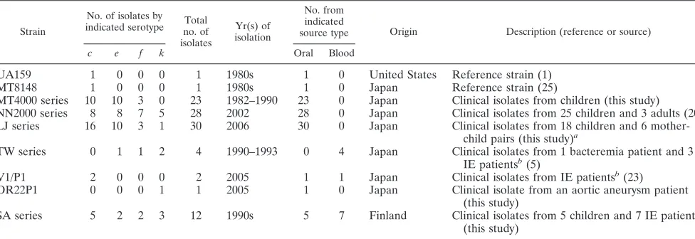
Bacterial isolates.A total of 102S. mutansstrains were examined in this study (Table 1). Strains MT8148 (serotypec) (26) and UA159 (c) (1) were used as the standard laboratory strains. Eighty-one clinical isolates ofS. mutanswere ob-tained from the Pedodontics Clinic of Osaka University Dental Hospital, Suita, Osaka, Japan, with the informed consent of patients obtained according to the protocol approved by the Ethics Committee of Osaka University Dental Hospi-tal. Four isolates from the blood of patients with bacteremia after tooth extrac-tion (TW295 [k]) or with infective endocarditis (TW871 [k], TW964 [f], and TW1378 [e]) were included (6). Blood and oral isolates from a patient with infective endocarditis (V1 [c] and P1 [c], respectively) (24) and another oral isolate collected in 2005 from a patient with an aortic aneurysm (strain OR22P1 [k]) were also included. We also examined seven blood isolates from Finnish patients with bacteremia or infective endocarditis and five oral strains isolated from Finnish subjects. All the strains were confirmed to beS. mutansby con-ventional physiological tests, including rough colony morphology on mitis-sali-varius agar (Difco Laboratories, Detroit, MI), bacitracin resistance, and fermen-tation of sorbitol, mannitol, raffinose, or melibiose (1% each) in a phenol red broth base (Difco).
Sucrose-dependent adhesion to glass surfaces. Sucrose-dependent cellular adhesion to glass surfaces was analyzed using a procedure described previously (12). Briefly, the test strains were grown at 37°C for 18 h at an angle of 30° in brain heart infusion broth (Difco) containing 1% sucrose. After incubation, the culture tubes were vigorously vortexed for 3 s, and the nonadhesive cells were transferred to fresh tubes. The cells remaining on the glass surfaces (adhesive cells) were removed using a rubber scraper and suspended in 3 ml of water. Both the adhesive and the nonadhesive cells were dispersed by ultrasonication, and their masses were determined from their optical densities at 550 nm (OD550). “Total cells” was defined as the OD550of the adhesive cells plus the nonadhesive cells, and “percentage adherence” as 100⫻(OD550of the adhesive cells)/(OD550 of the total cells). All assays were performed three times, and the means and standard deviations were calculated. Statistical analysis was performed using Prism4 software (version 4.0c; GraphPad Software, Inc., San Diego, CA).
Housekeeping loci used for MLST.Eight housekeeping gene loci were used in this study. The following sequences were obtained from the genome sequence of
S. mutansUA159 (NC_004350):tkt, which encodes transketolase (30);glnA,
which encodes glutamine synthetase subunit 1a (30);gltA, which encodes gluta-mate synthase (23);glk, which encodes glucose kinase (23);aroE, which encodes shikimate 5-dehydrogenase (15);gyrA, which encodes DNA gyrase subunit A (7);
murI, which encodes glutamate racemase (31); andlepC, which encodes signal peptidase I (39) (Table 1).
DNA isolation and sequencing.Chromosomal DNA was prepared from all isolates as described previously (5). Internal fragments of the MLST loci were PCR amplified (AmpliTaqGold, 10⫻PCR buffer, and 2 mM GeneAmp de-oxynucleotide triphosphate [dNTP] mix; Applied Biosystems) with the following cycling parameters: an initial denaturation at 94°C for 5 min and then 30 cycles consisting of 94°C for 30 s, 55°C for 30 s, and 72°C for 30 s, with a final extension at 72°C for 7 min. The amplified fragments were visualized after agarose gel electrophoresis on 1.5% agarose in the presence of 1g/ml ethidium bromide. They were then extracted from the agarose gel and cloned into the pGEM-T Easy vector (Promega). These plasmids were sequenced on both strands using vector-specific primers and BigDye Terminator version 3.1 (Applied Biosystems) by use of an ABI Prism 3110 genetic analyzer. In-frame internal fragments of the genes were selected for use in the MLST scheme.
Confirmation of serotypes.Serotyping was performed with the immunodiffu-sion method using rabbit antisera specific for serotypesc,e,f, andk, and with PCR methods using serotype-specific sets of primers, as described previously (22, 33). To determine serotypesc,e, andf, PCR amplification (TakaraExTaq, 10⫻
buffer, and 2.5 mM dNTPs; Takara) was performed with the following cycling parameters: initial denaturation at 96°C for 2 min and then 25 PCR cycles of 15 s of denaturation at 96°C, 30 s of annealing at 61°C, and 1 min of extension at 72°C. To identify serotypek, PCR amplification (AmpliTaqGold, 10⫻PCR buffer, and 2 mM GeneAmp dNTP mix; Applied Biosystems) was performed with the following cycling parameters: an initial denaturation at 94°C for 5 min and then 30 cycles of 94°C for 30 s, 60°C for 30 s, and 72°C for 30 s, with a final extension at 72°C for 7 min. The amplified fragments were separated by agarose gel electrophoresis on 1.5% agarose.
Alleles and ST assignments.For each locus, distinct allele sequences were assigned arbitrary allele numbers, with no weighting given to the degree of sequence divergence among the alleles. For each isolate, the alleles at each of the eight loci, defining the allelic profile or sequence type (ST), were analyzed with the nonredundant databases (http://linux.mlst.net/nrdb/). The STs were assigned arbitrary numbers in the order of their description. STs were grouped into lineages or clonal complexes with the program eBURST version 3, written and developed by E. Feil (http://eburst.mlst.net/), and the START version 2 package of programs, developed by K. Jolley (http://pubmlst.org/software/analysis/start2/) (11). Two or more independent isolates that shared identical alleles at six or more loci were defined as the members of a lineage. Each lineage was called a “group,” designated according to the ST identified as the putative ancestral type by use of eBURST.
[image:2.585.46.534.81.249.2]Computational analyses.The degree of clonality within the data set was estimated by calculating the index of association (IA) (34) between all of the STs in the data set by use of LIAN, version 3.5, written by B. Haubold and R. R.
TABLE 1. S. mutansstrains used in this study
Strain
No. of isolates by indicated serotype no. ofTotal
isolates
Yr(s) of isolation
No. from indicated
source type Origin Description (reference or source)
c e f k Oral Blood
UA159 1 0 0 0 1 1980s 1 0 United States Reference strain (1)
MT8148 1 0 0 0 1 1980s 1 0 Japan Reference strain (25)
MT4000 series 10 10 3 0 23 1982–1990 23 0 Japan Clinical isolates from children (this study) NN2000 series 8 8 7 5 28 2002 28 0 Japan Clinical isolates from 25 children and 3 adults (20) LJ series 16 10 3 1 30 2006 30 0 Japan Clinical isolates from 18 children and 6
mother-child pairs (this study)a
TW series 0 1 1 2 4 1990–1993 0 4 Japan Clinical isolates from 1 bacteremia patient and 3 IE patientsb(5)
V1/P1 2 0 0 0 2 2005 1 1 Japan Clinical isolates from IE patientsb(23)
OR22P1 0 0 0 1 1 2005 1 0 Japan Clinical isolate from an aortic aneurysm patient (this study)
SA series 5 2 2 3 12 1990s 5 7 Finland Clinical isolates from 5 children and 7 IE patientsb
(this study)
aClinical isolates LJ3 and LJ4, LJ11 and LJ12, LJ16 and LJ17, LJ24 and LJ25, LJ26 and LJ27, and LJ30 and LJ31 were from mother-child pairs. bIE, infective endocarditis.
on May 16, 2020 by guest
http://jcm.asm.org/
Hudson (http://adenine.biz.fh-weihenstephan.de/cgi-bin/lian/lian.cgi.pl) (10). We de-termined the number of polymorphic nucleotide sites and calculated thedN/dSratio
(wheredNvalues represent nonsynonymous base substitutions anddSvalues
represent synonymous base substitutions). We also performed phylogenic anal-yses of both individual genes and concatenated 3351-bp sequences by use of the DNAML, SeqBoot, and drawtree programs in the PHYLIP software package, written by J. Felsenstein (version 3.66; http://evolution.genetics.washington.edu /phylip.html), with the unweighted pair group method using arithmetic means.
Analysis ofcnmgenes ofS. mutansstrains.The prevalence of thecnmgene was evaluated by Southern blot analysis. Total genomic DNA from each strain was digested with HindIII (New England Biolabs, Beverly, MA). The DNA fragments were separated by 0.7% agarose gel electrophoresis and transferred to a nylon membrane (Hybond-N; Amersham Pharmacia Biotech). A 1,617-bpcnm
gene fragment containing the whole open reading frame was amplified by PCR from the genomic DNA ofS. mutansNN2072 with the primers cnm-F (5⬘-ATG AAAAGAAAAGGTTTACGAAGAC-3⬘) and cnm-R (5⬘-TCAGCTATGATA TTTACGGTAAAC-3⬘), designed based on the sequence of strain Z1 (GenBank accession no. AB102689) (29). The gene fragment was then labeled using a digoxigenin High-Prime DNA labeling and detection starter kit (Roche) accord-ing to the manufacturer’s instructions. The blotted membrane was prehybridized and then hybridized according to the protocol described by the manufacturer. After hybridization, the membrane was washed twice in 2⫻SSC (1⫻SSC is 0.15 M NaCl plus 0.015 M sodium citrate) containing 0.1% sodium dodecyl sulfate at room temperature and then washed twice in 0.5⫻SSC containing 0.1% sodium dodecyl sulfate for 15 min at 65°C. The washed membrane was visualized with the immunological method described by the manufacturer (Roche).
RESULTS
Development of an MLST scheme for S. mutans.
Chromo-somal DNA was obtained from 102 isolates. The eight houkeeping gene loci were amplified from 102 strains. The se-quences of the eight loci were determined and allelic profiles were assigned. The alleles defined for the MLST scheme were based on sequences of between 389 (gltA) and 462 (glnA) nucleotides. Between 14 (tkt) and 23 (gltAandlepC) alleles per locus were identified. The proportion of variable nucleotide sites present in the selected housekeeping genes ranged from 3.24% (glnA) to 5.43% (gltA) (Table 2). The proportion of nucleotide changes that altered the amino acid sequence (dN) and the proportion of silent changes (dS) were calculated for each gene, and thedN/dSratios for all eight loci were calcu-lated. All ratios were substantially less than 1 (Table 2). For the 102 S. mutans isolates, the mean number of alleles per locus was about 18, providing the theoretical potential to dis-tinguish more than 1.2⫻1010different allelic types. We also
compared a phylogenetic tree based on individual sequences with a phylogenetic tree based on the concatenated sequences of all eight alleles. However, no phylogenies constructed with these two data sets were incompatible (data not shown). Therefore, our MLST scheme for S. mutans showed a high discriminatory capacity.
Relatedness ofS. mutansisolates.Figure 1 shows a
dendro-gram constructed from the matrix of pairwise allelic differences between the STs of all 102 isolates. Even though the genetic variation at the MLST loci is relatively low (average, 4.2% nucleotide sites), these isolates were resolved into 92 STs, 85 of which (92.4%) were identified only once, indicating that theS.
mutans population displays diversification with little genetic
variation. Other STs contained between two and three mem-bers. The assignment of STs to lineages with BURST revealed that 36 STs were both unique and unrelated to any other STs, whereas the remaining 65 were assigned to 13 lineages (Fig. 1). Group 2 was the largest lineage and contained 24 isolates
TABLE 2. Primer sequences and characteristics of housekeeping gene loci included in the S. mutans MLST scheme Locus Gene locus tag and putative function of gene products Primer names Primer sequences (5 ⬘ to 3 ⬘ ) Size of
sequence fragment (bp)
No. of alleles identified No. (%) of polymorphic nucleotides dN / dS GenBank accession no. tkt Smu.291, transketolase tktF ACC CGG GTG TTG TCA TGG GCG CTG C tktR CAT AGG ATG ACG CTT CGC CAG AAA C 432 14 16 (3.70) 0.1032 AB281702–AB281802 glnA Smu.364, glutamine glnA-F CCT TGG GGA GAT GAA AAC GGA GCC G synthetase glnA-R TGG CCA TAA AGG TTG CAT ACA AAC C 462 16 15 (3.24) 0.0628 AB281803–AB281903 gltA Smu.365, glutamate gltA-F TGC CTT AAC GAT GTT AGA GAG AAT G synthase gltA-R AAA GAC TAT CTT CAA AAG CAC ACC C 387 23 21 (5.43) 0.2082 AB281904–AB282004 glk Smu.542, glucose glk-F GAC AAG TTC AGG AGA AAT GGG CTA T kinase glk-R CAG CAA CTC CAT GAA TAA GAT TGC C 402 19 16 (3.98) 0.6333 AB282005–AB282105 aroE Smu.778, Shikimate aroE-F GAT GAA GTA ACG AAA GCA GCA GAT T 5-dehydrogenase aroE-R TGC CAT CCA TAC CAA CAT TGG TCG C 396 21 19 (4.80) 0.4467 AB282207–AB282307 gyrA Smu.1114, DNA gyrase gyrA-F TAC AGG TGA TGT CAT GGG TAA ATA C subunit A gyrA-R CCG GGT AGT ACT TCC ATT AGG TCA C 432 17 17 (3.94) 0.1520 AB282106–AB282206 murI Smu.1718, glutamate murI-F TCC GGA GTG GGC GGT TTA ACG GTC G racemase murI-R TCA ACA ATA GGA ACA AAT TTG GGG C 423 15 17 (4.02) 0.0544 AB282308–AB282408 lepC Smu.1874, signal lepC-F CCG CGT CTC TTT ATC TGG TTT CTT G peptidase I lepC-R GAC AAT GCG ATC ATC ACC TAA AAG C 417 23 19 (4.56) 0.1305 AB282409–AB282509
on May 16, 2020 by guest
http://jcm.asm.org/
FIG. 1. Dendrogram of 102 isolates ofS. mutansbased on an MLST scheme with cluster analysis by the unweighted pair group method using arithmetic means. Clonal complex groups, STs, serotypes, and distribution of the collagen-binding protein gene (cnm) were determined as described in Materials and Methods.
on May 16, 2020 by guest
http://jcm.asm.org/
representing 19 STs. The remaining 12 groups contained from two to six member STs.
We also tried to determine the chronological relationships of the serotypes. However, no significant differences were ob-served in the distributions of the serotypes. Moreover, no geo-graphical relationships were identified among the isolates.
Evidence of recombination. The extent of recombination
within theS. mutanspopulation was assessed by determining the standardized IA(34) with LIAN. The IAfor the complete
data set was 0.0931 when randomized data sets (1,000 trials) were used. This value is significantly greater than zero, which is the value expected for a population in linkage equilibrium. However, in populations in which recombination is sufficient to randomize the alleles at different loci over long time periods, the appearance of multiple isolates with similar genotypes can result from the recent expansion of clones. Therefore, the SplitsTree program was used to detect recombination among the various STs. Allelic profile data were converted into dis-tance matrix values by use of START, and the resulting nexus file was analyzed with the split decomposition method. Splits-Tree analysis of the 102S. mutansstrains yielded a very low fit value (fit⫽22.2), which may have resulted from the program’s inability to analyze this large amount of information (14). We also analyzed several small subsets of randomly selected strains, which considerably improved the fit values. One exam-ple of a SplitsTree analysis displaying 10 parallelograms and a high fit value of ca. 55 was based on 18 randomly selectedS.
mutansisolates (data not shown).
Relationships between STs and serotypes.We then
deter-mined the serotypes of all the isolates by immunodiffusion and PCR-based typing (Table 3). Serotype c was dominant (43 isolates), followed bye(31 isolates),f(16 isolates), andk(12 isolates). In general, the serotypes appeared to be associated with the overall genotype defined by the STs, and only ST2 contained two serotypes (c and f). As stated above, closely related STs constituted clonal complexes. However, groups 1, 2, 3, and 4 contained isolates of more than one serotype, whereas the other groups contained only one serotype. Sero-type c, the major serotype ofS. mutansclinical isolates, was widely distributed among the clonal complex groups, such as in groups 1, 2, 3, 4, 6, 7, 8, 10, and 12, and in many singletons, indicating that serotypecis the ancestral serotype ofS. mutans. Group 2 contained 15 of the 31 serotypeeisolates (ST3, 5, 6, 22, 42, 71, 72, 73, 74, 75, 77, 83, and 84), and groups 3 and 4 contained three and six serotypeeisolates, respectively. Sero-typefisolates were found mainly in three groups (groups 5, 9, and 11), and approximately half the serotype f isolates oc-curred in these groups. Another three STs (ST48, 57, and 91) were not categorized within the same clonal complex but had closely related sequence homologies (Fig. 1). Only three STs (ST2, 21, and 85) that were serotypefwere located separately from the other serotypefgroups. Serotypek, newly identified in the last decade, was distributed among closely related STs (ST16, 36, 38, 66, 67, 68, and 79). These results indicate that the ancestral strain ofS. mutanswas serotypecand that the other serotypes have branched from the serotype c groups continuously during evolution up until the present.
Association between lineages and diseases.
Sucrose-depen-dent adhesion to glass surfaces is an important feature in the cariogenicity ofS. mutans. However, the serotype-specific
ad-hesion mechanism has still not been investigated. Therefore, we determined the in vitro adhesion capacities ofS. mutans
isolates. Serotypec isolates showed a slightly increased rate (84.01%⫾11.81%), and serotypekisolates showed a low rate (78.01%⫾6.94%) compared with those of the other serotype strains. Serotypeseandfshowed moderate rates of 81.49%⫾ 14.75% and 82.34%⫾6.39%, respectively. However, there was no statistically significant difference among the isolates. This observation indicates that the phylogenetic differences deter-mined with the eight housekeeping gene loci do not reflect the adhesion capacities of the S. mutans isolates. Three clinical isolates (MT4078, serotype c; MT4293, serotype e; and V1, serotypec) showed significantly lower adhesion than did the other isolates. However, we identified no relationship between serotype, phylogenetic position, regional specificity, and years of isolation.
We next determined the relationship between serotype and systemic infection, becauseS. mutansfrequently causes bacte-remia and infective endocarditis. However, the blood-derived isolates were widely distributed between the various STs and clonal complex groups (Fig. 1), and no significant relationship between STs and the blood-derived isolates was observed. Therefore, we conclude that theS. mutansblood isolates are not a specific strain and that each isolate has the potential to induce systemic infections such as bacteremia or infective en-docarditis.
Strain-specific collagen-binding adhesin (encoded bycnm) is a recently identified wall-anchored protein. However, its prev-alence amongS. mutansisolates has not been determined. To clarify the prevalence of thecnmgene, we analyzed the distri-bution of cnm-positive isolates by PCR and Southern blot analysis. Of the 102 strains examined, 22 werecnm positive (21.6%; see also data in Fig. 1). Thecnm-positive isolates were distributed in ST85; in ST45 and ST46 (group 11); in ST40; in ST14, 15, and 86 (group 5); in ST55; in ST34, 37, and 35 (group 9); in ST69; in ST26 and 27 (group 7); in ST57; in ST48; in ST80; in ST51; in ST88; in ST87; and in ST79 (Fig. 1). The predominantcnm-positive isolates were of serotypef(13cnm
positive of 18 total [81.3%]), followed by serotypek(5cnm -positive of 12 total [41.7%]). Three isolates of serotype c(3 [ST26, 27, and 51] of 43 total [7.0%]) and 1 isolate of serotype
e(1 [ST40] of 31 total [3.2%]) had thecnmgene. The preva-lence ofcnm gene in each serotype is statistically significant (between serotype c and serotype f, a P value of ⬍0.0001; between serotypescandk, aPvalue of 0.087; between sero-typeseandf, aPvalue of⬍0.0001; between serotypeseandk, aPvalue of 0.0087; and between serotypeseandk, aPvalue of 0.0042 [all by Fisher’s exact probability test]). These isolates were closely related to thecnm-positive serotypefandkstrains on phylogenetic analysis. Interestingly, the distribution ofcnm -positive strains was limited to closely related groups. These observations indicate that some clinical isolates have acquired the cnm gene, probably by horizontal gene transfer, during evolution.
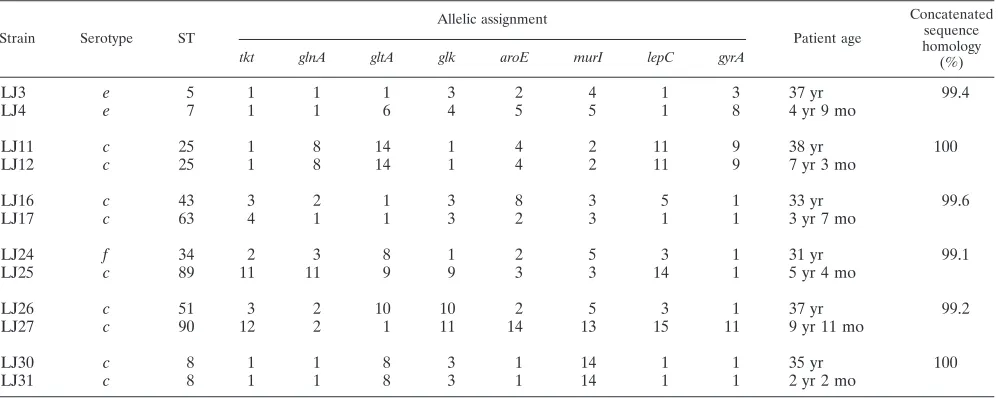
Streptococcus mutanstransmission from mother to child.In
the six mother-child pairs ofS. mutansisolates (Table 4), the STs of two pairs (LJ11 and 12 and LJ30 and 31) were com-pletely identical, indicating that theseS. mutansisolates were vertically transmitted from mother to child. Of the other four pairs, the LJ3-LJ4 and LJ16-LJ17 pairs had three and four
on May 16, 2020 by guest
http://jcm.asm.org/
TABLE 3. Allelic profiles of isolates belonging to the 92 STs identified in this study
Strain Serotype ST
Allelic assignment (arbitrary allele no.)
tkt glnA gltA glk aroE murI lepC gyrA
UA159 c 1 1 1 1 1 1 1 1 1
MT8148 c 63 4 1 1 3 2 3 1 1
MT4065 c 64 4 1 1 3 2 3 1 14
MT4071 c 92 14 2 4 1 2 3 1 1
MT4076 c 2 1 1 1 13 1 1 1 1
MT4078 c 20 1 5 9 1 4 3 11 9
MT4083 c 26 1 9 1 1 2 5 3 10
MT4087 c 29 1 15 15 3 4 2 19 1
MT4093 c 24 1 8 9 1 4 2 20 1
MT4112 c 61 3 14 1 17 20 15 21 1
MT4118 c 65 4 1 1 18 2 3 1 1
MT4164 c 23 1 8 9 1 4 2 11 9
MT4117 e 81 6 2 7 3 2 7 1 1
MT4119 e 59 3 10 15 3 21 2 1 1
MT4217 e 6 1 1 1 19 2 4 7 3
MT4245 e 72 6 1 1 3 2 4 1 3
MT4274 e 30 1 16 23 6 2 4 22 15
MT4278 e 84 6 5 15 3 2 4 1 3
MT4279 e 83 6 5 9 3 2 4 1 3
MT4293 e 73 6 1 1 3 14 4 1 16
MT4368 e 10 1 2 7 3 2 7 1 1
MT4369 e 11 1 2 7 3 2 7 1 17
MT4251 f 85 7 2 11 3 2 4 1 3
MT4333 f 46 3 2 5 1 2 3 23 1
MT4348 f 15 1 3 8 1 6 10 3 1
NN2098 c 31 2 1 1 2 2 2 1 2
NN2099 c 44 3 2 2 3 3 3 2 1
NN2092 c 9 1 2 1 3 2 4 1 3
NN2093 c 54 3 3 3 1 4 3 3 1
NN2025 c 70 5 4 1 4 5 5 4 4
NN2004 c 33 2 2 4 5 4 6 1 1
NN2085 c 39 2 5 5 6 2 3 3 1
NN2087 e 56 3 6 6 4 2 5 5 4
NN2089 e 42 3 1 1 1 2 4 1 3
NN2037 e 10 1 2 7 3 2 7 1 1
NN2044 e 22 1 7 1 3 2 3 1 4
NN2054 e 19 1 5 7 3 2 7 1 1
NN2076 e 75 6 1 1 3 2 8 1 1
NN2042 e 10 1 2 7 3 2 7 1 1
NN2053 e 76 6 1 6 4 5 5 1 4
NN2072 f 69 4 3 8 3 2 5 1 1
NN2165 f 57 3 8 1 1 6 3 3 1
NN2007 f 35 2 3 8 1 2 9 1 1
NN2117 f 48 3 2 9 1 7 4 6 1
NN2138-2 f 45 3 2 5 1 2 3 7 1
NN2168M-5 c 49 3 2 10 3 8 3 5 1
NN2121 f 85 7 2 11 3 2 4 1 3
NN2431M-2 f 86 8 3 8 1 6 10 1 1
NN2011 k 47 3 2 8 7 10 5 3 1
NN2111 k 67 4 2 12 1 7 5 8 6
NN2323M-1 k 66 4 2 12 1 4 5 8 6
NN2193-1 k 16 1 3 13 8 11 2 9 7
NN2105 k 68 4 2 12 1 4 5 10 6
LJ1 c 17 1 3 15 1 2 3 12 1
LJ2 e 40 2 9 5 1 4 3 7 1
LJ3 e 5 1 1 1 3 2 4 1 3
LJ4 e 7 1 1 6 4 5 5 1 8
LJ7 f 14 1 3 8 1 4 10 3 1
LJ11 c 25 1 8 14 1 4 2 11 9
LJ12 c 25 1 8 14 1 4 2 11 9
LJ13 c 58 3 10 15 4 13 11 13 1
LJ14 c 52 3 2 15 3 1 11 1 3
LJ16 c 43 3 2 1 3 8 3 5 1
LJ17 c 63 4 1 1 3 2 3 1 1
LJ18 e 77 6 1 16 3 2 4 1 3
LJ19 e 71 6 1 1 1 2 4 1 3
Continued on following page
on May 16, 2020 by guest
http://jcm.asm.org/
matches, respectively, in their ST profiles. However, on the MLST-based dendrogram and sequence-based phylogenetic tree, these two pairs were grouped onto different branches. The LJ24-LJ25 and LJ26-LJ27 pairs had only one allele match, and the LJ24-LJ25 members showed different serotypes. Al-though only a limited number of mother-child pairs were an-alyzed in this study, the MLST method allowed the strict dis-crimination of the strains from both individuals in each mother-child pair.
DISCUSSION
In developing the MLST in this study, target genes were selected based on the complete genome sequence ofS. mutans
UA159 (1) and the ongoing complete genome sequencing ofS.
mutansNN2025 in our laboratories, on the genomic
informa-tion from other bacteria, and on the published MLST schemes for other streptococci, such asS. pyogenes(4),S. uberis(44),S. suis(13), andS. pneumoniae(3), together with their locations in the genome. Finally, eight housekeeping genes were selected for this study. The eight loci were not subject to positive se-lection, as demonstrated by the dN/dS ratio for each locus, which was substantially less than 1 (Table 2). The mean num-ber of alleles identified per locus was 18.5, suggesting thatS.
mutans represents a genetically diverse species. The
[image:7.585.51.541.78.442.2]house-keeping genestkt (transketolase),glnA(glutamine synthetase 1a subunit), gyrA(DNA gyrase subunit A), murI(glutamate racemase), andlepC(signal peptidase I) were chosen because these genes are conserved across the genusStreptococcusand other genera. We also included three other genes,aroE (shiki-mate 5-dehydrogenase),glk(glucose kinase), andgltA (gluta-mate synthase), whose products are housekeeping proteins with sequence homologies that are specific toS. mutansstrains. Seven genes have usually been used in recent MLST schemes, because it is important that the selection of the genes to be sequenced in an MLST scheme developed for a partic-ular bacterial species should be optimized to save time and cost (40). However, the discrimination of each serotype, especially serotypesfandk, was not definitive in our MLST when seven genes were used in the analysis ofS. mutans. In addition, the combined analysis of housekeeping genes and some virulence genes has been also used to identify virulent or pandemic clones (35, 40). In this study, we used eight housekeeping loci because the virulence genes ofS. mutansare not as genetically diverse as those of other organisms. For example, glucosyl-transferases synthesize water-soluble and -insoluble glucans from a sucrose substrate, and these activities are important in
TABLE 3—Continued
Strain Serotype ST
Allelic assignment
tkt glnA gltA glk aroE murI lepC gyrA
LJ20 c 27 1 9 1 1 2 12 3 10
LJ22 c 82 6 4 1 4 5 5 1 4
LJ23 k 88 10 2 5 7 6 5 11 1
LJ24 f 34 2 3 8 1 2 5 3 1
LJ25 c 89 11 11 9 9 3 3 14 1
LJ26 c 51 3 2 10 10 2 5 3 1
LJ27 c 90 12 2 1 11 14 13 15 11
LJ30 c 8 1 1 8 3 1 14 1 1
LJ31 c 8 1 1 8 3 1 14 1 1
LJ32 f 37 2 3 17 1 2 5 3 1
LJ15 c 62 4 1 1 2 15 3 1 1
LJ29 c 12 1 2 7 3 16 7 1 1
LJ34 e 3 1 1 1 3 2 3 1 3
LJ36 e 72 6 1 1 3 2 4 1 3
LJ50 e 78 6 1 19 4 5 5 1 4
LJ59 e 72 6 1 1 3 2 4 1 3
LJ64 e 74 6 1 1 3 2 4 4 3
SA22 f 21 1 5 9 12 17 3 16 1
SA31 k 36 2 3 14 8 11 2 9 12
SA51 f 2 1 1 1 13 1 1 1 1
SA53 k 55 3 3 8 1 1 3 1 1
SA72 k 38 2 3 18 8 11 2 17 3
SA12 c 50 3 2 10 3 8 3 5 13
SA13 c 18 1 3 20 4 5 5 1 4
SA14 c 53 3 2 15 14 1 11 1 12
SA15 e 41 2 12 5 15 2 3 18 1
SA16 e 28 1 13 1 1 18 3 3 1
SA17 c 60 3 14 1 16 2 2 19 1
SA18 c 32 2 2 4 5 4 5 1 4
TW295 k 80 6 2 5 1 9 4 1 1
TW871 k 79 6 2 1 1 9 5 7 5
TW964 f 91 13 5 21 1 6 4 1 1
TW1378 e 13 1 2 22 1 19 3 13 1
V1 c 63 4 1 1 3 2 3 1 1
P1 c 4 1 1 1 13 7 3 11 1
OR22P1 k 87 9 8 14 1 12 4 11 1
on May 16, 2020 by guest
http://jcm.asm.org/
the colonization and biofilm formation ofS. mutans(27). Chia et al. (2) reported the existence of DNA polymorphisms in the 5⬘regions of thegtfBandgtfCgenes. However, only four or five variants have been observed in this region (2). This observation has been confirmed by use of restriction fragment length poly-morphisms of the gtfB gene (36). Therefore, these putative virulence genes ofS. mutansare not suitable for the analysis of the evolution or population biology of the species because their heterogeneity is low. The mean number of alleles iden-tified per locus was 18.5, providing the theoretical potential to distinguish more than 1.2⫻1010different genotypes. In fact, as
shown in Fig. 1 and Table 3, our MLST method divided the 102 strains into 92 STs, indicating that this MLST scheme has high discriminatory power.
The cell wall antigens of S. mutansare rhamnose-glucose polysaccharides, which are composed of␣1,2- and␣1,3-linked rhamnan backbones and glucose side chains. Bacterial cell wall polysaccharides decorate the cell surface and often play an important role in the colonization of the bacterial ecological niche. Each serotype-specific polysaccharide has a unique link-age to its glucose side chains. Serotypechas an␣1,2 linkage, serotype e has a 1,2 linkage, and serotype f has an ␣1,3 linkage (17). Serotypek strains have a unique “untypeable” phenotype in terms of known serotype antibodies because they lack glucose side chains linked to their rhamnose backbones (21). The genes related to serotype specificity have been de-termined, and each serotype has a specific and heterogenic region downstream from the conservedrgpA-rgpFoperon (33). On the basis of these specific regions, Shibata et al. suggested that none of the three serotypes could be defined as the an-cestral strain (33). However, in our MLST analysis, most of the serotypeeand serotypefstrains occur on their own branches on the dendrogram. Most serotypee strains are included in clonal complex group 2, and the root of this branch derives from serotype c (Fig. 1). The ST85 isolate in this group is serotypef, but the restriction fragment length pattern of the
rgpA-rgpFregion in the ST85 isolate seems to be that of
sero-typec(data not shown). Why these strains appear phenotyp-ically serotypef rather than serotype c is now under inves-tigation by complete nucleotide sequence analysis. The distri-bution of serotype f isolates is also restricted to limited branches derived from serotypec. These observations suggest that a serotypecstrain, the dominant serotype amongS. mu-tansisolates, is the ancestral phenotype of this organism and that serotypeeandfstrains acquired their strain-specific genes during evolution. It is still unclear whether these phenotypic changes are due to genetic transfer or genetic exchange. Therefore, these issues require further analysis.
Serotypek, recently identified as a new serotype ofS. mu-tans, also has an interesting distribution in our MLST analysis. All genes for the biosynthesis of serotype-specific polysaccha-rides are highly conserved relative to those of serotypec, and only the mRNA expression ofrgpEis diminished in serotypek
strains (25). Therefore, we hypothesized that serotypekmight be derived from serotypec. As we expected, many serotypek
isolates were derived from the serotype c branch (Fig. 1). However, some serotypekstrains seem to have been derived from serotypef(ST55 and ST80). Polysaccharide synthesis is a sequential reaction involving several gene products. Therefore, a defect in a gene or a loss of function of a gene product related to the biosynthesis of polysaccharides results in the formation of a serotype k strain. In fact, a gluA-destructive mutant of a serotypecstrain has the “serotypek” phenotype (21). Therefore, it is reasonable to infer thatS. mutansstrains that are phenotypically “serotypek” have arisen from the ge-netic dysfunction of serotype c or f strains in this era. It is possible that allS. mutansstrains may have the potential to become serotypekin the future.
[image:8.585.44.543.81.280.2]In this study, we compared the distribution and lineage dif-ferences of Japanese isolates with those of Finnish isolates because Japan is geographically and ethnically distinct from Finland. However, we found no regional bias in the distribu-tion of theS. mutansisolates (Fig. 1). Several lines of epide-miological evidence indicate that serotype c is predominant
TABLE 4. Mother-child transmission ofS. mutansby MLST analysisa
Strain Serotype ST
Allelic assignment
Patient age
Concatenated sequence homology (%)
tkt glnA gltA glk aroE murI lepC gyrA
LJ3 e 5 1 1 1 3 2 4 1 3 37 yr 99.4
LJ4 e 7 1 1 6 4 5 5 1 8 4 yr 9 mo
LJ11 c 25 1 8 14 1 4 2 11 9 38 yr 100
LJ12 c 25 1 8 14 1 4 2 11 9 7 yr 3 mo
LJ16 c 43 3 2 1 3 8 3 5 1 33 yr 99.6
LJ17 c 63 4 1 1 3 2 3 1 1 3 yr 7 mo
LJ24 f 34 2 3 8 1 2 5 3 1 31 yr 99.1
LJ25 c 89 11 11 9 9 3 3 14 1 5 yr 4 mo
LJ26 c 51 3 2 10 10 2 5 3 1 37 yr 99.2
LJ27 c 90 12 2 1 11 14 13 15 11 9 yr 11 mo
LJ30 c 8 1 1 8 3 1 14 1 1 35 yr 100
LJ31 c 8 1 1 8 3 1 14 1 1 2 yr 2 mo
a
For each pair of rows, the top row gives mother data and the bottom row gives child data.
on May 16, 2020 by guest
http://jcm.asm.org/
worldwide (70% to 80%), with serotypeethe next commonest (10% to 15%) and serotypefoccurring rarely (1% to 5%) (9). On the basis of these findings, we speculate that the serological differentiation ofS. mutansoccurred earlier than we had pre-viously inferred. To address this question further, we are con-tinuing to analyze worldwide serotype distributions using our MLST scheme.
Oral bacteria are considered to cause transient bacteremia, countered by professional dental treatment and daily oral care practices such as tooth brushing and flossing (32). However, the incidence of infective endocarditis estimated by a review of reports published between 1993 and 2003 was only 3.6 per 100,000 head of population per year (20). These observations suggest that some infective-endocarditis-specific or virulent strains exist and that these strains necessarily induce systemic infections. However, the specific distribution of S. mutans
strains related to systemic infections was not determined in our MLST analysis.
In contrast, the distribution of the collagen-binding protein gene (cnm) was clearly predominant in the clonal complex groups of serotypefstrains (Fig. 1). The gene product ofcnm
was first identified in the cold-agglutination phenotype ofS.
mutans, and thereafter the binding of collagen-binding adhesin
to collagen and laminin was reported (29). The binding of collagen-binding adhesin to specific extracellular matrix mole-cules may be important for the initial bacterial attachment to blood vessels. However, the distribution ofcnm-positive iso-lates does not exactly match that of isoiso-lates derived from bacteremia or infective endocarditis patients, so the contribu-tion of collagen-binding adhesin to the progression of systemic infections is not clearly explained.
The vertical transmission of S. mutans from mother to child is thought to be the major route for early acquisition (16). In contrast, the detection of genotypes in children that are not found in their mothers or other family members indicates that S. mutansmay also be acquired from other sources. In our MLST analysis, two of six mother-child pairs showed complete identity of ST profiles for the mother and the child, whereas the other pairs showed differing ST pro-files (Table 4). This result indicates that our MLST scheme is useful for epidemiological studies and is suitable for the long-term monitoring of S. mutans transmission. Mattos-Graner et al. reported at least two to five different genotypes in 30% of the children tested (18). In this study, we used only one isolate each from the mother and the child. Further prospective studies involving greater numbers ofS. mutans
isolates are required to explore the frequencies of vertical and horizontal transmission.
In conclusion, we have devised the first clear typing system
forS. mutans. In this study, we have provided new insight into
the lineages of the four serotypes, their regional specificities, and the distribution of a newly identified virulence gene and have demonstrated the applicability of the scheme to epidemi-ological studies. The superior discriminatory capacity of MLST, compared with that of classical serotyping or DNA-based genotyping methods, may have important practical im-plications. We hope that this MLST scheme will now be ex-panded to include isolates from other countries.
ACKNOWLEDGMENTS
This study was supported by the 21st Century COE program “Orig-ination of Frontier BioDentistry” at Osaka University Graduate School of Dentistry, supported by the Ministry of Education, Culture, Sports, Science and Technology of Japan; Grant-in-Aid for Scientific Research (B) 16390605 from the Japan Society for the Promotion of Science; Aid for Exploratory Research 17659647; Grant-in-Aid for Young Scientists (A) 18689050 Grant-in-Grant-in-Aid for Scientific Research on Priority Areas “Applied Genomics” from the Ministry of Education, Culture, Sports, Science and Technology; and PRESTO, the Japan Science and Technologies Agency.
REFERENCES
1.Ajdic, D., W. M. McShan, R. E. McLaughlin, G. Savic, J. Chang, M. B. Carson, C. Primeaux, R. Tian, S. Kenton, H. Jia, S. Lin, Y. Qian, S. Li, H. Zhu, F. Najar, H. Lai, J. White, B. A. Roe, and J. J. Ferretti.2002. Genome sequence ofStreptococcus mutansUA159, a cariogenic dental pathogen. Proc. Natl. Acad. Sci. USA99:14434–14439.
2.Chia, J. S., T. Y. Hsu, L. J. Teng, J. Y. Chen, L. J. Hahn, and C. S. Yang. 1991. Glucosyltransferase gene polymorphism amongStreptococcus mutans
strains. Infect. Immun.59:1656–1660.
3.Enright, M. C., and B. G. Spratt.1998. A multilocus sequence typing scheme forStreptococcus pneumoniae: identification of clones associated with serious invasive disease. Microbiology144:3049–3060.
4.Enright, M. C., B. G. Spratt, A. Kalia, J. H. Cross, and D. E. Bessen.2001. Multilocus sequence typing ofStreptococcus pyogenesand the relationships betweenemmtype and clone. Infect. Immun.69:2416–2427.
5.Fujiwara, T., T. Hoshino, T. Ooshima, S. Sobue, and S. Hamada.2000. Purification, characterization, and molecular analysis of the gene encoding glucosyltransferase fromStreptococcus oralis. Infect. Immun.68:2475–2483. 6.Fujiwara, T., K. Nakano, M. Kawaguchi, T. Ooshima, S. Sobue, S. Kawa-bata, I. Nakagawa, and S. Hamada.2001. Biochemical and genetic charac-terization of serologically untypableStreptococcus mutansstrains isolated from bacteremia. Eur. J. Oral Sci.109:330–334.
7.Gonzalez, I., M. Georgiou, F. Alcaide, D. Balas, J. Linares, and A. G. de la Campa.1998. Fluoroquinolone resistance mutations in theparC,parE, and
gyrAgenes of clinical isolates of viridans group streptococci. Antimicrob. Agents Chemother.42:2792–2798.
8.Gro¨nroos, L., M. Saarela, J. Matto, U. Tanner-Salo, A. Vuorela, and S. Alaluusua.1998. Mutacin production byStreptococcus mutansmay promote transmission of bacteria from mother to child. Infect. Immun.66:2595–2600. 9.Hamada, S., and H. D. Slade.1980. Biology, immunology, and cariogenicity
ofStreptococcus mutans. Microbiol. Rev.44:331–384.
10.Haubold, B., and R. R. Hudson.2000. LIAN 3.0: detecting linkage disequi-librium in multilocus data. Linkage analysis. Bioinformatics16:847–848. 11.Jolley, K. A., E. J. Feil, M. S. Chan, and M. C. Maiden.2001. Sequence type
analysis and recombinational tests (START). Bioinformatics17:1230–1231. 12.Kawabata, S., and S. Hamada.1999. Studying biofilm formation of mutans
streptococci. Methods Enzymol.310:513–523.
13.King, S. J., J. A. Leigh, P. J. Heath, I. Luque, C. Tarradas, C. G. Dowson, and A. M. Whatmore.2002. Development of a multilocus sequence typing scheme for the pig pathogenStreptococcus suis: identification of virulent clones and potential capsular serotype exchange. J. Clin. Microbiol.40:3671– 3680.
14.Kotetishvili, M., O. C. Stine, A. Kreger, J. G. Morris, Jr., and A. Sulakvelidze.2002. Multilocus sequence typing for characterization of clin-ical and environmentalSalmonellastrains. J. Clin. Microbiol.40:1626–1635. 15.Lee, H. Y., and J. C. Cote.2006. Phylogenetic analysis of␥-proteobacteria inferred from nucleotide sequence comparisons of the house-keeping genes
adk,aroEandgdh: comparisons with phylogeny inferred from 16S rRNA gene sequences. J. Gen. Appl. Microbiol.52:147–158.
16.Li, Y., and P. W. Caufield.1998. Arbitrarily primed polymerase chain reac-tion fingerprinting for the genotypic identificareac-tion of mutans streptococci from humans. Oral Microbiol. Immunol.13:17–22.
17.Linzer, R., M. S. Reddy, and M. J. Levine.1986. Immunochemical aspects of serotype carbohydrate antigens ofStreptococcus mutans, p. 29–38.InS. Hamada, S. M. Michalek, H. Kiyono, L. Manaker, and J. R. McGhee (ed.), Molecular microbiology and immunology ofStreptococcus mutans. Elsevier Science Publishers, Amsterdam, The Netherlands.
18.Mattos-Graner, R. O., Y. Li, P. W. Caufield, M. Duncan, and D. J. Smith. 2001. Genotypic diversity of mutans streptococci in Brazilian nursery chil-dren suggests horizontal transmission. J. Clin. Microbiol.39:2313–2316. 19.Mineyama, R., S. Yoshino, and N. Maeda.24 July 2006, posting date. DNA
fingerprinting of isolates ofStreptococcus mutansby pulsed-field gel electro-phoresis. Microbiol. Res. doi:101016/j.micres.2006.06.014.
20.Moreillon, P., and Y. A. Que.2004. Infective endocarditis. Lancet363:139– 149.
21.Nakano, K., R. Nomura, I. Nakagawa, S. Hamada, and T. Ooshima.2004. Demonstration ofStreptococcus mutanswith a cell wall polysaccharide
on May 16, 2020 by guest
http://jcm.asm.org/
cific to a new serotype,k, in the human oral cavity. J. Clin. Microbiol. 42:198–202.
22.Nakano, K., R. Nomura, N. Shimizu, I. Nakagawa, S. Hamada, and T. Ooshima.2004. Development of a PCR method for rapid identification of newStreptococcus mutansserotypekstrains. J. Clin. Microbiol.42:4925– 4930.
23.Nesbo, C. L., S. L’Haridon, K. O. Stetter, and W. F. Doolittle.2001. Phylo-genetic analyses of two “archaeal” genes inThermotoga maritima reveal multiple transfers between Archaea and Bacteria. Mol. Biol. Evol.18:362– 375.
24.Nomura, R., K. Nakano, H. Nemoto, K. Fujita, S. Inagaki, T. Takahashi, K. Taniguchi, M. Takeda, H. Yoshioka, A. Amano, and T. Ooshima.2006. Isolation and characterization ofStreptococcus mutansin heart valve and dental plaque specimens from a patient with infective endocarditis. J. Med. Microbiol.55:1135–1140.
25.Nomura, R., K. Nakano, and T. Ooshima.2005. Molecular analysis of the genes involved in the biosynthesis of serotype specific polysaccharide in the novel serotypekstrains ofStreptococcus mutans. Oral Microbiol. Immunol. 20:303–309.
26.Ooshima, T., A. Izumitani, S. Sobue, and S. Hamada.1983. Cariostatic effects of palatinose on experimental dental caries in rats. Jpn. J. Med. Sci. Biol.36:219–223.
27.Ooshima, T., M. Matsumura, T. Hoshino, S. Kawabata, S. Sobue, and T. Fujiwara.2001. Contribution of three glucosyltransferases to sucrose-depen-dent adherence ofStreptococcus mutans. J. Dent. Res.80:1672–1677. 28.Saarela, M., S. Alaluusua, T. Takei, and S. Asikainen.1993. Genetic
diver-sity within isolates of mutans streptococci recognized by an rRNA gene probe. J. Clin. Microbiol.31:584–587.
29.Sato, Y., K. Okamoto, A. Kagami, Y. Yamamoto, T. Igarashi, and H. Kizaki. 2004.Streptococcus mutansstrains harboring collagen-binding adhesin. J. Dent. Res.83:534–539.
30.Schenk, G., R. Layfield, J. M. Candy, R. G. Duggleby, and P. F. Nixon.1997. Molecular evolutionary analysis of the thiamine-diphosphate-dependent en-zyme, transketolase. J. Mol. Evol.44:552–572.
31.Schmidt, D. M., B. K. Hubbard, and J. A. Gerlt.2001. Evolution of enzy-matic activities in the enolase superfamily: functional assignment of un-known proteins inBacillus subtilisandEscherichia coli as L-Ala-D/L-Glu
epimerases. Biochemistry40:15707–15715.
32.Seymour, R. A., R. Lowry, J. M. Whitworth, and M. V. Martin.2000.
Infec-tive endocarditis, dentistry and antibiotic prophylaxis; time for a rethink? Br. Dent. J.189:610–616.
33.Shibata, Y., K. Ozaki, M. Seki, T. Kawato, H. Tanaka, Y. Nakano, and Y. Yamashita.2003. Analysis of loci required for determination of serotype antigenicity inStreptococcus mutansand its clinical utilization. J. Clin. Mi-crobiol.41:4107–4112.
34.Smith, J. M., N. H. Smith, M. O’Rourke, and B. G. Spratt.1993. How clonal are bacteria? Proc. Natl. Acad. Sci. USA90:4384–4388.
35.Sullivan, C. B., M. A. Diggle, and S. C. Clarke.2005. Multilocus sequence typing: data analysis in clinical microbiology and public health. Mol. Bio-technol.29:245–254.
36.Toi, C. S., P. Cleaton-Jones, and P. Fatti.2005. Characterization of Strep-tococcus mutansdiversity by determining restriction fragment-length poly-morphisms of thegtfBgene of isolates from 5-year-old children and their mothers. Antonie Leeuwenhoek88:75–85.
37.Tsukioka, Y., Y. Yamashita, Y. Nakano, T. Oho, and T. Koga.1997. Identi-fication of a fourth gene involved in dTDP-rhamnose synthesis in Strepto-coccus mutans. J. Bacteriol.179:4411–4414.
38.Tsukioka, Y., Y. Yamashita, T. Oho, Y. Nakano, and T. Koga.1997. Biolog-ical function of the dTDP-rhamnose synthesis pathway inStreptococcus mu-tans. J. Bacteriol.179:1126–1134.
39.van Roosmalen, M. L., N. Geukens, J. D. Jongbloed, H. Tjalsma, J. Y. Dubois, S. Bron, J. M. van Dijl, and J. Anne.2004. Type I signal peptidases of gram-positive bacteria. Biochim. Biophys. Acta1694:279–297. 40.Urwin, R., and M. C. Maiden.2003. Multi-locus sequence typing: a tool for
global epidemiology. Trends Microbiol.11:479–487.
41.Yamashita, Y., Y. Shibata, Y. Nakano, Y. Tsuda, N. Kido, M. Ohta, and T. Koga.1999. A novel gene required for rhamnose-glucose polysaccharide synthesis inStreptococcus mutans. J. Bacteriol.181:6556–6559.
42.Yamashita, Y., Y. Tsukioka, Y. Nakano, K. Tomihisa, T. Oho, and T. Koga. 1998. Biological functions of UDP-glucose synthesis inStreptococcus mutans. Microbiology144:1235–1245.
43.Yamashita, Y., Y. Tsukioka, K. Tomihisa, Y. Nakano, and T. Koga.1998. Genes involved in cell wall localization and side chain formation of rham-nose-glucose polysaccharide in Streptococcus mutans. J. Bacteriol. 180: 5803–5807.
44.Zadoks, R. N., Y. H. Schukken, and M. Wiedmann.2005. Multilocus se-quence typing ofStreptococcus uberisprovides sensitive and epidemiologi-cally relevant subtype information and reveals positive selection in the vir-ulence genepauA. J. Clin. Microbiol.43:2407–2417.