0095-1137/08/$08.00
⫹
0
doi:10.1128/JCM.02204-07
Copyright © 2008, American Society for Microbiology. All Rights Reserved.
Development and Validation of a One-Step Real-Time PCR Assay for
Simultaneous Detection of Subtype H5, H7, and H9
Avian Influenza Viruses
䌤
Isabella Monne,* Silvia Ormelli, Annalisa Salviato, Cristian De Battisti, Francesca Bettini,
Angela Salomoni, Alessandra Drago, Bianca Zecchin, Ilaria Capua, and Giovanni Cattoli
Istituto Zooprofilattico Sperimentale delle Venezie, Research and Development Department, OIE/FAO and National Reference Laboratory for
Newcastle Disease and Avian Influenza, OIE Collaborating Centre for Epidemiology, Training, and Control of
Emerging Avian Diseases, Viale dell’Universita
` 10, Legnaro, Padova, Italy
Received 14 November 2007/Returned for modification 9 January 2008/Accepted 17 March 2008
Among the different hemagglutinin (HA) subtypes of avian influenza (AI) viruses, H5, H7, and H9 are
of major interest because of the serious consequences for the poultry industry and the increasing
frequency of direct transmission of these viruses to humans. The availability of new tools to rapidly detect
and subtype the influenza viruses can enable the immediate application of measures to prevent the
widespread transmission of the infection. In this study, a novel one-step real-time reverse
transcription-PCR (RRT-transcription-PCR) was developed to detect simultaneously the H5, H7, and H9 subtypes of AI viruses from
clinical samples of avian origin. The sensitivity of the RRT-PCR assay was determined by using in
vitro-transcribed RNA and 10-fold serial dilutions of titrated AI viruses. High sensitivity levels were
obtained, with limits of detection ranging from 10
1to 10
3RNA copies and from 10
150% egg infectious dose
(EID
50)/100
l to 10
2.74
EID
50/100
l with titrated viruses. Excellent results were achieved in the
intra-and interassay variability tests. The comparison of the results with those obtained from the analysis of 725
avian samples by means of the reference method (virus isolation [VI]) showed a high level of agreement.
To date, this is the first real-time PCR protocol available for the simultaneous detection of AI viruses
belonging to subtypes H5, H7, and H9, and the results obtained indicate that this method is suitable as
a routine laboratory test for the rapid detection and differentiation of the three most-important AI virus
subtypes in samples of avian origin.
Influenza viruses are enveloped negative-strand RNA
vi-ruses belonging to the family of
Orthomyxoviridae
. Viruses
of the
Influenzavirus A
genus cause avian influenza (AI)
when infecting birds. AI is a disease of varying severity but
may be of great importance for animal health, with serious
implications for the poultry industry and, in some cases, for
human health. Subtyping of influenza A viruses is based on
antigenic differences between the two surface glycoproteins
hemagglutinin (HA) and neuraminidase (NA). To date, 16
HA and 9 NA subtypes of influenza A viruses have been
identified, and all of these subtypes have been isolated from
avian species (10) in most possible combinations. Influenza
A viruses infecting poultry can be divided into two distinct
groups on the basis of their ability to cause disease in
sus-ceptible birds: low-pathogenicity AI (LPAI) and highly
pathogenic AI (HPAI). The virulent viruses (HPAI) are
restricted to subtypes H5 and H7, although not all viruses of
these subtypes cause HPAI.
Only three HA and two NA subtypes (H1, H2, and H3 and
N1 and N2, respectively) of influenza A viruses have become
established in the human population, although in recent years
AI viruses of subtypes H5, H7, and H9 have caused an
increas-ing number of cases of infection in the human host (6, 16).
AI subtypes H5, H7, and H9 are also of major interest to the
poultry industry. The highly pathogenic subtype H5 and H7
viruses have caused several outbreaks with devastating
eco-nomic consequences (3). Viruses belonging to the H9 subtype
are LPAI viruses, but in the last decade, several outbreaks
caused by the H9N2 virus have occurred across a wide
geo-graphical area, causing serious disease problems in commercial
poultry in Iran, Pakistan, and the Middle East (1, 2, 3, 17). In
several geographical areas of Eurasia and Africa, these three
subtypes have been reported to cocirculate, particularly in
ar-eas in which live-bird markets are a common practice and
when a superinfection with a distinct subtype occurs in an area
in which another subtype is already endemic. This occurrence
poses problems for the correct diagnostic interpretation of
results in the case of the application of virus isolation (VI)
techniques or monovalent PCR assays, in which only the
pre-dominant virus may be detected. This can hamper the
appro-priate management of outbreaks and may result in the
appli-cation of incomplete intervention strategies.
The significant problems caused for the poultry industry by
subtype H5, H7, and H9 viruses and the increased risk of direct
transmission of these viruses to humans highlight the need for
a highly sensitive, accurate, and rapid test to reveal, as early as
possible, the circulation of these viral subtypes in the
suscep-tible avian population.
Conventional AI diagnostic tools (i.e., VI and hemagglutinin
* Corresponding author. Mailing address: Istituto Zooprofilattico
Sperimentale delle Venezie, Viale dell’Universita
` 10, 35020 Legnaro
(PD), Italy. Phone: 0039-0498084381. Fax: 0039-0498084360. E-mail:
[email protected].
䌤
Published ahead of print on 26 March 2008.
1769
on May 16, 2020 by guest
http://jcm.asm.org/
inhibition) are time consuming and require facilities not easily
available in some affected areas. Because of their rapidity and
sensitivity, molecular tests, such as reverse transcription-PCR
(RT-PCR) and real-time RT-PCR (RRT-PCR), are being
used more and more by medical and veterinary diagnosticians
for the diagnosis of AI (4, 24).
Recently, RRT-PCR assays have been developed for the
detection of type A influenza viruses (7, 23) and for the specific
diagnosis of H5 and H7 viruses (8, 12, 14, 15, 19, 21, 23, 27). To
date, the only published RRT-PCR assay designed for subtype
H7 was validated for viruses belonging to the American
lin-eage, and no primer and probe sets are currently available for
the identification of subtype H9 viruses.
Here we report the development and validation of a
sensi-tive and specific RRT-PCR assay with hydrolysis-type probes
to detect simultaneously subtypes H5, H7, and H9 of the AI
virus from clinical samples of avian origin.
MATERIALS AND METHODS
Viruses and bacterial strains.Selected avian viruses and bacteria were used to test the specificity and sensitivity of the RRT-PCR assay (Table 1).
To produce viral working stocks for the standardization of the assay, all avian viruses were propagated in the allantoic cavities of 9- to 11-day-old embryonated fowl eggs, whereas avian pneumovirus type A and B isolates were grown and harvested from tissue cultures. Bacterial strains were cultured and propagated using standard methods (25).
The median egg infectious dose (EID50) of each of the AI viruses used in the
sensitivity tests was calculated according to the Reed and Muench formula (18). RNA extraction.Viral RNA was extracted from clinical samples, supernatant of cell culture, and allantoic fluid by using a Qiagen RNeasy mini kit according to the manufacturer’s directions (Qiagen, Hilden, Germany). Two-hundred-microliter samples of allantoic fluid or of phosphate-buffered saline (PBS) sus-pensions of cloacal and tracheal swabs and samples of feces and organs, as described below, were used in the extraction. RNA was eluted in a final volume of 50l and stored at⫺80°C.
Primer and probe set design.Viral subtype H5-, H7-, and H9-specific primer and probe sets for conserved regions in the HA2 subunit of the H5, H7, and H9 HA gene sequences were designed (Table 2). Because of the significant sequence variability of the H5, H7, and H9 genes belonging to viruses isolated in different parts of the world, Eurasian and African H5, H7, and H9 influenza viruses were chosen as the main targets for primer and probe design. Multiple alignments of historical and recent H5, H7, and H9 subtypes were performed to minimize primer and probe mismatches. The alignment was performed using, respectively, 166, 81, and 131 HA nucleotide sequences for subtypes H5, H7, and H9. Primers and probes were designed and optimized to have compatible melting tempera-tures, enabling them to be used with identical thermal profiles. The hydrolysis probes for the H9 and H5 genes contained 6-carboxyfluorescein as a fluorescent reporter dye at the 5⬘end and 6-carboxytetramethylrhodamine as a quencher dye at the 3⬘end. The H7 hydrolysis probe was labeled with VIC at the 5⬘end, and the 3⬘-end label was 6-carboxytetramethylrhodamine.
[image:2.585.46.285.74.510.2]RRT-PCR.The reagents contained in a QuantiTect multiplex RT-PCR kit (Qiagen, Hilden, Germany) were used for RRT-PCRs. All but one of the prim-ers targeting the HA gene were applied to the PCR at the optimized concen-tration of 300 nM each. The exception was the H7-specific reverse primer, which was used at a concentration of 900 nM. Specific fluorescently labeled probes were used at a final concentration of 150 nM. The RRT-PCR took place in a final volume of 25l using a RotorGene 6000 (Corbett, Australia) apparatus. Each PCR tube contained a single primer/probe set (i.e., for H5 or H7 or H9). The identical thermal profile was adopted in order to detect the distinct subtypes simultaneously and within the same run. The following protocol was used for all
TABLE 1. Viral and bacterial strains used in this study
Pathogen Strain
Virusesa
AIV ...A/duck/Italy/1447/05 H1N1 AIV ...A/duck/Germany/1215/73 H2N3 AIV ...A/psitt/Italy/2073/00 H3N8 AIV ...A/cockatoo/England/72 H4N8 AIV ...A/mallard/Italy/3401/05 H5N1 AIV ...A/chicken/Italy/8/A98 H5N2 AIV ...A/duck/Italy/775/04 H5N3 AIV ...A/turkey/Italy/80 H5N2 AIV ...A/turkey/Canada/65 H6N2 AIV ...A/chicken/Italy/1067/V99 H7N1 AIV ...A/turkey/Italy/4580/99 H7N1 AIV ...A/chicken/Netherlands/03 H7N7 AIV ...A/avian/Pakistan/447/95 H7N3 AIV ...A/avian/Macaw/626/80 H7N7 AIV ...A/turkey/Italy/9289/02 H7N3 AIV ...A/mallard/Italy/4818-79/94 H7N4
AIV ...A/green winged teal/Egypt/778-14/2007 H7N1 AIV ...A/green winged teal/Egypt/778-17/2007 H7N1 AIV ...A/chicken/Italy/3981-90/2007 H7N3 AIV ...A/turkey/Italy/4527/2007 H7N3 AIV ...A/turkey/Ontario/6118/68 H8N4 AIV ...A/turkey/Scotland/1/70 H9N7
AIV ...A/chicken/Jordan/1436-1529V04/2003 H9N2 AIV ...A/chicken/Jordan/1436-554V04/2003 H9N2 AIV ...A/turkey/Wisconsin/66 H9N2
AIV ...A/ostrich/SA/01 H10N1 AIV ...A/duck/Memphis/546/174 H11N9 AIV ...A/duck/Alberta/60/76 H12N5 AIV ...A/gull/Maryland/704/77 H13N6 AIV ...A/mallard/Gurjev/263/82 H14N5 AIV ...A/shearwater/Aut/2576/79 H15N9 AIV ...A/gull/Denmark/68110/02 H16N3 APMV-1 ...Ulster 2C
PPMV-1 ...Pigeon/2875/00 APMV-2 ...Chicken/Yucaipa/56 APMV-3 ...Turkey/1087/82 APMV-4 ...Duck/Hong Kong/D3/75 APMV-6 ...Duck/Hong Kong/199/77 APMV-7 ...Dove/United Kingdom/4/75 APMV-8 ...Goose/1053/76
APMV-9 ...Pintail/Italy/493/04 IBV...793B serotype IBV...QX-like serotype APV ...Type A APV ...Type B
Bacteria Salmonellaspp. Campylobacterspp. Escherichia colistrains
aAIV, AI virus; APMV, avian paramyxovirus; PPMV, pigeon paramyxovirus;
[image:2.585.303.542.80.265.2]IBV, infectious bronchitis virus; APV, avian pneumovirus.
TABLE 2. RRT-PCR primer and probe sequences
Target Primer/probe Sequence (5⬘to 3⬘)aAI virus subtype
H5
H5-For
TTATTCAACAGTGG
CGAG
H5NE-Rev
CCAG(T)AAAGATAGAC
CAGC
H5 probe
CCCTAGCACTGGCAAT
CATG
AI virus subtype
H7-For
TTTGGTTTAGCTTCGGG
H7
H7-deg Rev
GAAGAA(C)AAGGCC(T)
CATTG
H7 probe
CATCATGTTTCATACTT
CTGGCCAT
AI virus subtype
H9-For
ATGGGGTTTGCTGCC
H9
H9-Rev
TTATATACAAATGTTGC
AC(T)CTG
H9 probe
TTCTGGGCCATGTCCA
ATGG
aParentheses show alternative nucleotides in degenerate primers/probes.
on May 16, 2020 by guest
http://jcm.asm.org/
primer/probe sets: 20 min at 50°C and 15 min at 95°C, followed by 40 cycles at 94°C for 45 s and 54°C for 45 s.
Analytical specificity and sensitivity of the method.The specificity of the primer/probe sets was tested on nucleic acids extracted from a diverse array of microorganisms that may be naturally present in samples of avian origin (Table 1). Each strain used was tested in triplicate.
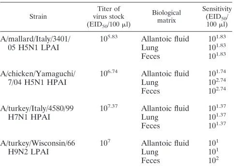
In the present study, the term “sensitivity of the method” reflects the efficacy of the entire method applied to recover the target organism in the field speci-mens, including the RNA extraction procedure and the RRT-PCR protocol (26). For this reason, allantoic fluid containing 10-fold serial dilutions of titrated AI viruses belonging to the H5, H7, and H9 subtypes was prepared, and the RNA was extracted and then used for the sensitivity test (Table 3). To establish whether the different types of sample matrices could influence the analytical sensitivity, lungs obtained from specific-pathogen-free chickens were weighed (0.1 g) and homogenized with sterile quartz sand in 1 ml (1:10 wt/vol) of PBS, pH 7.4. Lung homogenates were then blended with 10-fold dilutions of titrated subtype H5, H7, and H9 viruses and processed for RNA extraction. Similarly, feces obtained from specific-pathogen-free chickens were used for sensitivity tests. One-gram samples of feces were weighed and homogenized with sterile quartz sand to obtain a 1:5 wt/vol suspension in PBS. Blending and dilution were performed as described for lung samples.
Evaluation of the analytical sensitivity of the method was done by testing each dilution in five replicates. The sensitivity of the method was determined as the last dilution at which at least 4 of 5 replicates of each dilution was positive.
LoD of the RRT-PCR assay.In the present study, the PCR detection limit reflects the sensitivity of the RRT-PCR procedure, which includes the sensitivity of the primers and probes as well as the preparation of the master mix and the optimization of thermocycling conditions (26). To determine the limit of detec-tion (LoD) of the assay in terms of RNA copy numbers, in vitro-transcribed RNAs of the H5, H7, and H9 genes were analyzed. Briefly, the HA genes of A/chicken/Yamaguchi/7/04 (H5N1), A/turkey/Italy/4580/99 (H7N1), and A/tur-key/Wisconsin/66 (H9N2) strains were amplified by RT-PCR and the amplifica-tion products were cloned into the PCR-II vector using a dual-promoter TOPO TA cloning kit (Invitrogen, Carlsbad, CA) according to the manufacturer’s in-structions. Plasmids with the HA insert were isolated from positiveEscherichia colicolonies by using a GenElute plasmid miniprep kit (Sigma-Aldrich, St. Louis, MO). The H5, H7, and H9 insert control plasmids were linearized by using the restriction enzyme HindIII (MBI Fermentas, Lithuania) for the H9 and H7 genes and NotI (New England Biolabs, MA) for the H5 gene. The in vitro-transcribed RNA was generated from the T7 promoter by using a RiboMax kit (Promega, Madison, WI) according to the manufacturer’s recommendations and then quantified by using a UV BioPhotometer (Eppendorf, Hamburg, Ger-many).
The number of RNA copies was calculated by following the formula reported in a previous study (11). Tenfold dilutions of the RNA transcripts, ranging from 1 to 1010copies/l, were prepared. The LoD of the assay was determined from
three independent replicates.
Intra- and interassay variability.The repeatability of the H5, H7, and H9 RRT-PCR assay was determined using three different concentrations (high, medium, and low) of each viral subtype tested. The selected concentrations were
105.83
, 104.83
, and 103.83
(H5); 106.37
, 105.37
, and 103.37
(H7); and 106
, 104
, and 102
(H9) EID50/100l. For intra-assay variability, each dilution was analyzed in
triplicate. For interassay variability, each dilution was analyzed in six different runs performed by two distinct operators on different days. The coefficient of variation was determined in accordance with previously published guidelines (26).
Detection of virus RNA in samples collected from field-exposed and experi-mentally infected birds.To evaluate whether or not the RRT-PCR assay could be used as a diagnostic tool in surveillance programs for AI, we analyzed retro-spectively by VI and RRT-PCR 725 samples collected from different avian species of poultry (n⫽234) and wild birds (n⫽491) during field and laboratory investigations in Eurasia and Africa in 2006 to 2007. The samples consisted of tracheal swabs (n⫽114), cloacal swabs (n ⫽504), and organs (n⫽107; consisting of trachea, lungs, intestines, and brain) collected during necropsies. The number of samples analyzed for clinical validation is in accordance with the guidelines proposed in a previous study (5).
The agreement between VI and RRT-PCR results was investigated using Cohen’sKstatistics (a statistical measure of interrater agreement) and associated evaluation of statistical significance (20). The results were compared with the Cohen’sKstandard values proposed by Landis and Koch (13).
RESULTS
Specificity, analytical sensitivity, and intra-/interassay
vari-ability.
The H5, H7, and H9 primer and probe sets were able
to detect RNA of virus strains of their respective subtype only.
No positive results were obtained with any of the other
organ-isms listed in Table 1.
The sensitivity of the RRT-PCR assay was determined using
in vitro-transcribed RNA and titrated reference viruses.
In
terms of HA gene copy number, the LoDs for the H5, H7, and
H9 subtypes were 10
3, 10
1, and 10
3gene copies/
l of in
vitro-transcribed RNA, respectively. To determine the linearity of
the reaction and PCR efficiency, the threshold cycle values of
individual dilutions were plotted against the initial gene copy
number, leading to typical standard curves. The linear ranges
of the RRT-PCR assay span within 10
10and 10
2copies/
l for
the H7 gene and within 10
10and 10
4copies/
l for the H5 and
H9 genes. The reaction efficiencies for H5, H7, and H9 genes
were 0.97, 0.98, and 0.97, respectively. The correlation
coeffi-cient (
R
2) was higher than 0.99 in all measurements.
The sensitivity of the method relative to the detectable
in-fectious virus titer ranged between 10
2.74EID
50
/100
l and 10
1EID
50/100
l. The results obtained for the sensitivity of the
[image:3.585.43.285.79.252.2]method in different samples are summarized in Table 3.
To assess the intra- and interassay reproducibility, three
different concentrations (high, medium, and low) of each
ref-erence virus were tested in triplicate in six different runs
per-formed by two distinct operators on different days. The
coef-ficients of variation within runs (intra-assay variability) ranged
from 0.12% to 2.64%. The interassay variability was in the
range of 2.26% to 4.11% (Table 4).
TABLE 3. Sensitivity of the method
Strain
Titer of virus stock (EID50/100l)
Biological matrix
Sensitivity (EID50/
100l)
A/mallard/Italy/3401/
10
5.83Allantoic fluid
10
1.8305 H5N1 LPAI
Lung
10
1.83Feces
10
1.83A/chicken/Yamaguchi/
10
6.74Allantoic fluid
10
1.747/04 H5N1 HPAI
Lung
10
2.74Feces
10
2.74A/turkey/Italy/4580/99
10
7.37Allantoic fluid
10
1.37H7N1 HPAI
Lung
10
1.37Feces
10
1.37A/turkey/Wisconsin/66
10
7Allantoic fluid
10
1H9N2 LPAI
Lung
10
1 [image:3.585.298.543.81.161.2]Feces
10
2TABLE 4. Intra- and interassay coefficients of variation
Dilution
Coefficient of variation (%) for subtype:
H5 H7 H9
Intra-assay Interassay
Intra-assay Interassay
Intra-assay Interassay
High ⬍1.68 4.06 ⬍1.66 4.02 ⬍1.48 4.11
Medium ⬍1.58 3.81 ⬍1.35 3.02 ⬍1.74 3.19
Low ⬍1.16 2.76 ⬍2.64 2.26 ⬍4.11 3.92
on May 16, 2020 by guest
http://jcm.asm.org/
Detection of viral RNA in samples collected from
field-exposed and experimentally infected birds.
A total of 725
sam-ples was analyzed for subtypes H5, H7, and H9 by VI and
RRT-PCR (Table 5). Of these, 141 samples tested positive for
subtype H5 by means of RRT-PCR (4/114 tracheal swabs,
54/504 cloacal swabs, and 83/107 organ samples). For subtype
H7, the RRT-PCR assay identified 58 positive samples (44/114
tracheal swabs and 14/504 cloacal swabs), and 30 specimens
resulted in positive results for subtype H9 (21/114 tracheal
swabs and 9/504 cloacal swabs). The comparison of the results
of the two methods, summarized in Table 5, showed
agree-ments of 94.06%, 99.17%, and 98.89% for the H5, H7, and H9
subtypes, respectively. The Cohen’s
K
coefficients were 0.80,
0.94, and 0.85, respectively. The difference between the two
methods was not statistically significant (
P
⬍
0.01). The
per-centage of agreement between the results of RRT-PCR and VI
was influenced by the higher number of samples that tested
positive by RRT-PCR but negative by VI. However, 43/55
RRT-PCR-positive/VI-negative samples were sequenced and
their identities confirmed (data not shown). Only 2 of 26
sam-ples that were positive by VI for subtype H9 tested negative in
the molecular assay.
DISCUSSION
Several diagnostic methodologies are currently available for
the detection of AI infection, with VI in eggs universally
rec-ognized as the gold standard. However, this method is time
consuming and requires facilities (e.g., BSL3 laboratories) that
are not available in many affected areas. Recently, molecular
diagnostic tests have proven themselves to be invaluable as a
first step in the identification and control of disease outbreaks.
Conventional RT-PCR and RRT-PCR have been applied
suc-cessfully to the diagnosis of AI (7, 8, 12, 14, 15, 19, 21, 23, 27).
In this study, we present data on the development and
valida-tion of a real-time hydrolysis probe-based RT-PCR assay for
the simultaneous detection of AI viruses belonging to subtypes
H5, H7, and H9. Our results prove that the assay is highly
specific and sensitive. In a previous study (23), an RRT-PCR
assay was developed for the sequences of North American AI
virus H5 and H7 subtypes. In that assay, the sensitivity data
obtained were comparable to the results described here.
How-ever, the protocol described previously was a one-step
RT-PCR with different thermal profiles for H5 and H7 detection.
In addition to its sensitivity and specificity, the method
de-scribed in the present paper offers several advantages over
conventional diagnostic methods, including rapidity, flexibility,
and ease of use. This assay makes results for the three major
AI viruses currently prevalent in poultry in large areas of the
world available in approximately 3 h, and the use of a
single-step RRT-PCR procedure provides some protection against
contamination events. Based on its technical characteristics,
this assay could be used for large-scale screening and subtyping
of viral RNA during influenza A virus outbreaks and for
sur-veillance programs.
This RRT-PCR assay was developed and validated using the
same annealing temperature in order to identify the H5, H7,
and H9 subtypes in a single analytical session. In the literature,
the use of multiplexed PCRs has been reported as resulting in
a decreased sensitivity of the method (22, 28), and the
optimi-zation of the concentration of the multiplex PCR components
to achieve optimal amplification can pose several difficulties
(9). For these reasons, the development of a multiplex assay
was not attempted, as it was so important to identify the three
subtypes and maximize the assay’s performance. The
applica-tion of this RRT-PCR format was also due to the necessity of
having a cost-effective and flexible diagnostic tool that could be
easily switched to a single-subtype identification method that
would be applicable during the investigation of outbreaks
caused by only one of these subtypes. It should also be
con-sidered that not all of the existing real-time PCR platforms are
capable of detecting more than two fluorophores
simulta-neously, making a triplex PCR protocol inapplicable.
[image:4.585.42.285.82.153.2]The suitability of the RRT-PCR test described in the present
study as a diagnostic tool to rapidly recognize the three
most-important HA subtypes of the AI virus is confirmed by the
results obtained using samples from birds infected naturally.
These clinical samples were obtained from a wide range of
avian species and geographical areas during field and
labora-tory investigations. The assay has been used for monitoring the
AI virus in poultry and wild birds, and it has proved capable of
identifying the presence of several distinct genetic lineages of
subtype H5, H7, and H9 viruses, including the H5N1
sublin-eages circulating in Eurasia and Africa (data not shown). The
comparison of the results obtained from applying the
conven-tional diagnostic method (VI) and the RRT-PCR assay to
these clinical samples showed good agreement. The lowest
level of agreement was observed in the RRT-PCR/VI results
for subtype H5. Negative results by VI for H5 could be
ex-plained by considering the condition of the clinical samples at
the time of their arrival. Many of the samples that proved
positive for subtype H5 by RRT-PCR but negative by VI were
submitted to the OIE/FAO Reference Laboratory from Africa
and the Middle East, and in some cases, the cold chain was not
maintained during shipping, compromising the viability of the
viruses. Based upon the results obtained in the present study,
the RRT-PCR assay for simultaneous detection of subtypes
H5, H7, and H9 could be a useful instrument for rapid
screen-ing and surveillance in wild and domestic birds. Although this
method cannot replace the standard VI technique, this
RRT-PCR assay offers several advantages over standard methods,
and it could be used as a reliable tool for the rapid detection of
the three AI virus subtypes, including identification of
cocir-culating strains. Routine application in critical environments,
such as live-bird markets, or for samples obtained from wild
birds in their breeding or resting sites could give an indication
of the degree of coinfection with these subtypes, providing
insight into the complex ecoepidemiology of AI infections in
such birds.
TABLE 5. Results of RRT-PCR and VI from clinical samples
RRT-PCR result
VI/HI resulta
No. of samples of subtype:
H5 H7 H9
⫹
⫹
98
52
24
⫹
⫺
43
6
6
⫺
⫺
584
667
693
⫺
⫹
0
0
2
a
HI, hemagglutinin inhibition.
on May 16, 2020 by guest
http://jcm.asm.org/
ACKNOWLEDGMENTS
This study was supported by the European Union FLUTRAIN
project (training and technology transfer of AI diagnostics and disease
management skills) and by the Italian Ministry of Health (RF 2007).
We thank Laura Gagliazzo for statistical assistance.
REFERENCES1.Alexander, D. J.2003. Report on avian influenza in the Eastern Hemisphere during 1997–2002. Avian Dis.47:792–797.
2.Alexander, D. J.2007. Summary of avian influenza activity in Europe, Asia, Africa, and Australasia. 2002–2006. Avian Dis.51(Suppl. 1):161–166. 3.Capua, I., and D. J. Alexander.2004. Avian influenza: recent developments.
Avian Pathol.33:393–404.
4.Cattoli, G., and I. Capua.2006. Molecular diagnosis of avian influenza during an outbreak. Dev. Biol. (Basel)124:99–105.
5.Crowther, J. R., H. Unger, and G. J. Viljoen.2006. Aspects of kit validation for tests used for the diagnosis and surveillance of livestock diseases: pro-ducer and end-user responsibilities. Rev. Sci. Tech.25:913–935.
6.de Jong, M. D., and T. T. Hien.2006. Avian influenza A (H5N1). J. Clin. Virol.35:2–13.
7.Di Trani, L., B. Bedini, I. Donatelli, L. Campitelli, B. Chiappini, M. A. De Marco, M. Delogu, C. Buonavoglia, and G. Vaccari.2006. A sensitive one-step real-time PCR for detection of avian influenza viruses using a MGB probe and an internal positive control. BMC Infect. Dis.6:87.
8.Ellis, J. S., J. W. Smith, S. Braham, M. Lock, K. Barlow, and M. C. Zambon. 2007. Design and validation of an H5 TaqMan real-time one-step reverse transcription-PCR and confirmatory assays for diagnosis and verification of influenza A virus H5 infections in humans. J. Clin. Microbiol.45:1535–1543. 9.Elnifro, E. M., A. M. Ashshi, R. J. Cooper, and P. E. Klapper.2000. Mul-tiplex PCR: optimization and application in diagnostic virology. Clin. Micro-biol. Rev.13:559–570.
10.Fouchier, R. A., V. Munster, A. Wallensten, T. M. Bestebroer, S. Herfst, D. Smith, G. F. Rimmelzwaan, B. Olsen, and A. D. Osterhaus.2005. Charac-terization of a novel influenza A virus hemagglutinin subtype (H16) obtained from blackheaded gulls. J. Virol.79:2814–2822.
11.Fronhoffs, S., G. Totzke, S. Stier, N. Wernert, M. Rothe, T. Bruning, B. Koch, A. Sachinidis, H. Vetter, and Y. Ko.2002. A method for the rapid construction of cRNA standard curves in quantitative real-time reverse tran-scription polymerase chain reaction. Mol. Cell. Probes16:99–110. 12.Hoffmann, B., T. Harder, E. Starick, K. Depner, O. Werner, and M. Beer.
2007. Rapid and highly sensitive pathotyping of avian influenza A H5N1 virus by using real-time reverse transcription-PCR. J. Clin. Microbiol.45: 600–603.
13.Landis, J. R., and G. G. Koch.1977. The measurement of observer agree-ment for categorical data. Biometrics33:159–174.
14.Ng, L. F., I. Barr, T. Nguyen, S. M. Noor, R. S. Tan, L. V. Agathe, S. Gupta,
H. Khalil, T. L. To, S. S. Hassan, and E. C. Ren.2006. Specific detection of H5N1 avian influenza A virus in field specimens by a one-step RT-PCR assay. BMC Infect. Dis.6:40.
15.Payungporn, S., S. Chutinimitkul, A. Chaisingh, S. Damrongwantanapokin, C. Buranathai, A. Amonsin, A. Theamboonlers, and Y. Poovorawan.2006. Single step multiplex real-time RT-PCR for H5N1 influenza A virus detec-tion. J. Virol. Methods131:143–147.
16.Peiris, J. S. M., M. D. de Jong, and Y. Guan.2007. Avian influenza virus (H5N1): a threat to human health. Clin. Microbiol. Rev.20:243–267. 17.Perk, S., A. Panshin, E. Shihmanter, I. Gissin, S. Pokamunski, M. Pirak,
and M. Lipkind.2006. Ecology and molecular epidemiology of H9N2 avian influenza viruses isolated in Israel during 2000–2004 epizootic. Dev. Biol. (Basel)124:201–209.
18.Reed, L. J., and H. Muench.1938. A simple method of estimating fifty percent endpoints. Am. J. Hyg.27:493–497.
19.Rossi, J., S. Cramer, and T. Laue.2007. Sensitive and specific detection of influenza virus A subtype H5 with real-time PCR. Avian Dis.51(Suppl. 1):387–389.
20.Siegel, S., and N. J. Castellan.1988. Nonparametric statistics for behavioural sciences. McGraw-Hill, New York, NY.
21.Slomka, M. J., T. Pavlidis, J. Banks, W. Shell, A. McNally, S. Essen, and I. H. Brown.2007. Validated H5 Eurasian real-time reverse transcriptase-polymerase chain reaction and its application in H5N1 outbreaks in 2005– 2006. Avian Dis.51(Suppl. 1):373–377.
22.Spackman, E., D. A. Senne, L. L. Bulaga, S. Trock, and D. L. Suarez.2003. Development of multiplex real-time RT-PCR as a diagnostic tool for avian influenza. Avian Dis.47(Suppl. 3):1087–1090.
23.Spackman, E., D. A. Senne, T. J. Myers, L. L. Bulaga, L. P. Garber, M. L. Perdue, K. Lohman, L. T. Daum, and D. L. Suarez.2002. Development of a real-time reverse transcriptase PCR assay for type A influenza virus and the avian H5 and H7 hemagglutinin subtypes. J. Clin. Microbiol.40:3256–3260. 24.Suarez, D. L., A. Das, and E. Ellis.2007. Review of rapid molecular
diag-nostic tools for avian influenza virus. Avian Dis.51(Suppl. 1):201–208. 25.Swayne, D. E., J. R. Glisson, M. W. Jackwood, J. E. Pearson, and W. M. Reed
(ed.).1998. A laboratory manual for the isolation and identification of avian pathogens, 4th ed. University of Pennsylvania, Kennett Square, PA. 26.U.S. Environmental Protection Agency.2004. Quality assurance/quality
con-trol guidance for laboratories performing PCR analyses on environmental samples. U.S. Environmental Protection Agency, Washington, DC. 27.Xie, Z., Y. S. Pang, J. Liu, X. Deng, X. Tang, J. Sun, and M. I. Khan.2006.
A multiplex RT-PCR for detection of type A influenza virus and differenti-ation of avian H5, H7, and H9 hemagglutinin subtypes. Mol. Cell. Probes 20:245–249.
28.Zangenberg, G., R. K. Saiki, and R. Reynolds.1999. Multiplex PCR: opti-mization guidelines, p. 73–94.InM. A. Innis, D. H. Gelfand, and J. J. Sninsky (ed.), PCR applications: protocols for functional genomics. Academic Press, San Diego, CA.