0095-1137/08/$08.00⫹0 doi:10.1128/JCM.01737-07
Copyright © 2008, American Society for Microbiology. All Rights Reserved.
Differences in Virulence Markers between
Helicobacter pylori
Strains
from Iraq and Those from Iran: Potential Importance of Regional
Differences in
H. pylori-
Associated Disease
䌤
Nawfal R. Hussein,
1,2Marjan Mohammadi,
3Yeganeh Talebkhan,
3Masoumeh Doraghi,
3Darren P. Letley,
1Merdan K. Muhammad,
2Richard H. Argent,
1†
and John C. Atherton
1†*
Institute of Infection, Immunity, and Inflammation, Centre for Biomolecular Sciences, University of Nottingham, Nottingham NG7 2RD, United Kingdom, and Wolfson Digestive Diseases Centre, Queen’s Medical Centre,
University of Nottingham, Nottingham NG7 2UH, United Kingdom1; Azadi Teaching Hospital,
College of Medicine University of Dohuk, Dohuk, Kurdistan, Iraq2; and
Biotechnology Research Center, Pasteur Institute of Iran, 69 Pasteur Ave., Tehran 13164, Iran3
Received 31 August 2007/Returned for modification 19 November 2007/Accepted 10 March 2008
Helicobacter pyloricauses peptic ulceration and gastric adenocarcinoma; the latter is common in Iran but not
in Iraq. We hypothesized that more virulent H. pylori strains may be found in Iran than in Iraq and so compared established and newly described virulence factors in strains from these countries. We studied 59 unselected dyspeptic patients from Iran and 49 from Iraq.cagAwas found in similar proportions of strains from both countries (76% in Iran versus 71% in Iraq) and was significantly associated with peptic ulcer disease in Iraq (P<0.01) but not in Iran.cagAalleles encoding four or more tyrosine phosphorylation motifs were found in 12% of the Iranian strains but none of the Iraqi strains (P ⴝ 0.02). There were no significant differences in thevacAsignal-, middle-, or intermediate-region types between Iranian and Iraqi strains. Among the strains from Iran,vacAgenotypes showed no specific peptic ulcer associations, but among the strains from Iraq,vacAi1 strains were associated with gastric ulcer (P<0.02), mimicking their previously demonstrated association with gastric cancer in Iran.dupAwas found in similar proportions of Iranian and Iraqi strains (38% and 32%, respectively) and was associated with peptic ulceration in Iraqi patients (P <0.01) but not Iranian patients.H. pyloristrains from Iraq and Iran possess virulence factors similar to those in Western countries. The presence ofcagAwith more phosphorylation motifs in Iranian strains may contribute to the higher incidence of gastric cancer. However, the association between strain virulence markers and disease in Iraq but not Iran suggests that other host and environmental factors may be more important in the disease-prone Iranian population.
Helicobacter pyloriis a spiral-shaped, gram-negative bacillus which causes gastritis and peptic ulceration (18, 19, 36). Its treatment has become pivotal in the management of peptic ulcer disease (PUD).H. pyloriinfection is also an important risk factor for gastric adenocarcinoma, the second most impor-tant cause of cancer deaths worldwide. Gastric cancer is thought to have a multifactorial etiology; and bacterial strain type, host genotype, and environmental conditions are all thought to be factors contributing to gastric cancer (22). De-spite the geographical proximity of Iraq and Iran, the incidence of gastric cancer differs hugely between these countries; in Iran it ranges from 38 to 69 cases/105population (10, 21, 26, 27, 38), whereas in Iraq the incidence is 5 cases/105 population (10). We hypothesized that this difference may be due to differences in the virulence of the circulatingH. pyloristrains, and so we set out to type strains from these countries for their virulence.
We considered both well-established and more recently de-scribed virulence determinants.
Many strains of H. pylori produce the CagA protein, en-coded by the cagA gene within the cagpathogenicity island (PAI).H. pyloristrains possessing cagAare associated with a significantly increased risk for the development of atrophic gastritis, PUD, and gastric cancer (24, 32). ThecagPAI en-codes a type IV secretion system that facilitates the transloca-tion of CagA into the host epithelial cytosol, where it becomes phosphorylated with tyrosine at specific phosphorylation mo-tifs by the Src family of kinases (29, 30). Phosphorylated CagA forms a physical complex with SHP-2 phosphatase and stimu-lates cell signaling pathways, cytoskeletal changes, and abnor-mal cell proliferation (33). On the basis of the amino acid sequence of the SHP-2 binding site, CagA proteins can be subcategorized into Western and East Asian types. Both have type A and B phosphorylation motifs (usually one of each), but the Western types have additional C motifs (1–3) and the East Asian type has no C motifs but a D motif. The East Asian type CagA possesses stronger SHP-2 binding and transforming ac-tivities than the Western type CagA (11). The Western type CagA has a variable number of type C phosphorylation motifs, and the extent of cytoskeletal changes induced by CagA is
* Corresponding author. Mailing address: Wolfson Digestive Dis-eases Centre, C floor, South Block, Queen’s Medical Centre, Notting-ham NG7 2UH, United Kingdom. Phone: 44 115 8231034. Fax: 44 115 9422232. E-mail: [email protected].
† Richard Argent and John Atherton share senior authorship for this work.
䌤Published ahead of print on 19 March 2008.
1774
on May 16, 2020 by guest
http://jcm.asm.org/
dependent on this. Strains possessing CagA with greater num-bers of type C phosphorylation motifs are more closely asso-ciated with gastric carcinogenesis (9). Thus, determination of the degree of CagA phosphorylation or the number of phos-phorylation motifs appears to be more important than detec-tion ofcagAalone (4, 5).
The vacuolating cytotoxin (VacA) is a well-established H. pylori virulence factor which has multiple effects, including vacuolization of cultured epithelial cells, the induction of apoptosis, increases in the permeability of epithelial monolay-ers, the formation of pores in cells, and suppression of immune cell function (6, 13). ThevacAgene is polymorphic within its signal, intermediate, and middle regions. For the signal region, two distinct allelic sequences, s1 and s2, have been recognized. For the middle region, alleles can be categorized into two classes, m1 or m2. ThevacAgenotype is associated with in vitro cytotoxin activity (with s1 having greater cytotoxin activity than s2 and m1 having greater cytotoxin activity than m2) (8, 20, 34, 35). Rhead et al. have recently described a novel determinant of VacA toxicity, the intermediate or i region (23). They showed that two allelic variants of this region, i1 and i2, exist. Furthermore, they showed that only s1/m2 strains varied in their i types; s1/m1 and s2/m2 strains were exclusively i1 and i2, respectively. This novel region determines the vacuolating ac-tivity among these s1/m2 strains. Most importantly, a signifi-cant correlation was found between the i1 type ofvacAand gastric cancer in Iran (23).
The duodenal ulcer (DU)-promoting gene A (dupA) is a recently described virulence factor which comprises both jhp0917 and jhp0918 (16). Lu et al. found a significant rela-tionship betweendupAand DU, and the presence ofdupAwas related to neutrophil infiltration and a high level of interleu-kin-8 production by epithelial cells. Surprisingly, possession of this gene appeared to be protective against gastric adenocar-cinoma (16).
The object of this study was to type the virulence of un-selected strains from dyspeptic patients in Iran and Iraq. We aimed to compare the virulence of strains from these neigh-boring countries, which have very different incidences of gas-tric cancer, and to assess the association of virulence markers with PUD in each country.
MATERIALS AND METHODS
Patient-derived samples.Gastric biopsy specimens were obtained from 49 and
59 unselectedH. pylori-positive patients from Iraq and Iran, respectively,
under-going routine upper gastrointestinal endoscopy for investigation for dyspepsia.
The mean age⫾standard deviation of the Iraqi patients was 35⫾17 years, and
that of the Iranian patients was 40⫾14 years. All Iraqi patients were from the
five districts of city of Dohuk. The majority (48/59 [81%]) of Iranian patients were from the city of Tehran; other patients were referred from different regions in Iran. Endoscopic diagnoses were as follows: for DU, 15 in Iraq and 8 in Iran; for gastric ulcer (GU), 5 in Iraq and 9 in Iran; for no ulcer disease, 29 in Iraq and 42 in Iran. During gastroscopy, biopsy samples were taken and either placed in 1 ml of Iso-Sensitest broth (Oxoid, Basingstoke, United Kingdom) containing 15% (vol/vol) glycerol and stored in liquid nitrogen or cultured immediately for
H. pylori. In some cases, following prolonged storage and shipment to the United Kingdom, reculture was not possible. In these cases, DNA was extracted directly
from the biopsy specimens and used for PCR-basedH. pylorityping.
The study protocol was approved by the ethics and research committees of the individual hospitals, and all patients gave informed consent to participation in the study.
Culture.Each biopsy specimen was spread onto horse blood or Dent agar
plates and then incubated under microaerobic conditions generated with a CampyPak system (Becton Dickinson, Baltimore, MD) in an anaerobic jar at
37°C for 2 to 4 days. The organisms were identified asH. pyloriby colony
morphology, Gram stain, and urease activity. Cultures were harvested as sweeps rather than single colonies and were stored in Iso-Sensitest broth containing 15%
(vol/vol) glycerol at⫺80°C.
Genotyping ofH. pylori.PCR-based typing of theH. pyloriisolates was
per-formed with DNA extracted from bacteria or directly from the biopsy specimens.
PCR amplification ofcagAused previously described primers cag2 and cag4 (25)
to amplify the 3⬘variable region. PCR amplification of thecagPAI empty site
was performed as described previously (1). In the empty-site PCR, primers
anneal to sequences adjacent to thecagPAI insertion site in the genome and
allow amplification only of a DNA fragment of the expected size in the absence
of a complete or partialcagPAI at this locus. Genotyping of thevacAsignal,
intermediate, and middle regions was performed as described previously (7, 8,
23).dupAwas amplified with primer pairs DupAF113-DupAR1083 and
Du-pAF1202-DupA918R, as described previously (3) (Fig. 1). Determination of the number of CagA phosphorylation motifs and the types of motifs was carried out by using the forward primer cag2 and the reverse primers cagA-P1C, cagA-P2CG and cagA-P2TA (the B motif is polymorphic, and reverse primers cagA-P2CG and cagA-P2TA are designed to recognize all types described to date), and cagA-P3E, as described previously (5) (Fig. 2). Five microliters of the PCR products was electrophoresed in 1.5% (wt/vol) agarose gels for 40 min at 80 V in TAE (Tris-acetate-EDTA) buffer. All gels were stained with ethidium bromide (1 mg/liter) and photographed under UV light. A 1-kb DNA ladder (Gibco, Paisley, United Kingdom) was used as a size marker in all gels. Strains with previously determined genotypes were used as positive controls.
Data analysis.Statistical analysis of the data was performed by using logistic
[image:2.585.81.505.69.232.2]regression, the chi-square test, and Fisher’s exact test, with significance set at a
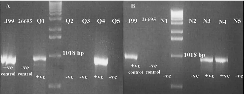
FIG. 1. Characterization of Iraqi (A) and Iranian (B) strains fordupAby PCR. Image shows the results of PCR typing ofdupAwith primers DupAF113 and DupAR1083.⫹ve, positive;⫺ve, negative.
on May 16, 2020 by guest
http://jcm.asm.org/
Pvalue of⬍0.05. Genotypes with mixed status forvacAwere excluded from the calculations of association.
RESULTS
Prevalence of cagAⴙstrains among dyspeptic Iranian and Iraqi populations.First, we assessed whether the prevalence of
cagA-positive (cagA⫹) strains was similar or different between the Iraqi and the Iranian populations. cagA⫹ strains were present in 76% (45/59) and 71% (35/49) of theH. pyloristrains from unselected Iranian and Iraqi patients with dyspepsia, respectively (Table 1). To exclude bias from disease associa-tion, we also compared subgroups of patients without peptic ulceration:cagAwas found in a higher proportion of Iranian strains than Iraqi strains (76% and 55%, respectively), al-though this did not quite achieve formal statistical significance (P ⫽ 0.06). In both countries, all cagA⫹ strains also typed positive forcagE. Among 14cagA-negative Iraqi strains, 7 were
cagPAI empty-site positive (implying that the wholecagPAI was absent) and 7 were empty-site negative (implying that there was still a partial cag PAI at this locus). Among 14
cagA-negative Iranian strains, 9 werecagPAI empty-site neg-ative. Thus, overall, thecagPAI appeared to be incompletely deleted in 16 strains.
No significant association was found between cagA status and clinical outcome for the Iranian patients, but a significant
correlation was found betweencagAand PUDs (Pⱕ0.01; odds ratio, 16.4) in Iraqi patients (Table 1). When DU and GU were considered separately for the Iraqi population, 14/15 (93%) patients with DU had cagA⫹ strains, whereas 16/29 (55%) patients with no ulcer (P⬍0.02) hadcagA⫹strains. All Iraqi GU patients hadcagA⫹strains (Pwas not significant compared with the results for patients with no ulcer, perhaps due to the low number of GU patients).
CagA phosphorylation motif numbers. Second, we turned our attention tocagA polymorphisms and, in particular, the number of CagA phosphorylation motifs, which we assessed using our recently described PCR-based typing system (5). Among thecagA⫹strains, 12% (7/49) of the strains from Iran carried acagAvariable region of⬎550 bp (when the region was amplified with primers cag2 and cag4), indicating the pres-ence of more than three CagA phosphorylation motifs. This was a significantly higher proportion than that found in the strains from Iraq, where all strains possessedcagAwith a vari-able-region size of 550 bp, indicating the presence of CagA with three phosphorylation motifs (P⫽0.02) (Table 1). In the analysis confined to patients without ulcers, 22% (7/32) of the
[image:3.585.84.502.69.233.2]cagA⫹Iranian strains had more than three phosphorylation sites, whereas none of the Iraqi strains did. Previous studies with other populations have linked multiple CagA phosphory-lation motif numbers with an increased risk of cancer but not
FIG. 2. Characterization of one Iraqi strain (A) and one Iranian strain (B) forcagAvariable-region tyrosine phosphorylation motifs. Genomic DNA samples fromH. pyloristrains were used to PCR amplify thecagAvariable-region EPIYA motifs by using forward primer cag2 and reverse primer cagA-P1C (A motif), reverse primers cagA-P2CG and cagA-P2TA (B motif), or reverse primer cagA-P3E (C motif). No Iraqi strain was found to have more than three phosphorylation motifs.
TABLE 1. cagAstatus,cagAphosphorylation motif number, anddupAstatus amongH. pyloristrains from unselected Iranian and Iraqi patients with dyspepsia
Country
% of strains positive for:
cagA More than threecagAphosphorylation motifs dupA
PUD NPUDa Total PUD NPUD Total PUD NPUD Total
Iraq 95 (19/20)b,c 55 (16/29) 71 (35/49) 0 (0/20) 0 (0/29) 0 (0/49) 55 (11/20)b 17 (5/29) 32 (6/49) Iran 76 (13/17) 76 (32/42) 76 (45/59) 0 (0/17) 17 (7/42) 12 (7/59)d 35 (6/17) 40 (17/42) 39 (23/59)
a
NPUD, no PUD.
b
P⬍0.05 for comparison of patients with PUD and patients without PUD.
c
Values in parentheses indicate the number of strains positive/total number of strains tested.
d
The presence ofcagAalleles with more than three phosphorylation motifs was significantly greater among Iranian strains than among Iraqi strains (P⫽0.02).
on May 16, 2020 by guest
http://jcm.asm.org/
[image:3.585.44.541.623.694.2]with an increased risk of ulcer (9, 15): in agreement with this, no strains with more than three phosphorylation motifs were found in the ulcer group from Iran (Table 1).
vacA polymorphism. We then turned to vacA polymor-phisms in Iranian and Iraqi strains, examining both established s and m genotypes, and also the recently described polymor-phic i-region type. Since individualH. pylori isolates possess only a single copy ofvacA, the presence of more than onevacA
s, i, or m genotype in a DNA sample indicates colonization by two or more strains with differentvacAgenotypes (8). Among the Iraqi isolates, a singlevacAsignal region was observed in all samples, but 8/49 (16%) of the specimens examined pos-sessed both middle-region types, and 9/49 (18%) pospos-sessed both i-region genotypes. Among the Iranian samples, all iso-lates possessed a single signal-region type, but 2/59 (3%) car-ried both m-region types and 7/59 (12%) possessed both i-region types. There was no difference in the prevalence of strains with different vacA genotypes among the unselected dyspeptic populations from Iran and Iraq, whether strains with multiple genotypes were excluded (planned analysis; Table 2) or classified as the more pathogenic or the less pathogenic type (exploratory analyses).
Next, we examined associations betweenvacAallelic varia-tion and peptic ulceravaria-tion within the Iranian and Iraqi popu-lations. For the Iranian strains, no significant associations were found. For the Iraqi strains, no significant association was found for duodenal ulceration, but 80% (4/5) of the strains isolated from GU patients were of thevacAi1 genotype, which was significantly greater than the 13% (4/29) of the strains from patients without ulcers (P ⬍ 0.02). Although this sub-group analysis is exploratory, it is interesting, given the de-scribed association betweenvacAi1 genotype and gastric can-cer and the similarities in epidemiology and pathogenesis between GU and gastric cancer. Associations were not seen between gastric ulceration and thevacAs and m types, again supporting the recent finding that thevacAi type is a better marker of strain virulence (23).
dupA status. Third, we examined strains for the recently described putative virulence genedupA. Similar proportions of Iranian and Iraqi strains possesseddupA(Table 1). Among the Iranian patients, we found no association betweendupAand the clinical outcome. However, among the Iraqi patients, 55% (11/20) of the peptic ulcer patients carrieddupA⫹strains, sig-nificantly more than the 17% (5/29) of the patients without ulcers who carrieddupA⫹strains (P⬍ 0.01; odds ratio, 6.2) (Table 1). When we looked at DU and GU separately, 60% (9/15) of theH. pylori isolates from DU patients weredupA⫹
(P⬍ 0.01 compared with the results for patients without ul-cers) and 40% (2/5) of theH. pyloriisolates from GU patients weredupA⫹(Pwas not significant compared with the results
for the patients without ulcers, but note the small number of GU patients).
Associations between virulence factors, particularly forcagA
phosphorylation motif number.Next, we assessed associations between virulence factors in strains from Iran and Iraq. As in virtually all strain populations worldwide, we found thatcagA⫹
strains were more likely to bevacAs1 than s2: in Iran, 37/45 (82%)cagA⫹strains werevacAs1, whereas 5/14 (36%) of the
cagA-negative strains were vacA s1 (P ⬍ 0.005); in Iraq, all
cagA⫹ strains typed s1, whereas 10/14 (71%) of the cagA -negative strains typed s1 (P⬍0.005). No significant associa-tions were found betweencagA status and other vacA poly-morphisms or betweencagAstatus anddupAstatus. As strains with a largercagA are thought to be more pathogenic than those with a smaller cagA, we examined the association be-tween the size of cagA and other virulence factors among Iranian strains. This analysis was not possible for Iraqi strains, as they all had the same number (n⫽3) of CagA phosphory-lation motifs. Seven of 45 (15%)cagA⫹Iranian strains carried a largercagA(with more than three phosphorylation motifs). In the association analysis withvacAgenotypes, we excluded patients with mixed genotypes. The small numbers of strains studied meant that most associations were not statistically sig-nificant, but for thevacAm region, 6/7 (86%) strains with more than three phosphorylation motifs were type m1, significantly more than the 10/37 (27%) strains with only three phosphory-lation motifs (P⫽0.01). Lastly, we examined the association between the size ofcagA anddupAstatus: 6/7 (86%) strains with more than three phosphorylation motifs were dupA⫹, significantly more than the 14/38 (36%) strain with only three phosphorylation motifs (P⫽0.03).
DISCUSSION
[image:4.585.43.542.81.136.2]The study of H. pylori virulence factors in populations is important, as they contribute to disease risk. For example, in Japan, where gastric cancer is common, more than 90% ofH. pyloristrains arecagApositive (17). The gastric cancer rate in Iraq is lower than that in Iran; we hypothesized that differences in the virulence factors of theH. pyloristrains between these two countries may partially explain this difference. We found no difference in the prevalence ofcagA⫹strains between un-selected dyspeptic populations from these countries, although among patients without ulcers,cagA⫹strains were 21% more prevalent in Iran (P ⫽ 0.06). Furthermore, Iranian patients withcagA⫹strains were more likely to have the more patho-genic forms ofcagAencoding four or more tyrosine phosphor-ylation sites, and among patients without ulcers, this difference was 22%. Taking these results together, this represents a con-siderable difference in potentialcagA-associated pathogenicity
TABLE 2. Distribution ofvacAallelic types amongH. pyloristrains isolated from unselected dyspeptic patients from Iraq and Iran
Country
No. of patients positive for the following allelic type/total no. (%):
s1/i1/m1 s1/i1/m2 s1/i2/m1 s1/i2/m2 s2/i2/m2 Mixed
Iraq 8/49 (16.3) 2/49 (4.1) 1/49 (2.0) 20/49 (40.8) 4/49 (8.2 ) 14/49 (28.5)
Iran 15/59 (25.4) 4/59 (6.7) 1/59 (1.7) 16/59 (27.1) 16/59 (27.1) 7/59 (11.9)
on May 16, 2020 by guest
http://jcm.asm.org/
which could contribute to the differences in gastric cancer rates seen between these populations: both cagA status and the number ofcagAphosphorylation motifs have been linked with cancer prevalence in a number of populations (4, 11). How-ever, we found no significant differences between Iranian and Iraqi populations in vacA types and, in particular, in the i-region type, which has recently been linked with gastric cancer risk in Iran (23). Also, we found no difference indupAstatus, which we studied becausedupAhas been reported to have a negative association with gastric cancer (16), although recent data from us dispute this (3).
In the present report, we have shown that 71% and 76% of theH. pyloristrains isolated from Iraqi and Iranian samples, respectively, werecagA⫹. This value is closer to the values for Western countries and Turkey than to the values for East or Southeast Asia (2, 14, 28, 31). Our strains had the Western type of CagA and the Western types ofvacA. Thus, it appears that the high cancer rate in Iran is not due to the presence of strains of the East Asian type in that country.
We looked within the Iranian and Iraqi populations for associations between virulence factors and PUD. Among the Iraqi strains but not the Iranian strains, we observed an asso-ciation betweencagA⫹status and PUD. Reports from a neigh-boring country, Turkey, have shown results similar to those from Iraq (28). No Iraqi strains hadcagAwith more than three phosphorylation motifs, so we could not perform an examina-tion for disease associaexamina-tions. The situaexamina-tion in Iran was inter-esting: no strains with more than three phosphorylation motifs were found in patients with peptic ulcer. This may imply that the presence of more than three phosphorylation motifs is protective against ulcers rather than being a specific predispo-sition to cancer, as reported previously (4, 11). ForvacA poly-morphisms, we found no association between thevacAi region and the clinical outcome for the Iranian samples. However, for the Iraqi specimens, a novel association was found between the presence ofvacAi1 strains and gastric ulcer. This is not unex-pected, as GU and gastric cancer are epidemiologically similar. However, our results need confirmation by the performance of studies with other populations, as only a small number of GU patients were enrolled in this study. FordupA, a significant link with PUD was present in the Iraqi population, but no associ-ation was found in Iranians. Thus, overall, we showed that the Iraqi population was similar to Western populations in terms of the association of many virulence factors with ulcer disease. In contrast, these associations were not seen in the Iranian population. This may imply that factors other than bacterial virulence are the most important for ulcer risk in Iran.
Many previous reports have shown a clustering of active virulence factors withinH. pyloristrains, for example, associa-tions betweencagA⫹status and thevacAs1 genotype (37). In agreement with the findings presented in those reports, we found a significant correlation betweencagA⫹status and the presence of thevacAs1 genotype in Iran and Iraq. In addition, we showed a significant association between the presence of a greater number ofcagAphosphorylation motifs and the pres-ence of both thevacA m1 genotype and dupA⫹ status. This further supports the concept of the clustering of virulence factors, such that the majority ofH. pyloristrains possess either many or a few, and the fact that it is favorable forH. pylorito be either strongly pathogenic or nonpathogenic.
Our study has several limitations. First, and perhaps most importantly, we studied H. pyloristrains from dyspeptic pa-tients rather than a random community sample of H. pylori
strains, and this may have introduced bias. The reason that we did this is that strain genotyping requires upper gastrointesti-nal endoscopy, which is difficult to perform with randomly selected asymptomatic individuals. We argue that the preva-lence of virulent strains in the dyspeptic group without ulcer disease is likely to be similar to that in asymptomatic commu-nity members: the association betweenH. pyloriinfection and nonulcer dyspepsia is controversial, and if such an association is present at all, it is weak; so any association with virulence factors is likely to be weaker still. Second, our study was not large. However, this is more likely to hide true-positive asso-ciations rather than to produce false-positive results and is most unlikely to have produced the multiple associations of virulence factors with disease that we have demonstrated. Third, we genotyped strains for virulence rather than perform virulence phenotyping. Many studies have shown good but imperfect associations between the genotype and the pheno-type of a strain, but any error is likely to be in favor of desig-nating a strain as virulent when, in fact, it is avirulent. For example, a seemingly virulentcagA⫹strain may not be able to translocate CagA into epithelial cells (if it has a mutation elsewhere in thecagPAI which inactivates the type IV secre-tion system) and so may be avirulent. In contrast, a seemingly avirulentcagA-negative strain will never be able to translocate CagA (as it lacks it), and so such a strain will never be virulent. Thus, any errors from genotyping rather than errors from phe-notyping are also likely to be conservative. Taken together these study deficiencies should encourage others to perform better studies to repeat and extend our investigation, but they do not negate our findings.
To summarize, the virulence factors of both Iraqi and Ira-nian H. pylori strains appear to be more closely related to strains from Western countries than to strains from Asian countries. Iranian strains appear to be more virulent, but the difference appears to be unlikely completely to explain the difference in disease prevalence between these countries. This suggests that unidentified strain, host, and environmental fac-tors may contribute to these differences. In the absence of an East Asian type ofcagAand almost universally virulent strains (as are found in Japan and parts of China), the very high gastric cancer rate in Iran remains largely unexplained. Simi-larly, the cancer rates in Iraq appear to be lower than those that would be expected from the circulating H. pyloristrain types, an enigma similar to that reported (controversially) in Africa (12).
ACKNOWLEDGMENTS
We thank theHelicobacter pylori Research Group at the Biotech-nology Research Center of the Pasteur Institute of Iran and the clinical staff at the Cancer Research Center of the Tehran University of Med-ical Sciences. We also thank the physicians and nurses in the gastro-enterology departments in both Iraq and Iran for assisting with biopsy specimen collection. We are grateful to Marouf Jaro, Karwan Fendi, and Halat Majed for their kind help in Azadi Hospital in Dohuk, Iraq.
REFERENCES
1.Akopyants, N. S., S. W. Clifton, D. Kersulyte, J. E. Crabtree, B. E. Youree,
C. A. Reece, N. O. Bukanov, E. S. Drazek, B. A. Roe, and D. E. Berg.1998.
on May 16, 2020 by guest
http://jcm.asm.org/
Analyses of thecagpathogenicity island ofHelicobacter pylori. Mol.
Micro-biol.28:37–53.
2.Arents, N. L., A. A. van Zwet, J. C. Thijs, A. M. Kooistra-Smid, K. R. van
Slochteren, J. E. Degener, J. H. Kleibeuker, and L. J. van Doorn.2001. The
importance ofvacA,cagA, andiceAgenotypes ofHelicobacter pyloriinfection
in peptic ulcer disease and gastroesophageal reflux disease. Am. J.
Gastro-enterol.96:2603–2608.
3.Argent, R. H., A. Burette, V. Y. M. Deyi, and J. C. Atherton.2007. The
presence ofdupAinHelicobacter pyloriis not significantly associated with
duodenal ulceration in Belgium, South Africa, China, or North America.
Clin. Infect. Dis.45:1204–1206.
4.Argent, R. H., M. Kidd, R. J. Owen, R. J. Thomas, M. C. Limb, and J. C.
Atherton.2004. Determinants and consequences of different levels of CagA
phosphorylation for clinical isolates ofHelicobacter pylori. Gastroenterology
127:514–523.
5.Argent, R. H., Y. Zhang, and J. C. Atherton. 2005. Simple method for
determination of the number ofHelicobacter pylori CagA variable-region
EPIYA tyrosine phosphorylation motifs by PCR. J. Clin. Microbiol.43:791–
795.
6.Atherton, J.2006. The pathogenesis ofH. pylori-induced gastro-duodenal
diseases. Annu. Rev. Pathol. Mech. Dis.1:63–96.
7.Atherton, J. C.1998.H. pylorivirulence factors. Br. Med. Bull.54:105–120.
8.Atherton, J. C., P. Cao, R. M. Peek, Jr., M. K. Tummuru, M. J. Blaser, and
T. L. Cover.1995. Mosaicism in vacuolating cytotoxin alleles ofHelicobacter
pylori. Association of specificvacA types with cytotoxin production and
peptic ulceration. J. Biol. Chem.270:17771–17777.
9.Azuma, T., A. Yamakawa, S. Yamazaki, K. Fukuta, M. Ohtani, Y. Ito, M.
Dojo, Y. Yamazaki, and M. Kuriyama.2002. Correlation between variation
of the 3⬘region of thecagAgene inHelicobacter pyloriand disease outcome
in Japan. J. Infect. Dis.186:1621–1630.
10.Globocan, IARC.2002. Cancer map: male stomach cancer, age-standardized
incidence rate per 100,000. http://www-dep.iarc.fr/2007/05/30.
11.Higashi, H., R. Tsutsumi, A. Fujita, S. Yamazaki, M. Asaka, T. Azuma, and
M. Hatakeyama.2002. Biological activity of theHelicobacter pylorivirulence
factor CagA is determined by variation in the tyrosine phosphorylation sites.
Proc. Natl. Acad. Sci. USA99:14428–14433.
12.Holcombe, C.1992.Helicobacter pylori: the African enigma. Gut33:429–431.
13.Iwamoto, H., D. M. Czajkowsky, T. L. Cover, G. Szabo, and Z. Shao.1999.
VacA fromHelicobacter pylori: a hexameric chloride channel. FEBS Lett.
450:101–104.
14.Kersulyte, D., A. K. Mukhopadhyay, B. Velapatino, W. Su, Z. Pan, C. Garcia,
V. Hernandez, Y. Valdez, R. S. Mistry, R. H. Gilman, Y. Yuan, H. Gao, T. Alarcon, M. Lopez-Brea, G. Balakrish Nair, A. Chowdhury, S. Datta, M. Shirai, T. Nakazawa, R. Ally, I. Segal, B. C. Wong, S. K. Lam, F. O. Olfat, T. Boren, L. Engstrand, O. Torres, R. Schneider, J. E. Thomas, S. Czinn,
and D. E. Berg.2000. Differences in genotypes ofHelicobacter pylorifrom
different human populations. J. Bacteriol.182:3210–3218.
15.Kidd, M., A. J. Lastovica, J. C. Atherton, and J. A. Louw.1999.
Heteroge-neity in the Helicobacter pylorivacAandcagAgenes: association with
gas-troduodenal disease in South Africa? Gut45:499–502.
16.Lu, H., P. I. Hsu, D. Y. Graham, and Y. Yamaoka.2005. Duodenal ulcer
promoting gene ofHelicobacter pylori. Gastroenterology128:833–848.
17.Maeda, S., K. Ogura, H. Yoshida, F. Kanai, T. Ikenoue, N. Kato, Y.
Shira-tori, and M. Omata.1998. Major virulence factors, VacA and CagA, are
commonly positive inHelicobacter pyloriisolates in Japan. Gut42:338–343.
18.Marshall, B. J., and J. R. Warren.2001. One hundred years of discovery and
rediscovery ofHelicobacter pyloriand its association with peptic ulcer
dis-ease, p. 19–24.InH. L. T. Mobley, G. L. Mendz, and S. L. Hazell. (ed.),
Helicobacter pylori: physiology and genetics. ASM Press, Washington, DC.
19.Marshall, B. J., and J. R. Warren.1984. Unidentified curved bacilli in the
stomach of patients with gastritis and peptic ulceration. Lanceti:1311–1315.
20.McClain, M. S., P. Cao, H. Iwamoto, A. D. Vinion-Dubiel, G. Szabo, Z. Shao,
and T. L. Cover.2001. A 12-amino-acid segment, present in type s2 but not
type s1Helicobacter pyloriVacA proteins, abolishes cytotoxin activity and
alters membrane channel formation. J. Bacteriol.183:6499–6508.
21.Nouraie, M., A. Pourshams, F. Kamangar, M. Sotoudeh, M. H. Derakhshan,
M. R. Akbari, H. Fakheri, M. J. Zahedi, K. Caldwell, C. C. Abnet, P. R.
Taylor, R. Malekzadeh, and S. M. Dawsey.2004. Ecologic study of serum
selenium and upper gastrointestinal cancers in Iran. World J. Gastroenterol.
10:2544–2546.
22.Peek, R. M., Jr., and M. J. Blaser.2002. Helicobacter pylori and
gastroin-testinal tract adenocarcinomas. Nat. Rev. Cancer2:28–37.
23.Rhead, J. L., D. P. Letley, M. Mohammadi, N. Hussein, M. A. Mohagheghi,
M. Eshagh Hosseini, and J. C. Atherton.2007. A new Helicobacter pylori
vacuolating cytotoxin determinant, the intermediate region, is associated
with gastric cancer. Gastroenterology133:926–936.
24.Rokkas, T., S. Ladas, C. Liatsos, E. Petridou, G. Papatheodorou, S.
Theo-charis, A. Karameris, and S. Raptis.1999. Relationship ofHelicobacter pylori
CagA status to gastric cell proliferation and apoptosis. Dig. Dis. Sci.44:487–
493.
25.Rudi, J., C. Kolb, M. Maiwald, D. Kuck, A. Sieg, P. R. Galle, and W.
Stremmel.1998. Diversity ofHelicobacter pylori vacAandcagAgenes and
relationship to VacA and CagA protein expression, cytotoxin production,
and associated diseases. J. Clin. Microbiol.36:944–948.
26.Sadjadi, A., R. Malekzadeh, M. H. Derakhshan, A. Sepehr, M. Nouraie, M.
Sotoudeh, A. Yazdanbod, B. Shokoohi, A. Mashayekhi, S. Arshi, A. Majid-pour, M. Babaei, A. Mosavi, M. A. Mohagheghi, and M. Alimohammadian.
2003. Cancer occurrence in Ardabil: results of a population-based cancer
registry from Iran. Int. J. Cancer107:113–118.
27.Sadjadi, A., M. Nouraie, M. A. Mohagheghi, A. Mousavi-Jarrahi, R.
Male-kezadeh, and D. M. Parkin.2005. Cancer occurrence in Iran in 2002, an
international perspective. Asian Pac. J. Cancer Prev.6:359–363.
28.Saribasak, H., B. A. Salih, Y. Yamaoka, and E. Sander.2004. Analysis of
Helicobacter pylorigenotypes and correlation with clinical outcome in
Tur-key. J. Clin. Microbiol.42:1648–1651.
29.Selbach, M., S. Moese, C. R. Hauck, T. F. Meyer, and S. Backert.2002. Src
is the kinase of theHelicobacter pyloriCagA protein in vitro and in vivo.
J. Biol. Chem.277:6775–6778.
30.Stein, M., F. Bagnoli, R. Halenbeck, R. Rappuoli, W. J. Fantl, and A.
Covacci.2002. c-Src/Lyn kinases activateHelicobacter pyloriCagA through
tyrosine phosphorylation of the EPIYA motifs. Mol. Microbiol.43:971–980.
31.Stephens, J. C., J. A. Stewart, A. M. Folwell, and B. J. Rathbone.1998.
Helicobacter pylori cagAstatus,vacAgenotypes and ulcer disease. Eur. J.
Gastroenterol. Hepatol.10:381–384.
32.Tomb, J. F., O. White, A. R. Kerlavage, R. A. Clayton, G. G. Sutton, R. D.
Fleischmann, K. A. Ketchum, H. P. Klenk, S. Gill, B. A. Dougherty, K. Nelson, J. Quackenbush, L. Zhou, E. F. Kirkness, S. Peterson, B. Loftus, D. Richardson, R. Dodson, H. G. Khalak, A. Glodek, K. McKenney, L. M. Fitzegerald, N. Lee, M. D. Adams, E. K. Hickey, D. E. Berg, J. D. Gocayne, T. R. Utterback, J. D. Peterson, J. M. Kelley, M. D. Cotton, J. M. Weidman, C. Fujii, C. Bowman, L. Watthey, E. Wallin, W. S. Hayes, M. Borodovsky,
P. D. Karp, H. O. Smith, C. M. Fraser, and J. C. Venter.1997. The complete
genome sequence of the gastric pathogenHelicobacter pylori. Nature388:
539–547.
33.Tsutsumi, R., H. Higashi, M. Higuchi, M. Okada, and M. Hatakeyama.2003.
Attenuation ofHelicobacter pyloriCagA⫻SHP-2 signaling by interaction
between CagA and C-terminal Src kinase. J. Biol. Chem.278:3664–3670.
34.van Doorn, L. J., C. Figueiredo, F. Megraud, S. Pena, P. Midolo, D. M.
Queiroz, F. Carneiro, B. Vanderborght, M. D. Pegado, R. Sanna, W. De Boer, P. M. Schneeberger, P. Correa, E. K. Ng, J. Atherton, M. J. Blaser, and
W. G. Quint.1999. Geographic distribution ofvacAallelic types of
Helico-bacter pylori. Gastroenterology116:823–830.
35.van Doorn, L. J., C. Figueiredo, R. Sanna, S. Pena, P. Midolo, E. K. Ng, J. C.
Atherton, M. J. Blaser, and W. G. Quint.1998. Expanding allelic diversity of
Helicobacter pylori vacA. J. Clin. Microbiol.36:2597–2603.
36.Whitfield, J.2003. The ulcer bug: gut reaction. Nature423:583–584.
37.Xiang, Z., S. Censini, P. F. Bayeli, J. L. Telford, N. Figura, R. Rappuoli, and
A. Covacci.1995. Analysis of expression of CagA and VacA virulence factors
in 43 strains ofHelicobacter pylorireveals that clinical isolates can be divided
into two major types and that CagA is not necessary for expression of the
vacuolating cytotoxin. Infect. Immun.63:94–98.
38.Yavari, P., T. G. Hislop, C. Bajdik, A. Sadjadi, M. Nouraie, M. Babai, and
R. Malekzadeh.2006. Comparison of cancer incidence in Iran and Iranian
immigrants to British Columbia, Canada. Asian Pac. J. Cancer Prev.7:86–90.