Brain Tumor Detection in MRI using Segmentation
and Classification Technique
Ashish V. Gore1, Prof. R. K. Kulkarni2
1,2Electronics and Telecommunication Engineering, Smt. Kashibai Navale College of Engineering Pune, India.
Abstract-: The large data set demands a highly productive segmentation and classification system. This system shows brain tumor classification in the form of different classes. This system depends on segmentation scheme. The segmentation stage partitioning a digital image to number of regions and extracting useful regions. Tumor tissues are differentiated in segmentation. The segmentation processes composed with the help of level set region based methodology. The segmented part is get ahead to feature extraction stage. At the finally stage outcome as a classification of segmented tumor images with the help of extracted features. Classification done with the help of support vector machine classifier. Support vector machine recuperate the classification over different classification techniques. The classification sureness achieved by the actualized method is better as compare to another’s.
Keywords—segmentation of magnetic resonance imaging, exracted features, region based level set method, support vector machine .
I. INTRODUCTION
Brain tumor classification remains to be a demanding part for scientists in this area on account of few decades due to different argumentations. Brain tumor classification technique is find in medical field for doctor’s help. Large database exhibit large variation in identification of tumor region and type. Identification accuracy of tumor affected by various methods of image captured such as CT scan, X-ray, and MRI. Classification of brain tumor is complex. Various reasons are due to high pixel values, quality, and clarity of captured images [5].
Different image processing in medical field is a essential and most beneficial research area in image processing for advancement of digital signal processing hardware’s. Medical diagnostics can easily provide image in digital formats. The investigators are trying to robotize the prognostics, helps to doctors for extraction of information correctly and with less efforts. That information aids doctors to recognize diseases and also to find solution over it. From this doctors recognize so many bizarreness like tumor, locating discontinuity inside the body etc. In human life relief from brain tumor has been a major design of medical analysers for decades, but progress in improvement of various medication takes much more period and money. Near about 40 percent of diseases are treated with successfully surgery [7].
The region growing based level set segmentation technique described here for identify the location of tumor . Level set method described on the basis of curves of the signal image. Level set method consider the topological changes to describe the curves [8]. The segmentation process involves the more than one two regions to be segmented. To beaten the human error, the resourceful categorization part is implemented which gives the accountability for categorization of image.
So many scientist have been successfully developed the categorization techniques for medical image categories. Like that here tumor is classified using SVM classifier.
It is supervised learning method gives outcome on the basis of extracted features. SVM generates mapping functions which is classification function. The mostly used medical image for this system is MRI. MRI images are ideal because of its painless natural property and less exposure to radiation. MRI captures high resolution images of soft tissues for the image processing. MRI provides details of unusualness that may not be located by X-rays and CT scan. The aim of this project is to choice the best segmentation outputs for efficient classification.
II. THEPROJECTEDSYSTEM
Fig 1 Block diagram of projected system.
The given considered systems execution path is given above in Fig.1. System classifies different tumor dataset as a tumorous or non-tumorous images.
At the start dataset are used for preprocessing stage. After that segmentation process is executed by using level set methodology. Then features are gathered through the segmented results which shows the location of tumorous part. Finally with the help of extracted features database will be classified by using support vector machine classifier.
The PNG format images are taken in to consideration for preprocessing. The preprocessing is required because of so many causes. Brain tumor images do not exhibit same size, color etc. Also images carries noise, for removal purpose filters also used. Most of images are color and for segmentation only gray images are used, for that color to gray conversion is used. Unwanted parts are removed with the help of morphological operations.
III. SEGMENTATIONANDFEATUREEXTRACTION
The segmentation identifies the position of tumor by considering level set approach. Segmentation contains the sub-division of image in to regions that are meaningful. Segmentation depends at one level where problems under consideration. Image segmentation is beneficial to use after surgery to conclude treatment progress.
Manual brain tumor segmentation need to train for processing information presented in the brain tumor images. The manual segmentation of the different sections of brain tumor will become a failure and time-wasting task for the adroit and produces improvised results in a way. Semi-automatic brain tumor segmentation chiefly subsist of the customer, synergy, and software figure out. The software computing is design at the recognition of brain tumor segmentation algorithms. In fully automatic brain tumor segmentation computer regulate the segmentation of brain tumor without any human cooperation. This segmentation algorithm combines artificial brilliance and previous knowledge.
Segmentation executed on the basis of region growing method as,
A. Region-Growing
Region-based segmentation approach audit pixels in an image and form dislocate regions by blending neighbourhood pixels with identity properties based on a predefined identity criterion. The region growing and the watershed segmentation methods are sector of the region domain. These are broadly included in the operation of tumor segmentation.
Compared to edge detection technique, segmentation algorithms depends on domain that are comparatively easy and most unaffected to unwanted signal. On the basis of edge methods allotment depends on an image based on accelerated advance in intensity closed edges whereas depending on region methods considered, separation an image into parts that are identical as per a set of initially defined principle. Region growing depends on splitting and merging of image.
B. Feature Extraction
Here different features are considered for further processing of image. Such as major axis, minor axis, eccentricity, area, variance, co-variance, mean and so on. Extracted area is located by using segmentation algorithm. With the help of features we conclude the classification of tumor.
IV. CLASSIFICATIONTECHNIQUE
Classifier regarding about segmentation and preprocessing methods. Segmentation is always depends on gray level pixel values.
Brain
Dataset Preprocessing
Classification is the identified as group of pixels. Image differentiation is most conspicuous as it is a analytical part for high-level processing like tumor differentiation.
Coordination is the final execution task in the system which contains brain tumor identification used to allocate the image into different classes. Here I have focus on the SVM classifier for MRI. SVM also belongs to kernel methods. In 1963, SVM classifier was first designed by Vapnik and Lerner [3]. SVM is a supervised information which gives best result comparatively to no of methods. With the help of hyper plane the SVM is differentiated in to the two types. Use of different kernel methods are done for SVM algorithm.
[image:4.612.218.434.243.335.2]In algorithm, each data types are plotted as n-dimensional space by considering each feature value as a particular coordinate. Then, differentiation got by finding the hyper-plane. The co-ordinates of individual observation are identified in SVM. Support Vector Machine is a bound which selects best two types (hyper-plane/ line). SVM classifiers are of linear, quadratic & polynomial kernel function. The SVM classifier results with kernel functions are shown in Table1 as below,
Table 1. SVM classifier result [5].
SVM gives low error and consume very less time with higher precision. SVM is alternative for ANN. SVM is a binary classifier. SVM is supervised classifier and used for MRI brain tumor classification because of computational efficiency and good performance. Working of structure risk reduction from the static learning theory. The SVM based on two steps such as training and testing. Primarily we train data in to the system for once & after that execute the system. SVM is the best method for MRI differentiation due to its bigger margin in a appearance space.
A. Linear SVM
In this part the training patterns are continuous separable. A continues function of the form is given by equation 1 as below [13],
1
...
...
)
(
y
W
y
a
f
T
Such that for each training sample
x
i the function yieldsf
(
y
)
0
forz
i
1
, andf
(
x
)
0
forz
i
1
. Training parts oftwo different types are differentiated by the hyper plane
f
(
y
)
W
Ty
c
0
, where weight vector is represented by w and normal to hyper plane, bias or threshold value is a and y, is the data point.B. Non-Linear SVM
In linear SVM straight line or hyper plane is used to distinguish between two classes. But data sets or data points are separated by drawing a straight line between two classes is not possible. In a nonlinear SVM classifier, a nonlinear operator is used to map the input pattern x into a higher dimensional space H. The nonlinear SVM classifier is defined by equation 2 below as [13],
2
...
...
)
(
)
(
y
W
Y
c
f
T
V. EXPERIMENTANDRESULTS
representation.
[image:5.612.221.405.386.600.2]
Fig 2: Original Image Fig 3: Segmented Image
Fig 4: Segmented Gray Image
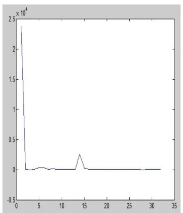
Fig 5: Tested results by Graphical representation
VI. CONCLUSION
It include the algorithm which gives the results of segmentation and classification of MRI brain tumor dataset can be benign or malignant. The classification result in the form of classes. Such as below 1 class, below2 class, below3 class. With help of more features we conclude the system more accurately.
REFERENCES
International Conference on Industrial and Information Systems, ICIIS 2009,28 - 31 December 2009, Sri Lanka
[3] Elsa D. Angelini, Glioma Dynamics and Computational Models: A Review of Segmentation, Registration, and In Silico Growth Algorithms and their Clinical Applications, Current Medical Imaging Reviews, 2007, 3, 262-276.
[4] Stefan Bauer, A survey of MRI-based medical image analysis for brain tumor studies, IOP PUBLISHING PHYSICS IN MEDICINE AND BIOLOGY Phys. Med. Biol. 58 (2013) R97–R129.
[5] C.L. Biji, Tumor Detection in Brain Magnetic Resonance Images Using Modified Thresholding Techniques, ECE Dept, Rajagiri School of Engineering & Technology, Kochi, India & ECE Dept. Mepco Schlenk Engineering College, Sivakasi, India.
[6] Anam Mustiqueem, An Efficient Brain Tumor Detection Algorithm Using Watershed & Thresholding Based Segmentation, I.J. Image, Graphics and Signal Processing, 2012, 10, 34-39 Published Online September 2012 in MECS (http://www.mecs-press.org/)
[7] Jin Liu, A Survey of MRI-Based Brain Tumor Segmentation Methods, TSINGHUA SCIENCE AND TECHNOLOGY ISSNll1007-0214ll04/10llpp578-595 Volume 19, Number 6, December 2014
[8] Chinnu A, MRI Brain Tumor Classification Using SVM and Histogram Based Image Segmentation, International Journal of omputer Science and Information Technologies, Vol. 6 (2) , 2015, 1505-1508.
[9] S.U.ASWATHY at el, A Survey on Detection of Brain Tumor from MRI Brain Images, 2014 International Conference on Control, Instrumentation, Communication and Computational Technologies (ICCICCT).
[10] Neha Tirpude & R. R. Welekar, A Study of Brain Magnetic Resonance Image Segmentation Techniques, International Journal of Advanced Research in Computer and Communication Engineering Vol. 2, Issue 1, January 2013.
[11] K.S.Deepak, AN EFFICIENT APPROACH TO PREDICT TUMOR IN 2D BRAIN IMAGE USING CLASSIFICATION TECHNIQUES, Final year students
BE-Computer Science And Engineering,K.S.Rangasamy College Of Technology,Truchengode **Assistant Professor K.S.Rangasamy College Of Technology,Truchengode.\
[12] Hari Babu Nandpuru, MRI Brain Cancer Classification Using Support Vector Machine, 2014 IEEE Students' Conference on Electrical, Electronics and Computer Science.
![Table 1. SVM classifier result [5].](https://thumb-us.123doks.com/thumbv2/123dok_us/8310253.857882/4.612.218.434.243.335/table-svm-classifier-result.webp)