Tropical Biomedicine 30(3): 459–466 (2013)
Comparison of two nested PCR methods for the detection
of human malaria
Anthony, C.1, Mahmud, R.1, Lau, Y.L.1*, Syedomar, S.F.2 and Sri La Sri Ponnampalavanar, S.2 1Department of Parasitology, Faculty of Medicine, University of Malaya, 50603 Kuala Lumpur, Malaysia 2Department of Medicine, Faculty of Medicine, University of Malaya, 50603 Kuala Lumpur, Malaysia *Corresponding author’s e-mail: [email protected]
Received 27 December 2012; received in revised form 29 March 2013; accepted 13 May 2013
Abstract. Battling malaria will be a persistent struggle without the proper means to diagnose the parasitic infection. However, the inherent limitations of microscopy, the conventional method of diagnosing malaria, affect the accuracy of diagnosis. The present study aimed to compare the accuracy of two different set of primers targeting the small subunit ribosomal RNA (ssRNA) and the dihydrofolate reductase-thymidylate synthase linker region (dhfr-ts) in detecting species specific malaria infections by nested PCR. The sensitivity and specificity of nested PCR assay using the two primers were calculated with reference to microscopy as the ‘gold standard’. The results show that 18S rRNA primers had 91.9% sensitivity and 100% specificity in detecting human Plasmodium species as opposed to dhfr-ts primers which had 51.4% sensitivity and 100% specificity. The higher sensitivity of 18S rRNA primers suggests that it may be a better diagnostic tool for detecting human malaria.
INTRODUCTION
The five species known to cause malaria in humans are Plasmodium falciparum,
Plasmodium vivax, Plasmodium ovale,
Plasmodium malariae, and the most recent
Plasmodium knowlesi. Despite various control efforts, malaria still remains a severe global health problem (Stratton et al., 2008). The incidences of malaria cases that occur annually vary from one report to another and this by itself reflects the lack of precision in malaria statistics (Wongsrichanalai et al., 2007).
Accurate diagnosis is imperative in the effective management of malaria (Tangpukdee et al., 2009). While microscopy has been and still remains the mainstay in malaria diagnosis, its limitations has often led to the misdiagnosis of Plasmodium
species and ultimately the mistreatment of malaria. Furthermore, the lack of skilled microscopists has massively hampered the efficacy of this traditional technique. It is also
important to note that low-level parasitemia which is below the detectable limit of blood smears is a norm under conditions of host acquired immunity or exposure to antimalarials (Taylor et al., 2010).
Rapid immunochromatographic tests were developed to aid better conduct of diagnosis. The basis of these tests is the detection of antigens in the blood of malaria patients (Di Santi et al., 2004). Monoclonal antibodies have been developed to target the conserved element of Plasmodium lactate dehydrogenase (PLDH) or specific regions which are unique to P. falciparum or P. vivax
(Murray et al., 2008). Unfortunately, in cases of low parasitemia, this method is somewhat insensitive. Another flaw of this method is the possibility of false positive results due to the persistence of antigenemia weeks beyond the actual infection (Mangold et al.,
2005).
reaction (PCR) was first applied to malaria diagnostics in the early 1990s (Erdman & Kain, 2008) and it was not long before this method was extensively used. In terms of sensitivity and specificity, comparative studies have consistently demonstrated the superiority of PCR-based diagnosis over microscopy-based diagnosis of malaria (Putaporntip et al., 2009). Apart from diagnosis, this DNA-based molecular method has also been commonly used in areas involving epidemiological studies and assessment of drug efficacy (Harris et al., 2010).
Nested PCR, multiplex PCR and real-time PCR are PCR based assays that have been developed to overcome the limitations and discrepancy of traditional diagnostic methods. Nested and multiplex PCR are able to shed light on species determination and differentiation when cumbersome morphological problems arise during attempts to identify parasites at species level (Chavalitshewinkoon-Petmitr, 2010). Real-time PCR assays have proven its ability in identifying mixed infections, detecting low parasitemia levels and also in the differentiation of Plasmodium species based on melting curve analysis (Mangold
et al., 2005). The common misdiagnosis of
P. knowlesi as P. malariae and P. falciparum
via microscopy can now be circumvented with the aid of these PCR based assays. The aim of this study was to compare the specificity and sensitivity of two nested-PCR methods using two different sets of primers for the molecular diagnosis of human malaria.
MATERIALS AND METHODS Patient samples
A total of 57 whole blood samples and 17 blood smears were obtained from the General Medical Ward, University Malaya Medical Centre (UMMC). The presence of malarial parasites in the specimens was determined by both microscopy and PCR. Twelve blood samples taken from healthy individuals with no history of malaria infection were used as negative controls in this study. Approval for
the use of these samples was obtained from the University of Malaya Medical Centre Ethics Committee (Reference no: 817.18). Microscopy
Giemsa (5%) stained blood smears, both thick and thin were prepared and examined under light microscope. Examination of these slides was carried out by skilled and experienced microscopists who were able to identify and differentiate the malaria parasites. At least 200 microscopic fields were examined before concluding the slide was negative for malaria parasites. Parasitaemia was calculated as number of infected red blood cells per 1000 red blood cells counted in the thin film.
DNA extraction from whole blood The template DNA required to carry out the nested PCR assay was prepared using the DNeasy Blood & Tissue Kit (QIAGEN, Valencia, CA). Parasite DNA was extracted from 100 µl of whole blood according to the manufacturer’s protocol. Purified DNA was eluted from the column with 50 µl elution buffer and this DNA was stored at -20ºC for further use.
DNA extraction from slides
Prior to the extraction of DNA, the slides were initially cleaned to remove oil residues. Approximately 50 µl of Tris-EDTA (TE) buffer was then pipetted onto the thin film. Whatman filter paper was cut into strips and placed on the slide to absorb the buffer. The smear was wiped off from the slide and placed in a 1.5 ml centrifuge tube using sterile forceps. The DNA from the filter paper was extracted using the DNeasy Blood & Tissue Kit (QIAGEN, Valencia, CA).
Nested PCR assay
genus-specific primers (rPLU1: 5’-TCA AAG ATT AAG CCA TGC AAG TGA-3’and rPLU5: 5’-CCT GTT GTT GCC TTA AAC TCC-3’), 2.5 µl 10X Buffer, 2.0 µl of 0.25 M dNTP and 0.2 µl of 1 u Taq polymerase and 15.3 µl of nuclease free water. The nest 1 amplification conditions were as follows: 1) Initial denaturation at 94ºC for 4 minutes, 2) 35 cycles of denaturation at 94ºC for 30 seconds, annealing at 55ºC for 1 minute and extension at 72ºC for 1 minute, 3) Final extension at 72ºC for 10 minutes and a hold temperature of 4ºC. Each of the nest 2 amplification mixture of 25 µl contained 4 µl of the nest 1 product and same amounts of buffer, dNTP,
Taq polymerase and nuclease free water as in nest 1. The nest 2 amplification conditions were identical to that of nest 1 except the annealing temperature of 58ºC for the species-specific primers.
The second nested PCR targeted at dihydrofolate reductase-thymidylate synthase linker region (dhfr-ts) was developed by Tanomsing et al. (2010). The amount of 10X buffer, dNTP, Taq polymerase and DNA template used were the same as the first set of nested PCR. Cycling conditions for both primary and secondary amplifications were followed as stated in Tanomsing et al. (2010). The amplified products were visualized through gel electrophoresis using a 2% agarose gel stained with Sybr Safe. A 100bp molecular marker (Fermentas) was used.
Analysis
The sensitivity and specificity of the 2 different primers were calculated with regards to microscopy as the reference. Percentage of sensitivity and specificity (%) were calculated as follows:
% Sensitivity = (number of true positives/ number of true positives + number of false negatives) x 100
% Specificity = (number of true negatives/ number of true negatives + number of false positives) x 100
RESULTS Microscopy analysis
In total, 74 patients were identified as being infected with malaria via blood film analysis. Out of this, 33 were identified as having P. falciparum infection, 23 were found to be P. vivax infection, 2 samples were found to have
P. ovale infection and 16 were identified as either P. malariae or P. knowlesi infection. The samples were then subjected to nested PCR using the 18S rRNA and dhfr-ts primers. Nested PCR was also carried out on 12 non malaria samples to determine specificity of the two different set of primers. The different species identified by microscopy and PCR are illustrated in Table 2.
Nested PCR
The comparison between microscopy and nested PCR assays using the 18S rRNA and dhfr-ts primes are shown in Table 1. The PCR analysis using the 18S rRNA primers detected 22 cases of P. falciparum, 21 cases of P. vivax, 2 cases of P. ovale, 14 cases of
P. knowlesi and 9 cases of mixed infections. There were 6 samples which were positive for P. falciparum via microscopy but produced negative results with PCR. Another 6 were found to have a different Plasmodium
species picked up by PCR when compared to microscopy.
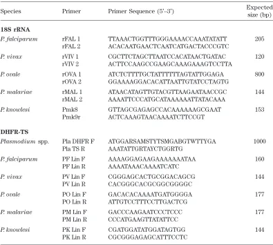
Table 1. Nested PCR primers targeting 18S rRNA and dhft-ts genes in malaria parasites
Species Primer Primer Sequence (5’–3’) Expected
size (bp)
18S rRNA
P. falciparum rFAL 1 TTAAACTGGTTTGGGAAAACCAAATATATT 205
rFAL 2 ACACAATGAACTCAATCATGACTACCCGTC
P. vivax rVIV 1 CGCTTCTAGCTTAATCCACATAACTGATAC 120
rVIV 2 ACTTCCAAGCCGAAGCAAAGAAAGTCCTTA
P. ovale rOVA 1 ATCTCTTTTGCTATTTTTTAGTATTGGAGA 800
rOVA 2 GGAAAAGGACACATTAATTGTATCCTAGTG
P. malariae rMAL 1 ATAACATAGTTGTACGTTAAGAATAACCGC 144
rMAL 2 AAAATTCCCATGCATAAAAAATTATACAAA
P. knowlesi Pmk8 GTTAGCGAGAGCCACAAAAAAGCGAAT 153
Pmk9r ACTCAAAGTAACAAAATCTTCCGT
DHFR-TS
Plasmodium spp. Pla DHFR F ATGGARSAMSTYTSMGABGTWTTYGA 1000
Pla TS R AAATATTGRTAYCTGGRTG
P. falciparum PF Lin F AAAAGGAGAAGAAAAAAATAA 160
PF Lin R AAAATAAACAAAATCATC
P. vivax PV Lin F CGGGAGCACTGCGGACAGCG 144
PV Lin R CACGGGCACGCGGCGGGGC
P. ovale PO Lin F GACACACAAAATGATGGGGA 177
PO Lin R ATTGTCCTTTCCTTGACTCG
P. malariae PM Lin F GACCCAAGAATCCCTCCC 177
PM Lin R CCCATGAAGTTATATTCC
P. knowlesi PK Lin F CGATGGATATGGATAGTGG 144
PK Lin R CGCGGGAGAGCATTTCCTC
Table 2. Number of samples with various malaria species detected by microscopy and PCR
Method/Species
Number of Samples
Pf Pv Po Pk Pm Pm/Pk Mixed Neg Total
Microscopy 33 23 2 0 0 16 0 12 86
Nested PCR (18 ssRNA) 22 21 2 14 0 0 9 18 86
Nested PCR (dhfr-ts) 7 11 1 17 0 0 2 48 86
Note: Pf = P. falciparum; Pv = P. vivax; Po = P. ovale; Pk = P. knowlesi; Mixed = Pv+Pk or Pv+Pf or Pk+Pf; Neg = Negative
Sensitivity and specificity
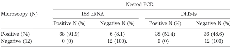
The sensitivity and specificity of nested PCR using the two sets of primers compared to microscopy are shown in Table 3. Of the 74
[image:4.579.92.487.515.591.2]specificity. Nested PCR with dhfr-ts primers on the other hand, showed only 51.47% sensitivity and 100% specificity.
DISCUSSION
In malaria studies, PCR is known to be a promising method especially in the identification of parasites in areas where four
Plasmodium species occur simultaneously (Snounou et al., 1993). However, it has been recognized that the success of the technique is dependable on the quality of DNA. Previous studies have shown that intrinsic (DNA amount or content of human DNA or haemoglobin) and extrinsic (use of heparin or inadequate conditions of blood collection, storage and amplification of samples) factors can inhibit the PCR assay (Barker et al., 1992; Snounou et al., 1993).
In general, the ribosomal ribonucleic acid (rRNA) has proven to be of benefit in molecular studies due to their cocktail of regions which have evolved at different rates and thus enabling them to be utilized at various taxonomic levels (Rubio et al., 2002). The decreased susceptibility and possible resistance to antifolate antimalarial drugs have led to the thorough investigation of sequence variations in the Plasmodium dhfr domain (Tanomsing et al., 2010).
While nested PCR assay using either the 18S rRNA or dhfr-ts primers were unable to pick up all the microscopically positive
Plasmodium samples, this assay was able to detect several mixed infections. The ability of the PCR assay to identify 2 and 9 mixed infections using dhfr-ts and 18S rRNA primers
respectively exhibits the greater sensitivity of the PCR assay over microscopy. With regards to mixed infections, it is possible for microscopic misdiagnosis to occur due to the domination of one species over the other (Ebrahimzadeh et al., 2007). Limitations of the light microscope could also serve as a reason in the misdiagnosis of mixed infections as a single infection (Ohrt et al., 2002).
The present study has demonstrated that nested PCR assay using the 18S rRNA primers are more sensitive and specific as opposed to carrying out the same assay with the dhfr-ts primers. While the 18S rRNA primers were able to identify the species of 68 (57 whole blood and 11 blood smears) samples, there were 6 P. falciparum positive blood smears that were not detected by PCR (18S rRNA). This may be an effect of a reduced number of parasites present in the sample, as some could have been lost during the process of scraping the slides. It is also known that factors pertaining to slide preparation for microscopic examination may contribute to the stability of the DNA template (Scopel et al., 2004). However, using this as reasons to justify 36 (20 whole blood and 16 blood smears) negative results obtained with the dhfr-ts primers would be farfetched. Tanomsing et al. (2010) reported that these newly developed primers are effective at detecting low level parasitemia, particularly
[image:5.579.91.486.108.189.2]P. falciparum. Interestingly, such was not the case in this study. Only 7 samples of 33 which were identified as P. falciparum gave the same results when tested by nested PCR with dhfr-ts primers. In fact these set of primers appear to be better at identifying P. knowlesi
Table 3. Sensitivity and specificity of 18S rRNA and dhfr-ts primers compared to microscopy
Nested PCR
Microscopy (N) 18S rRNA Dhfr-ts
Positive N (%) Negative N (%) Positive N (%) Negative N (%)
Positive (74) 68 (91.9) 06 (8.1)0 38 (51.4) 36 (48.6) Negative (12) 00 (0)00. 12 (100). 00 (0)00. 12 (100)
samples and to a lesser extent, P. vivax. Relative to 9 samples which were identified as mixed infections with 18S rRNA primers, the dhfr-ts primers were only able to detect 2 samples with mixed infections.
Of the 17 blood smears tested, the 18S rRNA primers were able to identify 11 malaria species while the dhfr-ts primers were able to identify only one infection. This is an interesting finding as the previous study by Tanomsing et al. (2010) did not test these primers with DNA extracted from blood smears. We also found that when the percentage parasitaemia of infection was above 1%, nested PCR methods using the dhfr-ts primers was able to identify the species of the malaria parasite. However, when the percentage parasitaemia of infection was below 1%, we observed that there were inconsistent results with this particular primer set. While nested PCR using the 18S rRNA primers was able to identify malaria species (68/74) with a parasitaemia as low as 0.03%, such was not the case with the dhfr-ts primers. There were instances where the dhfr-ts primers was able to detect an infection with a parasitaemia of 0.31% but was not able to identify malaria species with a parasitaemia of 0.63%.
There were 3 cases whereby the 18S rRNA primers detected both P. vivax and
P. knowlesi but only one species (either P. vivax or P. knowlesi) was detected by the dhfr-ts primers. While this may appear as though the 18s rRNA primers are more sensitive towards detecting mixed infections, a study carried out by Imwong et al. (2009) demonstrated otherwise. False positive amplification was observed when P. vivax
genomic DNA was used implicating stochastic cross reaction with the Pmk8-Pmkr9 primers. In this case, it is possible that the dhfr-ts primers are better at discriminating between P. vivax and P. knowlesi infections.
There were 6 blood smears which were positive for P. falciparum but negative when subjected to PCR with 18S rRNA primers. These results remained discordant upon re-examination of smears and repetition of
PCR. It is important to note that presence of artifacts in blood smears can lead to false positive readings. This may be due to the similar resemblance between artifacts and malaria parasites and as such would be a possibility why the PCR assay was not able to detect the parasite (Ohrt et al., 2002). The reasons could also be as what was explained by Scopel et al. (2004). The mis-identification of P. vivax as P. falciparum
could be attributed to the differences in smear preparation and also quality of the stain.
Based on this study, we found the 18S rRNA primers to be more effective at identifying the different species of human malaria. We also found that the dhfr-ts primers may not be as effective at identifying P. falciparum as initially proposed.
Acknowledgement. Special thanks to PARASEAD, University of Malaya for providing part of the blood samples for analysis. The authors would also like to thank the Ministry of Science, Technology and Innovation, E-Science Fund (Grant no: 12-02-03-2086) and UM Postgraduate Research Fund (PV044/2012A) for supporting this study.
REFERENCES
Barker, R.H.J., Banchongaksorn, T., Courval, J.M., Suwonkerd, W., Rimwungtragoon, K. & Wirth, D.F. (1992). A simple method to detect Plasmodium falciparum directly from blood samples using the polymerase chain reaction. American Journal of Tropical Medicine and Hygiene46: 416-426.
Chavalitshewinkoon-Petmitr P. (2010). Laboratory diagnosis of malaria. Siriraj Medical Journal62:98-102.
Ebrahimzadeh, A., Fouladi, B. & Fazaeli, A. (2007). High rate of detection of mixed infections of Plasmodium vivax and
Plasmodium falciparum in South-East Iran, using nested PCR. Parasitoly International56:61-64.
Erdman L.K. & Cain, K.C. (2008). Molecular diagnostic and surveillance tools for global malaria control. Travel Medicine and Infectious Disease6:82-99. Harris, I., Sharrock, W.W., Bain, L.M., Gray,
K-A., Bobogare, K-A., Boaz, L., Lilley, K., Krause, D., Vallely, A., Johnson, M-L., Gatton, M.L., Shanks, G.D. & Cheng, Q. (2010). A large proportion of asymptomatic Plasmodium infections with low and sub-microscopic parasite densities in the low transmission setting of Temotu Province, Solomon Islands: challenges for malaria diagnostics in an elimination setting. Malaria Journal 9: 254.
Imwong, M., Tanomsing, N., Pukrittayakamee, S., Day, N.P.J., White, N.J. & Snounou, G. (2009). Spurious amplification of a
Plasmodium vivax small-subunit RNA gene by use of primers currently used to detect P. knowlesi.Journal of Clinical Microbioly47: 4173-4175.
Mangold, K.A., Manson, R.U., Koay, E.S.C., Stephens, L., Regner, M., Thomson, R.B. Jr., Peterson, L.R. & Kaul, K.L. (2005). Real-time PCR for detetction and identification of Plasmodium spp.
Journal of Clinical Microbiology 43: 2435-2440.
Murray, C.K., Gasser, R.A. Jr., Magill, A.J. & Miller, R.S. (2008). Update on rapid diagnostic testing for malaria. Clinical Microbiology Reviews21: 97-110. Ohrt, C., Purnomo, Sutamihardja, M,A., Tang,
D. & Kain, K.C. (2002). Impact of microscopy error on estimates of protective efficacy in malaria-prevention trials. Journal of Infectious Diseases
186:540-546.
Putaporntip, C., Hongsrimuang, T., Seethamchai, S., Kobasa, T., Limkittikul, K., Cui, L. & Jongwutiwes, S. (2009). Differential prevalence of Plasmodium
infections and cryptic Plasmodium knowlesi malaria in humans in Thailand.
Journal of Infectious Diseases 199: 1143-1150.
Rubio, J.M., Post, R.J., Docters van Leeuwen, W.M., Henry, MC., Lindergard, G. & Hommel, M. (2002). Alternative polymerase chain reaction method to identify Plasmodium species in human blood samples: the semi-nested multiplex malaria PCR (SnM-PCR). Transactions of the Royal Society of Tropical Medicine and Hygiene96(Suppl 1): 199-204
Scopel, K.K.G., Fontes, C.J.F., Nunes, A.C., de Fatima Horta, M. & Braga, E.M. (2004). Low sensitivity of nested PCR using
Plasmodium DNA extracted from stained thick blood smears: An epidemiological retrospective study among subjects with low parasitaemia in an endemic area of the Brazilian Amazon region. Malaria Journal3:8. Singh, B., Bobogare, A., Cox-Singh, J.,
Snounou, G., Abdullah, M.S. & Rahman, H.A. (1993). A genus- and species-specific nested polymerase chain reaction malaria detection assay for epidemiologic studies. American Journal of Tropical Medicine and Hygiene60:687-692.
Snounou, G., Viriyakasol, S., Jarra, W., Thaithong, S. & Brown, K.N. (1993). Identification of the four human malaria parasite species in field samples by the polymerase chain reaction and detection of a high prevalence of mixed infections.
Molecular and Biochemical Para-sitology 58:283-289.
Tangpukdee, N., Duangdee, C., Wilairatana, P. & Krudsood, S. (2009). Malaria diagnosis: a brief review.Korean Journal of Parasitology 47:93-102.
Tanomsing, N., Imwong, M., Theppabutr, S., Pukrittayakamee, S., Day, N.P., White, N.J. & Snounou, G. (2010). Accurate and sensitive detection of Plasmodium
species in humans by use of the dihydrofolate reductase-thymidylate synthase linker region. Journal of Clinical Microbiology48: 3735-3737.
Taylor, S.M., Juliano, J.J., Trottman, P.A., Griffin, J.B., Landis, S.H., Kitsa, P., Tshefu, A.K. & Meshnick, S.R. (2010). High-throuput pooling and real-time PCR-based strategy for malaria detection.
Journal of Clinical Microbiology 48: 512-519.
Wongsrichanalai, C., Barcus, M,J., Muth, S., Sutamihardja, A. & Wernsdorfer, W.H. (2007). A Review of malaria diagnostic tools: microscopy and rapid diagnostic test (RDT). American Journal of Tropical Medicine and Hygiene