http://dx.doi.org/10.4236/ojgas.2014.42014 Published Online February 2014 (http://www.scirp.org/journal/ojgas/)
Primary cancers of the small bowel: About 20 cases
Noomen Haoues, Manel Mabrouk, Haithem Zaafouri*, Rabii Noomene, Ahmed Bouhafa, Anis Ben Maamer, Abderraouf Cherif
Department of General Surgery, Habib Thameur Hospital, Tunis, Tunisia Email: *[email protected]
Received 23 November 2013; revised 27 December 2013; accepted 6 January 2014
Copyright © 2014 Noomen Haoues et al. This is an open access article distributed under the Creative Commons Attribution License, which permits unrestricted use, distribution, and reproduction in any medium, provided the original work is properly cited. In accor-dance of the Creative Commons Attribution License all Copyrights © 2014 are reserved for SCIRP and the owner of the intellectual property Noomen Haoues et al. All Copyright © 2014 are guarded by law and by SCIRP as a guardian.
ABSTRACT
Background: Cancers of the small bowel are rare. Di- agnosis is late and difficult because of the lack of spe- cific signs. Treatment is surgical. Prognosis is usually poor and depends on the histological type of tumor. Aim of Study: To specify the epidemiological, clinical and therapeutic characteristics of small bowel prima- ry cancers in order to improve their prognosis. Ma- terial and Methods: This is a retrospective study about 20 cases of malignant tumors of the small bowel, col- lected in the department of general surgery of Habib Thameur Hospital in Tunis (Tunisia), from January 1994 through June 2011. Results: Our series involved 11 women and 9 men aged 62 on average (range: 44 - 80 years). In 45% of cases, the diagnosis was made in a patient rushed to hospital with clinical features of acute generalized peritonitis (66% of all surgical emer- gencies). Intestinal transit was performed in 5 pati- ents only. Ultrasound abdominal examination was per- formed in 11 patients. Abdominal CT scan was per- formed in 7 patients, but the results were conclusive in 4 cases only (57%). Small bowel scanning was done in 5 patients only, but led to a positive diagnosis in all of them. All of our patients underwent surgery. Tu- mors of the small bowel were histologically divided as follows: carcinoid tumor (8 cases), leiomyosarcoma (7 cases), giant B-cell lymphoma (2 cases), malignant stromal tumor (2 cases) and malignant myxoid sch- wannoma (1 case). Malignant tumors of the small bo- wel most commonly arise in the ileum (60%) followed by the jejunum (35%). As for the long-term course, there was a recurrence at one year of a leiomyosarco- ma and two recurrences of stromal tumors associated with liver metastases. Conclusion: Small bowel can- cers are rare. Time to consultation is long and diag- nosis is difficult and late due to the absence of typical
presentation. Treatment is surgical and progression depends essentially on histological findings.
KEYWORDS
Primary Cancers; Small Bowel; Epidemiology; Diagnosis
1. INTRODUCTION
Although the small bowel represents 75% of the length of the digestive tract and 90% of its absorptive mucosal surface area, primary cancers of the small bowel are un-common accounting for less than 6% of all gastrointes- tinal tract tumors. They are characterized by a lack of specific clinical features which are generally vague. Di- agnosis is therefore often made late following a compli-cation. Primary treatment remains surgical consisting of resection of the small bowel and of the regional metas-tatic lymph nodes.
2. AIM OF STUDY
Our objective is to define the different epidemiological, clinical, therapeutic and prognostic characteristics of these cancers so that their management can be improved.
3. MATERIAL AND METHODS
This is a retrospective study about 20 cases of malignant tumors of the small bowel, collected in the department of general surgery of Habib Thameur Hospital in Tunis (Tu- nisia), from January 1994 through June 2011. All patients with primary malignant tumor of the small bowel, from the duodeno-jejunal angle until the last ileal loop, were included. We excluded from the study cancers of the du-odenum and of Bauhin’s valve for they constitute a dif-ferent anatomico-pathological entity. We also excluded patients with small bowel metastases.
4. RESULTS
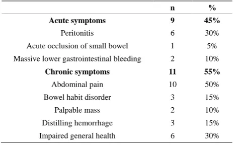
Our series involved 11 women and 9 men aged 62 on average (range: 44 - 80 years). In 45% of cases, the di-agnosis was made in a patient rushed to hospital with clinical features of acute generalized peritonitis (66% of all surgical emergencies). As for patients presenting to hospital for chronic symptoms, most of them primarily complained of abdominal pain (50%) and features of im- paired general health (30%) (Table 1).
Intestinal transit was performed in 5 patients only; the results were conclusive in 3 cases. Thus, it had a positive predictive value of 60% and gave false negative results in 2 patients out of five (40%).
Ultrasound abdominal examination was performed in 11 patients. It revealed a small bowel tumor in 5 cases (43.5%), among which there were 3 cases of hepatic me-tastasis.
Ultrasound findings were normal in 4 patients among which there were two cases of leiomyosarcoma, a case of carcinoid tumor and a case of lymphoma.
Abdominal CT scan was performed in 7 patients, but the results were conclusive in 4 cases only (57%). Small bowel scanning was done in 5 patients only, but led to a positive diagnosis in all of them.
All of our patients underwent surgery. Midline laparo- tomy was adopted in 17 cases. Laparoscopy had to be converted into laparotomy in a patient with a lymphoma due to the difficulties encountered. Assisted laparoscopy was done in 2 patients. The site, size and extension of le- sions are described in Table 2.
Curative resection was done in 15 patients (75% of cases). It consisted of surgical resection with end-to-end anastomosis in 9 cases and of resection with ileostomy in the cases of peritonitis.
Palliative resection was carried out in 5 patients (25% of cases) due to the extent of the local-regional involve- ment and to peritoneal carcinoses. Partial bladder remo- val was done in a patient with a leiomyosarcoma.
None of our patients received radiation therapy or hor- monotherapy.
Of the 20 patients of our series, 5 of them were given adjuvant chemotherapy: 3 cases of carcinoid tumor and 2 cases of lymphoma. Treatment by Gleevec was given to patients who were carriers of malignant stromal tumors.
In our series, tumors of the small bowel were histolo- gically divided as follows:
• Carcinoid tumor: 8 cases
• Leiomyosarcoma: 7 cases
• Giant B-cell lymphoma: 2 cases
• Malignant stromal tumor: 2 cases
• Malignant myxoid schwannoma: 1 case
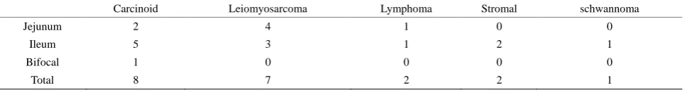
[image:2.595.307.541.111.255.2]Malignant tumors of the small bowel most commonly arise in the ileum (60%) followed by the jejunum (35%) (Table 3).
Table 1. Circumstances of discovery of malignant tumors of the small bowel.
n %
Acute symptoms 9 45%
Peritonitis 6 30%
Acute occlusion of small bowel 1 5%
Massive lower gastrointestinal bleeding 2 10%
Chronic symptoms 11 55%
Abdominal pain 10 50%
Bowel habit disorder 3 15%
Palpable mass 2 10%
Distilling hemorrhage 3 15%
Impaired general health 6 30%
Table 2.Surgery findings.
Malignant tumors
Site
Jejunum 7
Ileum 12
Bifocal 1
Size (cm) Variable: 3 - 60 cm Average: 31.5 cm
Serosa involvement 7 cases Metastatic lymph nodes 4 cases
Liver metastases 5 cases Peritoneal carcinosis 5 cases Bladder involvement 1 case
In our series there was only one death (5%). The vic-tim had a malignant myxoid schwannoma which metas-tasized to the lungs. Death occurred three days after sur-gery, from septic shock following infectious lung disease that didn’t yield to intensive care.
Immediate post operative course was uneventful for 17 patients (85% of cases). Apart from the above mentioned fatal complication, wound infection occurred in two pa-tients with leiomyosarcomas who had undergone elective surgery. They responded well to local treatment.
As for the long term course, there was a recurrence at one year of a leiomyosarcoma and two recurrences of stromal tumors associated with liver metastases.
5. DISCUSSION
5.1. Epidemiology
Small bowel tumors are the least frequent among gastro- intestinal tract tumors. Their epidemiological statistics, mainly based on hospital data, remain little known. They account for 6% of all GI tract tumors [1-5]. Incidence is 1.6 cases per 100.000 inhabitants per year [6].
Table 3. Distribution of small bowel malignant tumors according to sites.
Carcinoid Leiomyosarcoma Lymphoma Stromal schwannoma
Jejunum 2 4 1 0 0
Ileum 5 3 1 2 1
Bifocal 1 0 0 0 0
Total 8 7 2 2 1
versus 23% respectively [4]. In our department, we col-lected over 16 years, for the purpose of the study, 25 cases of small bowel tumors, 20 (80%) of which were malignant. Average age of occurrence of malignant tu- mors is between 50 and 60 years, but it was 62 in our series. It is 56 in North’s series [10].
According to the literature data, these cancers occur, in Africa, in patients aged between 34 and 42 and might therefore be correlated with living conditions and life expectancy of patients [11]. Nevertheless, the mean age depends on the histological type of tumor; it is lower for lymphomas and sarcomas [3,7]. Malignant tumors are more frequent in men than in women [12,13].
5.2. Etiology and Pathogenesis
Several hypotheses have been put forward to explain the relative infrequency of small bowel tumors in compari- son with other tumors of the digestive tract.
• Reduced transit time of food in the small intestine
which shortens the exposure time of the mucosal lin-ing to carcinogens, in addition to fluid circulation of alkaline intestinal chyme [14,15].
• Small amount of bacteria susceptible to produce
car-cinogens [7].
• Rapid turnover of the small bowel mucosa which in-
hibits the growth of cancer cells.
• High concentration of microsomal hydrolases likely
to inactivate some carcinogens.
• High level of Ig A, that is evidence of an important
anti-virus activity.
The corollary of all these hypotheses is that these tu-mors are more frequent in patients with congenital or ac- quired immuno-deficiency [7,16,17].
In the same way as we look for esophageal, gastric and colorectal cancers, precancerous lesions should be iden-tified for they pave the way for small bowel cancers [18]. Several authors have shown a link between adenoma and adenocarcinoma of the small intestine. In Perzin’s series [19], of the 51 patients involved in the study, 33 of them (65%) had concomitant adenomas and adenocarcinomas in the same lesions. Other precancerous lesions have been mentioned: Familial polyposis [20-22], Crohn’s di- sease [23-28], leiomyomes [29] and coeliac disease [30].
5.3. Clinical Presentation
Given the nonspecific and latent clinical manifestations
of small bowel cancers, diagnosis is made late with sev-eral month’s lag (5 to 11 months), and it is very difficult to suspect the site of a lesion and to foresee its nature. Moreover, clinical features very according to anatomic characteristics of the tumor (size, site, shape) [31].
Abdominal pain which was reported by half of our pa-tients, is the primary complaint made by more than 50% of patients [32]. It is due to obstruction of the intestinal lumen or to inflammatory manifestations secondary to tumor ulceration or necrosis [9].
Bowel habit disorders may reveal tumor of the small bowel in 6% to 30% of cases [33-35] in the form of al- ternating episodes of diarrhea and constipation especially in the presence of Koening’s syndrome. According to some authors, diarrhea is frequently encountered with lymphomas and carcinoid tumors [21,36]. In our Series, the three patients who had bowel habit disorders had car- cinoid tumors. Two of our patients complained of diarr- hea; a third patient had a sub-obstructive syndrome. Lo- wer G I bleeding is also noted but in patients with lei-omyomas and leiomyosarcomas [21,27]. Thus, in our series, there were only 3 cases of minor lower gastro-in- testinal bleeding: 2 patients with leiomyosarcoma and a third patient with a stromal tumor.
According to several authors, small bowel tumors are frequently revealed by a complication such as an ob- structive syndrome or massive G I bleeding or peritonitis [20,37,38]. Nine of our patients actually had to undergo emergency surgery.
Acute intestinal obstruction reveals tumor of the small bowel in 30% of cases [16,39]. This mechanical obstruc-tion results from obstrucobstruc-tion of the lumen or from stran-gulation. Among the 9 patients who underwent emergen- cy surgery, only one had acute intestinal obstruction.
According to Gore and Johnson, cases of peritonitis from tumoral perforation are rare [27]. On the contrary, such cases are frequent in Desa’s series [39]; they re- present 31% of all cases and they are essentially associ- ated with lymphomas and leiomyosarcomas [40,41]. In our series, 6 patients (30%) had peritonitis following tu- moral perforation, involving a lymphoma, a leiomyosar- coma, two carcinoid tumors and a schwannosarcoma.
[33,44].
In our series, emergency surgery was required for massive lower G I bleeding in two patients, a carrier of leiomyosarcoma and a carrier of stromal tumor.
Physical examination usually provides little informa-tion in case of small bowel tumor. Palpainforma-tion reveals an abdominal mass in only 30% to 50% of cases [45]. In our series, palpation disclosed an abdominal mass in 2 pa-tients only (10%): a carrier of leiomyoma and a carrier of malignant stromal tumor. Liver enlargement is rarely observed (7% of cases) [33]. This finding was encoun-tered in two of our patients who were both carriers of carcinoid tumors.
5.4. Special Investigations
Intestinal transit remains the best diagnostic procedure for small bowel tumors [9]. It is best performed by ente- roclysis employing double contrast method [46]. In addi- tion, it is the most reliable method for detecting small lesions in an early stage [47]. Small bowel scaning is a very efficient technique as it combines the advantages of the two other techniques enteroclysis and multibarette scanning [48]. It detects and defines small bowel tumors without yielding false negatives like other investigation procedures (sensitivity 100%, specificity 90%) [49].
In our series, small bowel scanning was done in 5 pa-tients and led to the diagnosis of small bowel tumor in all of them.
Video capsule endoscopy represents a major advance in the field of medical diagnostic investigation. This non- invasive technique makes it possible to visualize all the small bowel mucosal lining, even the zones that can’t be reached by other diagnostic methods [50].
5.5. Pathology
More than 2/3 of small bowel tumors are malignant. Most of them are adenocarcinomas [1,2,7,51] though they do not represent more than 1% of adenocarcinomas of the digestive tract. Like in other series, there were no adenocarcinomas in our own series. This finding might be explained by regional differences.
Carcinoid tumors rank second. They arise from the enterochromaffin cells of the neural crest. They represent 20% to 70% of tumors of the small bowel [44] and 13% to 34% of all endocrine tumors [52,53]. Most of them occur in the ileum. In our series, 5 patients out of 8 (62.5%) had ileal carcinoid tumors.
Malignant non-Hodgkin’s lymphomas come in third place. They represent 20% to 30% of all primary gastro- intestinal lymphomas and 12% to 31% of malignant small bowel tumors. They are located in the ileum in 53% of cases, in the jejunum in 35% of cases and in the duo- denum in 12% of cases [10,54]. Anatomic sites of gastro-
intestinal lymphomas vary according to geographic zones. Gastric lymphomas are thus three times more frequent than small bowel lymphomas in the West; it is the other way round that is observed in the East [55].
B-cell lymphomas are present in 65% of cases [25,56]. T-cell lymphomas are less common and represent less than 5% of non-Hodgkin’s lymphomas of the gastrointe- stinal track [56]. Nearly all of them are located in the small bowel and mainly in the jejunum [56].
Leiomyosarcomas account for 10 to 20% of cancers of the small bowel [27].
They are very often located in the ileum and mainly in Meckel’s diverticulum [56]. Schwannosarcomas are very rare as they represent no more than 4.9% of sarcomas [42].
5.6. Treatment
Primary treatment of small bowel tumors consists of wi- de segmental surgical resection (a 5 cm margin on either side of tumor) of the small bowel tumor and of its regio- nal metastatic lymph nodes [57,58].
Apart from lymphomas, chemotherapy is indicated for the treatment of primary small bowel tumors only if the disease is beyond all other means of treatment [18]. In our series, adjuvant chemotherapy was administered to 5 patients, two of whom were carriers of lymphomas. The other 3 patients had carcinoid tumors and liver metastas-es in addition to peritoneal carcinosis in one of them.
Radiation therapy as adjuvant treatment for small bo-wel tumors remains a matter for debate [59,60].
5.7. Prognosis
In Miles’s series, surgery-related mortality in cases of tumors of the small bowel is 10% [61,62]. The author doesn’t mention any specific cause. Death occurred fol- lowing general causes such as pulmonary embolism or myocardial infarction or after unspecified causes.
In our series, we had one surgery-related death (5%). The patient had a widespread malignant myxoid schwan- noma with pulmonary metastases. Death occurred from septic shock following a lung disease.
Prognosis is poor [27]. It depends on several factors: age and general condition of patient, age of symptoms, time of diagnosis, site of tumor, histological type of tu-mor, extent of invasion of surrounding tissues, presence of lymph nodes or distant metastases and type of surgery performed, palliative or curative.
survival is 19.7 months [63].
Five-year survival is lower for leiomyosarcomas than for other gastrointestinal tract cancers [64]; it ranges from 2% to 50% [54]. This survival rate is 25% after pal- liative surgery against 50% after curative operation [65].
Carcinoid tumors have the best prognosis [16]. Five- year survival ranges between 55% and 75% [51,52]. When they are localized in the small bowel, carcinoid cancers have a bleak prognosis in comparison with other locali-zation in the digestive tract.
6. CONCLUSION
Small bowel cancers are rare. Time to consultation is long and diagnosis is difficult and late due to the absence of typical presentation. Ultrasound scanning, CT scan- ning, enteroclysis and double contrast examination may reveal the tumor but positive diagnosis of cancer is de- termined by histology. Treatment is surgical and progres- sion depends essentially on histological findings.
REFERENCES
[1] Chow, J.S., Chen, C.C., Ahsan, H. and Neugut, A.I. (1996) A population-based study of the incidence of malignant small bowel tumors: SEER, 1973-1990. International Journal of Epidemiology, 25, 722-728.
http://dx.doi.org/10.1093/ije/25.4.722
[2] Cunningham, J.D., Aleali, R., Aleali, M. and Brower, S.T. (1997) Malignant small bowel neoplasms. Histopatologic determinants of recurrence and survival. Annals of Sur- gery, 225, 300-306.
http://dx.doi.org/10.1097/00000658-199703000-00010
[3] Disario, J.A., Burt, R.W., Vargas, H. and Mc Whorter, W.P. (1994) Small bowel cancer: Epidemiological and clinical characteristics from a population-based registry.
The American Journal of Gastroenterology, 89, 699-701. [4] Naef, M., Buhlman, M. and Baer, H.U. (1999) Small bo-
wel tumours: Diagnosis, therapy and prognostic factors.
Langenbeck’s Archives of Surgery, 384, 176-180. http://dx.doi.org/10.1007/s004230050188
[5] Schmutz, G., Chapuis, F., Morel, E., Maillet, L., Peron, J.M., N’Guyen, D., Régent, D. and Bwel, J.M. (1997) Tu- meurs et lymphomes du grêle. Encycl méd chir. Elseiver, Paris.
[6] Minardi, A.J., Zibari, G.B., Aultman, D.F., Mc Millan, R.W. and Mc Donald, J.C. (1998) Small bowel tumors.
Journal of the American College of Surgeons, 186, 664- 668. http://dx.doi.org/10.1016/S1072-7515(98)00092-1
[7] Gabos, S., Berkel, J., Band, P., Robson, D. and Whittaker, H. (1993) Small bowel cancer in Western Canada. Inter- national Journal of Epidemiology, 22, 198-206. http://dx.doi.org/10.1093/ije/22.2.198
[8] Zollinger, R.M., Sternfeld, W.C. and Schreiber, H. (1986) Primary neoplasms of the small intestine. The American Journal of Surgery, 151, 654-658.
http://dx.doi.org/10.1016/0002-9610(86)90035-8
[9] Bonnet, J. and Lémann, M. (1997) Tumeurs de l’intestin grêle. Encycl méd chir, Elseiver, Paris, Gastroentérologie, 9-067-C-10, 8.
[10] North, J.H. and Pack, M.S. (2000) Malignant tumors of the small intestine: A review of 144 cases. The American Journal of Surgery, 66, 46-51.
[11] Zongo, N., Sanou, A., Ouédraogo, T., Koama, A., Bonkoun- gou, G., Kaboré, R.A.F., Zida, M. and Sano, D. (2011) Cancers primitifs de l’intestin grêle: Aspects épidémiolo- giques et diagnostiques au CHUYO: A propos de dix cas et revue de la littérature. Journal of African Cancer, 3, 124-127. http://dx.doi.org/10.1007/s12558-011-0147-z
[12] Goumbri/Lompo, O.M., Traoré, S.S., Mendes Da Costa, P. and Beernaert, A. (1993) Benign tumor of the upper ga- stro-intestinal tract (stomach, duodenum, small bowel): A review of 178 surgical cases. Belgian multicentric study.
Acta Chirurgica Belgica, 93, 39-42.
[13] Kehila, M., Jerbi, A., Derbel, F., et al. (1990) Les tu- meurs primitives du grêle (lymphomes exclus). A propos de 19 cas (1978-1988). La Tunisie Médicale (Tunis Med), 68, 425-431.
[14] Lien, G., Mori, M. and Enjoji, M. (1988) Primary carci- noma of the small intestine. A clinicopathologic and im- munohistochemical study. Cancer, 61, 316-323.
http://dx.doi.org/10.1002/1097-0142(19880115)61:2<316 ::AID-CNCR2820610222>3.0.CO;2-O
[15] Lowenfels, A.B. (1973) Why are small-bowel tumours so rare? Lancet, 1, 24-25.
http://dx.doi.org/10.1016/S0140-6736(73)91228-2
[16] Fall, B., Thognon, P.H., Diop, R., et al. (1988) Les tu- meurs malignes primitives du grêle. Expérience dakaroise à propos de 16 observations. Chirurgie, 114, 69-75. [17] Turowski, G. and Abasson, D. (1995) Primary malignant
lymphoma of the intestine. The American Journal of Sur- gery, 169, 433-441.
http://dx.doi.org/10.1016/S0002-9610(99)80193-7
[18] Kitani, K., Yukawa, M., Fujiwara, Y., Tsujie, M., Hara, J. and Ikeda, M. (2013) Palliative surgery for malignant bo- wel obstruction in patients with advanced and recurrent gastroenterologicalcance. Gan To Kagaku Ryoho, 40, 1699-1701.
[19] Perzin, K.H. and Bridge, M.F. (1981) Adenoma of the small intestine: A clinico-pathologic review of 51 cases and a study of their relationship to carcinoma. Cancer, 48, 799-819.
http://dx.doi.org/10.1002/1097-0142(19810801)48:3<799 ::AID-CNCR2820480324>3.0.CO;2-Q
[20] Ryan, J.C. (1996) Premalignant conditions of the small intestine. Seminars in Gastrointestinal Disease, 7, 88-93. [21] Gore, R.M. (1997) Small bowel cancer. Clinical and pa-
thologic features. Radiologic Clinics of North America, 35, 351-360.
[22] Spigelman, A.D., Muraday, V. and Philips, R.K.S. (1989) Cancer and the Peutz-Jeghers syndrome. Gut, 30, 1588- 1590. http://dx.doi.org/10.1136/gut.30.11.1588
miology, Biomarkers & Prevention, 3, 205-207. [24] Williamson, R.C.N., Welch, C.E. and Malt, R.A. (1983)
Adenocarcinoma and lymphoma of the small intestine. Distribution and etiologic associations. Annals of Surgery, 197, 172-178.
http://dx.doi.org/10.1097/00000658-198302000-00008
[25] Domizio, P., Owen, R.A., shephered, N.A., Talbot, I.C. and Northon, A.J. (1993) Primary lymphoma of the small intestine. A clinicopathological study of 119 cases. The American Journal of Surgical Pathology, 17, 429-442. http://dx.doi.org/10.1097/00000478-199305000-00001
[26] Greenstein, A.J., Mullin, G.E., Strauchen, J.A., et al. (1992) Lymphoma in inflammatory bowel disease. Cancer, 69, 1119-1123. http://dx.doi.org/10.1002/cncr.2820690510
[27] Johnson, A.M., Harman, P.K. and Hankes, J.B. (1989) Primary small bowel malignancies. The American Jour- nal of Surgery, 51, 31-36.
[28] Savoca, P.E., Ballontyne, G.H. and Cahow, C.E. (1990) Gastrointestinal malignancies in crohn’s disease: A 20 year experience. Diseases of the Colon & Rectum, 33, 7- 11. http://dx.doi.org/10.1007/BF02053192
[29] Maanouni, A., Ben Mansour, A., Hamiani, O., Elaloui, M., Outarahout, O. and Souadka, A. (1980) Les tumeurs gastro-intestinales d’origine musculaires à propos de 11 observations. Chir, 106, 629-635.
[30] Carbonnel, F., Grollet-Bioul, L., Brouet, J.C., et al. (1998) Are complicated forms of celiac disease cryptic T-cell lymphomas? Blood, 92, 3879-3886.
[31] Penin, F., Serot, J.M., Cristinali, P., Boissel, P. and Gros- didier, J. (1980) Circonstances de diagnostic des tumeurs primitives du grêle après 70 ans. A propos de 8 obser- vations. Médecine et Hygiène, 38, 1802-1808.
[32] Garcia-Matcilla, J., Sanchez, F. and Parilla, P. (1994) Pri- mary small bowel malignant tumors. European Journal of Surgical Oncology, 20, 630-634.
[33] Brophy, C. and Cahaw, E. (1989) Primary small bowel malignant tumors. Unrecognized until emergent lapara- tomy. The American Journal of Surgery, 55, 408-412. [34] Ojha, A., Zachel, J., Scheuba, C., Zakez, R. and Wenzel,
E. (2000) Primary small bowel malignancies. Single-cen- ter results of three decades. Journal of Clinical Gastro- enterology, 30, 289-293.
http://dx.doi.org/10.1097/00004836-200004000-00017 [35] Trobertson, E.J., AI-Kaisi, N.K., Vareska, G.J. and Pon-
sky, J.L. (1986) Plasmacytoma of the ileum complicating crohn’s disease: Report of a case and review of the li- te-rature. Surgery, 100, 916-923.
[36] Auger, M.J. and Allan, N.C. (1990) Primary ileocecal lymphoma. A study of 22 patients. Cancer, 65, 358-361. http://dx.doi.org/10.1002/1097-0142(19900115)65:2<358 ::AID-CNCR2820650230>3.0.CO;2-0
[37] Jean, E., Gioan, J.A. and Manoli, P.H. (1980) Tumeurs du grêle. Aspect cliniques. A propos de 12 cas. Annals of Gastroenterology & Hepatology, 16, 91-96.
[38] Zollei, I. and Balogh, A. (1997) About the primary malig- nant tumors of small bowel. Acta Chirurgica Hungarica, 36, 406-408.
[39] Desa, L.A., Bridger, J., Grace, P.A., Krausz, T. and Spen- cer, J. (1991) Primary jejunoileal tumors: A review of 45 cases. World Journal of Surgery, 15, 81-86.
http://dx.doi.org/10.1007/BF01658970
[40] Finet, L., Brazier, F., Allace, J., Cencerie, R., Jolly, J. and Rule, N.C.E.A. (1993) Léiomyome du grele revele par une hemorragie digestive basse isolee a propos de 2 cas.
Annals of Gastroenterology & Hepatology, 29, 165-170. [41] Herbsman, H., Wetstein, L., Rosen, Y., Orces, H., Alfon-
so, A.E., Iyer, S.K. and Gardner, B. (1980) Tumors of the small intestine. Current Problems in Surgery, 17, 121- 182. http://dx.doi.org/10.1016/S0011-3840(80)80018-9
[42] Cervi, C. and Kanane, O. (1994) Neurosarcome de l`angle de treitz, à propos d’une observation. Journal de Chirurgie, 131, 355-357.
[43] Andrieu, G., Goldsat, D. and Chala, J. (1974) Schwan- nome du grele revele par des mélénas à répétition, diag- nosis arteriography. CNOE, 17, 2133-2144.
[44] Lambert, P., Minghini, A., Pincus, W., Kolm, P. and Perry, R.R. (1996) Treatment and prognosis of primary malig- nant small bowel tumors. American Surgeon, 62, 709- 715.
[45] Ouriel, K. and Adams, J.T. (1984) Adenocarcinoma of the small intestine. American Journal of Surgery, 147, 66-71. http://dx.doi.org/10.1016/0002-9610(84)90036-9
[46] Maglinte, D.T., Burney, B.T. and Miller, R.E. (1982) Lesions missed on small bowel follow through analysis and recommendations. Radiology, 144, 737-739.
[47] Fillippi dela Palavesa, M.M., Hannequin, F., Tuchman, C., Guth, S., Lahlou, D. and Roy, C. (1997) Imagerie de l’intestin grêle. Feuillets de Radiologie, 37, 91-102. [48] Boudhiaf, M., Soyer, P., Hamzi, L. and Enteroscanner,
R.R. (2006) Radiologie et imagerie médicale. Abdomi- nale Digestive, 6, 29705-29706.
[49] Orjollet-Lecoanet, C., Ménard, Y., Martins, A., Crombé- Ternamian, A., Cotton, F. and Valette, P.J. (2000) CT en- teroclysis for detection of small bowel tumors. Journal de Radiologie, 81, 618-627.
[50] Zagorowicz, E.S., Pietrzak, A.M., Wronska, E., Pachlew- ski, J., Rutkowski, P. and Kraszewska, E. (2013) Small bowel tumors detected and missed during capsule en- doscopy: Single center experience. World Journal of Gas-
troenterology, 19, 9043-9048.
http://dx.doi.org/10.3748/wjg.v19.i47.9043
[51] Bhutani, M.S. and Gopalswamy, N. (1994) A multicenter experience in the United States with primary malignant tumors of the small intestine. American Journal of Gas-
troenterology, 89, 460.
[52] Modlin, I.M. and Sandor, A. (1997) An analysis of 8305 cases of carcinoid tumors. Cancer, 79, 813-829.
[53] Perry, R.R. and Vinik, A.I. (1996) Endocrine tumors of the gastrointestinal tract. Annual Review of Medicine, 47, 57-68. http://dx.doi.org/10.1146/annurev.med.47.1.57
A. (1994) Lymphomes non hodgkiniens du tube digestif: Epidémiologie générale et données epidémiologiques sur 100 cas libanais recensés entre 1965 et 1991. Annales de Gastroénterologie et D’Hépatologie, 30, 283-236. [56] Ruskoné-Fourmestraux, A. (1988) Lymphomes non hodg-
kiniens primitifs du tube digestif. Encyclopédie Médico- Chirurgicale, Elsiveir, Paris, Gastroentérologie, 9-088-A-10, Hématologie, 13-018-A-10, 10.
[57] Brucher, B.L., Roder, J.D., Fink, U., Stein, H.J. and Bush, R. (1998) Prognostic factors in resected primary small bo- wel tumors. Digestive Surgery, 15, 42-51.
http://dx.doi.org/10.1159/000018585
[58] Aiello Crocifoglio, V. and Flores Flores, G. (1997) Tu- mors of the small intestine. Revista de Gastroenterología de México, 62, 167-174.
[59] Osias, G.L., Tepper, R.E., Zanzi, I. and Katz, S. (1998) Pseudogastroparesis as a presentation of adenocarcinoma of the proximal jejunum. American Journal of Gastro-
enterology, 93, 994-996.
http://dx.doi.org/10.1111/j.1572-0241.1998.00296.x
[60] Demetri, G.D. (2001) Targeting c-kit mutation in solid tumors: Scientific rational and therapeutic options. Semi-
nars in Oncology, 28, 19-26.
http://dx.doi.org/10.1016/S0093-7754(01)90099-5
[61] Miles, R., Crawford, D. and Duras, S. (1979) The small bowel tumor problem. The American Journal of Surgery, 189, 732-740.
[62] Peycelon, R. and Corread, R.F. (1970) Etude anatomo- clinique d’une serie de 29 tumeur de l’intestine grêle.
Annales de Chirurgie, 24, 261-272.
[63] Abrahams, N.A., Halverson, A., Fazio, V.W., Rybicki, L.A. and Goldblum, J.R. (2002) Adenocarcinoma of the small bowel: A study of 37 cases with emphasis on his- tologic prognostic factors. Diseases of the Colon & Rec-
tum, 45, 1496-1502.
http://dx.doi.org/10.1007/s10350-004-6457-9
[64] Emory, T.S., Sobin, L.H., Lukes, L., Lee, D.H. and O’Leary, T.J. (1999) Prognosis of gastrointestinal smooth-muscle (stromal) tumors. Dependence on anatomic site. Ameri-
can Journal of Surgical Pathology, 23, 82-87. http://dx.doi.org/10.1097/00000478-199901000-00009