R E S E A R C H A R T I C L E
Open Access
Modulation of transforming growth factor beta
signalling pathway genes by transforming growth
factor beta in human osteoarthritic chondrocytes:
involvement of Sp1 in both early and late
response cells to transforming growth factor beta
Catherine Baugé
1*, Olivier Cauvard
1, Sylvain Leclercq
2, Philippe Galéra
1, Karim Boumédiene
1*Abstract
Introduction:Transforming growth factor beta (TGFb) plays a central role in morphogenesis, growth, and cell differentiation. This cytokine is particularly important in cartilage where it regulates cell proliferation and extracellular matrix synthesis. While the action of TGFbon chondrocyte metabolism has been extensively
catalogued, the modulation of specific genes that function as mediators of TGFbsignalling is poorly defined. In the current study, elements of the Smad component of the TGFbintracellular signalling system and TGFbreceptors were characterised in human chondrocytes upon TGFb1 treatment.
Methods:Human articular chondrocytes were incubated with TGFb1. Then, mRNA and protein levels of TGFb receptors and Smads were analysed by RT-PCR and western blot analysis. The role of specific protein 1 (Sp1) was investigated by gain and loss of function (inhibitor, siRNA, expression vector).
Results:We showed that TGFb1 regulates mRNA levels of its own receptors, and of Smad3 and Smad7. It
modulates TGFbreceptors post-transcriptionally by affecting their mRNA stability, but does not change the Smad-3 and Smad-7 mRNA half-life span, suggesting a potential transcriptional effect on these genes. Moreover, the transcriptional factor Sp1, which is downregulated by TGFb1, is involved in the repression of both TGFb receptors but not in the modulation of Smad3 and Smad7. Interestingly, Sp1 ectopic expression permitted also to maintain a similar expression pattern to early response to TGFbat 24 hours of treatment. It restored the induction of Sox9 and COL2A1 and blocked the late response (repression of aggrecan, induction of COL1A1 and COL10A1).
Conclusions:These data help to better understand the negative feedback loop in the TGFbsignalling system, and enlighten an interesting role of Sp1 to regulate TGFbresponse.
Introduction
Transforming growth factor beta (TGFb) controls a
wide range of cellular responses, including differentia-tion, cell proliferadifferentia-tion, migradifferentia-tion, apoptosis, extracellular matrix remodelling and development. In cartilage, TGFb plays a crucial role by functioning as a potent regulator
of chondrocyte proliferation and differentiation, and of extracellular matrix deposition [1].
Biological effects of TGFbare mediated by two differ-ent serine/threonine kinase receptors, named type I (TbRI) and type II (TbRII), which are both required for inducing signal transduction. Following binding of TGFbto TbRII, the ligand-bound type II receptor forms an oligomeric complex with the type I receptor, result-ing in TbRI phosphorylation. Activated TbRI (also called ALK5) in turn transduces a number of secondary sig-nals, most notably the activation of Smad2/3. TbRI thus * Correspondence: [email protected]; karim.boumediene@unicaen.
fr 1
Université Caen, IFR ICORE 146, Laboratory of Extracellular Matrix and Pathology, Esplanade de la Paix, 14032 Caen cedex, France
Full list of author information is available at the end of the article
phosphorylates the receptor-regulated Smads (R-Smads) Smad2 and Smad3, which bind to Smad4, translocate into the nucleus and regulate gene expression in concert with other transcriptional factors, such as specific protein 1 (Sp1) [2,3]. Like R-Smads, the inhibitory Smad7 interacts with the activated type I TGFbreceptor. In con-trast to Smad2/3, however, Smad7 forms a stable associa-tion with the receptor complex and prevents receptor-mediated phosphorylation of pathway-restricted Smads, resulting in disruption of TGFbsignalling [4].
In the cartilage context, it is thought that TGFb sig-nalling pathway plays a critical role for maintenance of tissue homeostasis, and modification of TGFbsignalling gene expression may be a cause for articular diseases
such as osteoarthritis (OA) [5]. TbRII and Smad3, at
least, are mediators of OA, as established usingin vitro
and in vivo models. Indeed, Smad3 gene mutations in humans or targeted disruption in mice are associated with the pathogenesis of OA [6,7]. Similarly, mice that express a cytoplasmically truncated type II receptor, which acts as a dominant-negative mutant, develop a degenerative joint disease resembling human OA [8]. In addition,in vivoOA is associated with modifications of TbRII and Smad7 expression [9,10].
Several studies reported that TGFb levels are
increased, at least in the first stage of the disease [1,9]. We therefore wondered whether the modifications of
expression of TGFbsignalling mediators observed
dur-ing OA may be due, in part, to a feedback loop of TGFb.
Among numerous factors involved in the OA process and known to have the ability to regulate expression of
TGFb signalling genes, Sp1 seems to be particularly
interesting. This protein is a trans-activator of cartilage-specific genes. The Sp1 knockdown is thus associated with reduction of collagen expression [11]. Sp1 is also involved in the regulation of Sox9 [12]. This transcrip-tional factor also cooperates with Smads to regulate expression of multiple TGFbtarget genes [2,3,13].
In the present report, we have investigated the effect
of TGFb1 treatment on expression of TGFbsignalling
genes (receptors and Smads) and downstream genes (Sox9, COL2A1, aggrecan, COL10A1, COL1A1) in human articular chondrocytes. We demonstrate that
whereas TGFbtreatment upregulates its receptors and
Smad3 after short exposition time of TGFb1 (< 1 hour),
it causes a dramatic decrease of both TGFbreceptors,
and of Smad3 expression after longer incubation. In marked contrast, the levels of antagonistic Smad7 were increased in TGFb-stimulated cells in all our
experimen-tal conditions. In addition, we showed that TGFb1
induces a differential response according to the duration of treatment, with more beneficial effect for cartilage under short TGFbexposition. We also established a role
of Sp1 transcription factor in the downregulation
of TGFb receptors, and chondrocyte response to
TGFb. Taken together, these results provide novel
insights for the auto-modulation of TGFbsignalling in chondrocytes.
Materials and methods Reagents
Reagents were provided by Invitrogen (Bioblock
Scienti-fic, Illkirch, France) unless otherwise noted. TGFb1
(R&D Systems, Lille, France) was resuspended in PBS-HCl. Mithramycin and actinomycin D were obtained from Sigma-Aldrich Co. (St Quentin Fallavier, France). Oligonucleotides were supplied by Eurogentec (Angers, France).
Cell culture
OA human articular chondrocytes were prepared from femoral heads of patients who underwent hip replace-ment (ages between 63 and 81 years, median 77 years) as previously described [14]. All donors signed the agreement for this study according to the local ethical committee (Comité de protection des personnes). Cells
were seeded at 4 × 10⁴ cells/cm2 and cultured in
DMEM supplemented with 10% heat-inactivated FCS,
100 IU/ml penicillin, 100μg/ml streptomycin and 0.25
μg/ml fungizone, in a 5% CO2 atmosphere. Cells were
cultured for 5 to 6 days in 10% FCS-containing DMEM. Then, at confluence, the cells were incubated in
DMEM + 2% FCS for 24 hours before adding TGFb1
(1 to 10 ng/ml) in the same medium.
RNA extraction and real-time RT-PCR
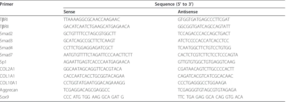
Total RNA from primary human articular chondrocyte cultures was extracted using Trizol. Following extrac-tion, 1μg DNase-I treated RNA was reverse transcribed into cDNA as previously described [14]. Amplification of the generated cDNA was performed by real-time PCR in Applied Biosystems SDS7000 apparatus (Applied Biosystems Inc., Courtaboeuf, France). The relative mRNA level was calculated with the 2-ΔΔCtmethod. Pri-mer sequences are presented in Table 1.
Protein extraction and western blot analysis
Cells were rinsed, and scrapped in RIPA lysis buffer supplemented with phosphatase and protease inhibitors. The extracts (50μg protein) were subjected to fractiona-tion in 10% SDS-PAGE, transferred to polyvinylidene fluoride membranes (Amersham Biosciences, Orsay,
France), and reacted with TbRI, TbRII, Smad2/3 or
SuperSignal West Pico Chemiluminescent Substrate (Pierce Perbio Science, Brébières, France) and exposed to X-ray film. The membranes were also reacted with antib-actin to verify equal loading.
Transfection experiments
Sp1 expression vector (pEVR2-Sp1) was obtained from Dr Suske (Institut fur Molekularbiologie and tumor-forschung, Marburg, Germany). Chondrocytes were transiently transfected by the nucleofection method as previously described [14]. After overnight transfection,
cells were treated with TGFb1 (5 ng/ml) in DMEM
con-taining 2% FCS. The silencing of Sp1 was performed using a siRNA targeting Sp1 (Tebu-Bio; Sp1 siRNA (h), sc-29487: AAUGAGAACAGCAACAACUCC) or a con-trol sequence (UUGUCCGAACGUGUCACGUdtdt), as previously described [13].
Statistical analysis
All experiments were repeated with different donors at least three times with similar results, and representa-tive experiments are shown in the figures. Data are presented as the mean ± standard deviation. Statistical significance was determined by Student’sttest. Differ-ences were considered statistically significant at
P< 0.05.
Results
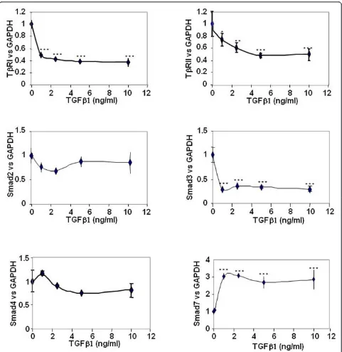
TGFb1 downregulates TGFbreceptors and Smad3, and upregulates Smad7
We investigated the effect of TGFb1 on mRNA
expres-sion of TGFbsignalling genes in a dose-dependent man-ner, using real-time RT-PCR (Figure 1). A 48-hour incubation with TGFb1 significantly reduced the
expres-sion of both TGFb receptors and Smad3, whereas the
Smad7 mRNA level was increased. These effects were
maximal at 1 ng/ml, except for TbRII for which the
maximal effect was observed only at doses above 5 ng/ml. No significant effect was observed on Smad2 and Smad4.
TGFb1 differentially regulates expression of its receptors and Smad3 according to duration of incubation
A time-course study (Figure 2a) revealed that, at mRNA levels, TGFb1 quickly upregulates its own receptors and Smad3, since it increases their expression as soon as 30 minutes of treatment. For longer treatments, TGFb1
exerted the opposite effect and downregulated TGFb
receptors (after 24 hours of incubation) as well as Smad3 (after 3 hours of incubation). On the contrary,
TGFb1 upregulated Smad7 expression whatever the
time of incubation.
Furthermore, western blot analysis (Figure 2b) showed
that TbRII is downregulated after 24 hours whereas
TbRI protein expression is decreased as soon as 1 hour after TGFb1 treatment. In addition, as expected, TGFb1 induced Smad2/3 phosphorylation - but this effect is transient since we were no longer able to detect phos-phorylated Smad2/3 after 3 hours or 24 hours of treat-ment with TGFb1.
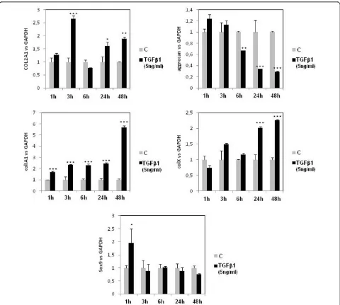
TGFbexerts differential effects on matrix genes and Sox9 according to duration of treatment
To evaluate the importance of the regulation of TGFb
pathways in cartilage homeostasis, we analysed mRNA expression of matrix genes (collagens type II, type I, and type X, and aggrecan) after increased duration of treat-ment (from 1 to 48 hours) (Figure 3). TGFb1 acted with various kinetics according to the considered genes. It induced COL2A1 expression in a biphasic manner (at 3 hours and after 24 hours of treatment, with no
stimu-lation for 6 hours of incubation). TGFb1 repressed
[image:3.595.57.541.100.273.2]aggrecan expression after 6 hours of treatment, and upregulated COL1A1 as soon as 1 hour of incubation. Table 1 Primer sequences for the present study
Primer Sequence (5’to 3’)
Sense Antisense
TbRI TTAAAAGGCGCAACCAAGAAC GTGGTGATGAGCCCTTCGAT
TbRII GACATCAATCTGAAGCATGAGAACA GGCGGTGATCAGCCAGTATT
Smad2 GCTGTTTTCCTAGCGTGGCTT TCCAGACCCACCAGCTGACT
Smad3 GCATCAGCCGCTTCTCAAGT ATCTCCCCACCATCACCTCC
Smad4 CCTTCTGGAGGAGATCGCT TCAATGGCTTCTGTCCTGTGG
Smad7 AATGTGTTTTCTAGATTCCCAACTTCTT CACTCTCGTCTTCTCCTCCCAGTA
Sp1 AGAATTGAGTCACCCAATGAGAACA GTTGTGTGGCTGTGAGGTCAAG
COL2A1 GGCAATAGCAGGTTCACGTACA CGATAACAGTCTTGCCCCACTT
COL1A1 CACCAATCACCTGCGGTACAGAA CAGATCACGTCATCGCACAAC
COL10A1 CCTGGTATGAATGGACAGAAAGG CCCTGAGGGCCTGGAAGA
Aggrecan TCGAGGACAGCGAGGCC TCGAGGGTGTAGCGTGTAGAGA
Concerning hypertrophic markers of cartilage, TGFb1 induced collagen type X expression after 24 hours of incubation. We also focused our attention on Sox9, a major transcription factor for the chondrocyte pheno-type, and found that TGFb1 induced its expression only for 1 hour of incubation.
TGFb1 enhances TGFbreceptor mRNA turnover, but does not modify that of Smads
Modifications of gene expression under TGFbtreatment
[image:4.595.58.539.87.582.2]Figure 2Transforming growth factor beta 1 regulation of receptors and Smad3 expression according to incubation duration.
Smad7. Human articular chondrocytes were incubated with actinomycin D, a transcription inhibitor, in
addi-tion to TGFb (Figure 4). The half-lives of Smad3 and
Smad7 mRNA, which were approximately 3.5 hours and 45 minutes, respectively, were not significantly
modified by TGFb. On the contrary, inhibition of de
novo transcription clearly showed that TGFb reduced
the mRNA half-life of both TGFb receptors. Indeed,
the TbRI half-life is about 20 minutes but was reduced to 10 minutes when chondrocytes were incubated with TGFb, and the TbRII mRNA half-life is 45 minutes for control cells and was reduced by almost 80% after
TGFbtreatment.
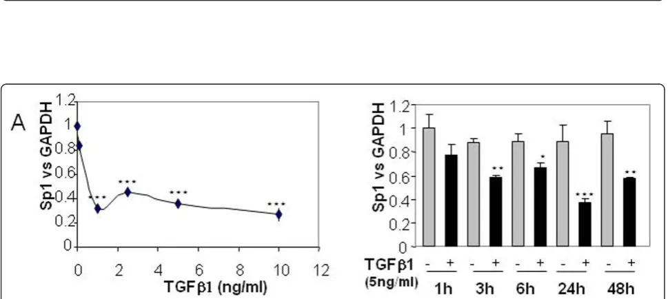
Sp1 mediates TGFb-induced modulation of TGFb receptors
As mentioned above, Sp1 is important for cartilage metabolism. We therefore analysed the effect of TGFb1
on Sp1 expression. We showed that TGFb strongly
reduces Sp1 mRNA levels in a dose-dependent and time-dependent manner (Figure 5).
[image:6.595.58.538.87.518.2]Figure 4 Transforming growth factor beta 1 (TGFb1) enhances TGFbreceptors mRNA turnover. Subconfluent human articular chondrocytes (HAC) were incubated with DMEM + 2% FCS for 24 hours. Thereafter, transforming growth factor beta 1 (TGFb1) or vehicle were added in the presence of actinomycin D (10μg/ml). Cells were then harvested at the indicated times for RT-PCR.
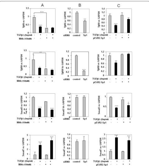
[image:7.595.56.541.444.661.2]To confirm the specific role of Sp1 in these regulations, gain and loss of function experiments were performed. First, silencing of Sp1 by siRNA for 24 hours led to inhibi-tion of both TGFbreceptor expression but did not modify Smad3 and Smad7 expression (Figure 6b). In contrast, forced expression of Sp1 for 24 hours did not change TbRI
and TbRII expression but counteracted TGFb-induced
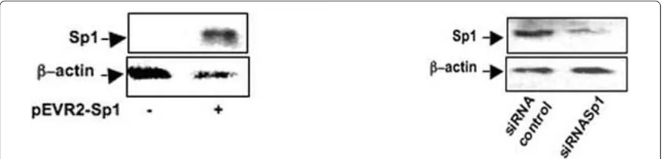
repression on these genes, whereas it did not affect Smad expression either in the presence or in the absence of TGFb(Figure 6c). The depletion of Sp1 by siRNA and the overexpression of Sp1 in pEVR2-Sp1 transfected cells were checked by western blot analysis (Figure 7) [13].
Sp1 ectopic expression permits maintaining a similar expression pattern as early response to TGFbeven after 24 hours of treatment
Since ectopic expression of Sp1 permits one to counter-act the inhibition of TbRI and TbRII expression induced
by long treatment with TGFb, we hypothesised that it
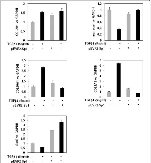
may also affect the expression of downstream genes. We therefore investigated the expression of matrix genes after 24 hours of incubation with TGFb1 in cells that had been transfected with Sp1 expression vector or con-trol vector. Ectopic expression of Sp1 modified cell
responses to TGFb. In Sp1 transfected chondrocytes,
24-hour treatment with TGFb induced COL2A1 and
Sox9 upregulation but was not able to downregulate aggrecan. Additionally, Sp1 ectopic expression blocked the upregulation of COL10A1 and COL1A1. Interest-ingly, the gene expression pattern induced by TGFb1 at 24 hours under Sp1 ectopic expression (Figure 8) is
similar to the early effect of TGFb1 at 1 hour in
untransfected cells (Figure 3).
Discussion
To our knowledge, the present study is the first systema-tic analysis of regulation by TGFbon gene expression of its own receptors and Smads, in human articular chon-drocytes. Our study shows that TGFbexerts a differential effect on the transcription of genes implicated in the
canonical Smads pathway. While TGFbupregulates its
receptors and Smad3 for short incubation (at least at mRNA level), it downregulates them in the long term. In addition, it upregulates Smad7 and does not significantly alter Smad2 and Smad4 expression. This positive and negative feedback loop of the TGFbpathway induces
dif-ferential response of chondrocytes to TGFb. The
mechanisms responsible for modulation of Smads and
for TGFb receptor expression seem to be different.
Indeed, TGFbdownregulates both receptors, at least by modifying the mRNA stability. This process appears slowly (after 24 hours of treatment). On the contrary,
TGFb1 quickly regulates Smad3 and Smad7 mRNA levels
by a mechanism independent of mRNA stability.
Our results suggest that following TGFb1 administra-tion a rapid activaadministra-tion of TGFbsignalling occurs, charac-terised by phosphorylation of Smad2/3 and upregulation
of TbRI, TbRII and Smad3 (at least at mRNA level).
Thereafter, a negative feedback loop of the TGFb1 signal-ling pathway occurs with a decline of these receptors and R-Smad expression and a simultaneous rise in the inhibi-tory Smad7 level. The activation of P-Smad2/3 and
upre-gulation of Smad7 after 30 minutes of TGFbtreatment
are consistent with observations from Jimenez’s group obtained with human and bovine chondrocytes [15].
The downregulation of TGFb receptors by its own
ligand is controversial, and is dependent on cell type as well as on duration of TGFb1 incubation. In lung
fibro-blasts, TGFb1 induced an increased type I receptor
expression by enhancing the transcription of this gene [16], whereas its expression is not modulated or down-regulated in osteoblasts [17,18]. Similarly, TbRII can be downregulated or upregulated by its own ligand [18-20].
In addition, in osteoblasts TGFb1 reduces the amount
of specific TbRII at the cell surface but does not affect the mRNA steady-state level [21].
We have established that, in human OA chondrocytes, TGFb acts, at least in part, by strongly decreasing the mRNA stability of its receptors. This rapid turnover potentially allows the receptor rate to change rapidly in response to its own ligand. We cannot, however,
exclude the possibility that TGFb downregulates its
receptors also at the transcriptional and translational levels.
Concerning Smad effectors, our results are consistent with data obtained in normal skin fibroblasts [22]
-which demonstrated that TGFb treatment causes an
upregulation of antagonistic Smad7, and a dramatic decrease in Smad3 mRNA expression. Interestingly, the mRNA level of the closely related Smad2 was not affected by 48 hours of treatment with TGFb1. A differ-ential regulation between R-Smads has already been described in lung epithelial and mesangial cells [23,24] and may lead to a variation in the cell response accord-ing to the level of TGFb. Similar to findings obtained in fibroblasts [22] or in mesangial cells [24], we established that the downregulation of Smad3 mRNA expression in
TGFb-treated chondrocytes was not due to decreased
transcript stability, suggesting a transcriptional effect of
TGFb. Further experiments, such as nuclear run-on or
gene reporter assays, would be required to definitively state this hypothesis.
In contrast to Smad3, Smad7 mRNA expression was
rapidly and markedly induced by TGFb. These findings
are agreement with reports describing Smad7 as an
immediate-early gene target of TGFbin MV1Lu cells,
with inhibition of TGFb signalling. Smad7 could nega-tively regulate TGFbsignalling; on one hand by
inhibit-ing R-Smad activation by TbRI or by enhancing TbRI
degradation in the cytoplasm, and on the other hand by
disrupting the formation of the TGFb-induced
func-tional Smad-DNA complex in the nucleus [25].
These TGFb-induced modifications on expression of
TGF receptors and Smads may participate in the chon-drocyte-phenotype changes observed in OA, a pathology associated, at least in the first stage, with an increase in
the TGFblevel [9]. Modifications of Smad3 expression
are associated with OA [6,7], and its expression stimu-lates type II collagen synthesis caused by TGFb1 [26]. Moreover, activation of Smad pathways by transfection with a dominant-negative Smad7 retroviral vector or
constitutively active TbRII abolished retinoic
acid-induced inhibition of chondrogenesis, suggesting that TGFbreceptor/Smad signalling is essential for this
pro-cess [27]. Furthermore, ectopic expression of TbRII
restores TGFbsensitivity and increases aggrecan and
col2 expression, in IL1-treated or passaged chondro-cytes, respectively ([14] and unpublished personal data).
Our experiments indicate that TGFb1 exerts a differ-ential effect on profiling of gene expression in chondro-cytes according to the duration of treatment. A short TGFb1 administration (1 hour) induces Sox9 expression, followed, after 3 hours, by induction of collagen type II expression. This effect was transient, but a second peak of collagen II expression appears after 24 hours of incu-bation of TGFb1. These data suggest that at least two different mechanisms are responsible for cell response to TGFb. A short TGFbadministration may activate the
Smad2/3 pathway (upregulation of TbRI, TbRII and
Smad3, and phosphorylation of Smad2/3), leading to an increase of Sox9, which, in turn, may induce collagen type II expression. Thereafter, a negative feedback loop occurs, characterised by a reduction of TbRI, TbRII and Smad3 expression and simultaneous induction of the inhibitory Smad7. This feedback leads to blockage of
Smad2/3-mediated TGFbsignalling and reduction of
Sox9, and furthermore to reduced collagen type II expression.
On the contrary, longer incubation leads an additional response to TGFbbut with a different pattern of matrix gene expression. This late response is associated with increased atypical collagen expression (COL1A1 and COL10A1) and reduction of aggrecan expression. These data suggest that a noncanonical pathway could be involved in this late response to TGFb. Several pathways
may be implied. In particular, the reduction of TbRI
expression may change the ratio between TbRI and
ALK1, another type I TGFbreceptor recently identified
in chondrocytes, favouring TGFb signalling via the
Smad1/5/8 route and, subsequently, chondrocyte term-inal differentiation [28,29].
Finally, in the present report we show that Sp1 is
involved in the regulation of TGFb receptors and cell
response to TGFb. TGFbacts controversially on Sp1
expression. Previous data obtained in rabbit
chondro-cytes showed that TGFbdecreases Sp1 expression and
binding activity [30], whereas recent studies indicate that TGFbinduces Sp1 in skin fibroblasts [31]. Our data show that Sp1 is downregulated in human chondrocytes, suggesting that this negative effect does not depend on the species but is cell-type specific.
The mechanism by which TGFbregulates Sp1
expres-sion is still unclear. In particular, the role of Smads in the regulation of Sp1 promoter activity is not known. Analy-sis of the Sp1 promoter (region -2,000 to +1) with Patch_Search [32], however, shows numerous putative binding sites for Smad3 and Smad4 in the 1,000 base pair upstream transcription initiation site of the Sp1 gene. An extensive study will be required to determine whether Smads directly or indirectly regulate Sp1 expression. Besides, a recent study shows that Smads bind in associa-tion with Sp1 to the CC(GG)-rich TGFb1 responsive
ele-ment of the human a1 type I collagen promoter that
[image:10.595.57.540.89.205.2]that Sp1 cooperate with Smads to regulate the expression of TGFbtarget genes [3,31,34,35].
Importantly, restoration by Sp1 of TGFb receptor
expression after inhibition by TGFb1 strongly suggests that inhibition of Sp1 by TGFb is a potential cause of
TGFb-mediated suppression. These results were in
agreement with previous reports that demonstrate Sp1
is a transactivator of both TGFb receptors [36,37].
Moreover, a key role of Sp1 in the Smad7 induction by TGFbwas recently established in pancreatic cancer cells [3]. In our study, however, Sp1 does not regulate Smad7 expression, suggesting that the regulatory mechanism of Smad7 is cell specific.
Interestingly, Sp1 ectopic expression permits one to maintain, even after 24 hours of treatment, the early cell
response to TGFb(induction of Sox9, COL2A1) and to
counteract the late response (upregulation of COL1A1, COL10A1, repression of aggrecan). These data suggest
that targeting Sp1 expression in association to TGFb
treatment might be an innovative strategy to maintain or induce the chondrocyte phenotype.
Conclusions
The present study enlightens a mechanism of feedback
loop controlling TGFb responses in human OA
chon-drocytes. Contrary to previous studies, which examined one particular gene, we investigated the TGFb-induced
expression of both TGFbreceptors and Smads, and the
molecular mechanism involved. We show that brief administration of TGFbinduces its signalling with
upre-gulation of TGFbreceptors and Smad3, which is
asso-ciated with Sox9 and COL2A1 induction. On the
contrary, a long incubation with TGFbdownregulates
its own receptors by decreasing the mRNA stability, reduces the Smad3 expression and upregulates the inhi-bitor Smad7. In addition, long treatments do not induce Sox9 expression but upregulate atypical cartilage matrix genes such as COL1A1 and COL10A1. We also provide information about the mechanism involved in this regu-lation. We showed the implication of the transcriptional factor Sp1 in the repression of both TGFbreceptors but not in the modulation of Smad3 and Smad7. In addi-tion, we demonstrated the involvement of Sp1 in both early and late response of these cells to TGFb. Sp1 ecto-pic expression permitted one to maintain the early
response of OA chondrocytes to TGFb at 24 hours of
treatment. Together, these data provide an overall view
of the feedback loop of the TGFb signal in human
articular chondrocytes, and highlight an interesting role of Sp1 in regulating the TGFbresponse.
Abbreviations
DMEM: Dulbecco’s modified Eagle’s medium; FCS: foetal calf serum; OA: osteoarthritis; PBS: phosphate-buffered saline; PCR: polymerase chain
reaction; R-Smads: receptor-regulated Smads; RT: reverse transcriptase; siRNA: small interfering RNA; Sp1: specific protein 1; TβRI: TGFβreceptor type I; TβRII: TGFβreceptor type II; TGFβ: transforming growth factor beta.
Acknowledgements
The authors thank Dr Suske (Institut fur Molekularbiologie and tumorforschung, Marburg, Germany) for providing pEVR2-Sp1. OC is a recipient of a fellowship from the Conseil Régional de Basse-Normandie.
Author details
1Université Caen, IFR ICORE 146, Laboratory of Extracellular Matrix and
Pathology, Esplanade de la Paix, 14032 Caen cedex, France.2Department of Orthopaedic Surgery, Saint-Martin Private Clinic, Rue Roquemonts, 14000 Caen, France.
Authors’contributions
CB conceived and carried out experiments, analysed data and wrote the paper. OC and SL participated in data collection and analysis. PG participated in data interpretation. KB conceived experiments, carried out experiments and analysed data. All authors were involved in writing the paper and had final approval of the submitted and published versions
Competing interests
The authors declare that they have no competing interests.
Received: 25 June 2010 Revised: 6 January 2011 Accepted: 15 February 2011 Published: 15 February 2011
References
1. Pujol JP, Chadjichristos C, Legendre F, Baugé C, Beauchef G, Andriamanalijaona R, Galéra P, Boumediene K:Interleukin-1 and transforming growth factorβ1 as crucial factors in osteoarthritic cartilage metabolism.Connective Tissue Res2008,49:293-297. 2. Docagne F, Gabriel C, Lebeurrier N, Lesné S, Hommet Y, Plawinski L,
Mackenzie ET, Vivien D:Sp1 and Smad transcription factors co-operate to mediate TGF-beta-dependent activation of amyloid-beta precursor protein gene transcription.Biochem J2004,383:393-399.
3. Jungert K, Buck A, Buchholz M, Wagner M, Adler G, Gress TM, Ellenrieder V:
Smad-Sp1 complexes mediate TGFβ-induced early transcription of oncogenic Smad7 in pancreatic cancer cells.Carcinogenesis2006,
27:2392-2401.
4. Nakao A, Afrakhte M, Morén A, Nakayama T, Christian JL, Heuchel R, Itoh S, Kawabata M, Heldin NE, Heldin CH, ten Dijke P:Identification of Smad7, a TGFβ-inducible antagonist of TGF-beta signalling.Nature1997,
389:631-635.
5. van der Kraan PM, van den Berg WB:Osteoarthritis in the context of ageing and evolution. Loss of chondrocyte differentiation block during ageing.Ageing Res Rev2008,7:106-113.
6. Yang X, Chen L, Xu X, Li C, Huang C, Deng CX:TGF-beta/Smad3 signals repress chondrocyte hypertrophic differentiation and are required for maintaining articular cartilage.J Cell Biol2001,153:35-46.
7. Yao JY, Wang Y, An J, Mao CM, Hou N, Lv YX, Wang YL, Cui F, Huang M, Yang X:Mutation analysis of the Smad3 gene in human osteoarthritis.
Eur J Hum Genet2003,11:714-717.
8. Serra R, Johnson M, Filvaroff EH, LaBorde J, Sheehan DM, Derynck R, Moses HL:Expression of a truncated, kinase-defective TGF-beta type II receptor in mouse skeletal tissue promotes terminal chondrocyte differentiation and osteoarthritis.J Cell Biol1997,139:541-552. 9. Boumediene K, Conrozier T, Mathieu P, Richard M, Marcelli C, Vignon E,
Pujol JP:Decrease of cartilage transforming growth factor-beta receptor II expression in the rabbit experimental osteoarthritis - potential role in cartilage breakdown.Osteoarthritis Cartilage1998,6:146-149.
10. Kaiser M, Haag J, Söder S, Bau B, Aigner T:Bone morphogenetic protein and transforming growth factor beta inhibitory Smads 6 and 7 are expressed in human adult normal and osteoarthritic cartilage in vivo and are differentially regulated in vitro by interleukin-1β.Arthritis Rheum
2004,50:3535-3540.
chondrocytes requires a decrease of Sp1.Sp3 ratio and of the binding activity of both factors to the COL2A1 promoter.J Biol Chem2008,
283:4850-4865.
12. Piera-Velazquez S, Hawkins DF, Whitecavage MK, Colter DC, Stokes DG, Jimenez SA:Regulation of the human SOX9 promoter by Sp1 and CREB.
Exp Cell Res2007,313:1069-1079.
13. Baugé C, Beauchef G, Leclercq S, Kim SJ, Pujol JP, Galéra P, Boumédiene K:
NFκB mediates IL-1β-induced down-regulation of TβRII through the modulation of Sp3 expression.J Cell Mol Med2008,12:1754-1766. 14. Baugé C, Legendre F, Leclercq S, Elissalde JM, Pujol JP, Galéra P,
Boumédiene K:Interleukin-1βimpairment of transforming growth factor beta1 signaling by down-regulation of transforming growth factor beta receptor type II and up-regulation of Smad7 in human articular chondrocytes.Arthritis Rheum2007,56:3020-3032.
15. Roman-Blas JA, Stokes DG, Jimenez SA:Modulation of TGFβsignaling by proinflammatory cytokines in articular chondrocytes.Osteoarthritis Cartilage2007,15:1367-1377.
16. Bloom BB, Humphries DE, Kuang PP, Fine A, Goldstein RH:Structure and expression of the promoter for the R4/ALK5 human type I transforming growth factor-beta receptor: regulation by TGF-beta.Biochim Biophys Acta1996,1312:243-248.
17. Kim KK, Ji C, Chang W, Wells RG, Gundberg CM, McCarthy TL, Centrella M:
Repetitive exposure to TGF-beta suppresses TGF-beta type I receptor expression by differentiated osteoblasts.Gene2006,379:175-184. 18. Centrella M, Ji C, Casinghino S, McCarthy TL:Rapid flux in transforming
growth factor-beta receptors on bone cells.J Biol Chem1996,
271:18616-18622.
19. Wakefield LM, Smith DM, Masui T, Harris CC, Sporn MB:Distribution and modulation of the cellular receptor for transforming growth factor-beta.
J Cell Biol1987,105:965-975.
20. Massagué J, Like B:Cellular receptors for type beta transforming growth factor. Ligand binding and affinity labeling in human and rodent cell lines.J Biol Chem1985,260:2636-2645.
21. Gebken J, Feydt A, Brinckmann J, Notbohm H, Müller PK, Bätge B: Ligand-induced downregulation of receptors for TGF-beta in human osteoblast-like cells from adult donors.J Endocrinol1999,161:503-510.
22. Mori Y, Chen SJ, Varga J:Modulation of endogenous Smad expression in normal skin fibroblasts by transforming growth factor-beta.Exp Cell Res
2000,258:374-383.
23. Yanagisawa K, Osada H, Masuda A, Kondo M, Saito T, Yatabe Y, Takagi K, Takahashi T:Induction of apoptosis by Smad3 and down-regulation of Smad3 expression in response to TGF-beta in human normal lung epithelial cells.Oncogene1998,17:1743-1747.
24. Poncelet AC, Schnaper HW, Tan R, Liu Y, Runyan CE:Cell phenotype-specific down-regulation of Smad3 involves decreased gene activation as well as protein degradation.J Biol Chem2007,282:15534-15540. 25. Zhang S, Fei T, Zhang L, Zhang R, Chen F, Ning Y, Han Y, Feng XH,
Meng A, Chen YG:Smad7 antagonizes transforming growth factor beta signaling in the nucleus by interfering with functional Smad-DNA complex formation.Mol Cell Biol2007,27:4488-4499.
26. Qiao B, Padilla SR, Benya PD:Transforming growth factor (TGF)-beta-activated kinase 1 mimics and mediates TGF-beta-induced stimulation of type II collagen synthesis in chondrocytes independent of Col2a1 transcription and Smad3 signaling.J Biol Chem2005,280:17562-17571. 27. Yu Z, Xing Y:All-trans retinoic acid inhibited chondrogenesis of mouse
embryonic palate mesenchymal cells by down-regulation of TGF-beta/ Smad signaling.Biochem Biophys Res Commun2006,340:929-934. 28. Blaney Davidson EN, Remst DF, Vitters EL, van Beuningen HM, Blom AB,
Goumans MJ, van den Berg WB, van der Kraan PM:Increase in ALK1/ALK5 ratio as a cause for elevated MMP-13 expression in osteoarthritis in humans and mice.J Immunol2009,182:7937-7945.
29. Hellingman CA, Blaney Davidson E, Koevoet W, Vitters EL, van den Berg WB, van Osch G, van der Kraan PM:Smad signaling determines chondrogenic differentiation of bone-marrow derived mesenchymal stem cells: inhibition of Smad 1/5/8P prevents terminal differentiation and calcification.Tissue Eng Part A2010.
30. Chadjichristos C, Ghayor C, Herrouin JF, Ala-Kokko L, Suske G, Pujol JP, Galéra P:Down-regulation of human type II collagen gene expression by transforming growth factor-beta 1 (TGF-β1) in articular chondrocytes involves SP3/SP1 ratio.J Biol Chem2002,277:43903-43917.
31. Ghosh AK, Mori Y, Dowling E, Varga J:Trichostatin A blocks TGF-beta-induced collagen gene expression in skin fibroblasts: involvement of Sp1.Biochem Biophys Res Commun2007,354:420-426.
32. Patch_Search.[http://www.gene-regulation.com/cgi-bin/pub/programs/ patch/bin/patch.cgi].
33. Sysa P, Potter JJ, Liu X, Mezey E:Transforming growth factor-beta1 up-regulation of human alpha(1)(I) collagen is mediated by Sp1 and Smad2 transacting factors.DNA Cell Biol2009,28:425-434.
34. Ihn H, Yamane K, Asano Y, Jinnin M, Tamaki K:Constitutively phosphorylated Smad3 interacts with Sp1 and p300 in scleroderma fibroblasts.Rheumatology (Oxford)2006,45:157-165.
35. Poncelet AC, Schnaper HW:Sp1 and Smad proteins cooperate to mediate transforming growth factor-beta 1-induced alpha 2(I) collagen expression in human glomerular mesangial cells.J Biol Chem2001,
276:6983-6992.
36. Periyasamy S, Ammanamanchi S, Tillekeratne MP, Brattain MG:Repression of transforming growth factor-beta receptor type I promoter expression by Sp1 deficiency.Oncogene2000,19:4660-4667.
37. Jennings R, Alsarraj M, Wright KL, Muñoz-Antonia T:Regulation of the human transforming growth factor beta type II receptor gene promoter by novel Sp1 sites.Oncogene2001,20:6899-6909.
doi:10.1186/ar3247
Cite this article as:Baugéet al.:Modulation of transforming growth factor beta signalling pathway genes by transforming growth factor beta in human osteoarthritic chondrocytes: involvement of Sp1 in both early and late response cells to transforming growth factor beta. Arthritis Research & Therapy201113:R23.
Submit your next manuscript to BioMed Central and take full advantage of:
• Convenient online submission
• Thorough peer review
• No space constraints or color figure charges
• Immediate publication on acceptance
• Inclusion in PubMed, CAS, Scopus and Google Scholar
• Research which is freely available for redistribution