Evaluation of Methods for Subtyping
Campylobacter jejuni
during
an Outbreak Involving a Food Handler
COLLETTE FITZGERALD,1* LETA O. HELSEL,1MABEL A. NICHOLSON,1SONJA J. OLSEN,1,2
DAVID L. SWERDLOW,1ROBERT FLAHART,3JUNE SEXTON,3ANDPATRICIA I. FIELDS1 Foodborne and Diarrheal Diseases Branch, Division of Bacterial and Mycotic Diseases, National Center for Infectious
Diseases,1and Epidemic Intelligence Service, Division of Applied Public Health Training, Epidemiology Program Office,2Centers for Disease Control and Prevention, Atlanta, Georgia, and Kansas Department of Health
and Environment, Division of Health and Environmental Laboratories, Topeka, Kansas3
Received 14 February 2001/Returned for modification 31 March 2001/Accepted 16 April 2001
In October 1998, the Centers for Disease Control and Prevention (CDC) assisted in an investigation of an outbreak of campylobacteriosis at a school in Salina, Kansas. Twenty-two isolates were submitted from the Kansas state public health laboratory to CDC, 9 associated with the outbreak and 13 epidemiologically
unrelated sporadic isolates. Pulsed-field gel electrophoresis (PFGE) usingSmaI andSalI was initially used to
validate the epidemiologic data. We then tested the ability of other subtyping techniques to distinguish the outbreak-associated isolates from unrelated sporadic isolates. The methods employed were somatic O
sero-typing, PCR-restriction fragment length polymorphism (RFLP) analysis offlaA, DNA sequence analysis of 582
bp offlaAthat included the short variable region (SVR), and sequencing of the entireflaAgene. PFGE was the
most discriminatory technique, yielding 11 SmaI and 10SalI restriction profiles. All outbreak isolates were
indistinguishable by PFGE, somatic O serotyping, and sequencing of the 582-bp region of theflaAgene.fla
typing by PCR-RFLP grouped one sporadic isolate with the outbreak strain. Analysis of the DNA sequence of
a 582-bp segment of flaAproduced strain groupings similar to that generated by PCR-RFLP but further
differentiated twoflaAPCR-RFLP types (with a 1-bp difference in the 582-bp region). Two sporadic strains
were distinct byflaAPCR-RFLP but differed only by a single base substitution in the 582-bp region. The entire
flaAgene was sequenced from strains differing by a single base pair in the 582-bp region, and the data revealed
that additional discrimination may in some cases be obtained by sequencing outside the SVR. PFGE was superior to all other typing methods tested for strain discrimination; it was crucial for understanding the
Kansas outbreak and, whenSmaI was used, provided adequate discrimination between unrelated isolates.
The significance of campylobacters as important human pathogens is now well established. In the United States, Campylobacter jejuniis the most common cause of bacterial enteritis; an estimated 2.5 million cases of human Campylo-bacterinfection occur each year (17). The rise in the number of Campylobacterinfections is most likely the result of increased case ascertainment (34) and a growing awareness of the or-ganism among the public, physicians, and the public health community. In 1998, 44% of laboratory-confirmed cases of bacterial gastroenteritis reported to the Centers for Disease Control and Prevention (CDC)-U.S. Department of Agricul-ture-Food and Drug Administration collaborating sites, food-borne disease active surveillance network (FoodNet) were caused byCampylobacterspecies (4).
Although outbreaks ofCampylobacterinfection occur (27), the majority of infections are sporadic. The control of Campy-lobacterinfection will ultimately depend on a more thorough understanding of sources, transmission routes, and pathogen-host interactions (1). Our current understanding of the epide-miology of Campylobacter infection remains incomplete. To gain more insight, laboratory methods are needed that
differ-entiate epidemiologically related isolates from unrelated iso-lates. Over the last 20 years, a large number of phenotypic and genotypic typing methods have been applied toCampylobacter isolates (25). Phenotypic techniques, such as biotyping (3, 16), serotyping (15, 28), and phage typing (7, 32), are useful for strain characterization and are still in widespread use, but they do have limitations. These include the considerable time and labor investment required for maintenance of reagents; cross-reactivity between antigens, notably in the somatic O (Penner) serotyping scheme; and the occurrence of nontypeable isolates (13, 25, 29). Genotyping offers greater capacity for differenti-ating strains and can be useful in making phylogenetic as well as epidemiologic inferences. Restriction endonuclease analy-sis, ribotyping, PCR-based methods, pulsed-field gel electro-phoresis (PFGE), and, more recently, DNA sequencing-based typing of theflaAgene have all been used to subtype Campy-lobacter(38). However, there are questions regarding the sta-bility of theCampylobacter genome (8, 37); at this time, no technique has been solely identified as the “gold standard” for typingCampylobacter.
In October 1998, CDC assisted the Kansas Department of Health and Environment in an investigation of an outbreak of campylobacteriosis at a school in Salina, Kansas, involving students, staff, and visitors. During the same period, additional persons with Campylobacter infection were identified in the surrounding community. It was unclear if they too were asso-ciated with the school outbreak, and PFGE was used to help * Corresponding author. Mailing address: NationalCampylobacter
andSalmonellaReference Laboratory, Foodborne and Diarrheal
eases Branch, Mailstop CO3, Division of Bacterial and Mycotic Dis-eases, National Center for Infectious DisDis-eases, Centers for Disease Control and Prevention, Atlanta, GA 30333. Phone: (404) 639-0838. Fax: (404) 639-3333. E-mail: [email protected].
2386
on May 15, 2020 by guest
http://jcm.asm.org/
determine that community cases were not linked to the out-break (21). In this study, we sought to assess the abilities of additional subtyping techniques to correctly characterize this epidemiologically well-defined collection ofCampylobacter iso-lates in terms of their abilities to distinguish between outbreak-associated isolates and non-outbreak-outbreak-associated isolates caus-ing sporadic infection in the Kansas community. In addition, a range of criteria, including the ease of use, cost, and rapidity of each method, was considered. The methods evaluated were somatic O serotyping, PCR-restriction fragment length poly-morphism (RFLP) analysis offlaA, DNA sequence analysis of 585 bp offlaAthat included the short variable region (SVR), sequencing of the entireflaAgene, and PFGE usingSmaI and SalI. Our findings show that PFGE is the most discriminatory subtyping method for molecular epidemiologic studies of Campylobacter.
MATERIALS AND METHODS
We tested 22 isolates from stool specimens submitted by the Kansas Depart-ment of Health and EnvironDepart-ment. They were nine outbreak-associated isolates from Saline County, five others from the same county, and eight epidemiologi-cally unrelated isolates from other counties in the same state isolated during the same period. All isolates were cultivated at 37°C for 48 h on heart infusion agar with 5% (vol/vol) defibrinated rabbit blood (Becton Dickinson Biosciences, Franklin Lakes, N.J.) under microaerobic conditions. Isolates were speciated by standard procedures (2).
Somatic O serotyping was performed as described by Penner and Hennessy (28) using a panel of 24 antisera which represent common serotypes in the United States (26). Isolates that were nontypeable underwent passage on blood agar an additional eight times until an antigen was detected. Flagellin PCR-RFLP analysis was done as previously described (19). Sequencing of 582 bp of
theflaAgene that included the SVR and the entire coding sequence was per-formed by the method of Meinersmann et al. (18).
Preparation ofC. jejuniDNA, macrorestriction analysis using the restriction enzymesSmaI andSalI, and PFGE were performed previously (21). Electro-phoresis was carried out for 22 h at 200 V and 14°C constant temperature in a CHEF-DRIII system (Bio-Rad, Richmond, Calif.) with pulse times ramped from 10 to 35 s forSmaI and 4 to 50 s forSalI. Simpson’s index of diversity was calculated as described previously (9).
Nucleotide sequence accession numbers.The sequences obtained in this study have been deposited in GenBank under accession numbers AF369577 to AF369587.
RESULTS
A summary of the typing results is given in Table 1.
Serotyping. Seven different somatic O (heat-stable)
sero-types were identified among the 22C. jejuniisolates (Table 1). Seven of nine isolates associated with the outbreak were sero-type O:19, and the remaining two were initially nonsero-typeable by standard techniques. Testing of the nontypeable isolates after eight transfers resulted in detection of the O:19 antigen. The isolates associated with sporadic infection exhibited a range of serotypes; none were serotype O:19 (Table 1).
Flagellin gene typing. All isolates produced an flaAPCR
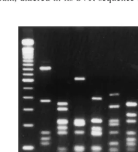
amplicon of the expected 1.7-kb size. Amplicons were digested withDdeI, andflaAtypes were assigned based on the different fragment patterns. Restriction analysis by DdeI gave seven differentflaAtypes among the 22C. jejuniisolates (Fig. 1). All outbreak-associated isolates had an indistinguishableflaAtype, designated D1 (Table 1). One sporadic strain (D5497; serotype O:4 complex) also had this flaAtype. The second most com-monflaAtype (D2) was seen in five of the seven O:4 complex isolates. The remaining strain with this serotype had a unique flaAtype. Of the four remainingflaAtypes, two (D4 and D5) were each seen in two isolates, and two (D3 and D7) were unique.
Flagellin gene sequencing.A 582-bp segment including the
[image:2.612.52.291.84.349.2]SVR offlaAwas sequenced and analyzed. A numerical desig-nation was assigned to each unique sequence (Table 1). The 582-bp sequences from all nine of the outbreak-associated isolates were indistinguishable. Strain D5497, which had aflaA PCR-RFLP pattern indistinguishable from that of the out-break strain, differed in its SVR sequence from the outout-break
[image:2.612.359.501.541.698.2]FIG. 1.DdeIflaAPCR-RFLP patterns of KansasC. jejunistrains. Lanes: 1, 100-bp ladder marker; 2, D5475; 3, D5478; 4, D5481; 5, D5488; 6, D5490; 7, D5492; 8, D5495.
TABLE 1. Typing results for KansasC. jejuniisolates
Strain no. Somatic Oserotype
Flagellin gene type PFGE type PCR-RFLP
(DdeI)
Sequencing
SmaI SalI SVR Full
Outbreak associated
D5475 19 D1 1 NTb A 1
D5477 19 D1 1 1 A 1
D5479 19 D1 1 NT A 1
D5482 19 D1 1 NT A 1
D5483 19 D1 1 NT A 1
D5484 19 D1 1 NT A 1
D5486 19 D1 1 NT A 1
D5476 19 D1 1 NT A 1
D5480 19 D1 1 NT A 1
Sporadic infection
D5478 4 complexa D2 2 2 B 2
D5487 4 complex D2 2 NT B 2
D5498 4 complex D2 2 NT B 2
D5493 4 complex D2 2b 2b H 7
D5494 4 complex D2 2 NT I 8
D5497 4 complex D1 1b 1b K 10
D5492 4 complex D6 5b 4 G 5
D5488 38, 29 D4 4 NT C 3
D5489 38 D4 4 NT C 3
D5490 8 D5 5 3 E 4
D5491 1, 8 D5 5 NT F 5
D5495 2 D7 6 NT J 9
D5481 5 D3 3 NT D 3
a4 complex consists of strains expressing any combinations of antigens 4, 13,
16, 43, and 50.
bNT, not tested.
on May 15, 2020 by guest
http://jcm.asm.org/
strain by a single base pair.flaAPCR-RFLP type D2 was also further differentiated by SVR sequencing; one strain (D5493) differed from the other four isolates with the sameflaAtype by a single base pair in the 582-bp region sequenced, which was outside the SVR. The SVR sequence of strain D5492 (flaA pattern D6) differed from the SVR sequence of strains D5490 and D5491 (flaApattern D5) by a single base pair, though their RFLP patterns were related but different (Fig. 1, lane 6 [D6] versus lane 7 [D7]).
The entire coding region of flaAwas sequenced from one isolate from each of the three groups of strains that differed by a single base pair in the 582-bp sequence. The outbreak strain and one sporadic strain, D5497, had indistinguishable flaA PCR-RFLP patterns and a 1-bp mismatch in the SVR. Two additional mismatches were found in the rest offlaA. Strains D5478 (as well as D5487, D5494, and D5498) and D5493 had identicalflaAPCR-RFLP patterns and a 1-bp mismatch in the 582-bp sequence that was outside the SVR. There were no additional base substitutions in the rest of the gene in these strains. Strains D5490 (and D5491) and D5492 were distinct by flaA DdeI RFLP but differed by 1-bp in the SVR. The flaA sequences from these strains were 99.9% identical for the first
approximately 800 bp offlaA(where the SVR is located) but more divergent at the 3⬘end of the gene (94% identity).
PFGE analysis.PFGE analysis ofSmaI-digested DNA from
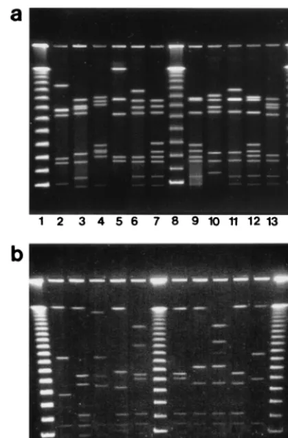
all isolates yielded between 5 and 10 fragments ranging in size from approximately 40 to 480 kb (Fig. 2a). All outbreak-asso-ciated isolates had indistinguishable macrorestriction profiles (pattern A). Ten different macrorestriction profiles were seen among the remaining 13 isolates from sporadic infections; none of them was identical to pattern A. Two macrorestriction profiles were observed among multiple strains, with all isolates of a given profile having identical serotypes andflaAtypes. The remaining eight strains all had unique macrorestriction pro-files. SalI digestion of DNA yielded up to eight fragments ranging in size from approximately 40 to 560 kb (Fig. 2b). The strains were grouped almost identically to those assigned using SmaI, except for two strains, D5491 and D5492. WithSmaI, each strain had a unique profile, with the two profiles differing from each other by three bands; withSalI, they were indistin-guishable.
Discrimination potentials of the different typing methods.A
summary of the discrimination among theC. jejunistrains by the different techniques used is shown in Table 2. The numer-ical index of discrimination (9) ranged from 0.749 for serotyp-ing up to 0.827 for PFGE usserotyp-ingSmaI.
DISCUSSION
We studied a collection of outbreak-associated and sporadic isolates from patients in Kansas who were all ill during the same time period in order to compare the relative usefulness of current subtyping techniques. The evaluation of the practical utility of these methods included both correct discrimination in this well-characterized event and the ability to perform the method rapidly and economically. Correct separation of out-break cases from sporadic cases was achieved by several of the methods: PFGE, serotyping, and sequencing of the SVR. PFGE was the most discriminatory technique used in this study, with a numerical index of discrimination of 0.827 for SmaI and 0.823 forSalI.
Although serotyping is a practical and valid phenotypic method for epidemiologic typing of Campylobacter and has been useful in both clinical (12) and outbreak (23, 36) inves-tigations, it can produce ambiguous results. This can be due to the occurrence of nontypeable strains, transient antigen ex-pression, and cross-reactivity between certain antigens (25, 29). The method requires a panel of antisera that is costly to main-tain; all these factors limit the use of this technique in surveil-FIG. 2. (a) PFGE restriction profiles ofSmaI-digested DNA of
Kan-sasC. jejunistrains. Lanes: 2, D5482; 3, D5487; 4, D5488; 5, D5481;
[image:3.612.68.278.72.391.2]6, D5490; 7, D5491; 9, D5492; 10, D5493; 11, D5494; 12, D5495; 13, D5497; 1, 8, and 14, 48.5-kb DNA ladder. (b) PFGE restriction profiles ofSalI-digested DNA of KansasC. jejunistrains. Lanes: 2, D5482; 3, D5487; 4, D5481; 5, D5488; 6, D5490; 8, D5491; 9, D5493; 10, D5494; 11, D5495; 12, D5497; 1, 7, and 13, 48.5-kb DNA ladder.
TABLE 2. Discrimination indices for methods used to typeC. jejuni
Typing method No. oftypes Isolates in maintype (%) Discriminationindex
Serotyping 7 41 0.749
PCR-RFLP offlaAgene 7 45 0.753
Sequencing offlaAgene
SVR 9 41 0.804
PFGE
SmaI 11 41 0.827
SalI 10 41 0.823
on May 15, 2020 by guest
http://jcm.asm.org/
[image:3.612.312.552.634.727.2]lance studies. In practical terms, serotyping is laborious and requires at least 5 to 7 days to complete, considering the need to repeatedly subculture isolates before testing. A number of studies have reported that repeated subculturing resulted in nontypeable strains becoming typeable by serotyping (10, 23). In this study, initial serotyping results identified two of nine outbreak isolates (D5476 and D5480) as nontypeable in the panel of 24 antisera used, yet epidemiologic data supported the fact that these isolates were part of the school outbreak. Only after repeated subculturing (eight transfers) were the two non-typeable isolates identified as serotype O:19.
Somatic serotype O:19 has been reported as the cause of a number ofCampylobacteroutbreaks (11, 24, 31). The preva-lence of serotype O:19 in sporadic cases of uncomplicated campylobacteriosis has been reported to be between 1 and 6% (20). Several studies suggest that Penner O:19 is overrepre-sented in Guillain-Barre´ syndrome-associatedC. jejuniisolates (S. Fujimoto, N. Yuki, T. Itoh, and K. Amako, Letter, J. Infect. Dis.165:183, 1992). No cases of Guillain-Barre´ syndrome were recognized in this outbreak.
Several subtyping methods based on flaA have been re-ported, including PCR-RFLP (19, 22) and flaA sequencing (18). They are generally simple, cost-effective, and relatively rapid (2 days). However, the use of a single genetic locus as an epidemiologic tool requires caution, especially when making inferences about clonal ancestry, since one gene may not be representative of the entire genome (38). Indeed, this has been reported previously for theflaAlocus (8) and is also demon-strated in our study. One sporadic strain (D5497) had aflaA PCR-RFLP profile indistinguishable from that of the outbreak pattern (D1), yet it had a different serotype (O:4) and different PFGE profiles. The occurrence of strains with different sero-types having identicalflaA types has been shown previously (22, 33). Thus, relying solely onflaAPCR-RFLP analysis can lead to misinterpretation of the data.
Sequence-based subtyping of the SVR, a 267-bp sequence located near the 5⬘end offlaAthat provides a level of discrim-ination similar to that detected in the entire flaAsequence (18), was more discriminatory than PCR-RFLP analysis (Table 2) and correctly differentiated the outbreak strain from the sporadic strains. Among the unrelated strains, sequencing of the SVR, as well as PFGE, further differentiated the O:4 com-plex strains. In our study, sequences outside the SVR provided additional discrimination not seen in the SVR. Although it is advantageous to sequence only a small region of theflaAgene, further evaluation may be necessary to clarify whether it is representive of the entire gene. Despite these limitations, mo-lecular characterization offlaAvia PCR-RFLP analysis or se-quencing may be useful for rapid, preliminary characterization of strains when the aim is to establish an epidemiologic link in a well-defined setting. Furthermore, sequencing of flaA pro-vides a precise measure of genetic variability, as it is based on the DNA sequence, not band matching. However, an initial investment in an automated DNA sequencer is necessary to carry out this method, making it less accessible for smaller laboratories. Recent advances in the development of high-throughput sequence capabilities, microarray technologies, and powerful bioinformatics tools means that sequence-based techniques are valuable and should be investigated further.
While PFGE is also somewhat labor-intensive, we regard it
as the current gold standard because it examines polymor-phisms throughout the genome and it has the highest discrim-inatory power of the typing methods tested. Recently CDC, in collaboration with state health departments and the Food and Drug Administration, established PulseNet, a computer net-work to rapidly analyze and compare PFGE patterns from different sources of several important food-borne pathogens (35). Until recently, one disadvantage of PFGE was the length of time required to perform the technique, which for Campy-lobacterwas typically 3 to 4 days. The development of a rapid PFGE protocol (24 to 30 h) forCampylobacter(30) and the addition of this important enteric organism to the PulseNet system, which is currently under way, will make this a rapid and standardized technique more amenable to routine use. To-gether with traditional epidemiologic methods, this genotypic database should enable us to make more accurate and relevant epidemiologic conclusions.
Interestingly, analysis of our study data revealed two small clusters of related isolates among the sporadic cases, one con-taining three and the other two isolates. This suggests that the Campylobacter isolates in both cases came from a common source, although we have no epidemiologic evidence to sup-port this. Epidemiologic and microbiological analysis of a larger panel of sporadic infections may be helpful in determin-ing whether these related isolates may have had a common source.
The observations described in this study provide an insight into the usefulness of some of the currently available subtyping methods. Two additional high-resolution genotyping tech-niques have recently been described that may have utility for the subtyping of Campylobacter: amplified fragment length polymorphism (AFLP) analysis (5) and multilocus sequence typing (MLST) (6). AFLP has the advantage of whole-genome analysis, as does PFGE, and provides automated data acquisi-tion and analysis. MLST, which involves comparative DNA se-quencing of several genetic loci, provides precise information regarding strain relationships and simplifies interlaboratory comparisons, both of which have proven difficult with PFGE. Further investigation into the utility of these methods is needed, and to address this, we have initiated a project to assess the application of DNA sequence-based subtyping methods for national and global epidemiologic studies ofCampylobacter.
REFERENCES
1.Advisory Committee on the Microbiological Safety of Food.1993. Interim report onCampylobacter. Her Majesty’s Stationery Office, London, United Kingdom.
2.Barrett, T. J., C. M. Patton, and G. K. Morris.1988. Differentiation of
Campylobacterspecies using phenotypic characterization. Lab. Med.19:96– 102.
3.Bolton, F. J., D. R. A. Wareing, M. B. Skirrow, and D. N. Hutchinson.1992. Identification and biotyping of campylobacters, p. 151–161.InR. G. Board, D. Jones, and F. A. Skinner (ed.), Identification methods in applied and environmental microbiology. SAB Technical Series 29. Academic Press, London, United Kingdom.
4.Centers for Disease Control and Prevention.1999. Incidence of foodborne illnesses: preliminary data from the Foodborne Diseases Active Surveillance Network (Foodnet)–United States. Morb. Mortal. Wkly. Rep.48:189–194. 5.de Boer, P., B. Duim, A. Rigter, J. van Der Plas, W. F. Jacobs-Reitsma, and
J. A. Wagenaar.2000. Computer-assisted analysis and epidemiological value of genotyping methods forCampylobacter jejuniandCampylobacter coli. J. Clin. Microbiol.38:1940–1946.
6.Dingle, K. E., F. M. Colles, D. R. A. Wareing, R. Ure, A. J. Fox, F. E. Bolton, H. J. Bootsma, R. J. L. Willems, R. Urwin, and M. C. J. Maiden.2001. Multilocus sequence typing system forCampylobacter jejuni. J. Clin. Micro-biol.39:14–23.
on May 15, 2020 by guest
http://jcm.asm.org/
7.Grajewski, B. A., J. W. Kusek, and H. M. Gelfand.1985. Development of a bacteriophage typing system forCampylobacter jejuniandCampylobacter coli. J. Clin. Microbiol.22:13–18.
8.Harrington, C. S., F. M. Thomson-Carter, and P. E. Carter.1997. Evidence for recombination in the flagellin locus ofCampylobacter jejuni: implications for the flagellin gene typing scheme. J. Clin. Microbiol.35:2386–2392. 9.Hunter, P. R., and M. A. Gaston.1988. Numerical index of the
discrimina-tory ability of typing systems: an application of Simpson’s index of diversity. J. Clin. Microbiol.26:2465–2466.
10. Jacobs-Reitsma, W. F., H. M. E. Maas, and W. H. Jansen.1995. Penner serotyping ofCampylobacter isolates from poultry with absorbed pooled antisera. J. Appl. Bacteriol.79:286–291.
11. Jones, D. M., E. M. Sutcliffe, and J. D. Abbott.1985. Serotyping of Campy-lobacterspecies by combined use of two methods. Eur. J. Clin. Microbiol.
4:562–565.
12. Karmali, M. A., J. L. Penner, P. C. Fleming, A. Williams, and J. N. Hen-nessy.1983. The serotype and biotype distribution of clinical isolates of
Campylobacter jejuniandCampylobacter coliover a three-year period. J. In-fect. Dis.147:243–246.
13. Khakhria, R., and H. Lior.1992. Extended phage-typing scheme for Campy-lobacter jejuniandCampylobacter coli. Epidemiol. Infect.108:403–414. 14. Kuroki, S., T. Saida, M. Nukina, T. Haruta, M. Yoshioka, Y. Kobayashi, and
H. Nakanishi.1993.Campylobacter jejunistrains from patients with Guillain-Barre´ syndrome belong mostly to Penner serogroup 19 and contain beta-N-acetylglucosamine residues. Ann. Neurol.33:570–576.
15. Lior, H., D. L. Woodward, J. A. Edgar, L. J. Laroche, and P. Gill.1982. Serotyping ofCampylobacter jejuni by slide agglutination based on heat-labile antigenic factors. J. Clin. Microbiol.15:761–768.
16. Lior, H.1984. New extended biotyping scheme forCampylobacter jejuni, Campylobacter coli, andCampylobacter laridis. J. Clin. Microbiol.20:636–640. 17. Mead, P. S., L. Slutsker, V. Dietz, L. F. McCaig, J. S. Bresee, C. Shapiro, P. M. Griffin, and R. V. Tauxe.1999. Food-related illness and death in the United States. Emerg. Infect. Dis.5:607–625.
18. Meinersmann, R. J., L. O. Helsel, P. I. Fields, and K. L. Hiett.1997. Discrimination of Campylobacter jejuni isolates by fla gene sequencing. J. Clin. Microbiol.35:2810–2814.
19. Nachamkin, I., H. Ung, and C. M. Patton.1996. Analysis of HL and O serotypes ofCampylobacterstrains by the flagellin gene typing system. J. Clin. Microbiol.34:277–281.
20. Nachamkin, I., B. M. Allos, and T. Ho.1998.Campylobacterspecies and Guillain-Barre´ syndrome. Clin. Microbiol. Rev.11:555–567.
21. Olsen, S. J., G. Hansen, L. Bartlett, C. Fitzgerald, A. Sonder, R. S. Manjrekar, T. Riggs, J. Kim, R. Flahart, G. Pezzino, and D. L. Swerdlow.
2001. An outbreak ofCampylobacter jejuniinfections associated with food handler contamination: the use of pulsed-field gel electrophoresis. J. Infect. Dis.183:164–167.
22. Owen, R. J., C. Fitzgerald, K. Sutherland, and P. Borman.1994. Flagellin gene polymorphism analysis ofCampylobacter jejuniinfecting man and other hosts and comparison with biotyping and somatic antigen serotyping. Epi-demiol. Infect.113:221–234.
23. Patton, C. M., T. J. Barrett, and G. K. Morris.1985. Comparison of the
Penner and Lior method for serotypingCampylobacterspp. J. Clin. Micro-biol.22:558–565.
24. Patton, C. M., I. K. Wachsmuth, G. M. Evans, J. A. Kiehlbauch, B. D. Plikaytis, N. Troup, L. Tompkins, and H. Lior.1991. Evaluation of 10 methods to distinguish epidemic-associatedCampylobacterstrains. J. Clin. Microbiol.29:680–688.
25. Patton, C. M., and I. K. Wachsmuth.1992. Typing schemes: are current methods useful? p. 110–128.InI. Nachamkin, M. J. Blaser, and L. S. Tomp-kins (ed.),Campylobacter jejuni: current status and future trends. American Society for Microbiology, Washington, D.C.
26. Patton, C. M., M. A. Nicholson, S. M. Ostroff, A. A. Ries, I. K. Wachsmuth, and R. V. Tauxe.1993. Common somatic O and heat-labile serotypes among
Campylobacterstrains from sporadic infections in the United States. J. Clin. Microbiol.31:1525–1530.
27. Pebody, R. G., M. J. Ryan, and P. G. Wall.1997. Outbreaks ofCampylobacter
infection: rare events for a common pathogen. Commun. Dis. Rep. Rev.
3:33–37.
28. Penner, J. L., and J. N. Hennessy.1980. Passive hemagglutination technique for serotypingCampylobacter fetussubsp.jejunion the basis of soluble heat-stable antigens. J. Clin. Microbiol.12:732–737.
29. Penner, J. L., J. N. Hennessy, and R. V. Congi.1983. Serotyping of Campy-lobacter jejuniandCampylobacter colion the basis of thermostable antigens. Eur. J. Clin. Microbiol.2:378–383.
30. Ribot, E. M., C. Fitzgerald, K. Kubota, T. J. Barrett, and B. Swaminathan.
2001. Rapid pulsed-field gel electrophoresis protocol for subtyping of
Campylobacter jejuni. J. Clin. Microbiol.39:1889–1894.
31. Sacks, J. J., S. Lieb, L. M. Baldy, S. Berta, C. M. Patton, M. C. White, W. J. Bigler, and J. J. Witte.1986. Epidemic campylobacteriosis associated with a community water supply. Am. J. Public Health76:424–428.
32. Salama, S. M., F. J. Bolton, and D. N. Hutchinson.1990. Application of a new phagetyping scheme to campylobacters isolated during outbreaks. Epi-demiol. Infect.104:405–411.
33. Santesteban, E., J. Gibson, and R. J. Owen.1996. Flagellin gene profiling of
Campylobacter jejuniheat-stable serotype 1 and 4 complex. Res. Microbiol.
147:641–649.
34. Skirrow, M. B., and M. J. Blaser.1992. Clinical and epidemiologic consid-erations, p. 3–8.InI. Nachamkin, M. J. Blaser, and L. S. Tompkins (ed.),
Campylobacter jejuni: current status and future trends. American Society for Microbiology, Washington, D.C.
35. Swaminathan, B., T. J. Barrett, and the CDC Pulsenet Task Force.2000. A national molecular subtyping network for food-borne bacterial disease sur-veillance in the United States, p. 529–535.InI. Nachamkin and M. J. Blaser (ed.),Campylobacter, 2nd ed. American Society for Microbiology, Washing-ton, D.C.
36. Vogt, R. L., A. A. Little, C. M. Patton, T. J. Barrett, and L. A. Orciari.1984. Serotyping and serology studies of campylobacteriosis associated with con-sumption of raw milk. J. Clin. Microbiol.20:998–1000.
37. Wassenaar, T. M., B. Geilhausen, and D. G. Newell.1998. Evidence of genomic instability inCampylobacter jejuniisolated from poultry. Appl. En-viron. Microbiol.64:1816–1821.
38. Wassenaar, T. M., and D. G. Newell.2000. Genotyping ofCampylobacter
spp. Appl. Environ. Microbiol.66:1–9.