0095-1137/05/$08.00⫹0 doi:10.1128/JCM.43.6.2616–2623.2005
Copyright © 2005, American Society for Microbiology. All Rights Reserved.
Development, Technical Performance, and Clinical Evaluation of a
NucliSens Basic Kit Application for Detection of Enterovirus RNA in
Cerebrospinal Fluid
Christine C. Ginocchio,
1,2* Frank Zhang,
1Amisha Malhotra,
3† Ryhana Manji,
1Peter Sillekens,
4Helma Foolen,
4Marlieke Overdyk,
4and Margot Peeters
4North Shore-Long Island Jewish Health System Laboratories, Department of Molecular Diagnostics, Lake Success,
New York,1North Shore University Hospital, Department of Laboratory Medicine,2and North Shore
University Hospital, Department of Pediatrics,3Manhasset, New York, and
bioMe´rieux, Boxtel, The Netherlands4
Received 13 November 2004/Returned for modification 19 January 2004/Accepted 4 March 2005
The combination of nucleic acid sequence-based amplification and electrochemiluminescence detection was used to develop an internally controlled, highly sensitive and specific assay for the detection of enterovirus (EV) RNA in cerebrospinal fluid (CSF). The analytical performance of the assay was determined using both in vitro-transcribed EV RNAs and viral culture isolates. The sensitivity of the assay was 10 EV RNA copies per amplification reaction. The assay detected all enteroviral isolates tested with no cross-reactivity to 21 nonen-teroviral species, including rhinovirus and parechovirus. The clinical performance of the assay was evaluated by testing 992 CSF specimens collected from adult and pediatric patients. NucliSens EV results from a subset of 327 CSF samples were compared to viral culture of nasopharyngeal specimens and rectal swabs (nⴝ195) and/or CSF (nⴝ212). Of the 212 CSF samples, 96 samples were positive by either the NucliSens EV assay (94/96; 97.9%) or culture (63/96; 65.6%), and 61/96 (63.5%) were positive by both methods. The inclusion of an EV-specific internal control monitored the entire process, including the efficiency of nucleic acid extraction, amplification, and detection. In total, only five blood-clotted CSF samples (0.5%) were inhibited. The NucliSens EV assay demonstrated superior sensitivity over viral culture (P< 0.001), excellent specificity, clear delinea-tion of positive samples, and minimal amplificadelinea-tion inhibidelinea-tion.
Enteroviruses (EV) are RNA viruses that are members of
thePicornaviridaefamily (6). The nonpolio enteroviruses,
in-cluding the coxsackieviruses, echoviruses, and numbered en-teroviruses, are responsible for approximately 5 to 10 million symptomatic infections per year in the United States (14, 20). Enteroviruses cause a diverse array of illnesses, including re-spiratory, cardiac, and neurologic ones, in both adults and children. Children under 5 years of age appear to be the most susceptible to infection, due partly to a lack of acquired im-munity and poor hygienic habits. In the neonate, enteroviruses can cause a sepsis-like picture (fever, irritability, failure to eat, poor responsiveness) or meningoencephalitis, which can be severe (14).
Enteroviral infections of the central nervous system will of-ten mimic bacterial infection, and therefore, children with meningitis or febrile infants are hospitalized until a bacterial etiology is either confirmed or ruled out. Many enteroviruses can be grown in primate cell lines; however, results of viral culture can take 3 to 8 days, and therefore, the results are generally not rapid enough to affect either treatment options
or length of hospitalization (4, 6, 16, 21). In addition, viral culture has a sensitivity of approximately 65 to 75%, in part because of the inability to culture all enteroviral serotypes, including several coxsackievirus group A strains that require mouse inoculation for detection (6). To increase the sensitivity of enteroviral detection and reduce the turnaround time for results, molecular methods, including those using reverse tran-scriptase PCR (RT-PCR) (2, 7, 10, 13–15, 18, 19, 21, 23–25) and nucleic acid sequence-based amplification (NASBA) (5, 9, 10), have been developed for the diagnosis of enteroviral in-fections.
This study describes the development, analytical perfor-mance, and clinical evaluation of an internally controlled Nu-cliSens basic kit assay (bioMe´rieux, Boxtel, The Netherlands) for the detection of EV from cerebrospinal fluid (CSF). The assay is a modification of a method first described by Fox et al. (5) and is based on NASBA and electrochemiluminescence (ECL) detection (3, 17). Total CSF nucleic acids and an EV-specific RNA internal control (IC) are coextracted according to the method of Boom (1). Single-tube coamplification of wild-type patient EV RNA (sense RNA) and an EV-specific IC is achieved through the coordinate activities of three en-zymes (avian myeloblastosis virus reverse transcriptase, RNase H, and T7 RNA polymerase) and two DNA oligonucleotides specific for the 5⬘-nontranslated region (NTR) of the entero-virus genome. One oligonucleotide (P1) contains the T7 RNA polymerase promoter sequence, and the second oligonucleo-tide probe (P2) contains a generic ECL detection probe
se-* Corresponding author. Mailing address: North Shore Long Island Jewish Health System Laboratories, 10 Nevada Drive, Lake Success, NY 11042. Phone: (516) 719-1079. Fax: (516) 719-1254. E-mail: cginocch @nshs.edu.
† Present address: University of Medicine and Dentistry of New Jersey, Robert Woods Johnson Medical School, New Brunswick, NJ 08903.
2616
on May 16, 2020 by guest
http://jcm.asm.org/
quence. The amplification results in the production of large amounts of single-stranded RNA that is antisense to the orig-inal target RNA and contains the ECL detection sequence. The single-stranded RNA product can then be readily detected using ECL after hybridization with a generic ruthenium (Ru2⫹) probe and a capture probe that is either EV or IC specific. Technical parameters analyzed in the study included assay sensitivity, specificity, IC validation, and the determina-tion of positive/negative cutoff values. Results from clinical specimens were evaluated based upon clinical presentation and other laboratory data, including bacterial and viral culture. (This study was presented in part at the 18th Annual Clinical Virology Symposium in 2002 and the 102nd General Meeting of the American Society for Microbiology in 2003.)
MATERIALS AND METHODS
Assay development.Construction and generation of the EV-specific external RNA standard and the EV internal RNA control were performed under good manufacturing practices conditions at bioMe´rieux, Boxtel, The Netherlands, using strict isolation precautions to prevent test sample contamination. Upon molecular cloning, a glycerol stock is produced from the transformant containing the plasmid, and portions of this glycerol stock are stored at⫺70°C as master bank stock and as master bank working stock. The transformant, stored under these conditions, is stable for years. During each large-scale production of the plasmid, genetic stability is checked by sequence analysis of a small part of the resulting plasmid DNA prior to the production of in vitro RNA. The external RNA standards and internal RNA controls were stored at⫺70°C until use.
Generation of an enterovirus-specific external RNA standard.For construc-tion of pG3O/POLIO-1 WT, nucleic acid was isolated from a cultured poliovirus type 1 vaccine strain using the silica/guanidine isothiocyanate-based extraction procedure as essentially described by Boom et al. (1). The extracted nucleic acid was used to amplify a 438-bp fragment of the 5⬘-noncoding region (5⬘-NCR) of the viral genome by RT-PCR. First-strand synthesis was performed using ran-dom hexamer primers and SuperScript II RT (Gibco BRL SuperScript pream-plification system). After reverse transcription, RNA from the cDNA:RNA hy-brids was degraded by RNase H incubation. The resulting first-strand cDNA was amplified directly by PCR using oligonucleotide primers POLIO-1 EcoRI and POLIO-1 Csp45I (Table 1). The amplified DNA fragment was digested with restriction enzymes EcoRI and Csp45I, whose restriction sites are located in the extensions of the oligonucleotide primers. Digested DNA was cloned into a modified pGEM vector (pG3O) after removal of a 300-bp fragment from this plasmid by EcoRI/Csp45I digestion. This resulted in recombinant plasmid pG3O/POLIO-1 WT. For large-scale plasmid production, the plasmid was trans-fected into MC1061 cells by electroporation. Cloning of the correct poliovirus type 1 5⬘-NCR fragment was confirmed by sequence analysis. The plasmid was linearized with BamHI, and in vitro RNA was generated using T7 RNA poly-merase. EV-specific RNA was purified, and the concentration of in vitro-gener-ated RNA solutions is based on spectrophotometry. An appropriate dilution of the RNA stock solution is prepared in water, and the optical density at 260 nm is measured. From the measured value for optical density at 260 nm, the RNA
concentration is calculated by making use of the dilution factor and the molar extinction coefficient of the in vitro RNA. This calculation results in a molar concentration. Using Avogadro’s number (6.022⫻1023
), this molar concentra-tion is transformed into copies per microliter. The molar extincconcentra-tion coefficient for a certain RNA is based on its base composition and standard molar extinction values for the nucleotides.
Generation of enterovirus-specific internal RNA control.For the construction of a plasmid from which IC RNA can be generated, a segment of 24 bp in the cloned poliovirus 5⬘-NCR wild-type sequence of plasmid pG3O/POLIO-1 WT was replaced by a fragment of 20 bp derived from the genome of potato leaf roll virus (PLRV; EMBL accession number Y07496, positions 4003 to 4022). Two PCR fragments were generated using the plasmid DNA of pG3/POLIO-1 WT as a template and utilizing oligonucleotide primer T7 PCR in combination with Entero/PLRV-R and SP6 PCR in combination with Entero/PLRV-F (Table 1). The resulting PCR products were combined and again amplified using the outer oligonucleotide primers T7 PCR and SP6 PCR of the first round of PCRs. The amplified DNA fragment from this second PCR was digested with EcoRI and Csp45I and cloned into EcoRI/Csp45I-digested pG3O vector, revealing plasmid pG3O/POLIO-1 SC1. Correct insertion of the PLRV fragment into the poliovi-rus 5⬘-NCR wild-type sequence was confirmed by sequence analysis. For large-scale plasmid production, recombinant plasmid pG3O/POLIO-1 IC1 was trans-fected into MC1061 cells by electroporation. The plasmid was linearized with BamHI, and in vitro RNA was generated using T7 RNA polymerase. The final RNA product is 1,271 nucleotides (with the T7 start at position 621 and the BamHI site at position 1735). The primer binding sites are the same for the IC and for wild-type EV, permitting coamplification in a single-tube format. Dif-ferential detection of wild-type EV and IC is possible using two different biotin-ylated capture probes, one specific for the 23-bp enterovirus sequence and one specific for the 20-bp PLRV sequence (Table 2). IC RNA was purified and quantitated by spectrophotometry, as previously described.
NASBA amplification and detection of wild-type enterovirus and IC RNAs. (i) Nucleic acid isolation.One-hundred-microliter aliquots of enteroviral or non-enteroviral viral culture isolates or 200-l CSF samples, 100l of base matrix (Boston Biomedica, Inc., West Bridgewater, MA), and 20l IC RNA were added directly to 0.9-ml NucliSens lysis buffer tubes containing guanidine thio-cyanate and Triton X-100 (bioMe´rieux, Durham, NC). Samples were lysed for 15 min at room temperature and then stored at⫺70°C until time of analysis. Total nucleic acids were isolated using the NucliSens basic kit isolation reagents (bi-oMe´rieux, Durham, NC) according to the manufacturer’s instructions.
(ii) Nucleic acid amplification. Amplification was performed as previously described (9) using enterovirus-specific primers and probes (Table 2). The P1.3 (antisense) oligonucleotide primer includes the sequence encoding the T7 poly-merase promoter that is required for the transcription-based NASBA process. The P2.2 (sense) oligonucleotide primer includes a sequence that is identical to the generic ECL detection probe supplied in the NucliSens basic kit. Amplifi-cation reactions were conducted at 41⫾0.5°C in a circulating water bath for 150 min. The EV amplicon size with the generic ECL capture probe sequence is 248 nucleotides. The IC amplicon size with the generic ECL capture probe sequence is 244 nucleotides.
(iii) Detection of NASBA amplicons.Products of the amplification reaction were detected using capture oligonucleotide sequences (capture probes) specific for either the EV RNA or the IC RNA with a generic ruthenium (Ru2⫹)-labeled
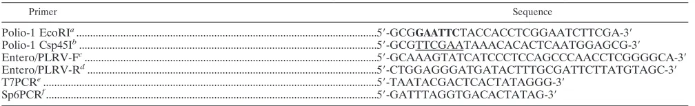
[image:2.585.42.546.77.154.2]ECL probe (Table 2) as previously described (9). After incubation, the detection reactions were analyzed in a NucliSens reader (bioMe´rieux, Durham, NC). TABLE 1. Primers used for the construction of the enterovirus-specific external RNA standard and enterovirus-specific internal control
Primer Sequence
Polio-1 EcoRIa...5⬘-GCGGAATTCTACCACCTCGGAATCTTCGA-3⬘ Polio-1 Csp45Ib...5⬘-GCGTTCGAATAAACACACTCAATGGAGCG-3⬘ Entero/PLRV-Fc...5⬘-GCAAAGTATCATCCCTCCAGCCCAACCTCGGGGCA-3⬘ Entero/PLRV-Rd...5⬘-CTGGAGGGATGATACTTTGCGATTCTTATGTAGC-3⬘ T7PCRe...5⬘-TAATACGACTCACTATAGGG-3⬘
Sp6PCRf...5⬘-GATTTAGGTGACACTATAG-3⬘ aUpstream polio primer.
bDownstream polio primer. cDownstream polio/PLRV primer. dUpstream polio/PLRV primer. eUpstream T7 PCR primer. fDownstrean SP6 PCR primer.
gBoldface denotes the EcoRI restriction site, and underlining denotes the Csp45I restriction site.
on May 16, 2020 by guest
http://jcm.asm.org/
Included in every run were two assay-negative (AN) controls, consisting of the ECL detection probe in combination with either the EV capture probe or the IC capture probe and 5l of specimen diluent, instead of diluted amplification products. The purpose of the EV and IC AN controls was to determine the magnitude of the background ECL signal for each probe and for each run. In addition, an internal reference solution (IRS) was included in every run to allow standardization of the ECL values from run to run.
(iv) Establishment of ECL positive/negative cutoff values.To determine the positive/negative cutoff values for the NucliSens EV assay, 60 negative control specimens consisting of 100l of base matrix and 200l of CSF were added to 900l of NucliSens lysis buffer, extracted, and amplified. The specimens were analyzed in a number of different runs, each run having its own value for the AN control and the IRS. Due to run-to-run variations observed for the IRS ECL values, all sample ECL values were normalized based on the mean value for all the IRS ECL values obtained for all runs included in this study. This normal-ization rules out any run-to-run variability in establishing the positive/negative ECL cutoff value. The positive/negative ECL cutoff value was reevaluated at the end of the entire study after consideration of the combined ECL results of the initial negative control studies, the sensitivity, specificity, and IC studies, and the ECL values obtained with the clinical samples tested in real time, for an overall total of 1,254 assay results.
(v) Determination of assay specificity.Serotype reactivity was tested using frozen clinical isolates obtained from the virology laboratories at Nassau Uni-versity Medical Center (East Meadow, NY), North Shore UniUni-versity Hospital (Manhasset, NY), and the Department of Health Services (Berkeley, CA). In total, 16 enteroviral isolates, including poliovirus Sabin 2, coxsackievirus types A7, A8, A9, A10, A15, B1, B2, B3, B4, and B5, echovirus types 6, 9, 11, and 30, and enterovirus type 71, and 22 nonenteroviral viral isolates, including adenovi-rus types 7 and 21, parainfluenza viadenovi-rus types 1, 2, and 3, influenza viadenovi-rus types A and B, rubella virus, mumps, herpes simplex virus type 1 and type 2, varicella virus, respiratory syncytial virus, cytomegalovirus, parechovirus 1, and 7 isolates of rhinovirus, were used. All isolates with ECL signals greater than or equal to the positive cutoff value (650 ECL units) were scored as positive, as determined by clinical samples and control EV RNAs as described in the previous section. Isolates with ECL values in the range of 350 to⬍650 ECL units were scored as indeterminate, and isolates with ECL signals of⬍350 ECL units were scored as negative.
(vi) Determination of assay sensitivity.Several different studies were con-ducted to determine the analytical sensitivity of the assay and to determine whether the IC RNA would affect the sensitivity of wild-type EV detection. For each study replicate samples were tested on multiple runs. Nucleic acid extrac-tion, amplificaextrac-tion, and detection were performed as described above. The fol-lowing samples were tested: (a) EV RNA control at input concentrations of 10, 50, 100, 500, and 5,000 copies (four tests per concentration) added directly to the NASBA amplification reaction mixture; (b) EV RNA control at input concen-trations of 100 copies (n⫽18), 200 copies (n⫽18), and 300 copies (n⫽12) added to NucliSens lysis buffer containing 100l of base matrix and IC RNA at concentrations of either 2,000 copies (n⫽12), 3,000 copies (n⫽12), or 4,000 copies (n⫽7); (c) EV RNA control at concentrations of 250, 500, 1,000, and 2,000 copies added to NucliSens lysis buffer containing 100l of base matrix and IC RNA at a concentration of 4,000 copies (four tests per EV RNA concentra-tion); (d) serial 10-fold dilutions of coxsackievirus B1 (range, 1⫻105.5
50% tissue culture infective doses [TCID50] to 1⫻101.5TCID50) and echovirus 30
(range, 1⫻106.5
TCID50to 1⫻10 1.5
TCID50) made using NucliSens lysis
buffer; (e) the strains included in the third European Union (EU) Concerted
Action for Quality Control (QCCA) proficiency panel for the molecular detec-tion of enteroviruses (issued in 2001), including diludetec-tion series of coxsackievirus A9 and echovirus 11, three individual samples containing coxsackievirus B5, echovirus 6, and enterovirus 71, and two negative control samples. The lyophi-lized samples were reconstituted in 1 ml water, and nucleic acids were extracted from 100l of the sample.
(vii) Detection of amplification inhibition.To determine the IC ECL cutoff value that would indicate amplification inhibition, the data obtained from two study sample groups were evaluated. The data included the range of IC ECL values and the mean IC ECL value obtained for the negative control samples tested in study iv and the results from 28 independent assay runs that detected EV RNA (100 copies per isolation) with IC RNA at a concentration of 2,000 copies per isolation. The IC ECL cutoff value for amplification inhibition was set at 50,000 IC ECL units, which was 2.6 times the minimum IC ECL value obtained for all the above samples. Samples positive for enterovirus can have IC ECL values above or below the IC ECL cutoff value. Enterovirus-negative samples with IC ECL values below the IC ECL cutoff value were considered inhibited and were reamplified from the original nucleic acid extraction. If inhibition was detected again and additional sample was available, the CSF was reextracted and reamplified. If inhibition was still detected with a negative EV ECL signal, the results were considered inhibited. This IC ECL cutoff value was reevaluated at the time of completion of the clinical studies and assessment of the IC ECL values for all negative clinical samples.
Clinical evaluation. (i) Clinical sample study 1.Initial clinical studies were performed using 144 CSF samples submitted to the North Shore University Hospital Clinical Virology Laboratory for routine viral culture. Samples were collected from pediatric patients admitted to the hospital with a diagnosis of sepsis and/or meningitis. Informed consent was obtained under an institutional review board-approved protocol. For 105 samples, routine CSF analysis includ-ing cell countinclud-ing; protein and glucose measurements; and bacterial and viral cultures, including routine CSF, nasopharyngeal (NP), and rectal specimens, were performed according to standard laboratory procedures.
(ii) Clinical sample study 2.From July 2001 through December 2003, 848 CSF samples (submitted from eight North Shore-Long Island Jewish Health System hospitals, Long Island, NY) were collected by lumbar puncture in accordance with routine diagnostic protocols from adult and pediatric patients with clinical symptoms suggestive of aseptic meningitis. A refrigerated or frozen aliquot of each CSF was submitted for routine enterovirus detection by the NucliSens EV assay. A subset of the patient samples was also submitted for routine viral cultures, including CSF (n⫽117), nasopharyngeal (n⫽102), and rectal (n⫽38) samples. For this subset, NASBA results were compared to viral culture results. (iii) Clinical sample processing.CSF samples were stored at⫺70°C until tested. A 200-l aliquot of CSF was added to NucliSens lysis buffer containing 100l of base matrix and 20l of IC RNA (2,000 copies) and tested as described above. Remaining CSF was stored in cryovials at⫺70°C in case additional or repeat testing was necessary.
(iv) Viral culture.A single tube of primary rhesus monkey kidney (RhMK), human embryonic lung fibroblast (MRC-5) and human epidermoid tumor (A549) cells (all from both Diagnostic Hybrids, Athens, Ohio, and Viromed Laboratories, Minneapolis, Minn.) were inoculated with 0.1 to 0.2 ml of CSF, depending on the available volume. NP aspirates, NP washes, and NP swabs submitted in viral transport medium (M4; MicroTest, Inc., Lilburn, Ga.) were inoculated onto HEp-2, A549, MRC-5, and RhMK cells and/or R-Mix trays (Diagnostic Hybrids, Athens, Ohio). Rectal swabs were submitted in VTM and inoculated onto RhMK, A549, and MRC-5 cells. Culture tubes were incubated TABLE 2. Enterovirus and IC primer and probe sequencesa
Primer or probe Sequence
P1.3b...5⬘-AATTCTAATACGACTCACTATAGGGCACCGGATGGCCAATCCA-3⬘ P2.2c...5⬘-GATGCAAGGTCGCATATGAGGGTGTGAAGAGCCTATTGAG-3⬘ Wild-type EV-specific capture probed...5⬘-biotin-CTCCGGCCCCTGAATGCGGCTAAT-3⬘
IC-specific capture probee...5⬘-biotin-GCAAAGTATCATCCCTCCAG-3⬘
Generic ECL detection probef...5⬘-ruthenium-labeled-GATGCAAGGTCGCATATGAG-3⬘ aLocated in the 5⬘-NCR of the enteroviral genome.
bUnderlining designates the overhang portion encoding the T7 RNA polymerase promoter. The primer is located at nucleotides 623 to 640 of V01150, poliovirus 1, strain Sabin 1.
cUnderlining denotes the generic ECL detection probe sequence. The primer is located at nucleotides 413 to 432 of V01150, poliovirus 1, strain Sabin 1. dThe capture probe is located at nucleotides 446 to 469 of V01150, poliovirus 1, strain Sabin 1.
eA potato leaf roll virus sequence is substituted for the enterovirus sequence located between nucleotides 446 and 469 of V01150, poliovirus 1, strain Sabin 1. fSupplied in the NucliSens basic kit.
on May 16, 2020 by guest
http://jcm.asm.org/
at 37°C for 14 days and examined daily for cytopathic effect (CPE). R-Mix trays were screened daily for 7 days for enterovirus CPE. Tubes or R-Mix trays exhibiting characteristic enteroviral CPE were identified by using pan-enterovi-rus fluorescent antibody stains (Chemicon International, Temecula, Calif., and DakoCytomation, Real Carpinteria, Calif.) and/or EV monoclonal antibody pools (Chemicon International, Temecula, CA).
(v) Resolution of discrepant results.Resolution of discrepant NucliSens EV and CSF culture results was based upon all available laboratory results and clinical findings. CSF specimens positive by NucliSens EV and negative by culture were considered true positives if (a) either the NP or rectal culture was positive for the isolation of an enterovirus, (b) an additional CSF aliquot was extracted and retested positive or, if sufficient CSF volume was not available, a repeat of the same nucleic acid eluate was NASBA positive, or (c) the CSF was from a patient with a concordant clinical presentation and CSF parameters were consistent with aseptic meningitis (negative Gram stain and bacterial cultures, normal glucose, elevated protein, leukocytosis, negative herpesvirus or other viral PCR). A negative NucliSens EV sample was considered a false negative if (a) the CSF culture was positive or (b) the NP or rectal specimen was positive, and the CSF was from a patient with a concordant clinical presentation and CSF parameters were consistent with aseptic meningitis.
(vi) Statistical analysis.The clinical sensitivity and specificity of the NucliSens EV assay and viral culture results were calculated according to standard formu-las. The McNemar test for paired sample nominal-scale-using pairs of data with a value for␣of 0.05 was used to determine the statistical difference between the results obtained by the EV NASBA assay and EV culture. This test results in a
Pvalue of 5.12E-04, which should be reported as aPvalue of⬍0.001.
RESULTS
Establishment of ECL positive cutoff values.Initially, assay positive/negative ECL cutoff values were determined by testing samples consisting of base matrix and IC RNA added to nor-mal CSF. The IRS and the AN controls were included with every test run. AN values varied from 77 ECL counts to 348 ECL counts. IRS values ranged from 27,631 ECL counts to 65,553 ECL counts. Due to the variations observed for the IRS, all EV ECL signals were normalized to the mean value for all the IRS values (ECL signal of 36,500 units). Raw EV ECL signals for the negative samples varied from 1 ECL count to 477 ECL counts. Upon normalization based on the corre-sponding IRS signal and subtraction of the AN value, cor-rected EV ECL signals varied from 0 (negative values, ob-tained for specimens for which the AN value exceeded the raw EV ECL signal, were set to 0) to 207 ECL counts. Based on these results the positive/negative EV ECL cutoff value was set atⱖ650 ECL units, which was three times the highest normal-ized EV ECL signal for a negative sample. After evaluation of the clinical sample data (992 CSF samples), an indeterminate zone was established for values ranging from 350 to⬍650 EV ECL units. This range was chosen because two samples (0.2%) had initial EV ECL values that fell within this range, and repeat testing of the samples yielded one positive result and one repeat indeterminate result.
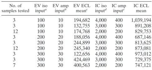
Evaluation of the IC. To monitor the fidelity of the Nu-cliSens EV assay, an EV-specific IC RNA was developed and evaluated. The IC RNA was designed to monitor all steps of the assay, including nucleic acid isolation, amplification, and detection. Importantly, the IC RNA should indicate the pres-ence of any amplification inhibitors in the sample but also not competitively inhibit the detection of any wild-type EV that would cause false-negative results. As shown in Table 3, all three IC RNA isolation input concentrations (2,000, 3,000, and 4,000 copies, which is equivalent to 200, 300 and 400 copies per amplification reaction) did not inhibit the detection of EV
RNA at the lowest number tested (100 copies per isolation; 10 copies per amplification reaction). IC RNA at a concentration of 2,000 copies per isolation gave a mean IC ECL signal of 845,461 ECL units. Therefore, 2,000 IC RNA copies per iso-lation was selected as the optimal input since it was the lowest value that gave positive results 100% of the time. The lower IC RNA copy number should also be a more sensitive detector of low levels of inhibition. Review of the data from 28 indepen-dent assay runs determined that EV RNA (100 copies per isolation) was always detected when the IC RNA (2,000 copies per isolation) was at least 19,000 ECL units. Therefore, to be conservative, the IC ECL threshold value for no amplification inhibition was set at 2.6 times the minimum value (equal to 50,000 IC ECL units). IC RNA aliquots, stored at⫺70°C, were highly stable and gave reproducible results for a minimum of 1 year. Each aliquot could be thawed and refrozen two times for a total of three uses per aliquot (data not shown).
Detection of amplification inhibition in clinical samples.In the presence of a positive EV ECL signal, the IC ECL may be positive, or it may be negative due to competition from the EV RNA. If the IC RNA was⬍50,000 ECL units and the EV ECL counts were⬍650 (negative result), the amplification was con-sidered inhibited, and the sample was reamplified from the original nucleic acid extraction. If inhibition was detected again and additional sample was available, the CSF was reex-tracted and reamplified. If inhibition was still detected with a negative EV ECL signal, the results were considered inhibited. The mean IC ECL reading was 948,513 ECL units for the 992 CSF samples tested. In total, only 5 (0.50%) of the 992 CSF samples demonstrated amplification inhibition. All five sam-ples consisted of⬍300l of CSF containing copious amounts of clotted blood. Sufficient sample material was not available for repeat extraction, and all five samples were inhibited upon repeat testing from the same extraction.
Assay analytical sensitivity.The analytical sensitivity of the assay was evaluated in four sets of experiments using either in vitro-transcribed EV RNA or viral stock cultures of known TCID50. Study I evaluated the assay sensitivity when EV RNA
[image:4.585.299.542.79.191.2]was added directly to the amplification reaction. All EV RNA samples at the concentrations tested (10 copies to 5,000 copies per amplification reaction) were detected (data not shown).
TABLE 3. Assay sensitivity
No. of samples tested
EV iso inputa
EV amp inputb
EV ECL meanc
IC iso inputd
IC amp inpute
IC ECL mean
3 100 10 194,682 4,000 400 1,039,194
3 100 10 132,755 3,000 300 891,208
12 100 10 174,768 2,000 200 829,753
3 200 20 188,056 4,000 400 687,146
3 200 20 244,899 3,000 300 813,625
12 200 20 245,340 2,000 200 873,081
3 300 30 122,656 4,000 400 973,012
3 300 30 424,469 3,000 300 729,375
7 300 30 400,563 2,000 200 747,121
a
Total number of enterovirus RNA copies in 200l of CSF per isolation (iso). b
Total number of enterovirus RNA copies in 5l of eluate used per ampli-fication reaction (amp).
c
ECL mean RLU value for samples tested. d
Total number of enterovirus IC RNA copies added to CSF extraction. e
Total number of enterovirus IC RNA copies in 5 l of eluate used per amplification reaction.
on May 16, 2020 by guest
http://jcm.asm.org/
Studies II and III evaluated the sensitivity of the assay when various concentrations of EV RNA (ranging from 100 to 2,000 copies) and IC RNA (2,000, 3,000, and 4,000 copies) were added to the lysis buffer and coextracted. For both studies, all input concentrations of EV RNA were detected, demonstrat-ing that the sensitivity of the assay was not affected by the extraction procedure nor the addition of IC RNA at all three concentrations (Table 3 and data not shown). The lowest con-centration of EV RNA detected in the amplification reactions was 10 copies (Table 3). Serial dilutions of the viral stock cultures demonstrated a detection level of approximately 101
to 101.5TCID
50(study IV) (data not shown). Results from the
third EU QCCA proficiency panel are summarized in Table 4 and were concordant with the expected results, with the excep-tion of two coxsackievirus type A9 samples (CO1, EV-CO2) containing less then 1.0 TCID50in the original sample.
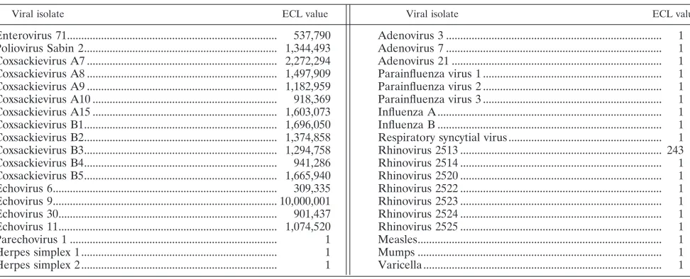
Assay specificity.To determine the specificity of the assay, stock isolates of 16 strains of enterovirus and 22 nonenteroviral isolates (Table 5) were tested with the assay. All enteroviral isolates gave EV ECL signals (range, 537,790 to 10,000,000
ECL units) greater than the positive cutoff value. All nonen-teroviral isolates gave EV ECL signals below the positive cutoff value (range, 1 to 243 ECL units). The one isolate with the 243-ECL-unit signal was a rhinovirus. Six additional rhinovirus isolates were then tested, and all demonstrated ECL signals of 1. The NucliSens assay was 100% specific for the detection of enterovirus.
Clinical evaluation. In the original pilot study, 144 CSF samples were tested with the NucliSens EV assay. Overall, there were 74 positive samples (51.4%), 68 negative samples (47.2%), and two grossly bloody CSF samples (1.4%) that were identified as inhibited. Results from 105 of the patients were compared to CSF, nasopharyngeal, and/or rectal viral culture results and assessed in combination with other laboratory data and clinical findings. In comparison to CSF viral culture, the NucliSens EV assay detected 14 more EV-positive samples (29.4%) (P⬍0.001). Ten of the 14 samples were EV culture positive from either an NP specimen or a rectal specimen. The remaining four NucliSens EV-positive samples were consid-ered true positives based on repeat positive NucliSens EV results from a stored frozen aliquot, CSF cell counts, negative bacterial cultures, and clinical symptoms.
In the supplemental clinical evaluation, 848 CSFs were tested, 603 from children⬍18 years of age and 245 from adults ⱖ18 years of age for the presence of enterovirus. There were 246 (29.0%) CSF samples positive for the detection of entero-virus. The overall detection rates were 31.5% for children and 22.6% for adults. During the peak enterovirus seasons (July through October) in 2001, 2002, and 2003, the percentages of CSF-positive samples were 50.6%, 31.7%, and 33.8%, respec-tively, with a mean of 36.4%. The detection rates were 43.5% for children and 25.0% for adults. August and September had the highest average positivity rates of 45.3% and 47.0%, re-spectively. Throughout the rest of the year the overall mean positivity rate was 13.7% (adults, 10%; children, 12.5%).
NucliSens EV results (n⫽227) were compared to a subset of the 848 CSF samples that had CSF viral culture (n⫽117) and/or viral culture from nasopharyngeal and/or rectal
speci-TABLE 4. Results of the third EU QCCA proficiency panel for the molecular detection of enterovirusesb
Sample code Enterovirus serotype
Virus titer (TCID50/ml)
Expected resulta
NucliSens EV result
EV-C03 Cox A9 3.6 Pos Pos
EV-C02 Cox A9 0.36 Pos Neg
EV-C01 Cox A9 0.036 Neg Neg
EV-C04 Neg Neg
EV-C09 Echo 11 25,000 Pos Pos
EV-C11 Echo 11 250 Pos Pos
EV-C06 Echo 11 25 Pos Pos
EV-C10 Neg Neg
EV-C07 Cox B5 320 Pos Pos
EV-C05 Echo 6 20,000 Pos Pos
Ev-C08 Entero 71 56 Pos Pos
aBased on results obtained by three reference laboratories.
[image:5.585.43.284.89.219.2]bAbbreviations: Cox, coxsackievirus; Echo, echovirus; Entero, enterovirus; Pos, positive; Neg, negative.
TABLE 5. Assay specificitya
Viral isolate ECL value Viral isolate ECL value
Enterovirus 71... 537,790 Poliovirus Sabin 2... 1,344,493 Coxsackievirus A7 ... 2,272,294 Coxsackievirus A8 ... 1,497,909 Coxsackievirus A9 ... 1,182,959 Coxsackievirus A10 ... 918,369 Coxsackievirus A15 ... 1,603,073 Coxsackievirus B1... 1,696,050 Coxsackievirus B2... 1,374,858 Coxsackievirus B3... 1,294,758 Coxsackievirus B4... 941,286 Coxsackievirus B5... 1,665,940 Echovirus 6... 309,335 Echovirus 9... 10,000,001 Echovirus 30... 901,437 Echovirus 11... 1,074,520
Parechovirus 1 ... 1
Herpes simplex 1 ... 1
Herpes simplex 2 ... 1
aSamples with ECL values ofⱖ650 are considered positive. Adenovirus 3 ... 1
Adenovirus 7 ... 1
Adenovirus 21 ... 1
Parainfluenza virus 1 ... 1
Parainfluenza virus 2 ... 1
Parainfluenza virus 3 ... 1
Influenza A ... 1
Influenza B ... 1
Respiratory syncytial virus ... 1
Rhinovirus 2513 ... 243
Rhinovirus 2514 ... 1
Rhinovirus 2520 ... 1
Rhinovirus 2522 ... 1
Rhinovirus 2523 ... 1
Rhinovirus 2524 ... 1
Rhinovirus 2525 ... 1
Measles... 1
Mumps ... 1
Varicella ... 1
on May 16, 2020 by guest
http://jcm.asm.org/
[image:5.585.47.533.520.717.2]mens (n⫽ 140) performed as part of the routine diagnostic work-up. The overall correlation of NucliSens EV results with CSF culture results was 82.1%. From the 117 samples with both NucliSens EV and CSF culture results, there was a total of 32 EV CSF-positive samples. The NucliSens EV assay de-tected 30 (93.8%), and CSF viral culture dede-tected 13 (40.6%) (P⬍0.001). The two CSFs negative by NASBA and positive by culture required repassaging in viral culture for sufficient growth. Testing of the culture isolate by the NASBA confirmed the presence of an enterovirus. Two additional samples origi-nally designated as culture positive stained poorly with the Chemicon Pan-enterovirus antibody stain, failed to stain with the enterovirus monoclonal pools and a supplemental direct stain (DakoCytomacon), and did not grow upon repassaging in culture. In addition, the culture supernatant was negative by the NucliSens EV assay. These two samples were then desig-nated as true negatives. The remaining 19 NASBA-positive and culture-negative samples were retested from a new aliquot (n ⫽16) or, if insufficient volume remained, from the same eluate (n⫽3). All 19 CSF samples were NucliSens EV positive by repeat testing. An additional 18 CSF samples that did not have CSF culture performed were positive by NucliSens EV and were classified as true positives because either rectal or nasopharyngeal specimens had tested culture positive for en-terovirus.
For the two studies combined, the overall concordance be-tween NucliSens EV and CSF culture was 88.2%. Of the 96 CSF EV-positive samples, NucliSens EV detected a signifi-cantly (P⬍0.001) greater number of positive samples (97.9%;
n⫽94) than did viral culture (65.6%;n⫽63).
DISCUSSION
This study describes the development, analytical perfor-mance, and clinical evaluation of an internally controlled Nu-cliSens basic kit application for the detection of a wide variety of enteroviruses in CSF. This assay is a modification of the protocol first described by Fox et al. (5). The first modification was the substitution of a new consensus primer set that did not incorporate mixed bases. The purpose of the mixed bases in the original primer set was to compensate for the sequence divergence in the 5⬘-NTR of the enterovirus genome, thus expanding the scope of the enterovirus subtype detection. However, preliminary studies from our laboratory (unpub-lished data) indicated that the amplification was more efficient without the mixed bases, and the scope of enteroviral detection was comparable. Positive NucliSens EV results were obtained using viral stocks of a variety of echoviruses, enteroviruses, and coxsackievirus group A and B isolates, including the coxsack-ievirus group A strains that cannot be grown in routine viral culture (6). Although all strains of clinically relevant enterovi-ruses were not tested, additional studies by Fox et al. (5) demonstrated broad reactivity with additional serotypes, in-cluding those endemic in Europe and the United Kingdom (11, 12). Enterovirus strains included in an EU QCCA proficiency panel were all detected by NucliSens EV. Two samples con-taining very small amounts of coxsackievirus type A9 originally tested negative (Table 4), but when higher input levels were used, positive results could be obtained for these samples as well (results not shown). In addition, the significantly better
detection rate of enterovirus by NucliSens EV versus viral culture of local clinical samples demonstrated the ability to improve the detection of, at minimum, the predominant strains prevalent in our patient population. All other nonenteroviral isolates, including rhinovirus, did not give ECL signals above the positive cutoff value. One rhinovirus at a titer of approxi-mately 106TCID
50gave a low-level ECL signal that was below
the positive cutoff value. Based on this result and reports of cross-reactivity of rhinovirus with other enterovirus primer pairs (10, 18, 19), we tested several other clinical isolates. All six additional isolates were negative by our assay. A study by Landry et al., which compared the NucliSens EV assay with the Argene Biosoft (Varhilles, France) enterovirus consensus RT-PCR assay, identified two isolates of rhinovirus that were neg-ative by the NucliSens EV assay but called positive by the Argene assay (10). Our laboratory currently uses the NucliSens EV assay to aid in the differentiation of rhinovirus from en-terovirus culture isolates. The assay did not detect parechovi-rus 1 (formerly echoviparechovi-rus 22) (8), a result consistent with re-ports by other investigators using the NucliSens EV primer set (5, 9) or other RT-PCR assays that use primers designed from the same 5⬘-NTR sequence region (2, 7, 10, 13, 15, 18, 19, 23–25).
The second improvement to the assay was the inclusion of an EV IC that is added prior to the isolation of the sample nucleic acids, coextracted, and coamplified in the same tube with the wild-type EV RNA. Our studies demonstrated that the three IC concentrations tested all gave positive results 100% of the time and did not have any effect on the sensitivity of the EV RNA detection. The combination of an optimal input of IC RNA and a standardized IC ECL cutoff value should clearly indicate low-level amplification due to the presence of inhibi-tion or a loss of RNA during the extracinhibi-tion process. Both of these factors are important for limiting the number of false-negative results and for improving the false-negative predictive value of the test. The ability to detect inhibition was demon-strated in five samples with significant amounts of clotted blood. Overall, the extraction and amplification methods were extremely robust since only 0.5% of the samples were inhib-ited.
The third modification to the test was the addition of base matrix to the sample lysis buffer. Preliminary studies (data not shown) demonstrated that the sensitivity of the assay was im-proved approximately 10-fold by the addition of the base ma-trix. Although the actual mechanism is not known, we can hypothesize that the base matrix may help stabilize the RNA after viral cell lysis and serve as an RNA carrier, similar to other nucleic acid extraction protocols that require the addi-tion of tRNA to enhance the sensitivity of the nucleic acid isolation. The sensitivity of the NucliSen EV assay was 100 copies per sample input, which is equivalent to 10 copies per amplification reaction.
Clinical studies were performed to assess the validity of the assay and to aid in establishing positive/negative ECL cutoff values. After a review of all ECL data for positive and negative CSF patient samples, it was determined that a value ofⱖ650 ECL units identified all true positives that were confirmed either by culture or by testing another sample aliquot. This positive cutoff value was also reaffirmed in the studies by Landry et al. (9, 10). An indeterminate zone was established to
on May 16, 2020 by guest
http://jcm.asm.org/
identify potential positive samples that may contain very low levels of EV RNA. Two CSF samples gave ECL values within this range and were retested. One sample retested as positive, and a nasopharyngeal culture was positive for enterovirus. The other sample retested as indeterminate, and the nasopharyn-geal sample was negative by culture. Subsequent to this study we have identified a small number of additional samples that fell within the indeterminate range, were positive by repeat testing and were confirmed by culture, clearly indicating the need to retest the samples.
The NucliSens EV assay showed superior ability to detect enterovirus in CSF when compared to what was previously considered the “gold standard,” i.e., viral culture. The assay detected 32.3% more positive CSF samples than viral culture did (P⬍0.001). Our data were consistent with two studies by Landry et al. (9, 10) that compared this assay to viral culture. In the first study a variety of clinical samples (CSF, nasopha-ryngeal, throat, rectal or stool) were tested, and NASBA de-tected 96% of all the EV-positive samples compared to 78% for viral culture (9). The second study compared the NucliSens EV assay to the Argene Biosoft enterovirus consensus RT-PCR assay and viral culture (10). Overall, the NucliSens EV assay and the Argene RT-PCR assay had comparable clinical sensitivities (92.9% and 88.1%, respectively), and both were superior to viral culture, which had a sensitivity of 60.5%. We identified two samples that were initially suspected to be en-terovirus culture positive based upon a low-level CPE and weak staining with the Chemicon Pan-enterovirus fluorescent antibody stain. However, the isolates were not confirmed by the EV monoclonal antibody pools or the DakoCytomacon EV direct stain, were not repassaged in tissue culture, and were NucliSens EV negative when the viral culture cell lysates were tested. This highlights the difficultly in confirming and differ-entiating some true low-level enterovirus culture positives from nonspecific cross-reactivity found with the current com-mercially available EV fluorescent antibody reagents. We have found testing the viral culture lysates with the NucliSens EV assay to be an important tool for refereeing these difficult cultures (i.e., determining if the result was falsely positive due to nonspecific antibody staining). Two specimens that were NucliSens negative and culture positive had very low titers and took 5 to 7 days to demonstrate CPE in tissue culture. Our negative results may have been due to either levels of the virus in CSF below our detection threshold or loss of viral RNA due to improper handling, shipment, or storage conditions.
In summary, the NucliSens EV assay was very sensitive and demonstrated superior sensitivity over viral culture, excellent specificity, and a clear delineation of positive samples. The extraction method resulted in highly purified, concentrated total nucleic acids with minimal inhibitory substances. One extraction can be performed, and the eluate can be used for the detection of both RNA and DNA targets. In particular, this is extremely useful for low-volume pediatric samples and when multiple analytes are requested. In addition, the extraction procedure is applicable to many different sample types (22). Our laboratory has also validated this assay for use with naso-pharyngeal specimens and stool samples and also as a method for culture confirmation (data not shown). The assay, including nucleic acid extraction, can be completed in approximately
6.5 h. Daily testing and the rapid turnaround time for results compared to routine viral culture has greatly impacted patient care in our institution by reducing hospitalization and the inappropriate use of antibiotics for viral infections. The cost of performing the assay was minimal compared to the savings in the treatment-associated costs (16, 21) and the emotional and traumatic impact of hospitalization for the children and par-ents.
ACKNOWLEDGMENTS
We sincerely thank the technical staff of the Molecular Diagnostics Laboratory, North Shore-Long Island Jewish Health System Labora-tories, Lake Success, NY, and the Clinical Virology Laboratory at North Shore University Hospital, Manhasset, NY, for all their techni-cal support. We thank Gary Leonardi, Nassau University Meditechni-cal Center, NY, and the Department of Health Services, Berkley, CA, for providing clinical viral isolates. We thank Julie Fox (University of Calgary and Molecular Diagnostics, Provincial Laboratory for Public Health, Calgary, Alberta, Canada) for her advice and collaborations. This study was funded in part by the Jane and Dayton Brown and Dayton Brown, Jr., Molecular Diagnostics Laboratory Research Fund. NucliSens basic kit reagents were kindly provided by bioMe´rieux, Box-tel, The Netherlands.
REFERENCES
1.Boom, R., C. J. Sol, M. M. Salimans, C. L. Jansen, P. M. E. Wertheim-van Dillen, and J. van der Noordaa.1990. Rapid and simple method for purifi-cation of nucleic acids. J. Clin. Microbiol.28:495–503.
2.Carroll, K. C., B. Taggart, B. Robison, C. Byington, and D. Hillyard.2000. Evaluation of the Roche AMPLICOR enterovirus PCR assay in the diag-nosis of enteroviral central nervous system infections. J. Clin. Virol.19:149– 156.
3.Compton, J.1991. Nucleic acid sequence-based amplification. Nature (Lon-don)350:91–92.
4.Dagan, R., and M. A. Menegus.1986. A combination of four cell types for rapid identification of enteroviruses in clinical specimens. J. Med. Virol. 19:219–228.
5.Fox, J. D., S. Han, A. Samuelson, Y. Zhang, M. L. Neale, and D. Westmore-land.2002. Development and evaluation of nucleic acid sequence based amplification (NASBA) for diagnosis of enterovirus infections using the NucliSens basic kit. J. Clin. Virol.24:117–130.
6.Hsiung, G.-D.1994.Picornaviridae, p. 119–140.InG.-D Hsiung, C. K. Y. Fong, and J. M. L. Landry (ed.), Hsiung’s diagnostic microbiology, 4th
ed. Yale University Press, New Haven, Conn.
7.Jacques, J., J. Carquin, V. Brodard, H. Moret, D. Lebrun, M. Bouscambert, J. Motte, G. Re´my, and L. Andre´oletti.2003. New reverse-transcriptase-PCR assay for rapid and sensitive detection of enterovirus genomes in cerebro-spinal fluid specimens from patients with aseptic meningitis. J. Clin. Micro-biol.41:5726–5728.
8.Joki-Korpela, P., and T. Hyypia.2001. Parechoviruses, a novel group of human picornaviruses. Ann. Med.31:466–471.
9.Landry, M., R. Garner, and D. Ferguson.2003. Rapid enterovirus RNA detection in clinical specimens by using nucleic acid sequence-based ampli-fication. J. Clin. Microbiol.41:346–350.
10.Landry, M., R. Garner, and D. Ferguson.2003. Comparison of the NucliSens basic kit (nucleic acid sequence-based amplification) and the Argene Biosoft enterovirus consensus reverse transcription-PCR assays for rapid detection of enterovirus RNA in clinical specimens. J. Clin. Microbiol.41:5006–5010. 11.Muir, P., U. Kammerer, K. Korn, M. N. Mulders, T. Poyry, B. Weissbrick, R. Kandolf, G. M. Cleator, and A. M. van Loon.1998. Molecular typing of enteroviruses: current status and future requirements. Clin. Microbiol. Rev. 11:202–227.
12.Nairn, C., and Clements, G. B.1999. A study of enterovirus infections in Glasgow from 1977 to 1997. J. Med. Virol.58:304–312.
13.Nijhuis, M., N. van Maarseveen, R. Schuurman, S. Verkuijlen, M. de Vos, K. Hendriksen, and A. M. van Loon.2002. Rapid and sensitive routine detec-tion of all members of the genus enterovirus in different clinical specimens by real-time PCR. J. Clin. Microbiol.40:3666–3670.
14.Pallansch, M. A., R. P. Roos.2001. Enteroviruses: polioviruses, coxsackievi-ruses, echovicoxsackievi-ruses, and newer enterovicoxsackievi-ruses, p. 723–775.InD. M. Knipe, P. M. Howley, D. E. Griffin, R. A. Lamb, M. A. Martin, B. Roizman, and S. E. Straus (ed.), Fields virology. Lippincott Williams & Wilkins, Philadel-phia, Pa.
15.Pozo, F., I. Casas, A. Tenorio, G. Trallero, and J. E. Echevarria.1998. Evaluation of a commercially available reverse transcriptase PCR assay for
on May 16, 2020 by guest
http://jcm.asm.org/
diagnosis of enteroviral infection in archival and prospectively collected cerebrospinal fluid specimens. J. Clin. Microbiol.36:1741–1745.
16.Ramers, C., G. Billman, M. Hartin, S. Ho, and M. H. Sawyer.2000. Impact of diagnostic cerebrospinal fluid enterovirus polymerase chain reaction test on patient management. JAMA283:2680–2685.
17.Romano, J. W., et al.1997. NASBA technology: isothermal amplification in qualitative and quantitative diagnostics. Immunol. Investig.26:15–28. 18.Rotbart, H. A.1990. Enzymatic amplification of the enteroviruses. J. Clin.
Microbiol.31:438–442.
19.Rotbart, H. A., M. H. Sawyer, S. Fast, C. Lewinski, N. Murphy, E. F. Keyser, J. Spardoro, S.-Y. Kao, and M. Loeffelholz.1994. Diagnosis of enteroviral meningitis by using PCR with a colorimetric microwell detection assay. J. Clin. Microbiol.32:2590–2592.
20.Rotbart, H. A., P. J. Brennan, K. H. Fife, J. R. Romero, J. A. Griffin, M. A. McKinlay, and F. G. Hayden.1998. Enterovirus meningitis in adults. Clin. Infect. Dis.27:896–898.
21.Stellrecht, K. A., I. Harding, A. M. Woron, M. L. Lepow, and R. A. Venezia.
2002. The impact of an enteroviral RT-PCR assay on the diagnosis of aseptic meningitis and patient management. J. Clin. Virol.25:S19–S36.
22.Tetali, S., E. M. Lee, J. Romano, M. H. Kaplan, and C. C. Ginocchio.2001. Chemokine receptor CCR5⌬32 genetic analysis using multiple specimen types and the NASBA-based NucliSens basic kit. Clin. Diagn. Lab. Immunol. 5:965–971.
23.Watkins-Riedel, T., M. Woegerbauer, D. Hollemann, and P. Hufnagl. 2002. Rapid diagnosis of enterovirus infections by real-time PCR on the Light-Cycler using the TaqMan format. Diagn. Microbiol. Infect. Dis. 42:99–105.
24.Yerly, S., A. Gervaix, V. Simonet, M. Caflisch, L. Perrin, and W. Wunderli. 1996. Rapid and sensitive detection of enteroviruses in specimens from patients with aseptic meningitis. J. Clin. Microbiol.34:199–201.
25.Zoll, G. J., W. J. Melchers, H. Kopecka, G. Jambroes, H. J. van der Poel, and J. M. Galama.1992. General-primer mediated polymerase chain reaction for detection of enteroviruses: application for diagnostic routine and persistent infections. J. Clin. Microbiol.30:160–165.