0095-1137/07/$08.00⫹0 doi:10.1128/JCM.01305-07
Copyright © 2007, American Society for Microbiology. All Rights Reserved.
Yield of Stool Culture with Isolate Toxin Testing versus a Two-Step
Algorithm Including Stool Toxin Testing for Detection of
Toxigenic
Clostridium difficile
䌤
Megan E. Reller,
1* Clara A. Lema,
1Trish M. Perl,
2Mian Cai,
1Tracy L. Ross,
1Kathleen A. Speck,
2and Karen C. Carroll
1Division of Medical Microbiology, Department of Pathology,1and Department of Hospital Epidemiology and Infection Control,2
The Johns Hopkins Medical Institutions, Baltimore, Maryland
Received 28 June 2007/Returned for modification 10 August 2007/Accepted 27 August 2007
We examined the incremental yield of stool culture (with toxin testing on isolates) versus our two-step algorithm for optimal detection of toxigenic Clostridium difficile. Per the two-step algorithm, stools were screened forC. difficile-associated glutamate dehydrogenase (GDH) antigen and, if positive, tested for toxin by a direct (stool) cell culture cytotoxicity neutralization assay (CCNA). In parallel, stools were cultured forC.
difficileand tested for toxin by both indirect (isolate) CCNA and conventional PCR if the direct CCNA was
negative. The “gold standard” for toxigenicC. difficilewas detection ofC. difficileby the GDH screen or by culture and toxin production by direct or indirect CCNA. We tested 439 specimens from 439 patients. GDH screening detected all culture-positive specimens. The sensitivity of the two-step algorithm was 77% (95% confidence interval [CI], 70 to 84%), and that of culture was 87% (95% CI, 80 to 92%). PCR results correlated completely with those of CCNA testing on isolates (29/29 positive and 32/32 negative, respectively). We conclude that GDH is an excellent screening test and that culture with isolate CCNA testing detects an additional 23% of toxigenicC. difficilemissed by direct CCNA. Since culture is tedious and also detects nontoxigenicC. difficile, we conclude that culture is most useful (i) when the direct CCNA is negative but a high clinical suspicion of toxigenicC. difficileremains, (ii) in the evaluation of new diagnostic tests for toxigenicC. difficile(where the best reference standard is essential), and (iii) in epidemiologic studies (where the availability of an isolate allows for strain typing and antimicrobial susceptibility testing).
Since its identification in 1978, Clostridium difficile has emerged as the predominant cause of antibiotic-associated co-litis and the leading cause of diarrhea in hospitalized patients (4). Strains of C. difficile can be toxigenic or nontoxigenic; however, only toxigenic strains produce disease. The two main virulence factors are toxin A, a 308-kDa enterotoxin with some cytopathic effects (TcdA), and Toxin B (TcdB), a potent 270-kDa cytotoxin that affects various tissue cell lines in vitro and inhibits bowel motility in vivo (3, 12). The genes encoding both toxins, tcdA and tcdB, have been sequenced, and both are known to disrupt the actin cytoskeleton of intestinal epithelial cells by modifying Rho family proteins. Toxin A has long been considered more important (19, 20), but an increasing number of reports of colitis due to TcdA-negative but TcdB-positive strains have now been made (5, 35). Furthermore, other viru-lence factors have now been identified. Recently, an increase in the frequency and severity of C. difficile-associated colitis, which is associated with a new strain that produces binary toxin (actin-specific ADP-ribosyltransferase) and is resistant to fluo-roquinolones in vitro, has refocused attention on early, accu-rate diagnosis (2, 9, 23, 29, 36).
Cell culture cytotoxicity neutralization assays (CCNA), which detect the cytopathic effects of toxins in monolayers of
cultured cells such as human diploid fibroblasts, are generally considered the best single standard for the detection of toxi-genicC. difficile. CCNA can detect quantities of toxin as low as 1 pg (1, 21); results are considered positive if characteristic changes are seen inⱖ50% of cells at 48 h and are neutralized byC. difficileantitoxin. However, CCNA testing is labor-inten-sive and subjective, and therefore it is not an ideal standard (1, 18). Consequently, we have limited CCNA testing to speci-mens that test positive for the glutamate dehydrogenase (GDH) common antigen of C. difficile, since this screen is ⬎99% sensitive in identifying CCNA-positive stool samples (34).
In response to the current epidemic, we studied the relative yield of culture with cytotoxin testing and characterization of isolates versus our standard two-step algorithm for the diag-nosis of epidemic and nonepidemic strains of toxigenic C. difficile.
(This work was presented in part at the 106th General Meet-ing of the American Society of Microbiology, Orlando, FL, 21 to 25 May 2006 [abstract C-031] [18a].)
MATERIALS AND METHODS
Study site and stool specimens.The study was performed at the Johns Hop-kins Medical Microbiology Laboratory from February 2005 through August 2006
as part of a coordinated effort to evaluate nosocomial transmission ofC. difficile.
Fifteen medical, surgical, and pediatric units were chosen for prospective sur-veillance. Stool samples submitted Monday to Friday from symptomatic patients
were cultured forC. difficileand tested by our current two-step algorithm (step
1, a direct stool test with an enzyme immunoassay [EIA] forC. difficilecommon
antigen [GDH]; step 2, CCNA on a stool specimen if the GDH screen is
* Corresponding author. Mailing address: Division of Medical Mi-crobiology, Department of Pathology, Johns Hopkins Medical Institu-tions, 600 N. Wolfe St., Baltimore, MD 21287. Phone: (410) 955-5077. Fax: (410) 614-8087. E-mail: [email protected].
䌤Published ahead of print on 5 September 2007.
3601
on May 16, 2020 by guest
http://jcm.asm.org/
positive). Recovered isolates ofC. difficilewere tested for toxin production by CCNA if no toxin had been detected by direct CCNA testing of the stool. Additionally, all available isolates were tested by an in-house conventional mul-tiplex PCR based on a modified CDC protocol (22).
Two-step algorithm: GDH testing and CCNA on stools.All stool specimens submitted were tested for GDH with the C. DIFF CHEK assay (an EIA made by TECHLAB and distributed by Inverness Medical Innovations, Inc., Princeton, NJ) in accordance with the manufacturer’s instructions. Specimens were
manu-ally washed using 1⫻wash solution. Results were quantified by measuring the
optical density at 450 nm on the BIO-TEK ELx800 microplate reader. A positive
result was defined as an optical density ofⱖ0.120 and a negative result as⬍0.120.
If the result was positive, a report was issued the same day stating that theC.
difficileantigen had been detected, that the presence of antigen may not correlate with disease, and that a toxin test was pending.
Stool samples negative for GDH were not tested further, since the negative predictive value of GDH-negative samples has been 99% (34). GDH-positive stool samples were tested directly for toxin by CCNA as described previously (34). Human foreskin fibroblasts (Diagnostic Hybrids, Inc. Athens, OH) were
seeded onto 96-well tissue culture microtiter plates at a concentration of 5⫻104
cells per well in fresh 10% minimum essential medium (0.2 ml per well). The
plates were incubated at 37°C under 5% CO2 until a confluent monolayer
formed. Dilutions of the stool filtrate (1:2 and 1:10) and positive-control toxin were prepared with sterile phosphate-buffered saline and antitoxin and were
incubated for 1 h at room temperature. Thereafter, 20l of each dilution of
control toxin, control toxin plus antitoxin, stool filtrate, and stool filtrate plus antitoxin were added to duplicate wells of the microtiter plate and incubated
overnight at 37°C under 5% CO2. Plates were read at 24 and 48 h. Wells with
ⱖ50% cell rounding were considered positive if the cytotoxic activity was also
neutralized by the antitoxin at either the 1:2 or the 1:10 dilution. The rare equivocal result at 48 h was read at 72 h and a final report of positive or negative issued.
Culture forC. difficile.The technologists responsible forC. difficileculture were different from those performing the two-step algorithm and were blinded to the two-step algorithm results. Stool samples were refrigerated before culture if not processed within 2 h. Fresh stool (1 ml) was incubated at 80°C for 10 min in a heat block to trigger the conversion of spores to vegetative bacteria and to promote toxin production. Next, 2 to 3 drops of liquid stool were inoculated onto cycloserine-cefoxitin-fructose agar (CCFA) alone and CCFA with horse blood
(Remel, Inc. Lenexa, KS). PresumptiveC. difficilewas identified by Gram
stain-ing (large gram-positive or gram-variable rods), colony morphology, and stan-dard phenotypic methods. Specifically, plates were incubated anaerobically for as long as 5 days at 35°C and were examined for suspicious colonies at 24-h
intervals. Suspicious colonies were subcultured toBrucellaagar and to CDC agar
for anaerobic incubation; a 5-g/ml vancomycin disk was placed on the CDC
agar to confirm vancomycin susceptibility. Suspicious colonies were also
subcul-tured to sheep blood agar for incubation under 5 to 7% CO2(aerotolerance
testing). TheBrucellaagar was examined for colony morphology (flat, yellow,
ground-glass-appearing colonies with a yellow halo), a “horse barn” odor, and yellow fluorescence with a Wood’s lamp. Colonies were confirmed to be proline positive with a proline-aminopeptidase disk. If a definitive identification was not possible by these methods, organisms underwent cell wall fatty acid analysis by
gas-liquid chromatography or were tested using Remel’s Rapid ANA II kit.C.
difficile strain ATCC 9689 and Clostridium perfringens ATCC strain 13124 (ATCC, Manassas, VA) were used as positive and negative controls, respectively.
The susceptibilities ofC. difficileisolates to ampicillin, amoxicillin-clavulanate,
cefoxitin, clindamycin, meropenem, metronidazole, moxifloxacin, and tetracy-cline were tested by agar dilution; plates were inoculated with a 0.5 McFarland suspension and incubated anaerobically according to the guidelines of the Clin-ical and Laboratory Standards Institute (CLSI; formerly NCCLS) (10).
Appro-priate ATCC strains of C.perfringens,C. difficile,Eubacterium lentum,Bacteroides
thetaiotaomicron, andBacteroides fragiliswere used for quality control of suscep-tibility testing according to the CLSI guidelines (10).
CCNA testing on isolates ofC. difficile.Single isolated colonies ofC. difficile
were inoculated into chopped-meat broth, incubated for 48 h, diluted 1:2 and
1:10, and incubated with human foreskin fibroblasts at 37°C under 5% CO2; in
each case, a second specimen was incubated with a toxin-specific antibody. The
plates were examined after 24 and 48 h of incubation with a microscope at⫻100
magnification;ⱖ50% cell rounding was interpreted as a positive result.
In-house multiplex conventional PCR for detection of toxigenicC. difficile.
PCR testing was performed by a person blinded to the results of all other testing. The in-house conventional PCR protocol was a modification of procedures used at the CDC (22) with primers based on published sequences (16, 32, 33). DNA
was extracted from a single colony ofC. difficilegrown for⬃48 h on an anaerobic
blood agar plate at 37°C. In the first reaction, primer targets included (i) a 204-bp region that codes for toxin B (16), (ii) a 247-bp sequence within which an 18- to
29-bp deletion indicates atcdC(repressor oftcdAandtcdB) deletion (32), and
(iii) a 377-bp internal control (derived from a 16S rRNA gene sequence forC.
difficileand synthesized by Invitrogen Corporation, Carlsbad, CA). A second reaction targeted a 510-bp product that is associated with binary toxin (33).
Data analysis.The “gold standard” for the diagnosis of toxigenicC. difficile
was detection ofC. difficile(either by GDH screen or by culture) and production
of toxin (detected either directly in a stool specimen or in cultured isolates by CCNA testing). Confidence intervals (CI) were calculated using exact methods. Dichotomous variables were compared with Fisher’s exact test. Only one spec-imen per patient was included in the analysis so as to avoid bias related to repeated sampling (correlated data).
RESULTS
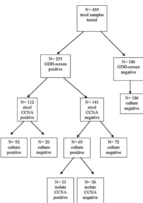
Figure 1 summarizes the testing results. Of 439 specimens tested, 186 were negative both by GDH screening and by culture, and 253 were GDH screen positive. Among the GDH screen-positive samples, 112 were positive for toxin by direct stool CCNA testing and 141 were negative. Of the 112 GDH-and toxin-positive (two-step algorithm-positive) samples, C. difficilewas isolated from 92. Of the 141 GDH screen-negative samples,C. difficilewas isolated from 69 samples; 33/69 isolates were confirmed to be toxigenic by CCNA testing of the isolate. The sensitivity of the two-step algorithm was 77% (112/145) (95% CI, 70 to 84%). Twenty samples determined to contain toxin-producingC. difficileby the two-step algorithm were not identified by culture; however, culture followed by CCNA test-ing on the isolate detected an additional 33 samples not
iden-FIG. 1. Results of testing for toxigenicClostridium difficile.
on May 16, 2020 by guest
http://jcm.asm.org/
[image:2.585.306.534.62.385.2]tified by the two-step algorithm (sensitivity of culture with CCNA testing, 87% [125 of 145] [95% CI, 80 to 92%]). Isolates confirmed to be toxigenic by CCNA were also toxin B positive by PCR (29/29). Demonstration of toxigenicity by stool CCNA usually correlated with PCR results (81 of 84 samples tested). Thirty-six patients were infected with nontoxigenicC. difficile
(confirmed nontoxigenic by PCR in 33 of 33 samples tested). Seventy-two specimens were GDH screen positive but negative forC. difficileby culture and CCNA.
Of the 110 isolates of toxigenic C. difficile tested by PCR, nearly one-third had either binary toxin or thetcdCdeletion, or both (features consistent with the outbreak strain NAP-1). The proportions of isolates with binary toxin by PCR were not significantly different for samples that tested positive by CCNA on stools and those that tested positive by CCNA on isolates (25/81 versus 7/29, respectively;P⫽0.63). The proportions of isolates with thetcdC deletion were also similar for the two groups (26/81 versus 6/29, respectively;P⫽0.34).
Complete PCR testing and antimicrobial susceptibility test-ing data were available for 108C. difficileisolates. All isolates were susceptible to metronidazole (MIC,ⱕ8g/ml). Among isolates found to harbor binary toxin and the tcdC deletion (suggestive of the epidemic strain NAP-1), 23/28 (82%) were resistant to moxifloxacin (MIC, ⱖ8g/ml) and 27/28 (96%) were resistant to or yielded intermediate results for clindamy-cin (MICs,ⱖ8g/ml and 4 g/ml, respectively). In contrast, among isolates without binary toxin and without thetcdC de-letion, only 33/76 (43%) yielded resistant or intermediate (MIC, 4g/ml) results for moxifloxacin and 66/76 (87%) were resistant to or intermediate for clindamycin.
DISCUSSION
Efficient and effective detection of C. difficile diarrhea re-mains a challenge in clinical medicine. The cytotoxin neutral-ization assay led to the identification ofC. difficilein 1978 and, nearly 30 years later, remains the benchmark for the detection of toxigenic C. difficile. During the 20 years following 1978, many laboratories began to switch to stand-alone EIAs for toxin A and/or toxin B because of their ease and speed. We used TECHLAB’s Tox A/B stand-alone EIA for toxin until 2003 based on data showing that its sensitivity was comparable to that of CCNA and because it was faster (1.5 h to results) (1). However, a surge of clinically suspected but Tox A/B EIA-negativeC. difficile-associated colitis prompted reevaluation of the Tox A/B-EIA, similar rapid assays, and a two-step algo-rithm (stool testing for GDH with CCNA only if samples were GDH positive) in comparison with stool CCNA. The two-step algorithm, which was evaluated in comparison with CCNA testing at 1:2 and 1:10 dilutions, was adopted when its sensi-tivity was found to be 96%, that of Tox A/B-EIA was found to be⬍40%, and other rapid toxin assays were found to be sim-ilarly inadequate (sensitivities,⬍70%) (34). Importantly, clini-cians welcomed the more sensitive algorithm despite its CCNA-related delay in final reporting. Inquiries for further explanation have been few. With the advent of a new epidemic strain ofC. difficile(alternately called BI, NAP-1, or ribotype 027) that appears to be more toxigenic and refractory to ther-apy (9, 22, 28, 29), reassessment of optimal diagnosis methods is critical.
CCNA, although considered the reference diagnostic stan-dard for toxigenicC. difficile, not only has practical drawbacks (it is time-consuming, requiring 24 to 48 h to results) but also is recognized to be an imperfect gold standard (17). We pre-viously found that repeating stool CCNA testing for patients with clinically suspected disease but negative test results does not increase the likelihood of detecting toxigenicC. difficile(7). Others have found, similarly, that repeating rapid EIA testing does not substantially increase sensitivity (25).
What about culture? Culture of toxin-positive stools and storage of isolates for future characterization and typing has now been endorsed by the European Society for Clinical Mi-crobiology and Infectious Diseases Study Group forC. difficile, the European Union Member States, and the European Cen-tre for Disease Prevention and Control (17). However, interest in culture is not new. Bond et al., among others, have long suggested that culture is more sensitive than stool cytotoxin testing for the detection of toxigenicC. difficileand therefore should be considered the gold standard (6, 11). Bouza and colleagues have reported that a 15% increase in sensitivity can be achieved by culture combined with cytotoxin testing of iso-lates (8). However, the data are hard to interpret, since actual and described culture techniques (such as the use of enrich-ment procedures, heat or alcohol shock, and the composition of CCFA) vary (14), as do the tests against which they are compared, the number of tests performed, and the populations tested. Comparative North American and European studies have demonstrated that prereduced CCFA with a high cy-closerine concentration, as proposed by George et al. (15), may be optimal (26, 30). To improve validity and comparability, we chose to use standard commercial media for culture, to assess toxigenicity by CCNA, to include only one observation per patient, and to do parallel testing (two-step algorithm and PCR) for all patients.
We found that the GDH screen identified 100% of culture-provenC. difficilecases. This highlights the high negative pre-dictive value of the GDH screen relative to culture, which has also been evaluated relative to CCNA (31, 34, 37). GDH screening is less specific, which may reflect antigenic homology among clostridia, as was suggested by a false-positive rapid stool toxin test result for a patient with Clostridium sordellii
bacteremia (13). Importantly, impaired specificity does not impact the performance of GDH as an excellent screening test, which needs only high sensitivity.
We found that neither the two-step algorithm nor culture is 100% sensitive relative to combined testing that includes both culture and CCNA testing (the composite gold standard). Cul-ture with CCNA testing of isolates identified an additional 33 cases of toxigenic C. difficile (23% of the total), which were corroborated as true positives by conventional PCR. However, culture and isolate CCNA testing alone (no two-step algo-rithm) would have missed at least 20 cases (14% of the total) and would take as long as 9 days for results (2 to 5 days for culture, 2 days for enrichment, and 1 to 2 days for the isolate CCNA) compared with the 1 to 2 days required for stool CCNA. The sensitivity of isolate CCNA testing could differ with the sampling technique, since 20 to 40% of hospitalized patients are colonized withC. difficile(24), and toxigenic and nontoxigenic C. difficile are likely to be morphologically indistinguishable on a plate. Although the sensitivity of
on May 16, 2020 by guest
http://jcm.asm.org/
ture for toxigenic C. difficile might be improved by doing isolate CCNA testing on a sweep of pure colonies, the time to results would not.
Nevertheless, culture does provide additional information of potential epidemiologic and therapeutic interest. Although metronidazole-resistant strains of C. difficile have been re-ported (27), none of ourC. difficile isolates was resistant to metronidazole, which supports the continued cost-effective use of metronidazole for empirical therapy in our population. In this study, susceptibility testing and PCR on isolates also showed that nearly one-third were the epidemic strain NAP-1, yet despite the association of NAP-1 strains with hyperproduc-tion of toxin, these isolates were not more likely to be recov-ered from CCNA-positive stools than from CCNA-negative stools (22). A limitation of the current study was that we were not able to compare the clinical severity of cases detected by stool or isolate CCNA that were or were not caused by the epidemic NAP-1 strain. To do this would require prospective collection of data with a validated clinical instrument, since information regarding diarrhea in clinical charts is notoriously inaccurate. Clinical correlation would be of great interest but was not part of the current study.
Our results do show that a combination of the two-step algorithm (with stool CCNA testing) and culture (with isolate CCNA testing) would increase the rate of detection of C. difficile. However, it is hard in practice to balance optimum sensitivity and timely diagnosis with workloads. In this study, culture was set up before the results of stool CCNA testing were known, which led ultimately to unnecessary testing of 112 of 253 stool CCNA-positive specimens. Ideally, culture would be done only if direct (stool) CCNA results were negative, since culture is unnecessary if stool CCNA testing has already identified toxigenicC. difficile. However, had culture been per-formed only when the stool CCNA result was known to be negative, results would have been further delayed by 24 to 48 h. Thus, although isolate CCNA testing may detect toxigenicC. difficilemissed by direct (stool) CCNA testing, if tests are done sequentially, the result will not be known until 6 to 11 days after sample submission.
In summary, we conclude that our two-step algorithm, al-though less sensitive than spore-enriched culture, is the pref-erable strategy for routine testing, because (i) more than half of the negative results can be reported the same day (GDH screen negative) and (ii) positive results can be reported within 24 to 48 h (after stool CCNA). In contrast, culture with CCNA testing requires 5 to 9 days. However, culture may be an im-portant tool to (i) aid diagnosis for patients with a high pretest probability ofC. difficiledespite negative two-step testing, (ii) evaluate new diagnostic tests for toxigenicC. difficile, and (iii) support epidemiologic assessments (by yielding an isolate for strain typing and for susceptibility testing). For example, cul-ture allowed us to diagnose toxigenicC. difficilefor some pa-tients for whom it would otherwise have been missed; to dem-onstrate that GDH is an ideal screening test; to confirm that metronidazole is still an appropriate empirical therapy; and to delineate the current molecular epidemiology ofC. difficileat our institution. Thus, we would recommend the two-step algo-rithm for routine testing, but we have also demonstrated that the combination of GDH screening, stool CCNA, and culture
with isolate CCNA has clinical, diagnostic, and epidemiologi-cal utility under selected circumstances.
ACKNOWLEDGMENTS
This work was supported in part by a grant (UR8/CCU315092) from the Centers for Disease Control and Prevention, Atlanta, GA.
We thank L. Clifford McDonald for critical review of the manu-script, Eileen Spinks and Jessica Blumenfeld for assistance with cul-ture, and the virology staff of the Johns Hopkins Hospital microbiology laboratory for completing the CCNA testing.
We report no potential conflicts of interest.
REFERENCES
1.Aldeen, W. E., M. Bingham, A. Aiderzada, J. Kucera, S. Jense, and K. C. Carroll.2000. Comparison of the TOX A/B test to a cell culture cytotoxicity
assay for the detection ofClostridium difficilein stools. Diagn. Microbiol.
Infect. Dis.36:211–213.
2.Barbut, F., D. Decre, V. Lalande, B. Burghoffer, L. Noussair, A. Gigandon, F. Espinasse, L. Raskine, J. Robert, A. Mangeol, C. Branger, and J. C. Petit.
2005. Clinical features ofClostridium difficile-associated diarrhoea due to
binary toxin (actin-specific ADP-ribosyltransferase)-producing strains.
J. Med. Microbiol.54:181–185.
3.Barroso, L. A., S. Z. Wang, C. J. Phelps, J. L. Johnson, and T. D. Wilkins.
1990. Nucleotide sequence ofClostridium difficiletoxin B gene. Nucleic Acids
Res.18:4004.
4.Bartlett, J. G.2006. Narrative review: the new epidemic of Clostridium difficile-associated enteric disease. Ann. Intern. Med.145:758–764. 5.Blake, J. E., F. Mitsikosta, and M. A. Metcalfe.2004. Immunological
detec-tion and cytotoxic properties of toxins from toxin A-positive, toxin B-positive
Clostridium difficilevariants. J. Med. Microbiol.53:197–205.
6.Bond, F., G. Payne, S. P. Borriello, and H. Humphreys.1995. Usefulness of
culture in the diagnosis ofClostridium difficileinfection. Eur. J. Clin.
Micro-biol. Infect. Dis.14:223–226.
7.Borek, A. P., D. Z. Aird, and K. C. Carroll.2005. Frequency of sample submission for optimal utilization of the cell culture cytotoxicity assay for
detection ofClostridium difficiletoxin. J. Clin. Microbiol.43:2994–2995.
8.Bouza, E., P. Munoz, and R. Alonso.2005. Clinical manifestations, treatment
and control of infections caused byClostridium difficile. Clin. Microbiol.
Infect.11(Suppl. 4):57–64.
9.Centers for Disease Control and Prevention.2005. SevereClostridium
diffi-cile-associated disease in populations previously at low risk—four states.
Morb. Mortal. Wkly. Rep.54:1201–1205.
10.CLSI.2007. Methods for antimicrobial susceptibility testing of anaerobic bacteria: Approved standard, 7th ed. CLSI document M11–A7. CLSI, Wayne, PA.
11.Delme´e, M., J. Van Broeck, A. Simon, M. Janssens, and V. Avesani.2005.
Laboratory diagnosis ofClostridium difficile-associated diarrhoea: a plea for
culture. J. Med. Microbiol.54:187–191.
12.Dove, C. H., S. Z. Wang, S. B. Price, C. J. Phelps, D. M. Lyerly, T. D. Wilkins, and J. L. Johnson.1990. Molecular characterization of the Clostridium difficiletoxin A gene. Infect. Immun.58:480–488.
13.Elsayed, S., and K. Zhang.2006. PositiveClostridium difficilestool assay in a
patient with fatalC. sordelliiinfection. N. Engl. J. Med.355:1284–1285.
14.Ferreira, C. E., V. Nakano, and M. J. Avila-Campos.2004. Cytotoxicity and
antimicrobial susceptibility ofClostridium difficileisolated from hospitalized
children with acute diarrhea. Anaerobe10:171–177.
15.George, W. L., V. L. Sutter, D. Citron, and S. M. Finegold.1979. Selective
and differential medium for isolation ofClostridium difficile. J. Clin.
Micro-biol.9:214–219.
16.Kato, H., N. Kato, K. Watanabe, N. Iwai, H. Nakamura, T. Yamamoto, K. Suzuki, S. M. Kim, Y. Chong, and E. B. Wasito.1998. Identification of toxin
A-negative, toxin B-positiveClostridium difficileby PCR. J. Clin. Microbiol.
36:2178–2182.
17.Kuijper, E. J., B. Coignard, and P. Tull.2006. Emergence ofClostridium difficile-associated disease in North America and Europe. Clin. Microbiol.
Infect.12(Suppl. 6):2–18.
18.Lee, S. D., D. K. Turgeon, C. W. Ko, T. R. Fritsche, and C. M. Surawicz.
2003. Clinical correlation of toxin and common antigen enzyme
immunoas-say testing in patients withClostridium difficiledisease. Am. J. Gastroenterol.
98:1569–1572.
18a.Lema, C., E. Spinks, J. Blumenfeld, A. Borek, D. Aird, K. Speck, T. Perl, and K. C. Carroll.2006. Comparison of bacterial culture with toxin testing to a two step algorithm using an enzyme immunoassay for glutamate
dehydro-genase with subsequent toxin testing for detection ofClostridium difficilein
stool samples, abstr. C-301. Abstr. 106th Gen. Meet. Am. Soc. Microbiol., Orlando, FL, 21–25 May 2006.
19.Lyerly, D. M., D. E. Lockwood, S. H. Richardson, and T. D. Wilkins.1982.
Biological activities of toxins A and B ofClostridium difficile. Infect. Immun.
35:1147–1150.
on May 16, 2020 by guest
http://jcm.asm.org/
20.Lyerly, D. M., K. E. Saum, D. K. MacDonald, and T. D. Wilkins.1985.
Effects ofClostridium difficiletoxins given intragastrically to animals. Infect.
Immun.47:349–352.
21.Lyerly, D. M., N. M. Sullivan, and T. D. Wilkins. 1983. Enzyme-linked
immunosorbent assay forClostridium difficiletoxin A. J. Clin. Microbiol.
17:72–78.
22.McDonald, L. C., G. E. Killgore, A. Thompson, R. C. Owens, Jr., S. V. Kazakova, S. P. Sambol, S. Johnson, and D. N. Gerding.2005. An epidemic,
toxin gene-variant strain ofClostridium difficile. N. Engl. J. Med.353:2433–
2441.
23.McEllistrem, M. C., R. J. Carman, D. N. Gerding, C. W. Genheimer, and L. Zheng.2005. A hospital outbreak ofClostridium difficiledisease associated
with isolates carrying binary toxin genes. Clin. Infect. Dis.40:265–272.
24.McFarland, L. V., M. E. Mulligan, R. Y. Kwok, and W. E. Stamm.1989.
Nosocomial acquisition ofClostridium difficileinfection. N. Engl. J. Med.
320:204–210.
25.Mohan, S. S., B. P. McDermott, S. Parchuri, and B. A. Cunha.2006. Lack of
value of repeat stool testing forClostridium difficile toxin. Am. J. Med.
119:356.e7–356.e8.
26.Mundy, L. S., C. J. Shanholtzer, K. E. Willard, D. N. Gerding, and L. R. Peterson.1995. Laboratory detection ofClostridium difficile. A comparison
of media and incubation systems. Am. J. Clin. Pathol.103:52–56.
27.Pela´ez, T., L. Alcala, R. Alonso, M. Rodriguez-Creixems, J. M. Garcia-Lechuz, and E. Bouza.2002. Reassessment ofClostridium difficile suscepti-bility to metronidazole and vancomycin. Antimicrob. Agents Chemother.
46:1647–1650.
28.Pe´pin, J., L. Valiquette, M. E. Alary, P. Villemure, A. Pelletier, K. Forget, K. Pepin, and D. Chouinard.2004.Clostridium difficile-associated diarrhea in a region of Quebec from 1991 to 2003: a changing pattern of disease severity.
CMAJ171:466–472.
29.Pe´pin, J., L. Valiquette, and B. Cossette. 2005. Mortality attributable to
nosocomial Clostridium difficile-associated disease during an epidemic
caused by a hypervirulent strain in Quebec. CMAJ173:1037–1042.
30.Peterson, L. R., P. J. Kelly, and H. A. Nordbrock.1996. Role of culture and
toxin detection in laboratory testing for diagnosis ofClostridium difficile
-associated diarrhea. Eur. J. Clin. Microbiol. Infect. Dis.15:330–336.
31.Snell, H., M. Ramos, S. Longo, M. John, and Z. Hussain.2004. Performance of the TechLab C. DIFF CHEK-60 enzyme immunoassay (EIA) in
combi-nation with theC. difficileTox A/B II EIA kit, the TriageC. difficilepanel
immunoassay, and a cytotoxin assay for diagnosis ofClostridium difficile
-associated diarrhea. J. Clin. Microbiol.42:4863–4865.
32.Spigaglia, P., and P. Mastrantonio.2002. Molecular analysis of the patho-genicity locus and polymorphism in the putative negative regulator of toxin
production (TcdC) amongClostridium difficileclinical isolates. J. Clin.
Mi-crobiol.40:3470–3475.
33.Stubbs, S., M. Rupnik, M. Gibert, J. Brazier, B. Duerden, and M. Popoff.
2000. Production of actin-specific ADP-ribosyltransferase (binary toxin) by
strains ofClostridium difficile. FEMS Microbiol. Lett.186:307–312.
34.Ticehurst, J. R., D. Z. Aird, L. M. Dam, A. P. Borek, J. T. Hargrove, and K. C. Carroll.2006. Effective detection of toxigenicClostridium difficileby a two-step algorithm including tests for antigen and cytotoxin. J. Clin.
Micro-biol.44:1145–1149.
35.van den Berg, R. J., E. C. Claas, D. H. Oyib, C. H. Klaassen, L. Dijkshoorn, J. S. Brazier, and E. J. Kuijper.2004. Characterization of toxin A-negative,
toxin B-positive Clostridium difficile isolates from outbreaks in different
countries by amplified fragment length polymorphism and PCR ribotyping.
J. Clin. Microbiol.42:1035–1041.
36.Warny, M., J. Pepin, A. Fang, G. Killgore, A. Thompson, J. Brazier, E. Frost, and L. C. McDonald.2005. Toxin production by an emerging strain of
Clostridium difficileassociated with outbreaks of severe disease in North
America and Europe. Lancet366:1079–1084.
37.Zheng, L., S. F. Keller, D. M. Lyerly, R. J. Carman, C. W. Genheimer, C. A. Gleaves, S. J. Kohlhepp, S. Young, S. Perez, and K. Ye.2004. Multicenter
evaluation of a new screening test that detectsClostridium difficilein fecal
specimens. J. Clin. Microbiol.42:3837–3840.