0095-1137/95/$04.0010
Copyrightq1995, American Society for Microbiology
Analysis of Clonal Relationships among Isolates of Shigella
sonnei by Different Molecular Typing Methods
PETER YUK-FONG LIU,* YEU-JUN LAU, BOR-SHEN HU, JAINN-MING SHYR, ZHI-YUAN SHI,
WEN-SHIH TSAI, YU-HUI LIN,
ANDCHING-YU TSENG
Section of Infectious Diseases, Taichung Veterans General Hospital, Taichung, Taiwan, Republic of China
Received 3 January 1995/Returned for modification 9 March 1995/Accepted 10 April 1995
Shigella sonnei
is a major cause of diarrheal disease in developed as well as in developing countries.
Epidemiologic studies of this organism have been limited by the lack of a simple and effective method for
comparing strains. In this study, we have compared different molecular typing methods, i.e., plasmid profile
analysis, restriction endonuclease analysis of plasmids, rRNA gene restriction analysis (ribotyping),
pulsed-field gel electrophoresis (PFGE), and enterobacterial repetitive intergenic consensus (ERIC) sequence-based
PCR (ERIC-PCR) for typing 20 clinical isolates of
S. sonnei
collected from six incidents of infection. PFGE and
ERIC-PCR fingerprintings had the highest discriminatory power for discrimination of epidemiologically
related isolates from epidemiologically unrelated strains of
S. sonnei
, and both gave seven distinct strain types
among these isolates and the type strain of the species. Plasmid study and ribotyping produced only six and
two distinct patterns, respectively, among these strains. All of these molecular typing techniques demonstrated
an identical fingerprint for eight temporally related sporadic isolates. It is possible that these temporally
related isolates belonged to a single bacterial clone and circulated obscurely through the community. Our
results indicate that the ERIC-PCR technique represents a rapid and simple means for typing
S. sonnei
with
a level of discrimination equivalent to that of PFGE but greater than those of plasmid profile analysis,
restriction endonuclease analysis of plasmids, and ribotyping.
Shigella sonnei has emerged as a major cause of diarrheal
disease in developed as well as in developing countries. It
accounts for about 60 to 80% of all cases of shigellosis
cur-rently reported in the United States (2). The study of
epide-miologic markers is important in an attempt to trace the source
of infection. Unlike the other Shigella spp., S. sonnei contains
only one serovar, and this has hindered the development of a
serologic typing schema. As a result, other typing procedures,
such as colicin typing, phage typing, biotyping, and drug
resis-tance pattern, have been used (15, 18, 24, 26). However, all of
these typing systems, which are based on the phenotypic
prop-erties of the microorganism, have some disadvantages or
lim-itations.
Recently, approaches at the molecular level have been used
to assess the relatedness of bacterial isolates. Plasmid pattern
analysis was found to be useful for the characterization of
epidemic strains harboring plasmids (8, 9, 19, 21). Its
discrim-inatory power is further increased by restriction endonuclease
digestion (20, 26). More recently, sensitive and reproducible
molecular markers, including those used in ribotyping (5, 11)
and pulsed-field gel electrophoresis (PFGE) (1, 16), have been
applied with success to S. sonnei and other microorganisms.
Despite the broad applicability of these techniques, their use in
clinical microbiology laboratories has been limited because
they are time-consuming and labor intensive. To circumvent
these problems, a novel DNA fingerprinting strategy based on
the PCR amplification of variable-length chromosomal
se-quences with a variety of primers was developed. One of these
approaches, known as the random-amplified polymorphic
DNA assay, is based on the use of simple arbitrary primers in
a PCR of low stringency to amplify segments of the genome
and has been used successfully for the typing of several
bacte-rial species (25). This method has the advantage that no prior
sequence information is required, but the fingerprint patterns
have a critical dependence on reaction conditions and
sub-strate concentration (3). On the other hand, Versalovic et al.
(23) used consensus primers in the PCR to amplify DNA
sequences located between successive repetitive elements, such
as the 126-bp enterobacterial repetitive intergenic consensus
(ERIC) sequence, in gram-negative bacteria, and they
sug-gested that this method may have the potential for subtyping
gram-negative enteric bacteria. We have applied this technique
successfully for typing strains of Serratia marcescens isolated in
nosocomial infections (10).
In the present study, we compared the results of plasmid
content analysis, restriction endonuclease digestion of
plas-mids, ribotyping, PFGE, and ERIC sequence-based PCR
(ERIC-PCR) as applied in studies of the molecular
epidemi-ology of S. sonnei isolated in Taiwan.
MATERIALS AND METHODS
Bacterial isolates.A total of 20 clinical isolates of S. sonnei from six different incidents of infection were examined in the study (Table 1). Incident 1 comprised five isolates (S1 to S5) collected during an outbreak that occurred in a primary school in Taichung City, Taiwan, in September 1993. Incident 2 comprised three isolates (S6 to S8) recovered from one household outbreak in Taichung City in February 1994. Two epidemiologically related isolates (S9 and S10) were col-lected during incident 3, which occurred in Hsinchu City, Taiwan, in November 1994. Incident 4 comprised eight isolates (S11 to S18) collected in Taipei City, Taiwan, during 1987 over a 3-month period (21 September to 19 December) when an unusually high frequency of isolations was noted. Two sporadic strains (S19 and S20) were isolated from Taichung City in March 1994 (incident 5) and December 1991 (incident 6), respectively. The type strain of the species, NCTC 9774, was also included in this study for comparison. All isolates were identified as S. sonnei by the analytical profile index procedure 20E system (API-Bio-Merieux, La Balme les Grottes, France) and serotyping.
Plasmid profile analysis.Lysates of S. sonnei isolates were prepared by the simplified alkaline lysis method of Kado and Liu (6). Plasmid DNA was detected by electrophoresis in 0.7% horizontal agarose gels containing 0.5mg of ethidium bromide per ml and photographed with UV light illumination. Plasmid size was
* Corresponding author. Mailing address: Section of Infectious Dis-eases, Taichung Veterans General Hospital, 160 Taichung Harbour Rd., Section 3, Taichung, Taiwan, Republic of China. Phone and fax: 886-4-4611523.
1779
on May 15, 2020 by guest
http://jcm.asm.org/
determined with a supercoiled DNA ladder (GIBCO-BRL Life Technologies, Gaithersburg, Md.). Only small plasmids, which appeared as bright bands below the band of chromosomal DNA on the gel, were used in the analysis. Large plasmids were not further investigated because of their instability (9). Faint bands seen below the chromosomal DNA were interpreted as relaxed forms of the brighter bands.
Restriction endonuclease analysis of plasmids (REAP).The restriction endo-nuclease EcoRI was used for digestion of the isolated plasmids. The restriction enzyme was obtained from GIBCO-BRL Laboratories, and digestion was per-formed according to the manufacturer’s instructions.
Ribotyping analysis.Total cellular DNA was extracted by guanidium thiocy-anate as previously described (13). The optical densities at 260 and 280 nm were used to estimate the DNA concentration and purity. DNA (5mg) was digested with 20 U of the restriction enzymes with the buffers and reaction conditions recommended by the manufacturer (GIBCO-BRL Laboratories). Restriction enzymes EcoRI, SalI, and HincII were used, according to the suggestions of Hinojosa-Ahumada et al. (5) and Nastasi et al. (11). The digested DNA was subjected to horizontal electrophoresis in 0.8% agarose gels in Tris-borate-EDTA buffer (pH 8.3) (TBE buffer) for 16 h at a constant 25 V. Southern blotting to a nylon membrane (Biodyne A; Pall Corp., East Hill, N.Y.) was performed with a vacuum pump unit (Hoeffer Scientific Instruments, San Fran-cisco, Calif.), and the DNA fragments were fixed to the membrane by exposure to UV light for 2 min. A biotin-labelled cDNA probe was made by reverse transcription of 16S plus 23S rRNA from Escherichia coli (Boehringer Mann-heim Biochemicals, MannMann-heim, Germany) as described previously (12). The membrane filter was soaked in prehybridization solution for 4 h at 428C and then in hybridization solution, containing 100ml of the biotinylated probe, for 16 h at the same temperature. Hybridization bands on the Southern blot membrane which contained digested Shigella DNA were detected with the streptavidin-alkaline phosphatase system with the BluGene kit (GIBCO-BRL Laboratories), following the procedures of the manufacturer. Ribosomal banding patterns were confirmed by repeated runs. Ribotypes were considered to be identical if they exhibited similar numbers and matching positions of bands.
PFGE of total genome DNA.Genomic DNA was prepared as described pre-viously (1, 16) but with some modifications. The bacterial suspension, prepared by scratching bacterial colonies directly from an overnight-incubated culture on blood agar, was adjusted to a concentration of 109
CFU/ml in SE buffer (75 mM
NaCl and 25 mM EDTA, pH 7.5) with a VITEK colorimeter (Hach Company, Loveland, Colo.). A portion of this bacterial suspension was then mixed with an equal volume of 2% low-melting-point agarose (Bio-Rad Laboratories, Rich-mond, Calif.), dispensed in a plug mold (Bio-Rad Laboratories), and allowed to solidify. For lysis, the resulting plugs were then placed in a mixture of 10 mM Tris-HCl (pH 7.6), 100 mM EDTA, 1 mM NaCl, 0.2% sodium deoxycholate, 0.5% sodium lauryl sarcosine, and 0.5 mg of lysozyme per ml. Following over-night incubation at 378C, the plugs were transferred into a solution which con-tained 1% sodium lauryl sarcosine, 0.5 M EDTA (pH 9.5), and 500mg of proteinase K per ml and incubated for 2 days at 568C under gentle shaking. The plugs were washed once for 1 h at room temperature in TE buffer (10 mM Tris-HCl [pH 7.5], 10 mM EDTA), once for 1 h at 378C in TE buffer containing 1 mM phenylmethylsulfonyl fluoride (Sigma Chemical Co., St. Louis, Mo.), and twice for 1 h at 48C in TE buffer. A slice of each plug (2.5 mm) was cut and incubated overnight with 20 U of XbaI (GIBCO-BRL Laboratories) with the buffers and the reaction conditions recommended by the manufacturer. The slices were then loaded into the wells of a 1.2% SeaKem GTG agarose plate (FMC Bioproducts, Rockland, Maine) in 0.53TBE buffer. Electrophoresis was done in a Bio-Rad contour-clamped homogeneous electric field (CHEF-DRII) apparatus for 24 h at 148C with an electric field of 6 V/cm, and the pulse time was increased from 5 to 35 s. A lambda ladder (Bio-Rad Laboratories) was used as the molecular size marker. Gels were stained with ethidium bromide (0.5mg/ml) for 30 min and destained in distilled water for 3 h. DNA bands were visualized under UV light and photographed. More than three band differences in the PFGE profile must be present for two strains to be considered different. This definition is based on the possibility that minor differences in the restriction patterns may occur secondary to a single base pair mutation in the chromosomal DNA (7).
ERIC-PCR analysis.ERIC-PCR was performed as previously described (10). Three colonies from a fresh 18-h culture on nutrient agar were harvested into 50 ml of sterile distilled water, boiled for 15 min, and then centrifuged for 5 min at 13,0003g. The supernatant fluid (1ml) was used as target DNA and added to a reaction mixture (final volume, 100ml) containing 1 U of Taq polymerase (Super Taq; HT Biotechnology Ltd., Cambridge, England), 10 mM Tris (pH 8.3), 50 mM KCl, 2.5 mM MgCl2, 0.01% (wt/vol) gelatin, 250mM (each)
[image:2.612.58.554.85.373.2]deoxynucleo-side triphosphates, and a 1mM concentration of a single primer. The primer used was ERIC1 (59-GTGAATCCCCAGGAGCTTACAT-39). Amplification was
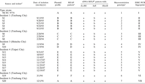
TABLE 1. Characteristics of S. sonnei isolates examined in the present study
Source and isolated Date of isolation (mo/day/yr)
Plasmid profile
REAP patterna
rDNA RFLPb
pattern with: Macrorestriction
genotypec ERIC-PCRfingerprint
EcoRI SalI HincII
Type strain
NCTC 9774 A A a a a 1 I
Incident 1 (Taichung City)
S1 9/13/93 B B a b a 2 II
S2 9/20/93 B B a b a 2 II
S3 9/21/93 B B a b a 2 II
S4 9/24/93 B B a b a 2 II
S5 9/15/93 B B a b a 2 II
Incident 2 (Taichung City)
S6 2/28/94 C C a b a 3 III
S7 2/28/94 C C a b a 3 III
S8 2/28/94 C C a b a 3 III
Incident 3 (Hsinchu City)
S9 11/9/94 D D a b a 4 IV
S10 11/9/94 D D a b a 4 IV
Incident 4 (Taipei City)
S11 9/21/87 E E a b a 5 V
S12 10/9/87 E E a b a 5 V
S13 11/4/87 E E a b a 5 V
S14 11/17/87 E E a b a 5 V
S15 11/17/87 E E a b a 5 V
S16 11/25/87 E E a b a 5 V
S17 11/26/87 E E a b a 5 V
S18 12/19/87 E E a b a 5 V
Incident 5 (Taichung City)
S19 3/1/94 F F a a a 6 VI
Incident 6 (Taichung City)
S20 12/1/91 A A a a a 7 VII
aDetermined by digestion with EcoRI.
bRFLP, restriction fragment length polymorphism. cDetermined by PFGE after digestion by XbaI.
dEach isolate was from the patient with the corresponding number.
on May 15, 2020 by guest
http://jcm.asm.org/
performed in a PHC-3 thermal cycler (Techne, Princeton, N.J.), with tempera-ture ramping as follows: 958C for 5 min to denature the template; four low-stringency cycles of 948C for 1 min, 268C for 1 min, and 728C for 2 min; 40 cycles of 948C for 30 s, 408C for 30 s, and 728C for 1 min; and finally, 728C for 10 min. Negative controls with no template DNA were included in each run. Amplified products (10ml) were resolved by agarose gel electrophoresis in 1.6% agarose gels in TBE buffer containing ethidium bromide (1mg/ml) at 30 V for 6 h and were visualized by UV transillumination. The PCR patterns were considered to be identical on the basis of similar numbers and matching positions of all major bands. Small differences in the intensities of faint bands were ignored.
RESULTS
The results with the different molecular typing techniques
are summarized in Table 1.
Plasmid profile analysis and REAP.
Some representative
profiles and patterns from REAP are illustrated in Fig. 1. The
clinical isolates and the type strain of the species exhibited six
distinct profiles of one to three small plasmids ranging from 5.0
to 10.1 kb. The plasmid profiles were identical within each
group of epidemiologically related isolates but were
distin-guishable from each other. Isolate S20 shared the same
plas-mid pattern with type strain NCTC 9774, and these two strains
also could not be differentiated by REAP with EcoRI (Table 1;
Fig. 1).
Ribotyping analysis.
For the 20 clinical isolates and the type
strain of the species, analysis of rRNA gene (rDNA) restriction
fragment length polymorphisms with EcoRI or HincII gave the
same single profile. With SalI, two distinct rDNA patterns
were observed (Fig. 2; Table 1).
PFGE of total genome DNA.
Some representative profiles
and patterns from PFGE are shown in Fig. 3. XbaI digestion
produced about 20 fragments; their sizes ranged from 32.4 to
582 kb. Seven distinct macrorestriction patterns were
gener-ated among the 20 clinical isolates and the type strain of the
species. The epidemiologically related isolates were found to
have identical PFGE patterns, and the pattern was distinct for
each incident of infection (Table 1).
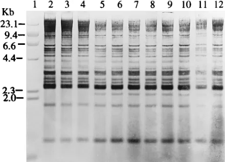
ERIC-PCR analysis.
The ERIC-PCR technique successfully
typed all isolates examined and produced bands in the 300- to
2,000-bp size range (Fig. 4). Seven different patterns were
observed among the 20 clinical isolates and the type strain of S.
sonnei (Table 1). The pattern was distinct for each incident of
infection.
DISCUSSION
Several molecular methods have been applied previously to
study the epidemiology of S. sonnei isolates. Plasmid profile
[image:3.612.62.298.71.166.2]analysis is the method used widely for the investigation of
outbreaks of shigellosis (8, 9, 19, 20, 21, 26). Previous reports
have shown that S. sonnei usually harbors a heterogenous
pop-ulation of plasmids, which may range in number from 2 to as
many as 10 (4). Tacket and Cohen (19) successfully used
plas-mid analysis to identify the epidemic strain which caused two
sequential outbreaks in two neighboring counties in Florida.
The plasmid profile of this outbreak strain was distinguished
from the profiles of three recent sporadic isolates from the
same place. In contrast, Prado et al. (14) performed plasmid
profile analysis on isolates of S. sonnei from Mexico and from
Houston, Tex., and found that the majority of strains had
identical or almost identical plasmid profile patterns. They
FIG. 1. Agarose gel electrophoresis of total plasmids and their EcoRI digests from S. sonnei isolates. Lane 1, supercoiled DNA size standard (GIBCO-BRL); lane 2, plasmid content of type strain NCTC 9774; lane 3, plasmid content of isolate S20; lane 4, plasmid content of isolate S19; lanes 5 to 7, plasmid contents of epidemiologically related isolates S6 to S8, respectively; lanes 8 to 12, plasmid contents of epidemiologically related isolates S1 to S5, respectively; lane 13,
HindIII restriction fragments of phage lambda; lanes 14 to 18, restriction
frag-ment patterns of EcoRI-digested total plasmid DNAs from type strain NCTC 9774 and isolates S20, S19, S6, and S1, respectively.
[image:3.612.323.543.73.232.2]FIG. 2. Ribotyping of S. sonnei (SalI digestion and Southern blotting). Lane 1, lambda DNA digested with HindIII; lane 2, DNA digest of type strain NCTC 9774; lane 3, DNA digest of isolate S20; lane 4, DNA digest of isolate S19; lanes 5 to 7, DNA digests of epidemiologically related isolates S6 to S8, respectively; lanes 8 to 12, DNA digests of epidemiologically related isolates S1 to S5, respec-tively.
FIG. 3. PFGE of XbaI-digested genomic DNAs from S. sonnei isolates. Lane 1, lambda ladder (Bio-Rad) which served as a molecular size marker; lane 2, DNA digest of type strain NCTC 9774; lane 3, DNA digest of isolate S20; lane 4, DNA digest of isolate S19; lanes 5 to 7, DNA digests of epidemiologically related isolates S6 to S8, respectively; lanes 8 to 12, DNA digests of epidemio-logically related isolates S1 to S5, respectively.
on May 15, 2020 by guest
http://jcm.asm.org/
[image:3.612.321.547.475.674.2]stated that S. sonnei with the same plasmid pattern can be
found within a wide geographic distribution, which will limit
the usefulness of the plasmid fingerprinting technique for
out-break investigation. A similar result was also found by
Yagupsky et al. (26). They suggested that REAP can
distin-guish more strains than the plasmid profile. In our study, all of
the epidemiologically unrelated strains except isolate S20 and
the type strain of the species were found to be different by both
plasmid profile analysis and REAP. Both of these techniques
are simple and easy to perform. However, they cannot be used
for isolates without plasmids. Instability of profiles or patterns
of digestion caused by the acquisition or loss of plasmids
rep-resents another disadvantage of these techniques.
Ribotyping has been applied for discriminating strains of S.
sonnei (5, 11). Hinojosa-Ahumada et al. (5) identified six
dis-tinct rDNA patterns by the use of SalI as the restriction
en-donuclease among 100 isolates from sporadic cases and 45
isolates from four different outbreaks of shigellosis which
oc-curred in the United States. Nastasi et al. (11) have tried
different enzymes for digestion of whole-cell DNA from S.
sonnei isolates, and they found that rDNA restriction patterns
generated by HincII provided the greatest strain
differentia-tion: 13 ribotypes among 432 isolates were identified after
digestion with this enzyme. However, 95% of the sporadic
strains were assigned to only four different ribotypes. Our
results showed the poor discriminatory power of ribotyping for
S. sonnei isolates. By using EcoRI, SalI, and HincII, we
iden-tified only two distinct ribotypes among 20 clinical isolates and
the type strain of the species, and this technique could not
discriminate the epidemiologically related isolates from the
epidemiologically unrelated strains. The fact that ribotyping is
both labor intensive and time-consuming further discourages
its use in clinical laboratories.
PFGE has been used successfully to identify genetic
sub-types among S. sonnei strains, and in one study, seven distinct
subtypes among nine epidemiologically unrelated strains were
identified by this technique (16). In addition, Brian et al. (1)
used PFGE to differentiate between outbreak-associated and
non-outbreak-associated isolates during an outbreak of
shigel-losis in a day-care center. In our present study, we showed the
usefulness of this technique for discrimination of
epidemiolog-ically related isolates from epidemiologepidemiolog-ically unrelated strains.
However, this technique is as labor intensive and
time-consum-ing as ribotyptime-consum-ing.
PCR-mediated genome fingerprinting based on ERIC has
been applied successfully to the epidemiologic typing of
me-thicillin-resistant Staphylococcus aureus (22), Acinetobacter
baumannii (17), and S. marcescens (10). A similar approach
was successfully used in the present study to characterize
epi-demiologically associated isolates of S. sonnei. To our
knowl-edge, this is the first report of the application of the PCR
technique for studying the epidemiology of S. sonnei. The
discriminatory power of ERIC-PCR was good. It provided a
result which agreed for all isolates examined to date with those
given by the more established PFGE technique. The
reproduc-ibility of this technique is good if the PCR protocol and
pa-rameters described above are strictly followed each time. In
comparison with ribotyping and PFGE, ERIC-PCR is
rela-tively easy to perform and less time-consuming. The results can
be obtained within 24 h. However, the intensities and the
patterns of the bands can be affected if there is any variation in
the parameters of PCR or conditions of gel running.
There-fore, it is much better to have all of the samples processed at
the same time and running on the same gel so that the banding
patterns of different isolates can be compared confidently.
Incident 4 comprised eight isolates that were collected from
Taipei City within a 3-month period. They were thought to be
temporally related sporadic isolates, but the frequency of
iso-lations was unusually high during this period. All of the isolates
showed identical plasmid profiles, rDNA restriction fragment
length polymorphism patterns, PFGE patterns, and
ERIC-PCR fingerprints. Brian et al. (1) also found that plasmid
analysis and PFGE sometimes could not differentiate
com-mon-source isolates from sporadic isolates in the same location
during the same period of time. It is possible that these
tem-porally related isolates belonged to a single bacterial clone and
circulated obscurely through the community.
In conclusion, we found that PFGE and ERIC-PCR have
the highest discriminatory power for differentiation of strains
of S. sonnei. ERIC-PCR fingerprinting is particularly useful
because of its simplicity and represents a less time-consuming
procedure. It provides a degree of discrimination equivalent to
that of PFGE but higher than those of plasmid profile analysis,
REAP, and ribotyping. However, the validation of the
discrim-inatory powers of REAP, ribotyping, PFGE, and ERIC-PCR
would be best established by analysis of strains which cannot be
differentiated by plasmid profiles, so future work on such a
comparison is necessary.
ACKNOWLEDGMENTS
We thank Meei-Fang Liu for her technical assistance. We also thank Hsin-Jyur Rehn, Meei-Rurng Liu, and Yu-Mei Huang for their help in outbreak investigation.
REFERENCES
1. Brian, M. J., R. Van, I. Townsend, B. E. Murray, T. G. Cleary, and L. K.
Pickering.1993. Evaluation of the molecular epidemiology of an outbreak of multiply resistant Shigella sonnei in a day-care center by using pulsed-field gel electrophoresis and plasmid DNA analysis. J. Clin. Microbiol. 31:128–133. 2. DuPont, H. L. 1990. Shigella species (bacillary dysentery), p. 1716–1722. In
G. L. Mandell, R. G. Douglas, Jr., and J. E. Bennett (ed.), Principles and practice of infectious diseases, 3rd ed. Churchill Livingstone Inc., New York. 3. Ellsworth, D. L., K. D. Rittenhouse, and R. L. Honeycutt. 1993. Artifactual variation in randomly amplified polymorphic DNA banding patterns. Bio-Techniques 14:214–217.
4. Haider, K., M. I. Huq, A. R. Samadi, and K. Ahmad. 1985. Plasmid charac-terization of Shigella species isolated from children with shigellosis and asymptomatic excretors. J. Antimicrob. Chemother. 16:691–698.
5. Hinojosa-Ahumada, M., B. Swaminathan, S. B. Hunter, D. N. Cameron, J. A.
Kiehlbauch, I. K. Wachsmuth, and N. A. Strockbine.1991. Restriction frag-ment length polymorphism in rRNA operons for subtyping Shigella sonnei. J. Clin. Microbiol. 29:2380–2384.
[image:4.612.64.288.74.217.2]6. Kado, C. I., and S. T. Liu. 1981. Rapid procedure for detection and isolation FIG. 4. ERIC-PCR products of S. sonnei analyzed by 1.6% agarose gel
elec-trophoresis. The primer was ERIC1 (see text). Lane 1, 1-kb molecular size marker (GIBCO-BRL); lane 2, negative control; lane 3, product of type strain NCTC 9774; lane 4, product of isolate S20; lane 5, product of isolate S19; lanes 6 to 8, products of epidemiologically related isolates S6 to S8, respectively; lanes 9 to 13, products of epidemiologically related isolates S1 to S5, respectively.
on May 15, 2020 by guest
http://jcm.asm.org/
of large and small plasmids. J. Bacteriol. 145:1365–1373.
7. Kaufmann, M. E., and T. L. Pitt. 1994. Pulsed-field gel electrophoresis of bacterial DNA, p. 83–92. In H. Chart (ed.), Methods in practical laboratory bacteriology. CRC Press, Inc., Boca Raton, Fla.
8. Litwin, C. M., K. J. Ryan, S. Chipowsky, A. Storm, and S. McCombie. 1990. Molecular epidemiology of Shigella sonnei in Pima County, Arizona: evi-dence for a Mexico-related plasmid. J. Infect. Dis. 161:797–800.
9. Litwin, C. M., A. L. Storm, S. Chipowsky, and K. J. Ryan. 1991. Molecular epidemiology of Shigella infections: plasmid profiles, serotype correlation, and restriction endonuclease analysis. J. Clin. Microbiol. 29:104–108. 10. Liu, P. Y. F., Y. J. Lau, B. S. Hu, et al. 1994. Use of PCR to study
epide-miology of Serratia marcescens isolates in nosocomial infection. J. Clin. Microbiol. 32:1935–1938.
11. Nastasi, A., S. Pignato, C. Mammina, and G. Giammanco. 1993. rRNA gene restriction patterns and biotypes of Shigella sonnei. Epidemiol. Infect. 110: 23–30.
12. Pitcher, D. G., R. J. Owen, P. Dyal, and A. Beck. 1987. Synthesis of a biotinylated DNA probe to detect ribosomal RNA cistrons in Providencia
stuartii. FEMS Microbiol. Lett. 48:283–287.
13. Pitcher, D. G., N. A. Saunders, and R. J. Owen. 1989. Rapid extraction of bacterial genomic DNA with guanidium thiocyanate. Lett. Appl. Microbiol.
8:151–156.
14. Prado, D., B. E. Murray, T. G. Cleary, L. K. Pickering. 1987. Limitations of using the plasmid pattern as an epidemiological tool for clinical isolates of
Shigella sonnei. J. Infect. Dis. 155:314–316.
15. Pruneda, R. C., and J. J. Farmer III. 1977. Bacteriophage typing of Shigella
sonnei. J. Clin. Microbiol. 5:66–74.
16. Soldati, L., and J. C. Piffaretti. 1991. Molecular typing of Shigella strains using pulsed field gel electrophoresis and genome hybridization with inser-tion sequences. Res. Microbiol. 142:489–498.
17. Struelens, M. J., E. Carlier, N. Maes, E. Serruys, W. G. V. Quint, and A. van
Belkum.1993. Nosocomial colonization and infection with multiresistant
Acinetobacter baumannii: outbreak delineation using DNA macrorestriction
analysis and PCR-fingerprinting. J. Hosp. Infect. 25:15–32.
18. Szturm-Rubinsten, S. 1968. Determination of biotype, phage type and co-licinogenic character of Shigella sonnei, and its epidemiologic importance. Arch. Immunol. Ther. Exp. 16:421–428.
19. Tacket, C. O., and M. L. Cohen. 1983. Shigellosis in day care centers: use of plasmid analysis to assess control measures. Peadiatr. Infect. Dis. J. 2:127– 130.
20. Tacket, C. O., N. Shaid, M. I. Huq, A. R. M. Alim, and M. L. Cohen. 1984. Usefulness of plasmid profiles for differentiation of Shigella isolates in Ban-gladesh. J. Clin. Microbiol. 20:300–301.
21. Tietze, E., H. Tschape, G. Horn, and F. Laue. 1984. Clonal distribution of multiple-drug-resistant Shigella sonnei strains: identification by means of plasmid pattern analysis. Ann. Microbiol. (Paris) 135b:155–164.
22. van Belkum, A., R. Bax, P. Peerbooms, W. H. F. Goessens, N. van Leeuwen,
and W. G. V. Quint.1993. Comparison of phage typing and DNA finger-printing by polymerase chain reaction for discrimination of methicillin-re-sistant Staphylococcus aureus strains. J. Clin. Microbiol. 31:798–803. 23. Versalovic, J., T. Koeuth, and J. R. Lupski. 1991. Distribution of repetitive
DNA sequences in eubacteria and application to fingerprinting of bacterial genomes. Nucleic Acids Res. 19:6823–6831.
24. Vlajinac, H., and S. Krajinovic. 1983. Colicine production as an epidemio-logical marker for Shigella sonnei. J. Hyg. 91:273–276.
25. Welsh, J., and M. McClelland. 1990. Fingerprinting genomes using PCR with arbitrary primers. Nucleic Acids Res. 18:7213–7218.
26. Yagupsky, P., M. Loeffelholz, K. Bell, and M. A. Menegus. 1991. Use of multiple markers for investigation of an epidemic of Shigella sonnei infec-tions in Monroe County, New York. J. Clin. Microbiol. 29:2850–2855.