Copyright © 1998, American Society for Microbiology. All Rights Reserved.
Typing of Dengue Viruses in Clinical Specimens and Mosquitoes by
Single-Tube Multiplex Reverse Transcriptase PCR
EVA HARRIS,
1* T. GUY ROBERTS,
1LEILA SMITH,
1JOHN SELLE,
1LAURA D. KRAMER,
2SONIA VALLE,
3ERICK SANDOVAL,
3ANDANGEL BALMASEDA
3Program in Molecular Pathogenesis, University of California, San Francisco, San Francisco, California 94143-0422
1;
Center for Vector-Borne Disease Research, University of California, Davis, Davis, California 95616
2; and
Centro Nacional de Diagno´stico y Referencia, Ministerio de Salud, Managua, Nicaragua
3Received 17 February 1998/Returned for modification 15 May 1998/Accepted 8 June 1998
In recent years, dengue viruses (serotypes 1 to 4) have spread throughout tropical regions worldwide. In
many places, multiple dengue virus serotypes are circulating concurrently, which may increase the risk for the
more severe form of the disease, dengue hemorrhagic fever. For the control and prevention of dengue fever, it
is important to rapidly detect and type the virus in clinical samples and mosquitoes. Assays based on reverse
transcriptase (RT) PCR (RT-PCR) amplification of dengue viral RNA can offer a rapid, sensitive, and specific
approach to the typing of dengue viruses. We have reduced a two-step nested RT-PCR protocol to a single-tube
reaction with sensitivity equivalent to that of the two-step protocol (1 to 50 PFU) in order to maximize
simplicity and minimize the risk of sample cross-contamination. This assay was also optimized for use with a
thermostable RT-polymerase. We designed a plasmid-based internal control that produces a uniquely sized
product and can be used to control for both reverse transcription or amplification steps without the risk of
generating false-positive results. This single-tube RT-PCR procedure was used to type dengue viruses during
the 1995 and 1997-1998 outbreaks in Nicaragua. In addition, an extraction procedure that permits the sensitive
detection of viral RNA in pools of up to 50 mosquitoes without PCR inhibition or RNA degradation was
developed. This assay should serve as a practical tool for use in countries where dengue fever is endemic, in
conjunction with classical methods for surveillance and epidemiology of dengue viruses.
Over the last 20 years, classic dengue fever and the more
se-vere form, dengue hemorrhagic fever-dengue shock syndrome
(DHF-DSS), have emerged as the most important
arthropod-borne viral diseases in humans (22). During this period,
den-gue fever has spread throughout tropical regions worldwide,
principally in urban settings. Up to 100 million cases of classic
dengue fever are estimated annually, and roughly 450,000 cases
of DHF-DSS are reported annually, while approximately 2.5
billion people live in areas at risk for epidemic dengue virus
transmission (9, 22). The dramatic spread of epidemic
den-gue fever and the emergence of DHF-DSS occurred after
World War II in Southeast Asia, where DHF is now one of the
leading causes of hospitalization and death. This pattern of
epidemic dengue fever and emerging DHF is being repeated in
Latin America (10), where it is spreading throughout the
re-gion at an alarming rate.
Dengue fever is caused by four distinct serotypes of dengue
virus, which are transmitted to humans by the domestic
mos-quitoes Aedes aegypti and Aedes albopictus (22). The lack of a
vaccine or a cure for dengue fever make the development of
laboratory-based surveillance systems all the more important
to provide an early warning of dengue fever epidemics and to
furnish information for effective vector control measures (9). It
is crucial to determine which serotypes of dengue virus are
circulating where and when since previous infection with one
of the four dengue serotypes can be an important risk factor
for developing DHF-DSS upon infection with a heterotypic
serotype (11, 23). The current “gold standard” for typing
den-gue virus involves isolation of the virus in cultured cells or
mosquitoes followed by indirect immunofluorescence.
How-ever, this requires cell culture facilities or mosquito colonies,
which are difficult to maintain in laboratories in developing
countries. The most rapid serological techniques, such as
im-munoglobulin M enzyme-linked immunosorbent assay with a
single serum sample, do not furnish information about the
se-rotype of the virus. The plaque reduction neutralization
tech-nique allows typing but is costly and difficult to perform.
Single-step reverse transcriptase (RT) PCR (RT-PCR)
de-tection and typing of dengue virus offers a sensitive, specific,
and rapid alternative that requires only one acute-phase serum
sample. This technique can be made cost-effective by following
a low-cost methodology (12–14). Early detection of dengue
virus in patient serum allows the possibility of mounting a rapid
response aimed at vector control in the affected areas. This
assay is also useful for typing the virus and providing important
information for epidemiological studies. In addition, rapid
as-says for the detection of dengue virus in mosquitoes are useful
for investigation of the virus and its vector in nature (6).
Re-cently, a number of molecular approaches to the detection and
characterization of dengue viral RNA have been described (8,
15, 18, 20, 24, 26, 28, 30, 34). Here we present a modified
RT-PCR assay for the single-step detection and typing of dengue
virus in clinical specimens and mosquitoes. This assay has been
simplified for use in countries where dengue fever is endemic.
MATERIALS AND METHODS
Virus strains and specimens.Virus stocks were kindly provided by the Centers
for Disease Control and Prevention (CDC) (serotype 1 dengue virus [dengue-1; strain Hawaii], serotype 2 dengue virus [dengue-2; strain 16681], serotype 3 dengue virus [dengue-3; strain H-87], and serotype 4 dengue virus [dengue-4; strain 703-4]) or were isolated in Nicaragua during the 1995 and 1997-1998 outbreaks. Human serum samples were obtained from patients clinically sus-pected of having dengue fever within 0 to 4 days from the time of onset of
* Corresponding author. Present address: Division of Public Health
Biology and Epidemiology, School of Public Health, University of
California, Berkeley, 140 Warren Hall, Berkeley, CA 94720. Phone:
(510) 643-9773. Fax: (510) 642-6350. E-mail: [email protected]
.edu.
2634
on May 15, 2020 by guest
http://jcm.asm.org/
symptoms. The samples had been collected for routine diagnosis of dengue fever at the Centro Nacional de Diagno´stico y Referencia in Managua, Nicaragua, in June and July 1995 and December 1997 to May 1998.
Mosquito inoculation.Mosquitoes (A. aegypti Rock) were inoculated with
approximately 103PFU of dengue-2 (strain 16681) by the method of Rosen and
Gubler (29). Adults were maintained at 27°C and were provided with 10% sucrose as nourishment. Live mosquitoes were frozen at270°C on days 1, 2, 3, 4, 6, 7, and 21. A group of mosquitoes that had died 3 days postinoculation were frozen on day 5. Uninfected A. aegypti were reared in the laboratory in Nicaragua from eggs laid by field-collected females.
Viral growth.A. albopictus C6/36 cells (16) were grown in minimal essential
medium (MEM) (Gibco BRL, Grand Island, N.Y.) containing Earle’s salts,
L-glutamine, and nonessential amino acids and supplemented with 0.11% sodium bicarbonate, 100 U of penicillin per ml, 75 U of streptomycin per ml, and 10% fetal bovine serum (FBS) (Gemini Bioproducts, Inc., Calabasas, Calif.). Baby hamster kidney cells (BHK21-15) (19) were grown in MEM as described above, except that 0.124% sodium bicarbonate and 5% FBS were used. Viruses were propagated in C6/36 cells, and after incubation at 28°C for 7 days, the cellular supernatant was clarified by centrifugation, supplemented with 20% FBS, and stored at270°C until use. For plaque assays, monolayers of BHK21-15 cells were grown to 90 to 95% confluence in six-well plates and were inoculated in duplicate with 200ml of cellular supernatant containing serial dilutions of the viral stock. After 2 h at 37°C, the cells were overlaid with MEM containing 1% SeaPlaque agarose (FMC BioProducts, Rockland, Maine) and 5% FBS and were incubated for 7 days at 37°C in 5% CO2. The cells were then fixed in 10% formadehyde for
2 h and stained with a solution containing 0.27% crystal violet. Virus isolation was performed by inoculating C6/36 cells with a 5- to 20-fold dilution of a serum specimen. After 7 days, isolated virus was serotyped with monoclonal antibodies and by RT-PCR.
RNA extraction.RNA was extracted from serum or the supernatant of
in-fected cells by combining 300ml of the sample sequentially with 300ml of lysis buffer (6 M guanidine isothiocyanate, 50 mM sodium citrate, 1% Sarkosyl, 20mg of Escherichia coli tRNA per ml, 100 mMb-mercaptoethanol), 60ml of 2 M sodium acetate (pH 4.0), 600 ml of water-saturated phenol, and 240 ml of chloroform and mixing after the addition of each of the reagents. After a 15-min centrifugation, the aqueous phase was transferred to a new tube and was mixed with an equal volume of isopropanol. Following a 20-min centrifugation at 4°C, the supernatant was removed and the pellet was washed in 75% ethanol, air dried, and resuspended in 25ml of RNase-free sterile distilled water.
RNA was extracted from pools of infected or uninfected mosquitoes macer-ated in 100ml of phosphate-buffered saline. Prior to maceration, pools of unin-fected mosquitoes were spiked with exogenous viral particles. The macerates were clarified by centrifugation, mixed with 100ml of lysis buffer (see above), and extracted with a 1:1 mixture of phenol and chloroform. Five microliters of acid-washed size-selected silica particles (13, 33) were added to each sample, and the mixture was incubated for 5 min, pelleted by centrifugation, and washed twice (50% ethanol, 10 mM Tris [pH 7.4], 1 mM EDTA, 50 mM NaCl). After resuspension in 10ml of RNase-free distilled water, the samples were incubated for 5 min at 50 to 55°C and centrifuged. The eluate supernatant was transferred to a new tube, and the pellet was resuspended in 5ml of water immediately prior to amplification. Alternatively, the washed pellet can be resuspended in 15ml of water and used directly for amplification.
Reverse transcription and PCR amplification. (i) Two-enzyme RT-PCR.The
reaction mixture contained 50 mM KCl, 10 mM Tris (pH 8.5), 0.1% Triton X-100, 0.01% gelatin, each of the four deoxynucleotide triphosphates at a con-centration of 200mM, 1.5 mM MgCl2, 30 mM tetramethylammonium chloride
(5), 0.5 M betaine (25), 5 mM dithiothreitol, 59primer D1 and 39primer TS1 at a concentration of 1mM each, 39primers TS2, TS3, and DEN4 at a concentration of 0.5mM each, 0.0017 to 0.025 U of RAV-2 RT (Amersham Corp., Arlington Heights, Ill.) perml, and 0.025 U of Taq DNA polymerase (Taq DNA polymerase [Promega Corp., Madison, Wis.]; AmpliTaq [Perkin-Elmer Corp., Foster City, Calif.]) perml. Reverse transcription was conducted at 42°C for 60 min, followed by 40 amplification cycles of 94°C for 30 s, 55°C for 1 min, and 72°C for 2 min, with a final extension at 72°C for 5 min. A total of 2.5 to 5ml of extracted RNA was used as a template in a 25-ml reaction volume. Amplification was conducted in 0.6-ml tubes (Robbins Scientific Corp., Sunnyvale, Calif.) with a model 480 thermal cycler (Perkin-Elmer, Norwalk, Conn.) or a PTC-200-60 thermocycler (MJ Research, Inc., Watertown, Mass.).
(ii) Single-enzyme (rTth) RT-PCR.The reaction mixture for rTth RT-PCR
contained 115 mM potassium acetate, 8% glycerol, 50 mM bicine (pH 8.2), the four deoxynucleotide triphosphates at a concentration of 200mM each, 2 mM manganese acetate, primers D1 and TS1 at a concentration of 0.5mM each, primers TS2, TS3, and DEN4 at a concentration of 0.25mM each, and 0.05 U of rTth DNA polymerase (Perkin-Elmer Corp.) perml. One cycle of 60°C for 30 min for the reverse transcription was followed by a 2-min incubation at 94°C and 40 cycles of 94°C for 45 s, 50°C for 1 min, and 60°C for 1 min, with a final extension at 60°C for 7 min.
Primer sequences are as follows: D1, 59-TCA ATA TGC TGA AAC GCG CGA GAA ACC G; TS1, 59-CGT CTC AGT GAT CCG GGG G; TS2, 59-CGC CAC AAG GGC CAT GAA CAG; TS3, 59-TAA CAT CAT CAT GAG ACA GAG C (18); and DEN4, 59-TGT TGT CTT AAA CAA GAG AGG TC. The expected sizes of the amplification products are 482 bp (dengue-1), 119 bp
(dengue-2), 290 bp (dengue-3), and 389 bp (dengue-4). Ten microliters of the 25-ml reaction mixtures was electrophoresed on 1.5% agarose gels in 13TBE (89 mM Tris borate, 2 mM EDTA [pH 8.3]) with a 100-bp ladder as a size standard (100, 200, 300, 400, 500, 600, 700, 800, 900, 1,000, 1,100, 1,200, 1,300, 1,400, 1,500, and 2,072 bp; Gibco BRL) or Amplisize DNA size standards (50, 100, 200, 300, 400, 500, 700, 1,000, 1,500, and 2,000 bp; Bio-Rad Laboratories, Richmond, Calif.).
Plasmid construction.To clone the dengue virus amplicons, dengue viral RNA
was amplified with 59and 39primers containing the restriction sites EcoRI and
BamHI, respectively, at their 59ends. The products were treated with proteinase K, digested with the appropriate restriction enzymes, gel purified, and ligated with pBluescript (KS II; Stratagene Cloning Systems, La Jolla, Calif.) that had been digested with EcoRI and BamHI and gel purified. To construct a control plasmid with a dengue-3 fragment;50 bp larger than the original amplicon, a 54-bp fragment was amplified from Leishmania braziliensis with primers 13A and MP3H (1), treated with proteinase K (Promega Corp.), and then incubated with T4 DNA polymerase (New England Biolabs, Inc., Beverly, Mass.) and the four nucleotides. The plasmid containing the dengue-3 insert (pDB3) was linearized within the insert by digestion with StyI, and the overhanging ends were filled in by incubation with T4 DNA polymerase and the four nucleotides. After treat-ment of the vector with calf intestinal alkaline phosphatase (New England Bio-labs, Inc.), the 54-bp L. braziliensis fragment was ligated into pBD3 to create pBD3L.
For in vitro transcription of the 350-bp dengue-3 fragment containing the insert, pBD3L was linearized with XbaI immediately downstream of the den-gue-3 insert and was incubated with T3 RNA polymerase (Promega Corp.) and the four nucleotides for 60 min at 37°C. To remove the plasmid DNA from the in vitro transcription reaction, the reaction mixture was treated with RQ1 DNase (Promega Corp.) in the presence of 10 mM calcium chloride for 60 min at 37°C. To confirm that only RNA remained, an aliquot was treated with RNase for 30 min at 37°C.
RESULTS
Single-tube RT-PCR assay.
A one-tube RT-PCR protocol
was developed to reverse transcribe dengue viral RNA and
amplify four differently sized type-specific products. This
pro-tocol is an adaptation of a two-step nested RT-PCR assay
described by Lanciotti et al. (18). Five oligonucleotide primers
are included in the one-step assay: one 5
9
primer that targets a
region of the capsid gene conserved in all four dengue virus
serotypes and four 3
9
primers, each of which is complementary
to sequences unique to each serotype. These primers are
po-sitioned such that a differently sized product is generated from
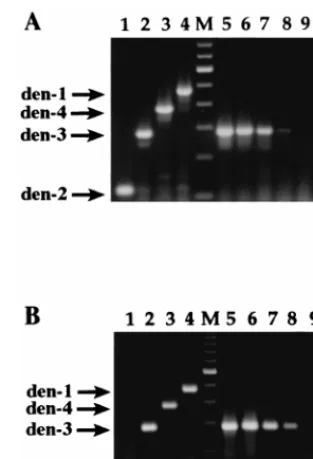
each type, as shown in Fig. 1A, lanes 1 to 4 (dengue-2, 119 bp;
dengue-3, 290 bp; dengue-4, 389 bp; dengue-1, 482 bp). Several
modifications were made to the original protocol. The
dengue-4-specific primer was redesigned to avoid hairpin formation so
as to increase the yield of the dengue-4 product. Cosolvents,
such as tetramethylammonium chloride (5) and betaine (25),
were included in the reaction mixture to improve the sensitivity
of the assay. The concentrations of the different primers were
adjusted to optimize the amplification of all four products.
To test the sensitivity of the single-tube RT-PCR assay,
plaque titration and RNA extractions were performed
simul-taneously with each dilution of a serial titration of viral stocks.
One-tenth of the extracted RNA was amplified by RT-PCR.
Plaque formation has commonly been used as a measure of the
sensitivity of dengue viral RT-PCR assays (4, 24, 26, 28, 30),
although it is not a direct indication of the number of viral
particles. An example of the sensitivity of this assay is shown in
Fig. 1A, lanes 6 to 9, where serial 10-fold dilutions of a
den-gue-3 stock were extracted, reverse transcribed, and amplified.
The limit of detection was approximately 1 PFU for dengue-1,
50 PFU for dengue-2, 1 PFU for dengue-3, and 30 PFU for
dengue-4. Side-by-side comparisons of the one-tube method
and the original two-step protocol revealed that the two
pro-cedures had similar sensitivities (data not shown).
The single-tube RT-PCR assay was adapted for use with the
thermostable RT-polymerase rTth (Fig. 1B, lanes 1 to 4),
ob-viating the need for two separate enzymes. Optimal results
were obtained with one-half of the amount of rTth
on May 15, 2020 by guest
http://jcm.asm.org/
mended by the manufacturer. The primer concentrations in
this assay were reduced by 50% compared to the
concentra-tions used in the two-enzyme assay described above, and no
cosolvents were necessary. The sensitivity of the rTth assay was
similar to that of the two-enzyme protocol (Fig. 1B and data
not shown).
Internal control plasmid.
As a positive control for the
RT-PCR assay, a plasmid containing a uniquely sized dengue-3
fragment was constructed. When amplified, this fragment can
be differentiated from the authentic viral amplification
prod-uct. The 290-bp dengue-3 product was cloned into pBluescript
(pBD3), and a 54-bp fragment was inserted to create a 350-bp
PCR substrate (pBD3L). Using this plasmid, an in vitro RNA
transcript can be generated for use as a positive control for
both the reverse transcription and amplification steps.
Alter-natively, the plasmid itself can be used as a positive control for
the amplification step only. Figure 2 (lanes 1 and 2) shows the
size difference between the original dengue-3 fragment and
the fragment containing the insert, excised from pBD3 and
pBD3L, respectively. The amplification products derived
di-rectly from plasmids pBD3 and pBD3L (lanes 3 and 4,
respec-tively) and from the DNase-treated in vitro transcript of
pBD3L (lane 5) are of the expected sizes. The last three lanes
demonstrate that the in vitro transcript of pBD3L (lane 6) is
sensitive to RNase (lane 8) but not DNase (lane 7).
RT-PCR detection and typing of dengue virus in clinical
specimens.
During outbreaks of dengue fever in Nicaragua in
1995 and 1997-1998, serum specimens referred to the National
Virology Laboratory at the Centro Nacional de Diagno´stico y
Referencia, Ministry of Health, were analyzed by the
two-en-zyme RT-PCR assay. Viral RNA was extracted in duplicate
directly from patient serum and was amplified in duplicate on
different days to minimize the risk of artifactual results. Figure
3 shows representative results obtained for specimens collected
in June and July 1995. Specimens were obtained from patients
in Bluefields, on the Atlantic coast of Nicaragua (Fig. 3A, lanes
2 to 8, 11, and 12, and Fig. 3B, lanes 1 to 7), and Chontales, in
central Nicaragua (Fig. 3B, lane 12). The results for positive
(Fig. 3A, lanes 9 and 10; Fig. 3B, lanes 8 to 11) and negative
(Fig. 3A, lane 1) controls were as expected. Duplicate aliquots
of each specimen yielded consistent and reproducible results.
Figure 3A demonstrates that two dengue virus serotypes (e.g.,
lane 4, dengue-3; lane 8, dengue-2) were circulating
simulta-neously in the same geographical area. Figure 3B shows that
dengue-3 was circulating in two different regions of the country
(lanes 3 and 5, Bluefields; lane 12, Chontales). This assay has
been used for routine epidemiological surveillance in
Nicara-gua since 1995 and has been implemented by collaborators at
the Centro Nacional de Enfermedades Tropicales in Santa
Cruz, Bolivia (27). The RT-PCR assay with rTth has also been
successfully used to analyze RNA extracted from clinical
spec-imens (data not shown).
[image:3.612.90.247.70.300.2]Detection of dengue virus in mosquitoes.
To prepare
mos-quito samples for RT-PCR amplification, a procedure for the
[image:3.612.309.546.70.209.2]FIG. 1. Detection and typing of dengue virus by using two versions of the RT-PCR assay. (A) Reverse transcription with RAV-2 RT and amplification with Taq DNA polymerase; (B) reverse transcription and amplification with the bifunctional enzyme rTth. (A and B) Lanes 1, dengue-2 (2); lanes 2, den-gue-3 (den-3); lanes 3, dengue-4 (den-4); lanes 4, dengue-1 (den-1); lanes M, 100-bp ladder (lowest band shown, 100 bp); lanes 5 to 8, dengue-3 at 1,000, 100, 10, and 1 PFU, respectively. (A) Lane 9, 0 pfu; lane 10, water (negative control). (B) Lane 9, water. Expected product sizes are as follows: dengue-2, 119 bp; dengue-3, 290 bp; dengue-4, 389 bp; dengue-1, 482 bp.
FIG. 2. Uniquely sized internal control. Lanes 1 and 2, authentic dengue-3 amplicon (den-3) and dengue-3 amplicon containing the 54-bp insert (den-3L), respectively, excised from pBD3 and pBD3L with EcoRI and BamHI; lanes 3 to 5, RT-PCR products derived from pBD3, pBD3L, and the DNase-treated in vitro transcript of pBD3L, respectively; lane M, 100-bp ladder (lowest band shown, 100 bp); lane 6, in vitro transcript of pBD3L; lane 7, DNase-treated in vitro transcript of pBD3L; lane 8, RNase-treated in vitro transcript of pBD3L. Expected product sizes are as follows: dengue-3, 290 bp; dengue-3L, 350 bp.
FIG. 3. RT-PCR detection and typing of dengue virus in serum from patients infected during the 1995 epidemic in Nicaragua. RNA was extracted from serum samples and was amplified by the two-enzyme single-tube RT-PCR assay as described in Materials and Methods. (A) Lane 1, negative control (water); lanes 2 to 8, 11, and 12, samples from patients from the Atlantic coast of Nicaragua (Bluefields); lane M, Amplisize DNA size standards (lowest band shown, 100 bp); lane 9, dengue-2 (den-2) RNA (positive control); lane 10, dengue-3 (den-3) RNA (positive control). (B) Lanes 1 to 7, samples from patients from the Atlantic coast of Nicaragua (Bluefields); lane M, Amplisize DNA size standards (lowest band shown, 100 bp); lane 8, dengue-2 (den-2) RNA (positive control); lane 9, dengue-3 (den-3) RNA (positive control); lane 10, dengue-4 (den-4) RNA (positive control); lane 11, dengue-1 (den-1) RNA (positive control); lane 12, sample from a patient from central Nicaragua (Chontales). Expected prod-ucts sizes are as follows: dengue-2, 119 bp; dengue-3, 290 bp; dengue-4, 389 bp; dengue-1, 482 bp.
on May 15, 2020 by guest
http://jcm.asm.org/
[image:3.612.94.247.577.658.2]extraction of viral RNA from pools of mosquitoes without
degradation of the RNA or inhibition of the PCR amplification
was required. With a combination of a guanidine-based lysis
buffer, organic solvents, and silica particles, a protocol that
allows the reproducible isolation of very small quantities of
viral RNA in the presence of up to 50 A. aegypti mosquitoes,
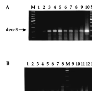
without RT-PCR inhibitors, was developed. Pools of
unin-fected mosquitoes were spiked with exogenous viral particles,
and extracts containing RNA from
,
100 PFU of dengue-3
were amplified (Fig. 4A). Eluates from silica particles and the
silica pellets themselves functioned equally well. RT-PCR
am-plification of RNA extracted from laboratory-infected
mosqui-toes demonstrated that dengue viral RNA could be detected in
a single mosquito at as early as 1 day postinoculation (Fig. 4B,
lane 1), and at as late as 21 days postinoculation, our last time
point for detection (lane 8). Pools of five mosquitoes yielded
the expected positive results (lanes 9 and 10) as well. Dengue
viral RNA was also amplified from infected mosquitoes frozen
2 days after natural death (Fig. 4B, lane 11). The results for
negative (Fig. 4A, lane 1; Fig. 4B lanes 1 and 13) and positive
(Fig. 4A, lane 10; Fig. 4B, lane 12) controls were as expected.
Extracts could be stored frozen for at least 6 months without
RNase inhibitors with no detectable loss of RNA integrity.
DISCUSSION
We have adapted a rapid RT-PCR assay for the typing of
dengue virus in patient serum and mosquitoes for use under
the difficult conditions often prevailing in countries where
den-gue fever is endemic. In general, PCR-based techniques can be
more rapid, sensitive, and specific than alternative techniques
when they are made accessible by a low-cost methodology that
involves the in-house preparation of reagents and materials,
recycling, simplification of procedures, and strict enforcement
of simple but effective procedures for minimizing DNA
con-tamination (12–14). Molecular techniques are particularly
use-ful for the detection and typing of dengue virus, and several
RT-PCR protocols have been described. Identification of the
four serotypes can be achieved by (i) nested amplification of a
primary product generated with universal dengue virus primers
(18, 20, 34), (ii) hybridization of a universal RT-PCR product
with type-specific probes (8, 15, 28), (iii) simultaneous
ampli-fication with four sets of type-specific primers (24), or (iv) use
of a single 5
9
universal primer and four type-specific 3
9
primers
(18, 30). However, the performance of multistep nested
am-plification increases the risk of cross-contamination, especially
during routine analyses, while hybridization procedures entail
additional cost and can be compromised by low water quality.
Therefore, we have simplified the reverse transcription and
amplification procedures and minimized the number of
prim-ers required for the detection and typing of dengue viruses in
a single tube.
The single-tube multiplex assay described herein was
adapt-ed from a previously reportadapt-ed nestadapt-ed RT-PCR protocol (18).
The conditions of sample extraction and amplification were
modified such that the sensitivity of the single-step assay was
comparable to that of the original nested protocol and to those
of other RT-PCR assays for dengue virus detection (18, 24,
30). For instance, one of the primers was redesigned to
im-prove the efficiency of amplification, and cosolvents were
in-cluded in the reaction mixture to optimize the sensitivity. In
addition, this RT-PCR assay was adapted for use with the
bifunctional RT-polymerase rTth (Perkin-Elmer Corp.). This
thermostable enzyme is easier to transport and store
in-coun-try than other RTs, such as RAV-2 (Amersham Corp.), which
are extremely labile. We constructed an internal amplification
control that generates a uniquely sized product and eliminates
the risk of false-positive results due to cross-contamination.
This control has been useful for on-site troubleshooting of
both the reverse transcription and the amplification processes.
The utility of this single-step assay has been demonstrated in
laboratories in developing countries, including Nicaragua (Fig.
3), Bolivia (27), Ecuador (21), and Guatemala (31). Prior to
the introduction of the simplified dengue virus RT-PCR assay,
the National Virology Laboratory at the Nicaraguan Ministry
of Health had been unable to type circulating dengue viruses
due to the lack of the cell culture facilities or mosquito colonies
necessary for classical viral isolation procedures. Only a small
percentage of serum samples had been sent to laboratories
outside the country for typing, a costly and lengthy process.
Serotype information is particularly important, since all four
dengue virus serotypes have been reported recently in
Nicara-gua (2, 17). Since 1995, RT-PCR has been used for
epidemi-ological surveillance of specimens from selected patients
sus-pected of dengue virus infection and for rapid diagnosis for
particular patients, while routine diagnosis is still performed by
the immunoglobulin M enzyme-linked immunosorbent assay.
Samples positive by RT-PCR are subsequently processed for
viral isolation in order to obtain viral stocks for future analysis,
now that the necessary facilities are available. We have found
that the virus in specimens that are received by the National
Virology Laboratory from regional health centers in
subopti-mal conditions for culture nonetheless can be successfully
am-plified, suggesting that although virus infectivity is
compro-mised, viral RNA can still be detected.
FIG. 4. (A) Detection of dengue virus in pools of mosquitoes by RT-PCR. A total of 350 PFU of dengue-3 (den-3) was added to pools of mosquitoes, which were then macerated. RNA was extracted as described in Materials and Meth-ods, and one-fourth of the extract was amplified by RT-PCR (RAV-2 RT–Taq polymerase). Lanes 2, 4, 6, and 8, silica particle eluate; lanes 3, 5, 7, and 9, pellet. Lane 1, water (negative control); lanes 2 and 3, 0 mosquitoes; lanes 4 and 5, 5 mosquitoes; lanes 6 and 7, 25 mosquitoes; lanes 8 and 9, 50 mosquitoes; lane 10, dengue-2 RNA (positive control); lane M, 100-bp ladder (lowest band shown, 200 bp). (B) Detection of dengue virus in laboratory-infected mosquitoes. Mos-quitoes were inoculated with dengue-2 (den-2; strain 16681) and were frozen at
270°C on the indicated days postinoculation. RNA was extracted and amplified by RT-PCR (RAV-2 RT–Taq polymerase). Lane 1, 5 uninfected mosquitoes; lane 2, one mosquito, one day postinoculation; lane 3, one mosquito, 2 days postinoculation; lane 4, one mosquito, 3 days postinoculation; lane 5, one mos-quito, 4 days postinoculation; lane 6, one mosmos-quito, 6 days postinoculation; lane 7, one mosquito, 7 days postinoculation; lane 8, one mosquito, 21 days postin-oculation; lane M, 100-bp ladder (lowest band shown, 100 bp); lane 9, five mosquitoes, 2 days postinoculation; lane 10, five mosquitoes, 7 days postinocu-lation; lane 11, five mosquitoes frozen 2 days after natural death; lane 12, dengue-2 RNA (positive control); lane 13, water (negative control).
on May 15, 2020 by guest
http://jcm.asm.org/
[image:4.612.76.262.65.248.2]This RT-PCR technique was also used by scientists at the
Nicaraguan Ministry of Health in October 1995 to investigate
an outbreak of hemorrhagic fever in northern Nicaragua which
was initially thought to be caused by dengue virus. When these
scientists demonstrated, using RT-PCR and other serological,
virological, and entomological methods, that dengue virus was
in fact not the cause, international interest was generated. Teams
of scientists from CDC and the Instituto de Medicina Tropical
“Pedro Kouri,” Havana, Cuba, collaborated with Nicaraguan
investigators, leading to the discovery that the etiological agent
was in fact Leptospira (3). In August 1997, Bolivian scientists at
the Centro Nacional de Enfermedades Tropicales used this
RT-PCR assay to type dengue virus in Bolivia for the first time
and to identify dengue-2 as the serotype responsible for the
1997 epidemic in Santa Cruz, corroborating the results
report-ed by CDC (27). Thus, in-country access to simplifireport-ed
PCR-based techniques is useful for immediate public health
pur-poses as well as for long-term epidemiological studies.
An RNA isolation method was required for the detection of
dengue virus in infected mosquitoes without RNA degradation
or PCR inhibition. The reported methods for the isolation
of RNA from mosquitoes require the use of costly reagents to
avoid inhibition (18), do not remove nucleases (7, 32), or
con-sume the entire extraction in a single amplification reaction
(7). Therefore, an extraction procedure that uses chaotropic
agents and organic extraction to remove nucleases and that
includes additional steps to remove substances potentially
in-hibitory to the amplification was developed. The resulting
ex-tracts are stable over time without the addition of expensive
nuclease inhibitors. RT-PCR conducted with RNA extracted
by this procedure exhibited excellent sensitivity and showed no
evidence of inhibition of amplification even in the presence of
large numbers of mosquitoes (Fig. 4A). Dengue viral RNA
from a single inoculated mosquito could be detected after as
little as 24 h and as many as 21 days. Furthermore, the
extrac-tion method was used to recover viral RNA from infected
mosquitoes frozen after natural death, suggesting that even
mosquitoes that have died during field collection can still
fur-nish valuable information about viral infection.
Due to the lack of a vaccine or a cure for dengue fever, the
development of laboratory-based surveillance systems is
criti-cal in order to provide early warning of dengue fever epidemics
(9). Such information can enable preventive measures (e.g.,
mosquito control) and enhance preparedness on the part of
physicians, hospitals, and the public. However, effective
sur-veillance requires that countries where dengue fever is
en-demic have access to appropriately adapted modern
technol-ogies. Therefore, we have modified a dengue virus RT-PCR
assay to make it suitable for use under existing conditions in
laboratories in countries where dengue fever is endemic.
ACKNOWLEDGMENTS
We thank Nina Agabian and Alcides Gonzalez for support; Dennis
Trent at the Center for Vector-Borne Diseases of CDC for prototype
viral strains and Robert Lanciotti for advice; Srisakul Kliks and Bob
Chiles for C6/36 and BHK21 cell lines and advice on cell culture; and
Deborah Lans and Gloria Guevara for technical assistance. We also
thank our collaborators Alberto Gianella and Carlos Peredo at the
Centro Nacional de Enfermedades Tropicales in Santa Cruz, Bolivia,
and Jose Pellegrino from the Instituto de Medicina Tropical “Pedro
Kouri.”
Financial support for this work was provided in part by the
Ameri-can Society for Biochemistry and Molecular Biology, the Fogarty
In-ternational Center of the National Institutes of Health (grant D43
TW00905), and donations from Perkin-Elmer Corp., Promega Corp.,
and Amersham Corp. for Applied Molecular Biology workshops.
REFERENCES
1. Belli, A., B. Rodriguez, H. Avile´s, and E. Harris. 1998. Simplified PCR detection of New World Leishmania from clinical specimens. Am. J. Trop. Med. Hyg. 58:102–109.
2. Centers for Disease Control and Prevention. 1995. Dengue type 3 infec-tion—Nicaragua and Panama, October-November, 1994. Morbid. Mortal. Weekly Rep. 44:21–24.
3. Centers for Disease Control and Prevention. 1995. Outbreak of acute febrile illness and pulmonary hemorrhage—Nicaragua, 1995. JAMA 274:1668. 4. Chan, S.-Y., I. M. Kautner, and S.-K. Lam. 1994. The influence of antibody
levels in dengue diagnosis by polymerase chain reaction. J. Virol. Methods
49:315–322.
5. Chevet, E., G. Lemaitre, and M. D. Katinka. 1995. Low concentrations of tetramethylammonium chloride increase yield and specificity of PCR. Nu-cleic Acids Res. 23:3343–3344.
6. Chow, V. T. K., Y. C. Chan, R. Yong, K. M. Lee, L. K. Lim, Y. K. Chung, S. G.
Lam-Phua, and B. T. Tan.1998. Monitoring of dengue viruses in field-caught
Aedes aegypti and Aedes albopictus mosquitoes by a type-specific
polymer-ase chain reaction and cycle sequencing. Am. J. Trop. Med. Hyg. 58:578– 586.
7. Chungue, E., C. Roche, M.-F. Lefevre, P. Barbazan, and S. Chanteau. 1993. Ultra-rapid, simple, sensitive, and economical silica method for extraction of dengue viral RNA from clinical specimens and mosquitoes by reverse tran-scriptase-polymerase chain reaction. J. Med. Virol. 40:142–145.
8. Deubel, V., M. Laille, J. P. Hugnot, E. Chungue, J. L. Guesdon, M. T.
Drouet, S. Bassot, and D. Chevrier.1990. Identification of dengue sequences
by genomic amplification: rapid diagnosis of dengue virus serotypes in pe-ripheral blood. J. Virol. Methods 30:41–54.
9. Gubler, D. J., and G. G. Clark. 1995. Dengue/dengue hemorrhagic fever: the emergence of a global health problem. Emerg. Infect. Dis. 1:55–57. 10. Gubler, D. J., and D. W. Trent. 1994. Emergence of epidemic dengue/dengue
hemorrhagic fever as a public health problem in the Americas. Infect. Agents Dis. 2:383–393.
11. Halstead, S. B. 1988. Pathogenesis of dengue: challenges to molecular biol-ogy. Science 239:476–481.
12. Harris, E. 1996. Developing essential scientific capability in countries with limited resources. Nat. Med. 2:737–739.
13. Harris, E. A low-cost approach to PCR: appropriate transfer of biomolec-ular techniques, in press. Oxford University Press, New York, N.Y. 14. Harris, E., M. Lo´pez, J. Are´valo, J. Bellatin, A. Belli, J. Moran, and O.
Orrego.1993. Short courses on DNA detection and amplification in Central
and South America: the democratization of molecular biology. Biochem. Educ. 21:16–22.
15. Henchal, E., S. Polo, V. Vorndam, C. Yaemsiri, B. Innis, and C. Hoke. 1991. Sensitivity and specificity of a universal primer set for the rapid diagnosis of dengue virus infections by polymerase chain reaction and nucleic acid hy-bridization. Am. J. Trop. Med. Hyg. 45:418–428.
16. Igarashi, A. 1985. Mosquito cell cultures and the study of arthropod-borne togaviruses. Adv. Virus Res. 30:21–39.
17. Kouri, G., M. Valdez, L. Arguello, M. G. Guzman, L. Valdes, M. Soler, and
J. Bravo.1991. Dengue epidemic in Nicaragua, 1985. Rev. Inst. Med. Trop.
Sa˜o Paulo 33:365–371.
18. Lanciotti, R., C. Calisher, D. Gubler, G. Chang, and V. Vorndam. 1992. Rapid detection and typing of dengue viruses from clinical samples by using reverse transcriptase-polymerase chain reaction. J. Clin. Microbiol. 30:545– 551.
19. MacPherson, I. A., and M. G. P. Stoker. 1962. Polyoma transformation of hamster cells—an investigation of genetic factors affecting cell competence. Virology 16:147–151.
20. Meiyu, F., C. Huosheng, C. Cuihua, T. Xiaodong, J. Lianhua, P. Yifei, C.
Weijun, and G. Huiyu.1997. Detection of flaviviruses by reverse
transcrip-tase-polymerase chain reaction with the universal primer set. Microbiol. Immunol. 41:209–213.
21. Moleon-Borodowsky, I., and L. E. Plaza. Personal communication. 22. Monath, T. P., and F. X. Heinz. 1996. Flaviviruses, p. 961–1034. In B. N.
Fields, D. M. Knipe, P. M. Howley, et al. (ed.), Fields virology. Lippincott-Raven Publishers, Philadelphia, Pa.
23. Morens, D. M. 1994. Antibody-dependent enhancement of infection and the pathogenesis of viral disease. Clin. Infect. Dis. 19:500–512.
24. Morita, K., M. Tanaka, and A. Igarashi. 1991. Rapid identification of den-gue virus serotypes by using polymerase chain reaction. J. Clin. Microbiol.
29:2107–2110.
25. Mytelka, D. S., and M. J. Chamberlin. 1996. Analysis and suppression of DNA polymerase pauses associated with a trinucleotide consensus. Nucleic Acids Res. 24:2774–2781.
26. Pao, C., D.-S. Yao, C.-Y. Lin, and C.-C. King. 1992. Amplification of viral RNA for the detection of dengue types 1 and 2 virus. J. Infect. 24:23–29. 27. Peredo, C., T. Garron, J. L. Pellegrino, E. Harris, and A. Gianella. Detection
and identification of dengue 2 virus from Santa Cruz, Bolivia, by a one-step RT-PCR method. Submitted for publication.
28. Pierre, V., M.-T. Drouet, and V. Deubel. 1994. Identification of mosquito-borne flavivirus sequences using universal primers and reverse transcriptase/
on May 15, 2020 by guest
http://jcm.asm.org/
polymerase chain reaction. Res. Virol. 145:93–104.
29. Rosen, L., and D. Gubler. 1974. The use of mosquitoes to detect and prop-agate dengue viruses. Am. J. Trop. Med. Hyg. 23:1153–1160.
30. Seah, C. L. K., V. T. K. Chow, H. C. Tan, and Y. C. Chan. 1995. Rapid, single-step RT-PCR typing of dengue viruses using five NS3 gene primers. J. Virol. Methods 51:193–200.
31. Torres, O. Personal communication.
32. Vodkin, M. H., T. Streit, C. J. Mitchell, G. L. McLaughlin, and R. J. Novak.
1994. PCR-based detection of arboviral RNA from mosquitoes homogenized in detergent. BioTechniques 17:114–116.
33. Vogelstein, B., and D. Gillespie. 1979. Preparative and analytical purification of DNA from agarose. Proc. Natl. Acad. Sci. USA 76:615–619.
34. Yenchitsomanus, P. T., P. Sricharoen, I. Jaruthasana, S. N. Pattanakitsakul,
S. Nitayaphan, J. Mongkolsapaya, and P. Malasit.1996. Rapid detection
and identification of dengue viruses by polymerase chain reaction (PCR). Southeast Asian J. Trop. Med. Public Health 27:228–236.