Copyright © 1998, American Society for Microbiology. All Rights Reserved.
Rapid and Specific Detection of Toxigenic Staphylococcus aureus:
Use of Two Multiplex PCR Enzyme Immunoassays for
Amplification and Hybridization of Staphylococcal
Enterotoxin Genes, Exfoliative Toxin Genes,
and Toxic Shock Syndrome Toxin 1 Gene
KARSTEN BECKER,* RICARDA ROTH,
ANDGEORG PETERS
Institute of Medical Microbiology, University of Mu¨nster,
48149 Mu¨nster, Germany
Received 18 December 1997/Returned for modification 10 April 1998/Accepted 22 June 1998
Two multiplex PCR enzyme immunoassays (PCR-EIAs) were developed for Staphylococcus aureus exotoxin
gene screening as an alternative to the conventional biological assays, which depend on detectable amounts of
toxins produced. One set of oligonucleotide primers and probes was designed to search for enterotoxin A to E
genes (entA, entB, entC, entD, and entE), and the other one was designed to detect the staphylococcal exfoliative
toxin genes (eta and etb) and the toxic shock syndrome toxin 1 gene (tst). Oligonucleotide primers were used
as published previously, modified or newly developed to meet the requirements of both good
size-distinguish-able amplification bands of multiplex PCR and the temperature limit of the uracil DNA glycosylase system for
carryover protection. Amplification products were visualized by agarose gel electrophoresis, and specificity was
controlled with the aid of a DNA EIA system using oligonucleotide probes derived from the sequences of the
S. aureus toxin genes. PCR procedures were performed by using template nucleic acids extracted from a panel
of S. aureus reference strains and from a collection of 50 clinical strains. The PCR results were compared with
those of immunological toxin production assays. This multiplex PCR-EIA system offers an alternative method
for the rapid, sensitive, specific, and simultaneous detection of the clinically important exotoxin potency of
isolated S. aureus strains for diagnostic purposes as well as research studies.
Many Staphylococcus aureus strains produce one or more of
a group of specific exotoxins that include staphylococcal
ente-rotoxins (SEs), staphylococcal exfoliative toxins (ETs), and
toxic shock syndrome toxin 1 (TSST-1). The SEs, which cause
staphylococcal food poisoning, are classified by serological
cri-teria into five major groups (A to E, referred to as SEA to
SEE, respectively), and SEC can be further subdivided into
SEC
1, SEC
2, and SEC
3, based on differences in minor epitopes
(1, 3, 4, 6, 11, 12, 23). The ETs are responsible for the
staph-ylococcal scalded-skin syndrome, and currently two different
toxin serotypes (A and B, referred to as ETA and ETB,
re-spectively) are known (16, 27). TSST-1 is the major exotoxin
etiologically involved in staphylococcal toxic shock syndrome,
especially in menstrual cases (5, 25).
Conventional methods for the detection of such
toxin-pro-ducing S. aureus strains are based on immunological
proce-dures measuring the toxin in the culture supernatants of
sus-pected S. aureus strains or in contaminated food extracts or in
patient specimens (26). These methods are always dependent
on detectable amounts of toxins. In recent years, protein
se-quences have been established for all these toxins, as well as
nucleotide sequences for the corresponding genes (tst, entA,
entB, entC
1to entC
3, entD, entE, eta, and etb) (2, 8, 10, 13, 14,
18, 20, 22). DNA-DNA hybridization techniques have been
developed that use gene fragments or oligonucleotide probes
that react with either one or more of the respective toxin genes
(24). Recently, specific oligonucleotide primers for PCR have
been described for analysis of S. aureus strains for the presence
of toxin genes, mainly for scientific reasons (9, 17). However,
there is still need for rapid, specific, and, especially,
simulta-neous detection of the genes for production of all of these
exotoxins of isolated S. aureus strains for diagnostic and
epi-demiological purposes.
Here we report two multiplex PCR enzyme immunoassays
(PCR-EIAs)—one designed to detect the staphylococcal
en-terotoxin genes, the other designed to detect the tst gene and
the et genes. The respective PCR protocols were evaluated by
using S. aureus template DNA extracted from a panel of 23
toxin-producing reference strains and a collection of 50
clini-cally isolated strains. The specificity was controlled by the aid
of a DNA EIA system using oligonucleotide probes derived
from the nucleotide sequences of the S. aureus toxin genes.
PCR results were compared with those of immunological toxin
production assays.
(This study was presented in part at the 35th Annual
Meet-ing of the Infectious Diseases Society of America, San
Fran-cisco, Calif., 1997.)
MATERIALS AND METHODS
Bacterial strains.The 23 toxin-producing reference strains (see Tables 3 and
4) were American Type Culture Collection strains, were part of the strain collection of our institute, or were kindly supplied by W. Witte (Robert-Koch-Institut, Wernigerode, Germany) and N. El Solh (Institut Pasteur, Paris, France) and had been previously defined by virtue of their respective toxin production. In addition to standard PCR controls for contamination events, S. aureus Cowan 1 and Staphylococcus epidermidis ATCC 20044 served as negative controls, with both run with each set of PCR and DNA-EIA reactions. All clinical strains were isolated in our diagnostic laboratory. A total of 50 isolates from 43 patients were identified biochemically by the automated ID 32 Staph system (API Systems
* Corresponding author. Mailing address: University of Mu¨nster,
Institute of Medical Microbiology, D-48149 Mu¨nster, Germany.
Phone: (49) 251 83-55360. Fax: (49) 251 83-55350. E-mail: kbecker
@uni-muenster.de.
2548
on May 15, 2020 by guest
http://jcm.asm.org/
S. A., Montalieu Vercieu, France) and confirmed as S. aureus by Pastorex Staph-Plus (Sanofi Diagnostics Pasteur, Marnes-la-Coquette, France). For nucleic acid (NA) isolation, S. aureus strains were subcultured on blood agar. A single colony was then inoculated into brain heart infusion broth (BHI, Merck, Darmstadt, Germany) and incubated overnight with shaking at 37°C.
DNA isolation procedures.Total NAs were isolated from 0.5 ml of a brain
heart infusion broth culture. Cells were pelleted by centrifugation at 5,0003g for 20 min (Biofuge 13; Heraeus, Hanau, Germany), resuspended in 185ml of TE buffer (20 mM Tris chloride, 2 mM EDTA [pH 8.0]) with 15ml of recombinant lysostaphin (15 mg/ml) from AMBI (Tarrytown, N.Y.), and incubated at 37°C for 0.5 h. NAs were subsequently extracted with a QIAamp tissue kit (QIAGEN, Hilden, Germany) according to the manufacturer’s recommendations. NA sam-ples were eluted with distilled water and adjusted to a final concentration of 1
mg/ml according to A260values.
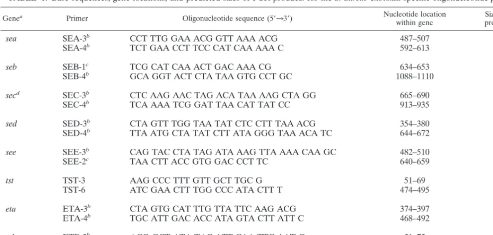
PCR.Oligonucleotide primers are listed in Table 1. Primarily, we tested primer sequences published by Johnson et al. (17). Modifications or newly designed PCR primers were tested to meet the requirements of a multiplex PCR to obtain good size-distinguishable amplification bands as well as the require-ments of the uracil DNA glycosylase (UNG) system with an inferior limit of annealing temperature at 55°C. These new or modified primers and the biotin-ylated oligonucleotide probes were designed by computerized sequence analysis with PC/Gene, release 18.0 (IntelliGenetics, Mountain View, Calif.) and Primer Premier 4.04 (Premier Biosoft, Palo Alto, Calif.). The oligonucleotides were obtained from MWG Biotech (Ebersberg, Germany).
First, preliminary trials with different annealing temperatures (50, 52.5, 55, and 57.5°C), cycle numbers (25, 30, and 35), amounts (1.0, 1.25, 1.5, and 2.0 U), and brands of DNA polymerases (Taq DNA polymerase; Boehringer, Mannheim, Germany; Expand high-fidelity PCR system, Boehringer; PrimeZyme, Biometra, Go¨ttingen, Germany; AmpliTaq DNA polymerase, Applied Biosystems, Foster City, Calif.; and AmpliTaq Gold DNA polymerase, Applied Biosystems) and with various concentrations of magnesium chloride (1.0, 1.5, 2.0, 2.5, 3.0, 3.5, and 4.0 mM) were performed to define the optimal PCR conditions (data not shown). The amplification was performed in an automated thermocycler with a hot bonnet (Hybaid, Teddington, United Kingdom). The optimized thermal cycling conditions for both multiplex PCRs were 30 cycles of denaturation at 95°C for 1 min (2 min for the first cycle), annealing at 55°C for 1 min, and polymerization at 72°C for 2 min (5 min for the last cycle). Optimized amplifications were carried out in 0.5-ml tubes in a reaction volume of 50ml containing 200mM dATP, dCTP, and dGTP. Instead of dTTP, dUTP was used in a concentration of 600
mM. The master mix (PCR Core Kit Plus; Boehringer) contained 10 mM Tris-HCl (pH 8.9), 50 mM KCl, 3 mM magnesium chloride, 1.0 U of UNG, and 0.5 pmol of primer. This reaction mixture was incubated for 10 min at room tem-perature (UNG reaction condition). For UNG inactivation, prevention of
prim-er-dimer formation and denaturation of template DNA, after addition of tem-plate DNA (5 to 10 ng), the mixture was incubated at 95°C for 5 min and stored on ice before addition of 1.25 U of Taq DNA polymerase (Boehringer, Mann-heim, Germany).
Amplified products (10ml) were resolved by 3.0% agarose gel electrophoresis at 150 V for 1 to 2 h. The gel was then stained with ethidium bromide, and the bands were visualized under UV illumination at 254 nm.
Hybridization procedures. Designed 59-biotinylated oligonucleotide probes
for the eight toxins tested (Table 2) were separately used for hybridization of amplified DNA from the multiplex PCRs in a generic DNA EIA (GEN-ETI-K DEIA; Sorin, Saluggia, Italy) according to the manufacturer’s recommendations. Briefly, strips coated with streptavidin were incubated with an optimized con-centration of the biotinylated probes (20 pg/ml for each probe) at 5°C overnight. After addition of 100ml of hybridization buffer, the strips were incubated with 20
ml of the 1:10-diluted denatured PCR products at the optimal hybridization temperature (55°C for all used probes) for 1 h. Following hybridization, the samples were treated with 100ml of a 1:50-diluted anti-double-stranded DNA mouse antibody solution for 1 h. The detection of the DNA-antibody bond was performed by addition of an enzyme tracer system (antimouse antibody labelled with horseradish peroxidase), and the results were measured after incubation with the chromogen-substrate solution by using a vertical reading photometer. The optical density at 450/630 nm (OD450/630ratio) of the resulting color was
measured, and the A630was subtracted from the A450. A cutoff of 0.150
absor-bance units above the mean value of determinations of toxin-negative reference strains was defined as a positive reaction.
Sensitivity testing.For determination of the sensitivities of the PCR assays and
hybridization procedures, the minimal amount of staphylococcal DNA that would be successfully detected was determined. Purified DNA of reference strains was quantitated by using a spectrophotometer, and 10-fold serial dilutions starting at 1mg/ml were subsequently subjected to multiplex PCR and DNA EIA. Upon completion of PCR, an aliquot from each tube was analyzed by electro-phoresis and placed into the DNA EIA as described above. To compare the influence of the multiplex procedure versus that of a single-PCR application, DNA of the toxin-positive reference strains was tested as a single target versus equal amounts of a mixture of all gene targets.
Specificity testing.Samples of DNA from the target toxin-negative S. aureus
strain Cowan 1 and from a set of coagulase-negative staphylococci, from pyro-genic exotoxin and streptolysin O-producing strains of Streptococcus pyogenes, and from other gram-positive microorganisms (microcooci, stomatococci, and bacilli) of the strain collection of our institute were tested. Ten nanograms of template DNA was used per reaction.
Determination of staphylococcal toxins.Culture filtrates of the S. aureus
[image:2.612.53.549.80.317.2]strains were tested for the presence of SEA to SEE by an EIA (Ridascreen set
TABLE 1. Base sequences, gene locations, and predicted sizes of PCR products for the S. aureus exotoxin-specific oligonucleotide primers
Genea Primer Oligonucleotide sequence (59339) Nucleotide location
within gene product (bp)Size of PCR
sea
SEA-3
bCCT TTG GAA ACG GTT AAA ACG
487–507
127
SEA-4
bTCT GAA CCT TCC CAT CAA AAA C
592–613
seb
SEB-1
cTCG CAT CAA ACT GAC AAA CG
634–653
477
SEB-4
bGCA GGT ACT CTA TAA GTG CCT GC
1088–1110
sec
dSEC-3
bCTC AAG AAC TAG ACA TAA AAG CTA GG
665–690
271
SEC-4
bTCA AAA TCG GAT TAA CAT TAT CC
913–935
sed
SED-3
bCTA GTT TGG TAA TAT CTC CTT TAA ACG
354–380
319
SED-4
bTTA ATG CTA TAT CTT ATA GGG TAA ACA TC
644–672
see
SEE-3
bCAG TAC CTA TAG ATA AAG TTA AAA CAA GC
482–510
178
SEE-2
cTAA CTT ACC GTG GAC CCT TC
640–659
tst
TST-3
AAG CCC TTT GTT GCT TGC G
51–69
445
TST-6
ATC GAA CTT TGG CCC ATA CTT T
474–495
eta
ETA-3
bCTA GTG CAT TTG TTA TTC AAG ACG
374–397
119
ETA-4
bTGC ATT GAC ACC ATA GTA CTT ATT C
468–492
etb
ETB-3
bACG GCT ATA TAC ATT CAA TTC AAT G
51–75
262
ETB-4
AAA GTT ATT CAT TTA ATG CAC TGT CTC
286–312
aSequences and locations were derived from the published nucleotide sequences for SEA (7), SEB (18), SEC
1to SEC3(10, 13), SED (2), SEE (14), TSST-1 (8),
and ETA and ETB (20).
bModified oligonucleotide primers based on primer sequences published by Johnson et al. (17). cOligonucleotide primer sequences as described by Johnson et al. (17).
dThe primer sequences used here cover all three subtypes of SEC.
on May 15, 2020 by guest
http://jcm.asm.org/
A, B, C, D, E; R-Biopharm, Darmstadt, Germany), and, except for SEE, by a semiquantitative reversed passive latex agglutination test (SET-RPLA; Unipath, Basingstoke, Hampshire, United Kingdom). The presence of TSST-1 was deter-mined by the same method (TST-RPLA; Unipath). An immunoblot with sheep anti-ETA antibody (Toxin Technology, Sarasota, Fla.), alkaline phosphatase-conjugated donkey anti-sheep immunoglobulin G (Sigma, St. Louis, Mo.), and purified ETA (Toxin Technology) as a positive control was used for the detection of ETA production.
RESULTS
Optimization of PCR conditions.
Titration of Mg
21concen-tration of the reaction buffer revealed an optimum of 3 mM for
both multiplex PCRs. The primer sets allowed PCR
amplifi-cation of the target gene fragments with 25, 30, or 35 cycles.
Thirty cycles was found to be optimal for both multiplex
sys-tems. The optimal thermal profile was 95°C for 1 min, 55°C
for 1 min, and 72°C for 2 min for both multiplex PCRs. The
Expand High Fidelity PCR system (Boehringer, Mannheim,
Germany) and the use of other DNA polymerases had no
advantages over our multiplex PCR systems.
DNA EIA.
Analysis of the nucleotide sequences published
for the target genes indicated that the 24- to 26-base
oligonu-cleotide probes (Table 2) encode sequences unique to the
toxins from which they were deduced. Hybridization
experi-ments with these gene probes were performed with PCR
am-plification products derived from single and multiplex PCRs as
described above. The optimal concentration for coating the
microtiter plates with the biotinylated probes was 20 pg/ml for
each probe. The results indicated a complete correlation
be-tween the amplification products and their corresponding
hy-bridization. No unspecific hybridization was observed among
the different PCR products of all staphylococcal exotoxins
tested.
[image:3.612.52.550.81.182.2]Sensitivity of multiplex PCR and DNA EIA.
Both multiplex
primer sets successfully amplified the appropriate regions of
the target toxin genes from a DNA dilution containing as little
as 50 pg (ranging from 1 to 50 pg) for single PCRs and in
multiplex PCRs as little as 100 pg (ranging from 1 to 100 pg) of
DNA of a DNA mixture isolated from reference strains
con-taining the test toxin genes. In a given multiplex PCR mixture
consisting of DNA from every type of target gene, the resulting
band intensities of the larger amplicons were reduced
com-pared to the intensities of the smaller amplicons and to the
TABLE 2. Base sequences and gene locations for the S. aureus exotoxin-specific 5
9
-biotinylated oligonucleotide probes
Genea Probeb Oligonucleotide sequence (59339) Nucleotide location
within gene
sea
SEA-7B
GGA GTT GGA TCT TCA AGC AAG ACG
531–554
seb
SEB-3B
GAG AAT AGC TTT TGG TAT GAC ATG
901–924
sec
cSEC-5B
AAC GGC AAT ACT TTT TGG TAT GAT
772–795
sed
SED-5B
TAA AGC CAA TGA AAA CAT TGA TTC A
382–406
see
SEE-5B
CTT TGG CGG TAA GGT GCA AAG AGG C
594–618
tst
TST-5B
AAG CCA ACA TAC TAG CGA AGG AAC
348–371
eta
ETA-5B
CCA TGC AAA AGC AGA AGT TTC AGC
414–437
etb
ETB-5B
TAC CAC CTA ATA CCC TAA TAA TCC AA
190–215
aSequences and locations were derived from the published nucleotide sequences for SEA (7), SEB (18), SEC
1to SEC3(10, 13), SED (2), SEE (14), TSST-1 (8),
and ETA and ETB (20).
b59biotinylated.
cThe probe sequence used here covers the SEC
1to SEC3subtypes.
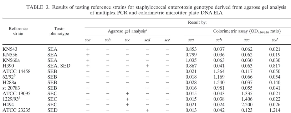
TABLE 3. Results of testing reference strains for staphylococcal enterotoxin genotype derived from agarose gel analysis
of multiplex PCR and colorimetric microtiter plate DNA EIA
Reference
strain phenotypeToxin
Result by:
Agarose gel analysisa Colorimetric assay (OD
450/630ratio)
sea seb sec sed see sea seb sec sed see
KN543
SEA
1
2
2
2
2
0.853
0.037
0.062
0.021
0.018
KN556
SEA
1
2
2
2
2
0.799
0.036
0.062
0.019
0.019
KN560a
SEA
1
2
2
2
2
1.035
0.063
0.030
0.030
0.018
H390
SEA, SED
1
2
2
1
2
0.867
0.041
0.063
0.817
0.019
ATCC 14458
SEB
2
1
2
2
2
0.021
1.364
0.117
0.050
0.022
62/92
bSEB
2
1
2
2
2
0.018
1.169
0.066
0.054
0.036
H288a
SEB
2
1
2
2
2
0.028
1.540
0.037
0.140
0.018
st 20783
SEB
2
1
2
2
2
0.016
0.981
0.055
0.041
0.021
ATCC 19095
SEC
2
2
1
2
2
0.015
0.043
1.335
0.021
0.025
1229/93
bSEC
2
2
1
2
2
0.015
0.038
1.406
0.022
0.020
H494
SEC
2
2
1
2
2
0.021
0.024
2.200
0.026
0.018
ATCC 23235
SED
2
2
2
1
2
0.013
0.042
0.123
1.214
0.021
1634/93
bSED
2
2
2
1
2
0.019
0.024
0.128
1.475
0.018
ATCC 27664
SEE
2
2
2
2
1
0.021
0.034
0.099
0.020
1.819
Cowan 1
c2
2
2
2
2
0.014
0.083
0.039
0.019
0.017
ATCC 20044
d2
2
2
2
2
0.020
0.031
0.110
0.024
0.018
a2, negative;1, positive (judged by eye).
bKindly provided by W. Witte (Robert Koch-Institut Wernigerode, Germany). cS. aureus negative control strain.
dS. epidermidis negative control strain.
on May 15, 2020 by guest
http://jcm.asm.org/
[image:3.612.49.549.498.693.2]artificially pooled single PCR, but this does not affect the test
in practice. The sensitivity of subsequent DNA EIAs was
al-ways 10 to 100 times higher than that of both the single and
multiplex PCRs (data not shown).
Specificity of the staphylococcal oligonucleotide primers
and probes.
Initially, the specific primer pairs published by
Johnson et al. (17) were tested for their applicability to
mul-tiplex PCR for detection of the staphylococcal toxin genes. To
meet the requirements of good size-distinguishable
amplifica-tion bands of multiplex PCR and of the temperature limit of
the UNG system for carryover protection, it was necessary to
modify and partly select new primers directed to specific
nu-cleotide sequences within the target genes. The eight modified
or newly developed pairs of toxin-specific oligonucleotide
prim-ers and the eight newly derived toxin-specific biotinylated
oli-gonucleotide probes were selected by computer analysis with
previously described nucleotide sequences for the
correspond-ing genes of SEA (7), SEB (18), SEC
1to SEC
3(10, 13), SED
(2), SEE (14), TSST-1 (8), and ETA and ETB (20). A total of
23 reference strains of S. aureus were used as templates for the
primer sets, each with 5 to 10 ng of bacterial DNA in the PCR
mixture. The DNA of all S. aureus exotoxin-producing
refer-ence strains was specifically amplified by the primers and
con-firmed by oligonucleotide probes of the DNA EIA; thus, the
corresponding genes were correctly recognized (Tables 3 and
4). The sizes of the amplicons were identical to those predicted
from the primer design (Table 1). Figures 1 and 2 show
rep-resentative gels for the PCR product patterns of both multiplex
PCRs. In addition, none of the primer pairs and the
corre-sponding probes reacted with any strains of non-SE-producing
S. aureus strains, other coagulase-positive staphylococci (S.
in-termedius or S. schleiferi subsp. coagulans), a spectrum of
co-agulase-negative staphylococcal species (S. epidermidis, S.
hae-molyticus, S. hominis, S. warneri, S. capitis, and S. simulans),
and other related gram-positive genera, including
stomato-cocci, microstomato-cocci, and Bacillus spp. (data not shown). Because
of the close relationship between SEB and SEC and other
members of the pyrogenic exotoxin family, we tested pyrogenic
exotoxin and streptolysin O-producing strains of S. pyogenes in
our multiplex PCR system, and no specific amplification and
hybridization were observed.
Testing of S. aureus isolates by multiplex PCR-EIA and
comparison with toxin phenotype.
Amplicons with predicted
sizes and a positive hybridization reaction were detected in all
reference strains analyzed (Tables 3 and 4). Also, there was
100% correlation between the results of the multiplex
PCR-EIA and the toxin phenotype testing of the reference strains.
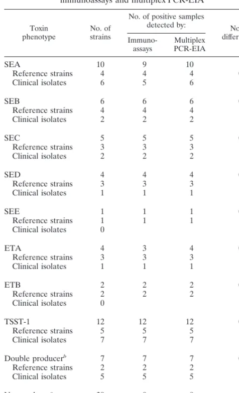
Within 50 clinical S. aureus isolates obtained from 43
pa-tients, a total of 19 positive amplification results were found.
All could be confirmed by the respective hybridization
proce-dure. In 11 clinical strains from different patients, we detected
various genes for enterotoxins, in 7 strains we detected the
TSST-1 gene, and in 1 strain we detected the ETA gene (Table
5).
Both multiplex PCR systems could detect exotoxin double
producers in reference strains (one SEA plus SED and one
ETA plus ETB) in one step. In the clinical strains, we observed
five strains carrying two exotoxin genes: one strain carried the
sec and tst genes, and four strains carried the sea and tst genes
[image:4.612.307.545.69.211.2](Table 5). Thus, an excellent correlation was found between
TABLE 4. Results of testing reference strains for staphylococcal
ETs and TSST-1 genotype derived from agarose gel analysis
of multiplex PCR-EIA and colorimetric
microtiter plate DNA EIA
Reference strain
Toxin pheno-type(s)
Result by: Agarose gel
analysisa Colorimetric assay(OD
450/630ratio)
eta etb tst eta etb tst
1930/95b ETA 1 2 2 1.308 0.017 0.013 BM 10458c ETA 1 2 2 1.284 0.028 0.028 BM 10431c ETA, ETB 1 1 2 0.961 1.932 0.017 BM 10143c ETB 2 1 2 0.045 1.922 0.031 161/93b TSST-1 2 2 1 0.019 0.019 1.450 150 TSST-1 2 2 1 0.024 0.021 2.166 235 TSST-1 2 2 1 0.017 0.018 1.705 KN813 TSST-1 2 2 1 0.020 0.022 1.870 KN820 TSST-1 2 2 1 0.020 0.019 1.792 Cowan 1d 2 2 2 0.034 0.020 0.036 ATCC 20044e 2 2 2 0.024 0.019 0.024
a2, negative;1, positive (judged by eye).
bKindly provided by W. Witte (Robert Koch-Institut Wernigerode, Germany). cKindly provided by N. El Solh (Centre National de Re´fe´rence des
Staphy-locoques, Institut Pasteur, Paris, France).
[image:4.612.320.536.524.667.2]dStaphylococcus aureus, negative control strain. eStaphylococcus epidermidis, negative control strain.
FIG. 1. Agarose gel electrophoresis patterns showing PCR-amplified prod-ucts in multiplex PCR for the staphylococcal enterotoxin genes. Lanes 1, DNA molecular weight markerlHindIII; 2, sea; 3, seb; 4, sec; 5, sed; 6, see; 7, multiplex PCR with all enterotoxin genes simultaneously (sea to see); 8, artificial arrange-ment of the amplification fragarrange-ments of sea to see; 9, DNA molecular weight markerlBstE II. Sizes are marked in base pairs on the left and right.
FIG. 2. Agarose gel electrophoresis patterns showing PCR-amplified prod-ucts in multiplex PCR for the staphylococcal ET genes (eta and etb) and TSST-1 gene (tst). Lanes 1: DNA molecular weight markerlHindIII; 2, eta; 3, etb; 4, tst; 5, multiplex PCR with both ET genes and the TSST-1 gene simultaneously; 8, artificial arrangement of the amplification fragments of eta, etb, and tst; 9, DNA molecular weight markerlBstE II. Sizes are marked in base pairs on the left and right.
on May 15, 2020 by guest
http://jcm.asm.org/
the results of genotype and phenotype testing. In only one
strain positive for sea by multiplex PCR-EIA could SEA toxin
production not be detected by both immunological tests used.
DISCUSSION
The ability of S. aureus strains to produce SEs, ETs, and/or
TSST-1 is an important property with various clinical
implica-tions. Determination is still mainly based on immunological
methods for toxin detection which are time- and
labor-con-suming. Furthermore, these methods depend on the
concen-tration of toxin expressed and thus can be negatively
influ-enced by various factors. Also, for enterotoxins, differences in
the toxin production levels by S. aureus strains grown in natural
substrate and laboratory media have been described previously
(15).
We have developed a multiplex PCR-EIA test system which
allows the simultaneous detection of the enterotoxin (sea to
see), ET (eta and etb), and TSST-1 (tst) genes. We could prove
this system to be sensitive and specific for the respective genes
by using staphylococcal cultures. Because our objective was to
develop a simple and fast method for use in routine diagnosis,
we preferred an NA preparation procedure that was simpler
than the classical phenol-chloroform extraction method.
Sam-ples for PCR can be prepared in 1 h without an obvious risk of
sample cross-contamination. The use of alternative DNA
poly-merase systems, so-called expanded and/or high-fidelity
sys-tems, had no advantages over our multiplex PCRs, although
they are described as being able to increase the yield of
am-plification by reducing the mismatch pausing associated with
Taq DNA polymerase.
Strict precautions must be taken to avoid false-positive
am-plification because of contamination of the reaction mixture
both with target DNA from staphylococcal cells and by
car-ryover of amplified target DNA from previous PCR cycles.
Therefore, to prevent false-positive results, we used the UNG
system (21). This enzyme can be used with dUTP to eliminate
PCR contaminations from previous DNA synthesis reactions
by degrading uracil-containing DNA. Since it has been
report-ed that UNG from Escherichia coli remains partially active or
regains activity leading to degradation of the dU-containing
PCR product, it is necessary to keep the reaction mixture at a
higher temperature (70°C) or to freeze the product
imme-diately after the last amplification step (19). Because UNG
shows activity only below 55°C, it is recommended to choose
annealing temperatures at or above this temperature in order
to avoid degradation of newly synthesized dU-containing
am-plicons by possible residual UNG activity after the inactivation
step of this enzyme. To meet this recommendation, we had to
modify or design new primers composed of nucleotides
allow-ing such temperatures. A higher annealallow-ing temperature is also
recommended to increase the specificity of the PCR. The use
of a DNA EIA system as a hybridization step after the PCR
procedure offers a simple and reliable specificity control of the
amplicons.
The multiplex PCR-EIA test system described here was
pri-marily designed for the detection of exotoxin genes in S. aureus
in culture. Research is in progress to apply this system to the
direct detection of toxigenic S. aureus strains in human
speci-mens and food material.
ACKNOWLEDGMENTS
We thank Susanne Deiwick and Martina Schulte for excellent
tech-nical assistance. We are grateful to M. Herrmann for critical
sugges-tions on the manuscript.
REFERENCES
1. Avena, R. M., and M. S. Bergdoll. 1967. Purification and some physicochem-ical properties of enterotoxin C, Staphylococcus aureus strain 361. Biochem-istry 6:1474–1480.
2. Bayles, K. W., and J. J. Iandolo. 1989. Genetic and molecular analyses of the gene encoding staphylococcal enterotoxin D. J. Bacteriol. 171:4799–4806. 3. Bergdoll, M. S., C. R. Borja, and R. M. Avena. 1965. Identification of a new
enterotoxin as enterotoxin C. J. Bacteriol. 90:1481–1485.
4. Bergdoll, M. S., C. R. Borja, R. N. Robbins, and K. F. Weiss. 1971. Identi-fication of enterotoxin E. Infect. Immun. 4:593–595.
5. Bergdoll, M. S., B. A. Crass, R. F. Reiser, R. N. Robbins, and J. P. Davis. 1981. A new staphylococcal enterotoxin, enterotoxin F, associated with toxic-shock-syndrome Staphylococcus aureus isolates. Lancet i:1017–1021. 6. Bergdoll, M. S., M. J. Surgalla, and G. M. Dack. 1959. Staphylococcal
enterotoxin: identification of a specific precipitating antibody with entero-toxin neutralizing property. J. Immunol. 83:334–338.
7. Betley, M. J., and J. J. Mekalanos. 1988. Nucleotide sequence of the type A staphylococcal enterotoxin gene. J. Bacteriol. 170:34–41.
8. Blomster-Hautamaa, D. A., B. N. Kreiswirth, J. S. Kornblum, R. P. Novick,
[image:5.612.49.287.97.488.2]and P. M. Schlievert.1986. The nucleotide and partial amino acid sequence
TABLE 5. Detection of exotoxin-producing S. aureus reference
strains (n
5
23) and clinical isolates (n
5
50) by
immunoassays and multiplex PCR-EIA
Toxin
phenotype No. ofstrains
No. of positive samples
detected by: No. of differencesa
Immuno-assays PCR-EIAMultiplex
SEA 10 9 10 1
Reference strains 4 4 4 0 Clinical isolates 6 5 6 1
SEB 6 6 6 0
Reference strains 4 4 4 Clinical isolates 2 2 2
SEC 5 5 5 0
Reference strains 3 3 3 Clinical isolates 2 2 2
SED 4 4 4 0
Reference strains 3 3 3 Clinical isolates 1 1 1
SEE 1 1 1 0
Reference strains 1 1 1 Clinical isolates 0
ETA 4 3 4 0
Reference strains 3 3 3 Clinical isolates 1 1 1
ETB 2 2 2 0
Reference strains 2 2 2 Clinical isolates 0
TSST-1 12 12 12 0
Reference strains 5 5 5 Clinical isolates 7 7 7
Double producerb 7 7 7 0 Reference strains 2 2 2
Clinical isolates 5 5 5
Nonproducerc 39 0 0 0
Reference strains 3 0 0 Clinical isolates 36 0 0
aDifferences between the test results from immunological and PCR methods. bDouble exotoxin producers: one reference strain with SEA and SED, one
reference strain with ETA and ETB, four clinical strains with SEA and TSST-1, and one clinical strain with SEC and TSST-1, respectively.
cNonproducer of staphylococcal exotoxins tested.
on May 15, 2020 by guest
http://jcm.asm.org/
of toxic shock syndrome toxin-1. J. Biol. Chem. 261:15783–15786. 9. Bohach, G. A., B. N. Kreiswirth, R. P. Novick, and P. M. Schlievert. 1989.
Analysis of toxic shock syndrome isolates producing staphylococcal entero-toxins B and C1 with use of southern hybridization and immunologic assays. Rev. Infect. Dis. 11(Suppl. 1):S75–81.
10. Bohach, G. A., and P. M. Schlievert. 1987. Nucleotide sequence of the staphylococcal enterotoxin C1 gene and relatedness to other pyrogenic tox-ins. Mol. Gen. Genet. 209:15–20.
11. Casman, E. P. 1960. Further serological studies of staphylococcal entero-toxin. J. Bacteriol. 79:849–856.
12. Casman, E. P., R. W. Bennett, A. E. Dorsey, and J. A. Issa. 1967. Identifi-cation of a fourth staphylococcal enterotoxin, enterotoxin D. Biochemistry
94:1875–1882.
13. Couch, J. L., and M. J. Betley. 1989. Nucleotide sequence of the type C3
staphylococcal enterotoxin gene suggests that intergenic recombination causes antigenic variation. J. Bacteriol. 171:4507–4510.
14. Couch, J. L., M. T. Soltis, and M. J. Betley. 1988. Cloning and nucleotide sequence of the type E staphylococcal enterotoxin gene. J. Bacteriol. 170: 2954–2960.
15. Gomez-Lucı´a, E., J. Goyache, J. A. Orden, J. L. Blanco, J. A. Ruiz
Santa-Quiteria, L. Domı´nguez, and G. Sua´rez.1989. Production of enterotoxin A
by supposedly nonenterotoxigenic Staphylococcus aureus strains. Appl. En-viron. Microbiol. 55:1447–1451.
16. Johnson, A. D., L. Spero, J. S. Cades, and B. T. de Cicco. 1979. Purification and characterization of different types of exfoliative toxin from Staphylococ-cus aureus. Infect. Immun. 24:679–684.
17. Johnson, W. M., S. D. Tyler, E. P. Ewan, F. E. Ashton, D. R. Pollard, and
K. R. Rozee.1991. Detection of genes for enterotoxins, exfoliative toxins, and
toxic shock syndrome toxin 1 in Staphylococcus aureus by the polymerase chain reaction. J. Clin. Microbiol. 29:426–430.
18. Jones, C. L., and S. A. Khan. 1986. Nucleotide sequence of the enterotoxin B gene from Staphylococcus aureus. J. Bacteriol. 166:29–33.
19. Kwok, S., and R. Higuchi. 1989. Avoiding false positives with PCR. Nature
339:237–238.
20. Lee, C. Y., J. J. Schmidt, A. D. Johnson-Winegar, L. Spero, and J. J. Iandolo. 1987. Sequence determination and comparison of the exfoliative toxin A and toxin B genes from Staphylococcus aureus. J. Bacteriol. 169:3904–3909. 21. Longo, M. C., M. S. Berninger, and J. L. Hartley. 1990. Use of uracil DNA
glycosylase to control carry-over contamination in polymerase chain reac-tions. Gene 93:125–128.
22. O’Toole, P. W., and T. J. Foster. 1987. Nucleotide sequence of the epider-molytic toxin A gene of Staphylococcus aureus. J. Bacteriol. 169:3910–3915. 23. Reiser, R. F., R. N. Robbins, A. L. Noleto, G. P. Khoe, and M. S. Bergdoll. 1984. Identification, purification, and some physicochemical properties of staphylococcal enterotoxin C3. Infect. Immun. 45:625–630.
24. Rifai, S., V. Barbancon, G. Prevost, and Y. Piemont. 1989. Synthetic exfoli-ative toxin A and B DNA probes for detection of toxigenic Staphylococcus aureus strains. J. Clin. Microbiol. 27:504–506.
25. Schlievert, P. M., K. N. Shands, B. B. Dan, G. P. Schmid, and R. D.
Nishimura.1981. Identification and characterization of an exotoxin from
Staphylococcus aureus associated with toxic-shock syndrome. J. Infect. Dis.
143:509–516.
26. Thompson, N. E., M. Razdan, G. Kuntsmann, J. M. Aschenbach, M. L.
Evenson, and M. S. Bergdoll.1986. Detection of staphylococcal enterotoxins
by enzyme-linked immunosorbent assays and radioimmunoassays: compari-son of monoclonal and polyclonal antibody systems. Appl. Environ. Micro-biol. 51:885–890.
27. Wiley, B. B., L. A. Glasgow, and M. Rogolsky. 1976. Staphylococcal scalded-skin syndrome: development of a primary binding assay for human antibody to the exfoliative toxin. Infect. Immun. 13:513–520.