www.impactjournals.com/oncotarget/ Oncotarget, Vol. 7, No. 35
GDF11 administration does not extend lifespan in a mouse
model of premature aging
Sandra Freitas-Rodríguez1, Francisco Rodríguez1 and Alicia R. Folgueras1
1 Departamento de Bioquímica y Biología Molecular, Facultad de Medicina, Instituto Universitario de Oncología del Principado
de Asturias (IUOPA), Universidad de Oviedo, Oviedo, Spain
Correspondence to: Alicia R. Folgueras, email: [email protected] Keywords: progeria, accelerated aging, GDF11, longevity
Received: May 25, 2016 Accepted: July 22, 2016 Published: August 05, 2016
AbstrAct
GDF11 has recently emerged as a powerful anti-aging candidate, found in young blood, capable of rejuvenating a number of aged tissues, such as heart, skeletal muscle and brain. However, recent reports have shown contradictory data questioning its capacity to reverse age-related tissue dysfunction. The availability of a mouse model of accelerated aging, which shares most of the features occurring in physiological aging, gives us an excellent opportunity to test in vivo therapies aimed at extending lifespan both in pathological and normal aging. On this basis, we wondered whether the proposed anti-aging functions of GDF11 would have an overall effect on longevity. We first confirmed the existence of a reduction in GDF11/8 levels in our mouse model of accelerated aging compared with wild-type littermates. However, we show herein that GDF11 daily administration does not extend lifespan of premature-aged mice.
IntroductIon
The existence of “rejuvenating” factors in young blood capable of improving the function of aging stem
cells was first demonstrated in 2005 by the group of Tom
Rando [1]. A decade after this seminal contribution, the new wave of studies has been on the search for those circulating regulatory molecules that can restore the regenerative function of old stem cells and reverse aging
[2-4]. Among several cell-extrinsic factors and metabolites identified to date, GDF11 has been found to be one of the most powerful anti-aging candidates. Thus, it has been shown that GDF11 levels in blood decline with age, and
that its supplementation to reach young physiological range in old mice improved the features and function
of a number of age-deteriorated tissues, including heart, skeletal muscle and brain [5-8].
Many of the symptoms associated with normal aging have also been reported in human accelerated
aging syndromes, such as Hutchison-Guilford progeria,
a devastating disease caused by alterations in the
nuclear envelop architecture [9-11]. Those include skin
wrinkling, hair loss, muscle atrophy, osteoporosis and a premature cardiovascular disease, which is responsible for the death of the patients in the childhood due to
myocardial infarction or stroke [12]. The generation of
mouse models that phenocopy most of the features of this
syndrome, such as mice deficient in the metalloproteinase Zmpste24, which is required for nuclear lamin A
maturation, represents useful tools for studying not only the mechanisms underlying this disease, but also those
common to normal aging process [12-14].
Our previous studies with a mosaic mouse model
in which deficient cells coexist with Zmpste24-proficient cells in similar proportion showed a complete correction of the progeroid phenotype [15]. These results demonstrated the relevance of cell-extrinsic mechanisms
in the establishment of this pathology and suggested that a therapeutic approach based on the administration of key systemic factors may be an avenue to improve the progression of this disease. On this basis, and given the
proposed anti-aging functions of GDF11, we analyze
herein the in vivo effect of GDF11 administration on the lifespan of premature-aged mice.
results
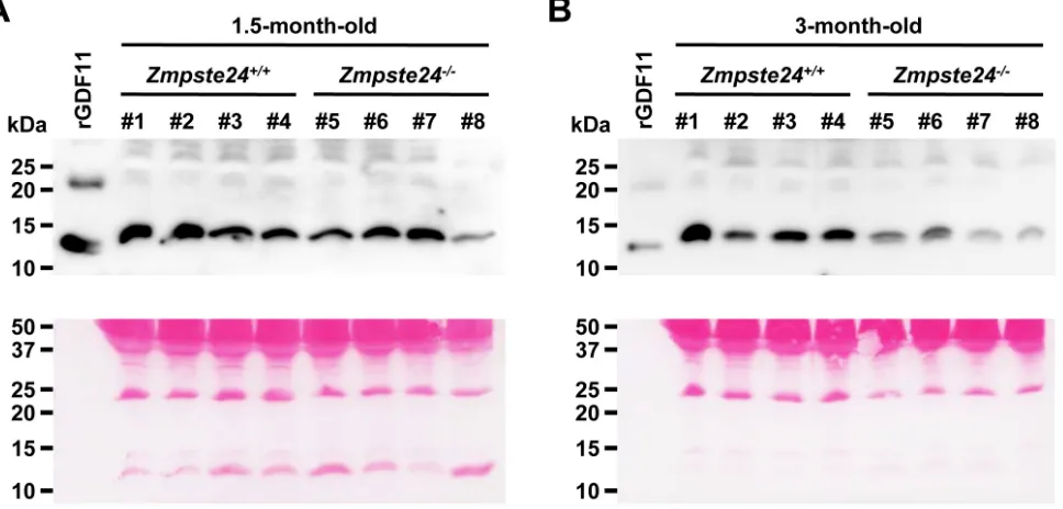
To evaluate whether all attributed anti-aging properties of GDF11 may have an overall effect on longevity, we first determined whether GDF11 levels
aging [6, 8]. We performed western-blot analyses with plasma samples obtained from the same wild-type and Zmpste24-/- mice at the age of 1.5 months and 3 months,
to monitor a possible decline over time, considering that
average lifespan of these mutant mice is 4 months and
that accelerated aging symptoms start to manifest around
the age of 2 months. We used the same commercial
antibody as the one previously reported in the original
study by Loffredo et al., where GDF11 was first identified as an anti-aging factor [6]. Importantly, recent findings demonstrated that this antibody also recognizes GDF8 (myostatin), a closely related member of the TGF-β superfamily that shares 89% identity in amino acid sequence in the mature active form, questioning those previous published data that showed a GDF11 decline
with age [16]. However, to date, no alternative reliable
assay capable of detecting endogenous GDF11 in mouse
serum has been described [16, 17]. On the basis of these
premises, we observed a marked decrease in GDF11/8
plasma levels in Zmpste24-/- mice compared with
wild-type littermates at the age of 3 months. Interestingly, no significant differences were found when analyzing plasma
samples that had been obtained from the same individuals
1.5 months earlier, prior to the development of any aging phenotype (Figure 1A-1B). Ponceau S staining from the corresponding western-blot showed equivalent loading
in all lanes. Altogether, these results indicate that the
reduction in GDF11/8 blood levels observed in Zmpste24
-/-versus wild-type mice occurs upon the manifestation of
the progeroid phenotype.
We next evaluated whether GDF11 could be one of
the circulating factors capable of slowing down the aging symptoms and extending the lifespan of Zmpste24-/- mice,
considering its overall effect on a number of aged tissues
and the above-mentioned observations (Figure 1B). To
test this hypothesis, we did use the same commercial
recombinant GDF11 protein (PreproTech) and at the same dosage (0.1 mg/kg, daily) that had been reported to have an anti-aging effect [5-7]. By using this approach, we were able to detect by western-blot an increase in circulating GDF11/8 plasma levels in progeroid Zmpste24-/- mice 1
to 2 hours after rGDF11 injection (Figure S1), similar to
what it had previously been described (see Loffredo et
al. Figure S5). Even though the used antibody was able to recognize both GDF11 and GDF8, it was reasonable
to speculate that the protein increase we observed in the
blood of the same individual within a 1-2 hour temporal window corresponded to rGDF11. Moreover, alternative
detection methods have also demonstrated that daily
intraperitoneal injection of rGDF11 (0.1 mg/kg) increased circulating levels of GDF11 above endogenous plasma
levels, which were below the detection limit of the assay [17]. Therefore, considering that our goal was to restore
progeroid GDF11 plasma levels to a wild-type condition, we were confident that this dose was adequate to test
our hypothesis. Thus, male and female Zmpste24-/- mice
received a daily intraperitoneal injection of rGDF11 (0.1 mg/kg) or vehicle, starting at the age of 2.5 months,
once the accelerated aging symptoms started to manifest.
rGDF11 treatment did not increase survival of Zmpste24
-/-Figure 1: Progeroid Zmpste24-/- mice show reduced GdF11/8 plasma levels. Western-blot analysis of GDF11/8 plasma levels
of young (1.5-month-old) A. and aged (3-month-old) b. Zmpste24-/- and wild-type mice. Note that plasma samples were obtained from
[image:2.612.68.552.436.668.2]mice compared with vehicle-treated animals (Figure 2A-2B). These results were in line with recent reports showing no effect of GDF11 on cardiac or skeletal muscle function,
arguing against the “rejuvenating” potential of this protein
[16-19].
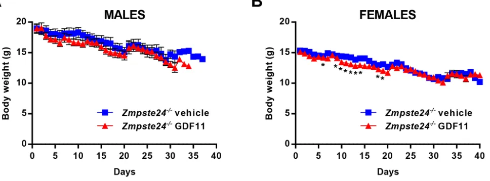
Additionally, it has recently been suggested that
high doses (0.5 mg/kg) of rGDF11 may decrease body weight [20]. In our experiment, we observed that long-term rGDF11 administration at a lower dose (0.1 mg/kg,
daily) did not dramatically decrease the body weight of Zmpste24-/--treated mice compared with vehicle-treated
littermates, although the average body weight was slightly
lower in rGDF11-treated mice, being this difference statistically significant at some points of the treatment period in female mice (Figure 3A-3B).
dIscussIon
Studies based on heterochronic parabiosis, where a
young and an old individual share a circulatory system,
have demonstrated that certain age-associated dysfunctions
may be rescued or ameliorated by the exposure to a young
systemic environment. Interestingly, even the earliest studies using this technique provided evidence of lifespan extension of the older parabiont [21, 22]. Current findings have supported the notion that this beneficial effect has its
basis on the restoration of the regenerative capacity of the stem cell pool in the old individual, which suggests that boosting tissue regeneration may slow down organismal
aging [23]. In the search for those circulatory factors
[image:3.612.73.554.259.445.2]that decline with age and that are responsible for the
Figure 2: rGdF11 therapy does not extend longevity of Zmpste24-/- mice. A. Kaplan-Meier survival plot of rGDF11-treated (n
= 5) and vehicle-treated (n = 6) male Zmpste24-/- mice. b. Kaplan-Meier survival plot of rGDF11-treated (n = 8) and vehicle-treated (n = 6)
female Zmpste24-/- mice. Mice received a daily intraperitoneal dose of rGDF11 (0.1 mg/kg) or vehicle.
Figure 3: rGdF11 therapy does not prevent body weight loss of Zmpste24-/- mice. Body weights of rGDF11- and
[image:3.612.72.556.509.688.2]maintenance of stem cell performance over time, GDF11 has been identified as a powerful anti-aging candidate
with a broad effect on a number of tissues, including cardiac and skeletal muscle and the cerebral vasculature
[5-7]. This apparent pleiotropy prompted us to hypothesize
that this factor might contribute to extend lifespan,
considering the number of key tissues/organs that could benefit. To test this hypothesis, we used a murine model of accelerated aging (Zmpste24-deficient mice), given that
it shares most of the features occurring in natural aging
[12]. In particular, it has been reported that many of the
alterations observed in these progeroid mice are caused
by an impaired stem cell function [13, 24, 25]. Moreover,
we have previously demonstrated that the coexistence of
Zmpste24-deficient cells and Zmpste24-proficient cells
(mosaic mice) completely prevented the development
of the premature aging phenotype, suggesting that
cell-extrinsic mechanisms exerted by the “healthy” cells were responsible for the full reversion of progeroid features
[15].
Nevertheless, in addition to the above-mentioned
data, to evaluate the possibility that our mouse model of
premature aging was suitable to test the effect of GDF11 on longevity, we first determined whether GDF11 levels
decline with age in Zmpste24-/- mice. Unfortunately, recent studies have reported that current immunoreagents,
due to the high similarity between GDF11 and GDF8 (myostatin) amino acid sequences, cannot discriminate
between these two closely related members of the
TGF-β superfamily in plasma samples [16]. In the light of these findings, the original papers have recently been revisited [20]. Therefore, consistent with what it has
now been reported to occur in natural aging, we only observed a marked decrease in the circulating pool of
GDF11/8 proteins in Zmpste24-/- compared with wild-type littermates when the aging symptoms had already started to manifest in these mice. Thus, no differences where observed when comparing plasma samples from the same individuals obtained at an early age, prior to the development of the progeroid phenotype. These results further support the similarities between our mouse model of premature aging and the process of physiological aging. However, in addition to the criticisms raised about
the original observation of a GDF11 decline in aged mice, recent studies have also questioned the capacity
of this circulating factor to reverse cardiac and skeletal
age-related tissue dysfunction [16-19]. Dosing and the
source of the recombinant protein have been claimed as possible factors to explain the discrepancies between
the data obtained by different investigators [20, 26]. In
this regard, to test our hypothesis about a possible role
for GDF11 on lifespan extension, we did use the same commercial rGDF11 protein that has been used in those studies describing its anti-aging properties, and at a
dosage capable of raising its levels in Zmpste24-/- plasma samples. However, rGDF11 daily treatment did not extend
the lifespan of progeroid mice compared with
vehicle-treated Zmpste24-/- littermates. It has been suggested that some of the original conclusions about GDF11
cardioprotective effects could be due to the decrease in
body weight observed as a secondary effect of rGDF11 daily administration [17, 20, 26]. Our results showed that rGDF11 treatment only caused a slightly reduction
in the body weight of female Zmpste24-/- mice compared
with vehicle-treated littermates during the first days of the experiment, whereas no significant differences were
observed in the male cohort.
In conclusion, our results demonstrate that circulating GDF11/8 levels are reduced in our mouse
model of premature aging, which shares most of the symptoms that occur in normal aging. However,
GDF11 protein administration is not sufficient to extend longevity in these progeroid mice. Although
accelerated-aging mouse models can serve as powerful tools to
test and develop anti-aging therapies common to both
physiological and pathological aging, the existence of certain differences between the two processes implies that
further investigation is still required to determine whether long-term GDF11 administration has a pro-survival effect
on normal aged animals.
MAterIAls And Methods
Animal experiments
Zmpste24-/- mice have previously been described [27]. Zmpste24-/- mice were backcrossed 10 generations to C57BL/6N background. All procedures involving
mice were conducted in accordance with the guidelines
of the Committee for Animal Experimentation of the Universidad de Oviedo. Blood was extracted from the facial veins after anaesthetizing mice and collected into EDTA-coated tubes. Blood was centrifuged at 1000 g at 4ºC, and the supernatant was stored at -80ºC until analysis.
Mice were given a daily intraperitoneal injection of either
rGDF11 (PeproTech) at 0.1 mg/kg or vehicle.
Western-blot
1.5% nonfat dry milk in TBS-T buffer with 1:2000 goat anti-rabbit IgG horseradish peroxidase (Cell Signaling, cat. #7074S), washed and developed with Immobilon Western Chemiluminescent HRP substrate (Millipore). Chemiluminescent images were acquired with a Fujifilm LAS3000 mini apparatus.
statistical analysis
Statistical analyses were performed by using GraphPad Prism 6 software. Log-rank (Mantel-Cox) test was used for Kaplan-Meier survival analysis. Student’s t-test was used for the analysis of body weight differences
between mouse cohorts.
AcknoWledGMents
We thank Dr. C. López-Otín for his support and advice and A. Moyano and R. Feijoo for their excellent
technical assistance.
FundInG
This work was supported by grants from Ministerio
de Economía y Competitividad (Spain) and RTICC-Spain. S.F-R. is recipient of an FPU Fellowship. A.R.F. is supported by the Juan de La Cierva Program. The IUOPA is supported by Fundación Cajastur-Asturias.
conFlIcts oF Interest
The authors declare no competing financial interests.
Author contributions
A.R.F. designed the experiments. S.R-F., A.R.F. and F.R. performed the experiments and analyzed the data. A.R.F. wrote the manuscript.
reFerences
1. Conboy IM, Conboy MJ, Wagers AJ, Girma ER, Weissman IL and Rando TA. Rejuvenation of aged progenitor cells by exposure to a young systemic environment. Nature. 2005; 433:760-764.
2. Brack AS, Conboy MJ, Roy S, Lee M, Kuo CJ, Keller C and Rando TA. Increased Wnt signaling during aging alters muscle stem cell fate and increases fibrosis. Science. 2007; 317:807-810.
3. Ruckh JM, Zhao JW, Shadrach JL, van Wijngaarden P, Rao TN, Wagers AJ and Franklin RJ. Rejuvenation of regeneration in the aging central nervous system. Cell stem cell. 2012; 10:96-103.
4. Villeda SA, Plambeck KE, Middeldorp J, Castellano JM, Mosher KI, Luo J, Smith LK, Bieri G, Lin K, Berdnik D, Wabl R, Udeochu J, Wheatley EG, et al. Young blood reverses age-related impairments in cognitive function and synaptic plasticity in mice. Nature medicine. 2014; 20:659-663.
5. Katsimpardi L, Litterman NK, Schein PA, Miller CM, Loffredo FS, Wojtkiewicz GR, Chen JW, Lee RT, Wagers AJ and Rubin LL. Vascular and neurogenic rejuvenation of the aging mouse brain by young systemic factors. Science. 2014; 344:630-634.
6. Loffredo FS, Steinhauser ML, Jay SM, Gannon J, Pancoast JR, Yalamanchi P, Sinha M, Dall’Osso C, Khong D, Shadrach JL, Miller CM, Singer BS, Stewart A, et al. Growth differentiation factor 11 is a circulating factor that reverses age-related cardiac hypertrophy. Cell. 2013; 153:828-839.
7. Sinha M, Jang YC, Oh J, Khong D, Wu EY, Manohar R, Miller C, Regalado SG, Loffredo FS, Pancoast JR, Hirshman MF, Lebowitz J, Shadrach JL, et al. Restoring systemic GDF11 levels reverses age-related dysfunction in mouse skeletal muscle. Science. 2014; 344:649-652. 8. Olson KA, Beatty AL, Heidecker B, Regan MC, Brody
EN, Foreman T, Kato S, Mehler RE, Singer BS, Hveem K, Dalen H, Sterling DG, Lawn RM, et al. Association of growth differentiation factor 11/8, putative anti-ageing
factor, with cardiovascular outcomes and overall mortality
in humans: analysis of the Heart and Soul and HUNT3 cohorts. Eur Heart J. 2015; 36:3426-3434.
9. López-Otín C, Blasco MA, Partridge L, Serrano M and Kroemer G. The hallmarks of aging. Cell. 2013; 153:1194-1217.
10. Aliper AM, Csoka AB, Buzdin A, Jetka T, Roumiantsev S, Moskalev A and Zhavoronkov A. Signaling pathway activation drift during aging: Hutchinson-Gilford Progeria Syndrome fibroblasts are comparable to normal middle-age and old-age cells. Aging. 2015; 7:26-37.
11. Pacheco LM, Gomez LA, Dias J, Ziebarth NM, Howard GA and Schiller PC. Progerin expression disrupts critical adult stem cell functions involved in tissue repair. Aging. 2014; 6:1049-1063.
12. Gordon LB, Rothman FG, López-Otín C and Misteli T. Progeria: a paradigm for translational medicine. Cell. 2014; 156:400-407.
13. Espada J, Varela I, Flores I, Ugalde AP, Cadiñanos J, Pendás AM, Stewart CL, Tryggvason K, Blasco MA, Freije JM and López-Otín C. Nuclear envelope defects cause stem cell dysfunction in premature-aging mice. J Cell Biol. 2008; 181:27-35.
15. de la Rosa J, Freije JM, Cabanillas R, Osorio FG, Fraga MF, Fernández-García MS, Rad R, Fanjul V, Ugalde AP, Liang Q, Prosser HM, Bradley A, Cadiñanos J, et al. Prelamin A causes progeria through cell-extrinsic mechanisms and prevents cancer invasion. Nat Commun. 2013; 4:2268. 16. Egerman MA, Cadena SM, Gilbert JA, Meyer A, Nelson
HN, Swalley SE, Mallozzi C, Jacobi C, Jennings LL, Clay I, Laurent G, Ma S, Brachat S, et al. GDF11 Increases with age and inhibits skeletal muscle regeneration. Cell metabolism. 2015; 22:164-174.
17. Smith SC, Zhang X, Zhang X, Gross P, Starosta T, Mohsin S, Franti M, Gupta P, Hayes D, Myzithras M, Kahn J, Tanner J, Weldon SM, et al. GDF11 does not rescue aging-related pathological hypertrophy. Circulation research. 2015; 117:926-932.
18. Hinken AC, Powers JM, Luo G, Holt JA, Billin AN and Russell AJ. Lack of evidence for GDF11 as a rejuvenator of aged skeletal muscle satellite cells. Aging cell. 2016; 15:582-584.
19. Rodgers BD and Eldridge JA. Reduced circulating GDF11 is unlikely responsible for age-dependent changes in mouse heart, muscle, and brain. Endocrinology. 2015; 156:3885-3888.
20. Poggioli T, Vujic A, Yang P, Macias-Trevino C, Uygur A, Loffredo FS, Pancoast JR, Cho M, Goldstein J, Tandias RM, Gonzalez E, Walker RG, Thompson TB, et al. Circulating growth differentiation factor 11/8 levels decline with age. Circulation research. 2016; 118:29-37.
21. Conboy MJ, Conboy IM and Rando TA. Heterochronic parabiosis: historical perspective and methodological
considerations for studies of aging and longevity. Aging
cell. 2013; 12:525-530.
22. Ludwig FC and Elashoff RM. Mortality in syngeneic rat
parabionts of different chronological age. Transactions of
the New York Academy of Sciences. 1972; 34:582-587. 23. Signer RA and Morrison SJ. Mechanisms that regulate stem
cell aging and life span. Cell stem cell. 2013; 12:152-165. 24. Song M, Lavasani M, Thompson SD, Lu A, Ahani B and
Huard J. Muscle-derived stem/progenitor cell dysfunction in Zmpste24-deficient progeroid mice limits muscle regeneration. Stem cell research & therapy. 2013; 4:33. 25. Liu B, Ghosh S, Yang X, Zheng H, Liu X, Wang Z, Jin G,
Zheng B, Kennedy BK, Suh Y, Kaeberlein M, Tryggvason K and Zhou Z. Resveratrol rescues SIRT1-dependent
adult stem cell decline and alleviates progeroid features
in laminopathy-based progeria. Cell metabolism. 2012; 16:738-750.
26. Walker RG, Poggioli T, Katsimpardi L, Buchanan SM, Oh J, Wattrus S, Heidecker B, Fong YW, Rubin LL, Ganz P, Thompson TB, Wagers AJ and Lee RT. Biochemistry and Biology of GDF11 and Myostatin: Similarities, Differences, and Questions for Future Investigation. Circulation research. 2016; 118:1125-1142.
27. Pendás AM, Zhou Z, Cadiñanos J, Freije JM, Wang J, Hultenby K, Astudillo A, Wernerson A, Rodríguez F, Tryggvason K and López-Otín C. Defective prelamin
A processing and muscular and adipocyte alterations in