JOURNAL OFCLINICALMICROBIOLOGY,
0095-1137/97/$04.0010
Oct. 1997, p. 2573–2579 Vol. 35, No. 10
Copyright © 1997, American Society for Microbiology
Characterization of
Streptococcus agalactiae
Strains by
Randomly Amplified Polymorphic DNA Analysis
SONIA CHATELLIER, CELINE RAMANANTSOA, PATRICK HARRIAU, KARINE ROLLAND, AGNES ROSENAU,ANDROLAND QUENTIN*
De´partement de Microbiologie Me´dicale et Mole´culaire, Unite´ de Bacte´riologie, Centre National de la Recherche Scientifique EP 117, Centre Hospitalier Universitaire Bretonneau, 37044 Tours Ce´dex, France
Received 24 December 1996/Returned for modification 18 March 1997/Accepted 17 July 1997
A collection of 54 unrelatedStreptococcus agalactiaestrains isolated from cerebrospinal fluid samples from
neonates and 60 unrelated strains isolated from carriers that had been previously studied by multilocus enzyme electrophoresis (R. Quentin, H. Huet, F.-S. Wang, P. Geslin, A. Goudeau, and R. K. Selander, J. Clin. Microbiol. 33:2576–2581, 1995) were characterized by randomly amplified polymorphic DNA (RAPD) assay.
Four primers, 5*AGGGGGTTCC3*, 5*AACGCGCAAC3*, 5*GCATCAATCT3*, and 5*AGTCGGGTGG3*, named
OPS16, AP42, A4, and OPS11, respectively, were selected from 29 primers tested. This investigation identified 71 RAPD types. The three families of strains defined by multilocus enzyme electrophoresis analysis, which contain most of the cerebrospinal fluid isolates, were also identified by clustering analysis of RAPD data. Each of these three groups exhibits specific RAPD patterns or fragments. The discriminatory power of the RAPD typing method was also evaluated. The simplest typing scheme was obtained by the combination of RAPD typing done with primers AP42 and OPS11 and serotyping (index of discrimination, 0.97).
Streptococcus agalactiaeis the most common cause of inva-sive bacterial disease in neonates and is also an occasional pathogen of adults. Reported attack rates range from 0.7 to 3.7 per 1,000 live births for neonatal early-onset disease and 0.5 to 1.8 per 1,000 live births for late-onset disease (1). Neonatal acquisition ofS. agalactiaeis mostly from the mother’s vagina in early-onset disease, although nosocomial, community, and breast milk transmissions have been reported (1, 2, 11).
In the United States (10), Denmark (5), and France (13),S. agalactiae has a clonal genetic population structure demon-strated by multilocus enzyme electrophoresis (MLEE). In each study, the isolates fell into two phylogenetic groups. However, the clonal composition of populations varies geographically. In the United States, serotype III strains belong to two distantly related evolutionary lineages of clones that apparently differ in their ability to invade the central nervous system. One of these two groups was considered to be a high-virulence clone be-cause it accounts in large part for the high morbidity and mortality caused by serotype III isolates (10). The Danish S. agalactiaepopulation includes a major phylogenetic group of serotype III strains, not found in the North American collec-tion, and strains isolated from cerebrospinal fluid (CSF) sam-ples from neonates with meningitis did not cluster in a partic-ular group (5). In France, invasive isolates are equally distributed among two phylogenetic groups and mostly belong to three families of virulent clones (13).
As a basis for the development of specific diagnostic proce-dures for rapid identification of virulent clones, several char-acteristics of phylogenetic lineages have been reported. For example, in the United States, the serotype III strains belong-ing to the high-virulence clone produce more extracellular neuraminidase than the other serotype III strains (10). In ad-dition, strains of this group do not grow at 40°C in a chemically
defined medium because of the temperature sensitivity of fruc-tose-1,6-biphosphate aldolase (8, 9). These results offer sup-port that based on the genetic determinants of the unique temperature sensitivity of fructose-1,6-biphosphate aldolase, highly specific probes to identify this high-virulence clone can be developed (8).
In the present study, we further examined the French CSF collection ofS. agalactiae, previously studied by MLEE (13), by randomly amplified polymorphic DNA (RAPD) analysis, a simple DNA-based typing method valuable for detecting DNA sequence diversity among isolates (19, 20). The objectives of this genetic characterization were (i) to examine if particular RAPD patterns are markers of the three potentially virulent families of strains observed in France by MLEE and (ii) to determine if RAPD analysis is a sufficiently discriminatory method to investigate epidemic or nosocomial S. agalactiae neonatal invasive infections.
MATERIALS AND METHODS
Bacterial isolates.A national collection of 54 strains isolated from CSF sam-ples from neonates suffering from meningitis was collected from 25 general hospitals throughout France. All CSF strains were isolated from neonates with clinical and biological signs of meningitis. Forty-five strains were from newborn infants with early-onset disease, and 9 strains were from babies with late-onset disease. Fifty-nine unrelated strains isolated from asymptomatic patients were used for comparisons with invasive strains: 37 strains were from the vaginas of asymptomatic women, and 22 strains were from gastric fluid samples from asymptomatic neonatal carriers. The type strain (NCTC 8181) was used as a reference.
Strains had previously been serotyped for capsular polysaccharide and for protein antigens by double diffusion and/or polystyrene-latex agglutination (4, 13). Fourteen serotypes were identified (Fig. 1): serotypes Ia (6 strains), Ia/c (10 strains), Ib (4 strains), Ib/c (8 strains), II (9 strains), II/c (7 strains), II/R (3 strains), III (18 strains), III/R (37 strains), IV/c (2 strains), IV/R (1 strain), V (1 strain), V/c (1 strain), and V/R (1 strain). Five strains were not typeable for capsular polysaccharide antigens (NT) and were typeable for protein antigen (c or R): 2 strains were NT/c, and 3 strains were NT/R. One strain was nontypeable. All isolates were also previously characterized by MLEE analysis (13). Isolates were assigned to two major phylogenetic divisions (divisions I and II) at a genetic distance of 0.6. The genetic diversity of the total population was 0.404. Division I was a homogeneous group of strains in which CSF isolates were closely related (genetic diversity of CSF strains of division I, 0.046). Most CSF division I strains are members of two electrophoretotypes (ETs); 67% of all the strains of this division cluster into these two ETs. Therefore, the detection of strains of this * Corresponding author. Mailing address: De´partement de
Micro-biologie Me´dicale et Mole´culaire, Unite´ de Bacte´riologie, Centre Na-tional de la Recherche Scientifique EP 117, Centre Hospitalier Uni-versitaire Bretonneau, 37044 Tours Ce´dex, France. Phone: 33-2 47 47 80 56. Fax: 33-2 47 47 38 12.
2573
on May 15, 2020 by guest
http://jcm.asm.org/
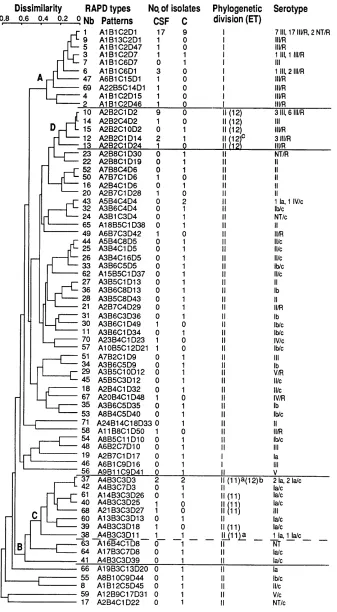
FIG. 1. Genetic relationships among 71 RAPD types. The dendrogram was generated by the unweighted pair group method with arithmetic means. The phylogenetic division (I or II) and ETs (ET11 and ET12) reported here were identified previously by MLEE as described by Quentin et al. (13). Strains belonging to the three virulent groups defined by MLEE (phylogenetic division I, ET11, and ET12) also clustered by RAPD data analysis (groups A, C, and D, respectively). Most of the strains of serotype Ia clustered in a particular group (group B). The nine strains isolated from cases of late-onset disease were in RAPD type 1 (two strains), type 5 (one strain), type 6 (one strain), type 69 (one strain), type 10 (two strains), type 30 (one strain), and type 57 (one strain). The superscript letters in the phylogenetic
division (ET) column indicate the following:a, one strain;b, one strain;c, two strains. Nb, designation number of RAPD type.
2574
on May 15, 2020 by guest
division is an indication of a high risk of invasive infection. Division II is more polymorphic, and CSF strains are more distantly related (genetic diversity of CSF strains of division II, 0.131). In this division, most of the CSF strains belong to two ETs: ET11 and ET12. Therefore, three clusters of strains may be considered to be associated with a high risk of invasive infection: division I, ET11, and ET12. These MLEE characteristics of strains are shown in Fig. 1.
RAPD fingerprinting.RAPD analysis was performed as described by Williams et al. (20) with some modifications. The PCR mixture consisted of buffer (10 mM Tris-HCl, 50 mM KCl, 2.5 mM MgCl2; pH 8.3), 100 mM each of the four deoxynucleotide triphosphates (Boehringer, Mannheim, Germany), 0.2 mM primer, 25 ng of DNA extracted and purified as previously described (3), and 0.5 U ofTaqDNA polymerase (Appligene, Pleasanton, Calif.) in a total volume of 25ml under a drop of mineral oil. A group of twenty 10-mers was acquired in Oligo 10 mer Kit S from Operon Technologies, Inc. (Alameda, Calif.). Nine other primers were synthesized by Eurogentec (Seraing, Belgium). Each sample was subjected to the first cycle of amplification (4 min at 94°C, 1 min at 36°C, and 2 min at 72°C) in a DNA Thermal Cycler 480 (Perkin-Elmer Cetus, Norwalk, Conn.). Each of the 44 subsequent cycles consisted of denaturing at 94°C for 1 min, annealing at 36°C for 1 min, and extension at 72°C for 2 min (for the last cycle, extension at 72°C for 10 min).
Amplified products (22ml) were separated by electrophoresis in a 1.4% aga-rose gel (SeaKem ME; FMC BioProducts, Rockland, Maine) and visualized by UV transillumination following ethidium bromide staining. A 1-kb DNA ladder (Life Technologies, Gaithersburg, Md.) was used in each gel as molecular size standards. A negative control, consisting of the same reaction mixture but with water instead of template DNA was included in each run.
Pattern analysis.Photographs of each gel were digitized with a video camera connected to a microcomputer (Bio-Profil; Vilber-Lourmat, Marne la Valle´e, France). The Taxotron package (Taxolab; Institut Pasteur, Paris, France),
in-cluding RestrictoScan, RestrictoTyper, Adanson, and Dendrograf, was used for numerical analysis. The images, in tagged image file format, were transferred to a Macintosh microcomputer, and the band migration distances for each lane were determined in pixel units with RestrictoScan. The molecular size of each fragment was calculated from migration distances by using cubic spline algo-rithms with RestrictoTyper. This software also generates a schematic represen-tation of electrophoretic patterns and produces a distance matrix. The relation-ships between RAPD types were calculated by the unweighted pair group method with arithmetic means (15) with the Adanson clustering program (dis-similarity) (Fig. 1). A dendrogram of the tree description file was drawn with Dendrograf.
D.The discrimination index (D) of the RAPD technique was calculated by application of Simpson’s numerical index of diversity (6).
RESULTS
Identification of informative primers. To identify primers
[image:3.612.72.552.83.461.2]that generate informative arrays of PCR products, 15 unre-latedS. agalactiaestrains were selected from the entire panel of isolates: they were isolated from various different sites and belong to different MLEE phylogenetic divisions and serotypes (13) as follows. Ten strains were isolated from the vaginas of asymptomatic women, and five strains were from CSF samples from neonates suffering from meningitis. Seven strains, includ-ing five CSF and two vaginal isolates, belonged to the MLEE phylogenetic division I. Six of these strains were of serotype III, TABLE 1. Primers tested by RAPD analysis ofS. agalactiaea
Pattern and primer Sequence
(59339) % G1C
Fragments
No. of patterns Size range
(kbp) No.
No more than five patterns generated
A1 TCACGATGCA 50 0.7–2 3–5 5
A2 ACGTATCTGC 50
A3 GTGACGTAGG 60 0.7–2 1–2 2
A5 CCGAGTCCA 66 0.7–3 2–4 2
A6 ACGCGCCCT 77 1.5–3.5 2 1
AP40 CCGCAGCCAA 70 0.4–4 2–7 4
AP44 AGCCAGTTTC 50 0.7–4 3–6 3
OPS02 CCTCTGACTG 60
OPS05 TTTGGGGCCT 60
OPS06 GATACCTCGG 60
OPS07 TCCGATGCTG 60 0.7–2 1–4 5
OPS09 TCCTGGTCCC 70 1.4 1 2
OPS10 ACCGTTCCAG 60 1.5–4 2–4 5
OPS12 CTGGGTGAGT 60 1–1.5 1–4 5
OPS13 GTCGTTCCTG 60 0.5–2 3–5 4
OPS14 AAAGGGGTCC 60 0.5 1 1
OPS15 CAGTTCACGG 60
OPS19 GAGTCAGCAG 60 0.5–3 5–6 5
OPS20 TCTGGACGGA 60 0.5–2 1–3 5
At least six patterns generated Small size range and/or several
low-intensity bands
OPS01 CTACTGCGCT 60 0.5–2 1–7 6
OPS03 CAGAGGTCCC 70 1–2 1–5 7
OPS04 CACCCCCTTG 70 0.5–2 1–5 8
OPS08 TTCAGGGTGG 60 0.7–1.6 1–6 8
OPS17 TGGGGACCAC 70 0.5–3 4–7 6
OPS18 CTGGCGAACT 60 0.7–3 1–3 6
Most informative patterns
OPS16 AGGGGGTTCC 70 0.6–3 3–6 7
AP42 AACGCGCAAC 60 0.6–4 3–5 6
A4 GCATCAATCT 40 0.5–1.5 2–4 8
OPS11 AGTCGGGTGG 70 0.1–4.5 2–11 10
aFifteen representative strains were examined.
VOL. 35, 1997 RAPD FINGERPRINTING OFS. AGALACTIAE STRAINS 2575
on May 15, 2020 by guest
http://jcm.asm.org/
and one was of serotype NT/R. Eight strains, isolated from vaginal samples, were in MLEE phylogenetic division II and were of various serotypes (Ia/c, Ib, II, II/c, II/R, III/R, IV/c, and V/R).
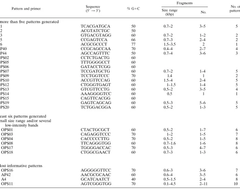
Twenty-nine oligonucleotides were selected according to the following constraints: 9 or 10 nucleotides in length, between 40 and 77% G1C in composition, and containing no palindromic sequence. Performance data for each primer in PCR with the 15 DNA samples are presented in Table 1. Nineteen primers with G1C contents of 50 to 77% resulted in no amplification product or in less than six different patterns. Six other primers gave a large number of patterns with only a small range of fragment sizes and/or a large number of low-intensity bands; the intensities of the minor fragments varied in repeated tests (Table 1). The remaining four primers (OPS11, OPS16, A4,
and AP42) with G1C contents of 40 to 70% were selected, as they gave reproducible patterns comprising fragments with a large size range and a small number of low-intensity bands (Fig. 2). They also gave the best differentiation of the 15 epi-demiologically unrelated strains. The reproducibility of the RAPD patterns obtained with these four primers was verified by repeating experiments under the same conditions. Each strain was tested at least three times.
Genetic diversity of isolates as defined by RAPD fingerprint-ing. For the whole panel of 114 strains, 24 RAPD patterns composed of 2 to 7 bands with sizes between 0.6 and 3 kbp were obtained with primer OPS16, 14 patterns composed of 1 to 5 bands of 0.5 to 1.5 kbp were observed with A4, 18 patterns composed of 1 to 6 bands of 0.6 to 4 kbp were achieved with AP42, and 50 patterns composed of 1 to 11 bands of 0.1 to 4.5 kbp resulted with OPS11. Schematic representations of all patterns are presented in Fig. 3. Each pattern contained 1 to 38 isolates.
By combination of RAPD patterns obtained with the four primers, 71 RAPD types were observed for the 114 strains. Genetic relationships among the 71 RAPD types are repre-sented in the dendrogram shown in Fig. 1. This clustering analysis was able to differentiate the group of strains belonging to MLEE phylogenetic division I (group A [Fig. 1]) from the strains belonging to MLEE phylogenetic division II with the exception of two strains (RAPD types 19 and 46 [Fig. 1]). Strains of group A are divergent from the other strains with dissimilarities of 54 to 75% (Fig. 1).
Genetic variations of strains in relation to serotypes. All
serotypes were represented by strains of a variety of RAPD types, with the exception of serotypes V, V/c, and V/R, each of which included only one strain (Fig. 1). The numbers of RAPD types per polysaccharide serotype were as follows: 12 for the 16 strains of serotype Ia (Ia and Ia/c), 12 for the 12 strains of sero-type Ib (Ib and Ib/c), 19 for the 19 strains of serosero-type II (II, II/c, and II/R), 19 for the 55 strains of serotype III (III and III/R), and 3 for the 3 strains of serotype IV (IV, IV/c, and IV/R).
RAPD clustering analysis (Fig. 1) showed that all polysac-charide serotypeable strains of group A which were previously assigned to MLEE phylogenetic division I were of serotype III and that 13 of the 16 strains of serotype Ia were in a particular group (group B [Fig. 1]). Clustering was not observed for the other serotypes.
RAPD particularities of potentially virulent groups of
strains defined previously by MLEE. French CSF isolates
mostly belong to three virulent clone families: MLEE phylo-genetic division I and two ETs (ET11 and ET12) in MLEE phylogenetic division II (13). These MLEE characteristics of strains are indicated in Fig. 1. These three particular groups of strains were also identified by RAPD clustering analysis: 38 of the 40 strains of the MLEE division I were in RAPD group A, strains of MLEE ET11 were assigned to a closely related group of strains (group C), and strains of MLEE ET12 were in a particular cluster of strains (group D) (Fig. 1).
These three clusters of strains were relatively homogeneous, with dissimilarities between strains below 35% for the strains in groups A and C and 22% for the strains in group D. CSF isolates were also assigned to a smaller number of RAPD types than were carrier isolates. Twelve patterns were identified with primer OPS16 for CSF isolates versus 18 patterns for carrier isolates, 9 patterns were identified with A4 for CSF isolates versus 13 for carrier isolates, 8 patterns were identified with AP42 for CSF isolates versus 15 for carrier isolates, and 21 patterns were identified with OPS11 for CSF isolates versus 36 for carrier isolates.
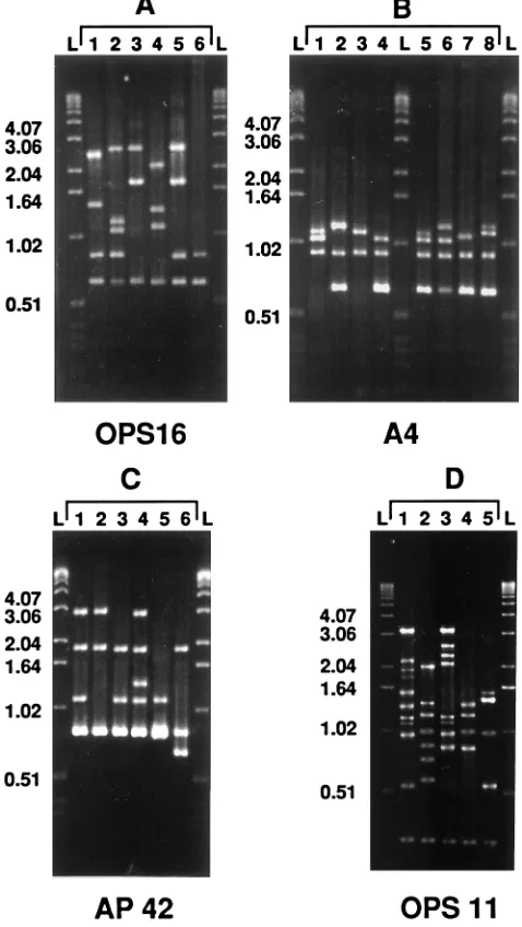
[image:4.612.58.297.74.498.2]The three virulent groups of strains exhibited specific RAPD characteristics. Three fragments, one of 0.64 kbp obtained with FIG. 2. Illustration of the RAPD patterns most frequently generated with
primers OPS16 (A1 to A6), A4 (B1 to B8), AP42 (C1 to C6), and OPS11 (D1 to D5), which include at least four strains ofS. agalactiae. These primers were chosen because they generate patterns which contain a large range of fragment sizes and a small number of minor fragments. L, 1-kb DNA ladder (DNA molecular size markers).
on May 15, 2020 by guest
http://jcm.asm.org/
FIG. 3. Schematic representations of RAPD patterns generated with primers OPS16 (A1 to A24), A4 (B1 to B14), AP42 (C1 to C18), and OPS11 (D1 to D50) for
114 S. agalactiae strains. Three amplified fragments (2) (Table 2) were able to differentiate strains belonging to virulent group A (Fig. 1) from the other isolates. RAPD
pattern B3 identified strains of virulent group C (Fig. 1). Combination of RAPD patterns A2 and B2 (A2B2) characterized the strains of virulent group D (Fig. 1). The
numbers (n) of cerebrospinal fluid strains (CSFa) and carrier strains (Cb) are shown to the right of the schematic representation of RAPD patterns.
VOL. 35, 1997 RAPD FINGERPRINTING OF S. AGALACTIAE STRAINS 2577
on May 15, 2020 by guest
http://jcm.asm.org/
primer A4, one of 1.2 kbp generated with primer AP42, and one of 2.4 kbp produced with primer OPS 16 (Fig. 3) were able to identify 37 of the 38 strains (97.3%) of virulent group A (Table 2). The 12 strains of group C all gave RAPD pattern B3 (Fig. 1) with primer A4 (Fig. 2 and 3), a pattern which was observed only for 3 other strains which did not belong to group C. The 15 strains of group D were characterized by a particular combination of patterns A2 and B2 (Fig. 1) obtained with primers OPS16 and A4, respectively (Fig. 2 and 3).
The nine CSF strains isolated from late-onset disease cases did not exhibit particular RAPD characteristics (Fig. 1). Five of these nine isolates were in group A, which contained 27 CSF strains. Two strains were in group D, which included 13 CSF strains. Of the 8 CSF strains which did not cluster with other
CSF strains, two were from late-onset disease cases. None of the 6 CSF strains of CSF group C was isolated from late-onset disease cases.
RAPD typing as an epidemiological tool.An
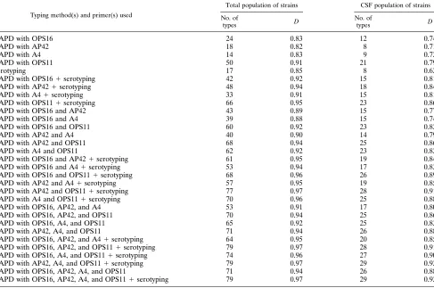
epidemiologi-cal tool would be valuable in investigating cross-colonization or possible nosocomial or epidemic invasive S. agalactiae infec-tions. Therefore, we evaluated the discriminatory power of the RAPD typing method for two classes of isolates: all strains and the 54 invasive CSF strains (7). By combining data obtained with the four primers, it was possible to define 71 RAPD types for the 114 clinical isolates and 26 types for the 54 CSF strains. D was calculated for serotyping and for each primer, either alone or combined (Table 3). The simplest typing scheme which had an index greater than 0.90, the lowest threshold value for interpreting typing results with confidence (6), was obtained by RAPD done with primers AP42 and OPS11 and serotyping (D5 0.97 for the total population of strains and D50.91 for the CSF population of strains).
DISCUSSION
[image:6.612.59.298.101.171.2]We evaluated the effectiveness of a simple and fast tech-nique called the RAPD assay (20) for characterizing a collec-tion ofS. agalactiaestrains previously studied by MLEE (13), a well-known method for studying the genetic structure of bacterial populations (14). There was general agreement be-tween groupings obtained by RAPD and MLEE analyses. This agreement of results, which is consistent with previous studies of other bacterial pathogens (18) and parasitic protozoa (16), TABLE 2. Frequencies of three specific fragments amplified by
RAPD analysis allowing differentiation of virulent group A strains from other isolates
Primer Size of fragment (kbp)
No. of isolates (%) exhibiting the fragmenta
Group A Other strains
A4 0.64 1 (2.6) 57 (75.0)
AP42 1.2 0 73 (96.0)
OPS16 2.4 37 (97.3) 3 (3.9)
aThe values for group A strains were significantly different from the values for
the other strains (P,0.001 by thex2test).
TABLE 3. NumericalDs calculated from RAPD data (with one or more of the four primers) used alone or in combination with serotype data to type the total population of 114 strains and the 54 CSF strains
Typing method(s) and primer(s) used
Total population of strains CSF population of strains
No. of
types D
No. of
types D
RAPD with OPS16 24 0.83 12 0.74
RAPD with AP42 18 0.82 8 0.71
RAPD with A4 14 0.83 9 0.72
RAPD with OPS11 50 0.91 21 0.79
Serotyping 17 0.85 8 0.62
RAPD with OPS161serotyping 42 0.92 15 0.81
RAPD with AP421serotyping 48 0.94 18 0.84
RAPD with A41serotyping 33 0.91 15 0.81
RAPD with OPS111serotyping 66 0.95 23 0.86
RAPD with OPS16 and AP42 43 0.89 15 0.77
RAPD with OPS16 and A4 39 0.88 15 0.74
RAPD with OPS16 and OPS11 60 0.92 23 0.82
RAPD with AP42 and A4 40 0.90 14 0.79
RAPD with AP42 and OPS11 68 0.94 25 0.86
RAPD with A4 and OPS11 62 0.92 23 0.82
RAPD with OPS16 and AP421serotyping 61 0.95 19 0.84
RAPD with OPS16 and A41serotyping 53 0.94 17 0.83
RAPD with OPS16 and OPS111serotyping 68 0.96 26 0.89
RAPD with AP42 and A41serotyping 57 0.95 19 0.85
RAPD with AP42 and OPS111serotyping 77 0.97 28 0.91
RAPD with A4 and OPS111serotyping 70 0.96 25 0.88
RAPD with OPS16, AP42, and A4 53 0.91 17 0.80
RAPD with OPS16, AP42, and OPS11 70 0.94 25 0.86
RAPD with OPS16, A4, and OPS11 65 0.92 25 0.83
RAPD with AP42, A4, and OPS11 71 0.94 26 0.88
RAPD with OPS16, AP42, and A41serotyping 64 0.95 20 0.85
RAPD with OPS16, AP42, and OPS111serotyping 79 0.97 28 0.91
RAPD with OPS16, A4, and OPS111serotyping 74 0.96 27 0.90
RAPD with AP42, A4, and OPS111serotyping 79 0.97 29 0.92
RAPD with OPS16, AP42, A4, and OPS11 71 0.94 26 0.88
RAPD with OPS16, AP42, A4, and OPS111serotyping 79 0.97 29 0.92
on May 15, 2020 by guest
http://jcm.asm.org/
[image:6.612.66.553.405.728.2]was observed only when the data obtained with the four prim-ers were combined so as to be able to differentiate isolates efficiently (D.0.90).
Although the general genetic structure of ourS. agalactiae collection as determined by numerical analysis of MLEE was comparable to that determined from RAPD data, RAPD anal-ysis allowed further subdivision of strains that are not distin-guishable by MLEE analysis. Indeed, the virulent groups of strains identified by MLEE analysis were each apparently very homogeneous (13). In contrast, the RAPD method, which identified the same virulent families in cluster analysis, was able to discriminate strains inside each cluster. This indicates that RAPD typing is a far more sensitive method than MLEE typing for epidemiological investigations as previously indi-cated by Wang et al. (18) for other species. In addition, the RAPD technique is a more rapid and accessible system than MLEE, which is nevertheless a good reference method used by a few laboratories for defining the phylogenetic structure of a microbial population. The simplest typing scheme including RAPD which has acceptable discriminatory power for all S. agalactiaestrains and CSF isolates combines two independent typing systems: RAPD done with primers AP42 and OPS11 and serotyping (Table 3). Because community transmissions or cross-colonizations ofS. agalactiaestrains are possible (2, 11), such an accessible tool would be a great improvement for microbiologists. The good correlation between groupings obtained with MLEE and RAPD data analyses also indicates that the nu-merical analysis of RAPD data and the four primers used here may be proposed for the delineation of clones within S. aga-lactiae populations. Interestingly, the three major families of virulent clones of S. agalactiae strains identified by MLEE (division I, ET11, and ET12) (13) were assigned to three dis-tinct RAPD analysis groups (A, C, and D, respectively [Fig. 1]) exhibiting specific RAPD characteristics. Thus, the extensive similarities among genes coding for metabolic enzymes (10, 13) and among rRNA genes (3) observed for CSF S. agalactiae isolates are also found throughout the microbial genome ofS. agalactiaerandomly explored by RAPD analysis. No consistent difference was observed between strains isolated from early-and late-onset disease. This demonstrates a strong homogene-ity in the genomes of S. agalactiaestrains able to invade the central nervous system of neonates and extends earlier results (3, 10, 13) indicating that strains belonging to some genotypes are more likely than others to cause early or late neonatal meningitis. Particularities of CSF isolates shown by MLEE (13) or iden-tified by ribotyping (3) are not suitable for screening for po-tentially highly virulent isolates among strains colonizing preg-nant women because these methods are neither rapid nor widely accessible. In contrast, RAPD is a rapid and simple technique which may be used for prenatal screening programs to assess the pathogenic potential ofS. agalactiaestrains iso-lated from the vagina of the mother and/or from the gastric fluid samples from asymptomatic neonates. The presence of the particular patterns or amplified fragments that characterize the virulent clones would indicate a high risk of disease. This would be very useful for planning prophylactic strategies. Nev-ertheless, PCR may not be within the diagnostic scope of some laboratories, and interlaboratory reproducibility of RAPD pat-terns has still not been clearly demonstrated (12, 17). There-fore, the specific fragments amplified by the RAPD system that are characteristic of particular groups of strains which contain most of the CSF isolates should be studied further so that specific DNA sequences can be designed for a PCR-based detection system of S. agalactiaevirulent-clone families. This information could also be used for the development of probes suitable, for example, for DNA immunoassays. In addition and on
account of the geographical variations of the genetic structure of S. agalactiae, other national collections should be examined.
ACKNOWLEDGMENTS
This research was supported by grants from the Centre Hospitalier Universitaire of Tours.
We thank the Colle`ge de Bacte´riologie Virologie et Hygie`ne des Hoˆpitaux de France for supplying the CSF strains via Pierre Geslin.
REFERENCES
1.Baker, C. J., and M. S. Edwards.1995. Group B streptococcal infections, p. 980–1054.InJ. S. Remington and J. O. Klein (ed.), Infectious diseases of the fetus and newborn infant, 4th ed. The W. B. Saunders Co., Philadelphia, Pa. 2.Bingen, E., E. Denamur, N. Lambert-Zechovsky, Y. Aujard, N. Brahimi, P. Geslin, and J. Elion.1992. Analysis of DNA restriction fragment length polymorphism extends the evidence for breast milk transmission in Strepto-coccus agalactiaelate-onset neonatal infection. J. Infect. Dis.165:569–573. 3.Chatellier, S., H. Huet, S. Kenzi, A. Rosenau, P. Geslin, and R. Quentin.
1996. Genetic diversity of rRNA operons of unrelatedStreptococcus agalac-tiaestrains isolated from cerebrospinal fluid of neonates suffering from meningitis. J. Clin. Microbiol.34:2741–2747.
4.Geslin, P., G. Sissia, J. Jelinkova, A. Fremaux, and J. Motlova.1992. Serotype distribution of group B streptococci isolated from human sources in France over a 10-year period (1980–1989). Zentralbl. Bakteriol. Suppl.22:484–485. 5.Helmig, R., N. Uldbjerg, J. Boris, and M. Kilian.1993. Clonal analysis of
Streptococcus agalactiaeisolated from infants with neonatal sepsis or men-ingitis and their mothers and from healthy pregnant women. J. Infect. Dis.
168:904–909.
6.Hunter, P. R., and M. A. Gaston.1988. Numerical index of the discrimina-tory ability of typing systems: an application of Simpson’s index of diversity. J. Clin. Microbiol.26:2465–2466.
7.Maslow, J. N., M. E. Mulligan, and R. D. Arbeit.1993. Molecular epidemi-ology: application of contemporary techniques to the typing of microorgan-isms. Clin. Infect. Dis.17:153–164.
8.Mattingly, S. J., and E. K. Eskew.1993. Temperature sensitivity of fructose-1,6-biphosphate aldolase accounts for the inability of the high-virulence clone of Streptococcus agalactiaeto grow at 40°C. Curr. Microbiol.26:147–150. 9.Mattingly, S. J., J. J. Maurer, E. K. Eskew, and F. Cox.1990. Identification
of a high-virulence clone of serotype IIIStreptococcus agalactiaeby growth characteristics at 40°C. J. Clin. Microbiol.28:1676–1677.
10. Musser, J. M., S. J. Mattingly, R. Quentin, A. Goudeau, and R. K. Selander.
1989. Identification of a high-virulence clone of type IIIStreptococcus aga-lactiae(group B streptococcus) causing invasive neonatal disease. Proc. Natl. Acad. Sci. USA86:4731–4735.
11. Noya, F. J. D., M. A. Rench, T. J. Metzger, G. Colman, J. Naidoo, and C. J. Baker.1987. Unusual occurrence of an epidemic of type Ib/c group B strepto-coccal sepsis in a neonatal intensive care unit. J. Infect. Dis.155:1135–1144. 12. Penner, G. A., A. Bush, R. Wise, W. Kim, L. Domier, K. Kasha, A. Laroche,
G. Scoles, S. J. Molnar, and G. Fedak.1993. Reproducibility of random amplified polymorphic DNA (RAPD) analysis among laboratories. PCR Methods Applications2:341–345.
13. Quentin, R., H. Huet, F.-S. Wang, P. Geslin, A. Goudeau, and R. K. Selander.
1995. Characterization ofStreptococcus agalactiaestrains by multilocus enzyme genotype and serotype: identification of multiple virulent clone families that cause invasive neonatal disease. J. Clin. Microbiol.33:2576–2581.
14. Selander, R. K., D. A. Caugant, H. Ochman, J. M. Musser, M. N. Gilmour, and T. S. Whittman.1986. Methods of multilocus enzyme electrophoresis for bacterial population genetics and systematics. Appl. Environ. Microbiol.
51:873–884.
15. Sneath, P. H. A., and R. R. Sokal.1973. Numerical taxonomy. W. H. Free-man and Co., San Francisco, Calif.
16. Tibayrenc, M., K. Neubauer, C. Barnabe, F. Guerrini, D. Skarecky, and F. J. Ayala.1993. Genetic characterization of six parasitic protozoa: parity be-tween random-primer DNA typing and multilocus enzyme electrophoresis. Proc. Natl. Acad. Sci. USA90:1335–1339.
17. Van Belkum, A., J. Kluytmans, W. Van Leeuwen, R. Bax, W. Quint, E. Peters, A. Fluit, C. Vandenbroucke-Grauls, A. Van Den Brule, H. Koeleman, W. Melchers, J. Meis, A. Elaichouni, M. Vaneechoutte, F. Moonens, N. Maes, M. Struelens, F. Tenover, and H. Verbrugh.1995. Multicenter eval-uation of arbitrarily primed PCR for typing ofStaphylococcus aureusstrains. J. Clin. Microbiol.33:1537–1547.
18. Wang, G., T. S. Whittam, C. M. Berg, and D. E. Berg.1993. RAPD (arbitrary primer) PCR is more sensitive than multilocus enzyme electrophoresis for distinguishing related bacterial strains. Nucleic Acids Res.21:5930–5933. 19. Welsh, J., and M. McClelland.1990. Fingerprinting genomes using PCR
with arbitrary primers. Nucleic Acids Res.18:7213–7218.
20. Williams, J. G. K., A. R. Kubelik, K. J. Livak, J. A. Rafalski, and S. V. Tingey.1990. DNA polymorphisms amplified by arbitrary primers are useful as genetic markers. Nucleic Acids Res.18:6531–6535.
VOL. 35, 1997 RAPD FINGERPRINTING OFS. AGALACTIAE STRAINS 2579