Prolactin and prolactin receptors are expressed
and functioning in human prostate.
M T Nevalainen, … , P M Martikainen, P L Harkonen
J Clin Invest.
1997;
99(4)
:618-627.
https://doi.org/10.1172/JCI119204
.
Prolactin is widely expressed in different tissues, and it is presumed to have both local and
systemic actions. In males it is known to influence reproductive functions but the
significance and mechanisms of prolactin action in male accessory reproductive tissues are
poorly understood. Here we show that prolactin acts as a direct growth and differentiation
factor for human prostate, as measured by changes in DNA synthesis and epithelial
morphology of organ cultures. Furthermore, we report the expression in human prostate of a
short prolactin receptor form in addition to the long form, based upon ligand cross-linking
studies and RT-PCR analysis of mRNA expression. The highest density of prolactin
receptors was detected in the secretory epithelial cells by immunohistochemistry. Finally,
we report that prolactin is locally produced in human prostate epithelium, as evidenced by
marked prolactin immunoreactivity in a significant portion of prostate epithelial cells, with
parallel expression of prolactin mRNA in human prostate. Collectively, these data provide
significant support for the existence of an autocrine/paracrine loop of prolactin in the human
prostate and may shed new light on the involvement of prolactin in the etiology and
progression of neoplastic growth of the prostate.
Research Article
Find the latest version:
J. Clin. Invest.
© The American Society for Clinical Investigation, Inc. 0021-9738/97/02/0618/10 $2.00
Volume 99, Number 4, February 1997, 618–627
Prolactin and Prolactin Receptors Are Expressed and Functioning in
Human Prostate
Marja T. Nevalainen,* Eeva M. Valve,* Patricia M. Ingleton,‡ Martti Nurmi,§ Paula M. Martikainen,iand Pirkko L. Härkönen*
*Institute of Biomedicine, Department of Anatomy and the Medicity Research Laboratory, University of Turku, FIN-20520 Turku, Finland; ‡Institute of Cancer Studies, University of Sheffield, Medical School, United Kingdom; §Department of Surgery, University of
Turku, FIN-20520 Turku, Finland; and iDepartment of Pathology, University of Turku, FIN-20520 Turku, Finland
Abstract
Prolactin is widely expressed in different tissues, and it is presumed to have both local and systemic actions. In males it is known to influence reproductive functions but the sig-nificance and mechanisms of prolactin action in male acces-sory reproductive tissues are poorly understood. Here we show that prolactin acts as a direct growth and differentia-tion factor for human prostate, as measured by changes in DNA synthesis and epithelial morphology of organ cultures. Furthermore, we report the expression in human prostate of a short prolactin receptor form in addition to the long form, based upon ligand cross-linking studies and RT-PCR analy-sis of mRNA expression. The highest density of prolactin receptors was detected in the secretory epithelial cells by immunohistochemistry. Finally, we report that prolactin is locally produced in human prostate epithelium, as evi-denced by marked prolactin immunoreactivity in a signifi-cant portion of prostate epithelial cells, with parallel expres-sion of prolactin mRNA in human prostate. Collectively, these data provide significant support for the existence of an autocrine/paracrine loop of prolactin in the human pros-tate and may shed new light on the involvement of prolactin in the etiology and progression of neoplastic growth of the prostate. (J. Clin. Invest. 1997. 99:618–627.) Key words: hu-man prostate • prolactin effects • prolactin receptors •
pro-lactin expression
Introduction
Prolactin (Prl)1 is a peptide hormone of numerous and distinct
bioactive and immunoreactive variants (1). Its versatile func-tions are known to include effects on the immune system, on osmoregulation, and on behavior (1, 2). In reproduction Prl has been shown to have luteotropic actions and to regulate tes-ticular function by increasing the binding of luteinizing
hor-mone to Leydig cells (1, 2). Furthermore, Prl stimulates lacta-tion and growth in mammary gland (1, 2) by activating the expression of milk protein genes (3, 4) and genes presumably necessary for proliferation of mammary cells (5–7). In prostate Prl has been presumed to have a role in the regulation of pros-tatic development, growth, and differentiation (8–11). Our previous results showed that in organ culture of rat dorsal and lateral prostate Prl has a direct effect on morphology, on DNA labeling with [3H]thymidine, and on the expression of prostatic
tissue specific genes M-40 and RWB encoding secretory pro-teins (12). The dorsal and lateral lobes are the parts of rat prostate that are considered most homologous to human pros-tate (13). Prl has also been suggested to be involved in the reg-ulation of abnormal prostatic function (8, 14–16), but little is known, however, about the specific and direct effects of Prl on human prostate. The source of Prl and the mechanisms under-lying the responses of prostatic cells to Prl have remained un-clear (8, 10, 17–21).
The gene for Prl receptor (22) belongs to the cytokine-hematopoietin receptor superfamily encoding receptors for growth hormone, granulocyte colony-stimulating factor, gran-ulocyte-macrophage colony-stimulating factor, erythropoie-tin, leukemia inhibitory factor, oncostatin M, ciliary neu-rotrophic factor, and interleukins 2–7 (23), based on conserved sequences in their extracellular domains (24). In rat, three iso-forms (short, long, and intermediate) of Prl receptor have been identified (22, 25, 26). The short and long forms differ from each other in the length of their cytoplasmic domain resulting from alternative splicing of a single primary transcript (22, 25). The third rat Prl receptor, which is intermediate in size and has first been identified in the Nb2 pre-T rat lymphoma cell line (26), possibly (27, 28), results from a partial deletion in the Prl receptor gene within the cytoplasmic region of the long recep-tor form (26). In humans only a single Prl receprecep-tor cDNA, with a considerable homology to the rat long receptor form, has been cloned (29). However, the expression of also the in-termediate form of Prl receptor in human mammary tissue, re-sulting from RNA processing, was reported recently (30). Of interest, only the long and intermediate receptors have been reported to be able to transduce the hormonal message to the expression of milk protein genes (4), whereas the mitogenic signal has been reported to be equally transmitted by both the long and the short Prl receptor forms (31). Receptors for Prl are widely expressed in different tissues (32–36), and extrapi-tuitary production of Prl has been shown in lymphocytes, pla-centa, and mammary tissue (30, 37–42).
The purpose of this work was to characterize the effects of Prl on human prostatic tissue, to study its receptors, and to evaluate whether Prl is synthesized locally in human prostate. The Prl regulation of prostatic tissue was studied in organ cul-tures. This method was used because in primary culture nor-mal prostatic epithelial cells frequently dedifferentiate and
Address correspondence to Dr. Pirkko Härkönen, Department of Anatomy, Institute of Biomedicine, University of Turku, Kiinamyl-lynkatu 10, FIN-20520 Turku, Finland. Phone: 358-2-3337379; FAX: 358-2-3337352; E-mail: [email protected]
Received for publication 13 September 1996 and accepted in re-vised form 3 December 1996.
lose androgen sensitivity (43), whereas organ culture of human prostate (44) as well as rat dorsolateral (12, 36) and ventral prostate (45–47) has been shown to maintain hormone respon-siveness and specific tissue function. This is probably due to the presence of all tissue components, which allows the inter-actions between epithelium and stroma, shown to be impor-tant for the maintenance of the differentiated state of prostatic epithelium (48, 49). The prostatic expression of Prl receptors and the local synthesis of Prl were studied at both protein and mRNA levels.
The results show that Prl has a characteristic effect on the morphology of human prostate and that it increases prostatic DNA synthesis. In this work we demonstrate, for the first time, that in addition to the long Prl receptors also the short form Prl receptors are expressed in human prostate. The Prl receptors are localized to the prostatic secretory epithelium. We also demonstrate the extrapituitary synthesis of Prl within prostate tissue which may attribute Prl a specific function as an auto-crine/paracrine mediator within human prostate, in addition to its endocrine function.
Methods
Tissue samples and organ culture.Human prostate tissues were ob-tained from seven patients (age 39–76 yr; mean age6SE: 58.864.4) undergoing cystoprostatectomy because of bladder carcinoma. The tissues were used in the experiments after approval by the Local Eth-ical Committee. A 1-cm-thick horizontal section of each prostate tis-sue was transported to the laboratory in ice cold medium 199 with Earle’s salts (Flow Laboratories, Newcastle, United Kingdom) sup-plemented with penicillin (100 IU/ml), streptomycin sulfate (100 ml/ ml), and glutamine (100 mg/ml). A thin slice of tissue was taken for histopathologic examination to confirm the diagnosis of each speci-men, and the samples were taken for immunohistochemical stainings, membrane preparations, and mRNA determination. The rest of the prostate tissue was cultivated as explant culture which were started all within 1–3 h after surgery.
The organ cultures were performed as described earlier (12, 36, 44–47). Briefly, prostatic tissue was cut into 1-mm3 pieces and
trans-ferred onto lens papers lying on stainless steel grids in Petri dishes. The medium used was phenol-free medium 199 with Earle’s salts (Flow Laboratories) containing dialyzed 5% fetal calf serum, penicil-lin (100 IU/ml), streptomycin sulfate (100 ml/ml), and glutamine (100
mg/ml). The basal medium also contained the combination of insulin (insulin Lente; Novo Industries, Copenhagen, Denmark) and dexa-methasone (9-fluoro-16-methylprednisolone; Sigma Chemical Co., St. Louis, MO) at concentrations of 0.08 IU/ml and 1027 M,
respec-tively. The gas atmosphere was a mixture of O2, CO2, and air (40:5:
55), and the temperature was 378C. The explants were cultured for 7 d and the medium was changed daily. Dihydrotestosterone (DHT) (5-androstan-17b-ol-3-one; Sigma Chemical Co.) (final concentra-tion 1027 M) was dissolved in ethanol. The final concentration of
eth-anol in the culture medium was 0.1%. Human prolactin (hPrl) (a gift from Dr. Parlow, National Institute of Diabetes and Digestive and Kidney Diseases, Baltimore, MD) was diluted in 0.01 M NaHCO3,
pH 8.0, and used at the concentration of 45 nM, which has been proved to be suitable for the tissue culture conditions (12, 46, 47). Four to five parallel dishes were always cultured for each hormone combination in each experiment.
Morphology. For morphological evaluation three to five explants from every dish were taken randomly in formalin at the end of the culture and embedded in paraffin. Serial sections of 7 mm were cut from each piece and stained with hematoxylin and eosin.
[3H]Thymidine incorporation and autoradiography. The incorpor-ation of [3H]thymidine into DNA was measured at the end of each
culture. The 40 explants of two Petri dishes were incubated with 2
mCi/ml methyl [3H]thymidine (Amersham, Aylesbury, United
King-dom) for 2 h at 378C and washed thereafter with ice-cold medium to terminate the labeling reaction. 20 explants were taken for autora-diography and 20 explants were homogenized in 400 ml water. The ho-mogenate was precipitated with 20% TCA, washed three times with 5% TCA, and hydrolyzed in 5% TCA at 908C. The supernatant was used for DNA determination (50), and its radioactivity was measured by a liquid scintillation counter. The incorporation of radioactive thy-midine into DNA is expressed as counts per minute per 100 mg of DNA. The acid-soluble radioactivity was counted from the superna-tant before hydrolysis of the TCA-precipitated DNA. Descriptive statistics was used to analyze the differences in DNA labeling (cpm/ 100 mg DNA) between the explants cultured with or without Prl for 7 d. One sample t test was used to analyze the difference between ra-tio IDP (cpm/100 mg DNA) and ID (cpm/100 mg DNA) from one. The 95% confidence interval for mean was also calculated. P values , 0.05 were interpreted as statistically significant. The statistical computa-tions were performed using SAS program package (51).
For autoradiography, slices of the labeled explants were coated with Kodak nuclear emulsion NTB 3 after routine histologic process-ing. The exposure was carried out at 2158C for 7 d. The autoradio-grams were developed in Kodak D19 developer and stained with he-matoxylin and eosin.
Prl receptor immunohistochemistry.Human prostatic tissues were fixed in 4% formalin overnight, dehydrated, cleared, and embedded in paraffin. Sections were cut at 7 mm and mounted on poly-l-lysine–
coated slides. The procedure for immunohistochemical analysis of prostatic tissue is based on that of Sternberger (52) and has been de-scribed in detail by Danks et al. (53). Briefly, sections were rehy-drated, nonspecific peroxidase activity was blocked by immersion in methanol-peroxide, nonspecific binding sites were blocked by normal goat serum, and sections were incubated in specific primary antibody for 2 h at the dilution of 1:200 at room temperature. The specific poly-clonal anti-rPrl receptor antibody (R120) (36) recognizing the extra-cellular domain of both the short and long Prl receptors (residues 53– 68 of the external domain common to both receptor forms) was used. The production and use of polyclonal anti-Prl receptor antibodies has been described previously (36). After incubation with goat anti–rab-bit serum (Serotec Ltd., Oxford, United Kingdom) the rabanti–rab-bit peroxi-dase-antiperoxidase (Dakopatts a/s, Copenhagen, Denmark) binding was detected using 3,3-[prime] diaminobenzidine as chromogen. For controls, primary antibody was replaced by nonimmune rabbit serum (NRS) or conjugate-absorbed serum; absorption with thyroglobulin alone did not affect specific staining. Rat kidney, human chorion leave, human breast, and rat and human female liver tissues were used as positive control tissues.
Prl immunohistochemistry. Paraffin sections (7 mm) of human prostates fixed in 4% formalin were used for Prl immunohistochemis-try. A polyclonal anti-hPrl antibody and biotin-streptavidin amplified immunodetection technology (both obtained from BioGenex Labo-ratories, San Ramon, CA) with minor modifications were used as the detection system. After deparaffination of the slides and rehydration in graded alcohol, the sections were treated with 0.4% pepsin (Merck, Darmstadt, Germany) in 0.01 N HCl in 378C to unmask the antigen epitopes. The endogenous peroxidase activity was blocked by incu-bating the slides in 3% peroxide in water, and the nonspecific binding of the Igs was minimized by 1 h of preincubation of the slides in nor-mal goat serum (BioGenex Laboratories) at room temperature. The primary antibody was applied at a dilution of 5:100 in 1% BSA in PBS overnight. Antigen-antibody complexes were detected by a bio-tinylated anti–rabbit secondary antibody and a streptavidin-biotin-horseradish-peroxidase complex with 3,3-[prime] diaminobenzidine as chromogen and Mayer hematoxylin as a counterstain. Negative controls were done either without the primary antibody or with NRS (BioGenex Laboratories). Human prolactinomas were used as posi-tive control tissues.
human prostates were prepared according to the method of Ben-David et al. (54). Briefly, tissues were homogenized at four times their volume in homogenization buffer [0.3 M sucrose, 25 mM Tris, 0.25 mM PMSF, 10 mM monothioglycerol, 1.0 mM EGTA, 0.1% Tween 20 (Merck), and aprotinin (100,000 kallikrein inactivator units/liter)] with an Ultraturrax homogenizer (Janke & Kunkel, IKA-Labortechnik, Staufen, Germany). The homogenate was centrifuged at 500 g for 15 min at 48C, after which the supernatant containing the crude membrane fractions was again centrifuged at 25,000 g for 30 min at 48C. The pellet was washed once with a wash buffer [25 mM Tris, 10 mM MgCl2, 1.0 mM EGTA, 0.1% Tween 20, 10 mM
mono-thioglycerol, 0.25 mM PMSF, and aprotinin (100,000 kallikrein inacti-vator units/liter)], then resuspended in it, and used for determination of protein (55) and for receptor binding studies.
hPrl was iodinated using the IODO-GEN method (56) as de-scribed previously (36). The specific activity of labeled Prl prepared in this manner ranged from 200 to 225 mCi/mg, as determined by a self-displacement assay. The fraction of the labeled hormone deriva-tives capable of binding to an excess of estrogen-treated female rat liver membranes was 20–30%. The binding assays were performed according to the method of Ben-David et al. (54). Briefly, 100 mg of membrane proteins, pretreated with 3 M MgCl2 (36), and 1 ng of
[125I]hPrl (300,000 cpm) were incubated in 0.5 ml incubation buffer
(0.1% BSA in above described wash buffer) at 208C for 16 h in the absence or presence of unlabeled hPrl (5 mg). After binding equilib-ria were established the entire reaction mixture was centrifuged at 12,000 g for 4 min to separate the free iodine from receptor-bound [125I]hPrl. Freshly prepared disuccimidylsuberate in DMSO was then
added to labeled membranes to a final concentration of 0.5 mM in wash buffer. After an incubation of 15 min in an ice bath, ice-cold wash buffer was added to the samples to terminate the reaction, and the reaction mixture was then centrifuged at 12,000 g at 48C for 15 min. Each pellet was then applied to a 12% vertical slab SDS poly-acrylamide gel, and electrophoresis was performed according to the method of Laemmli (57) under reducing conditions (in the presence of 5% 2-mercaptoethanol and 100 mM DTT), after which autoradiog-raphy was carried out. The experiment was repeated six times.
Northern blot analysis. RNA was isolated according to the method of Chomczynski and Sacchi (58). A PstI-EcoRV cDNA fragment (243 bp) of the rat liver Prl receptor cDNA clone F3 (25, 59) (a gift
from Dr. Paul Kelly, INSERM U-344, Faculty of Medicine, Necker, Paris, France), corresponding to region unique to the short form of rat Prl receptor cDNA (see Fig. 5),was labeled by a random oligonu-cleotide priming method (60), and the Northern blotting and hybrid-ization were performed as described previously (36). The experiment was repeated six times. The expression of the short Prl receptor mRNAs in liver from estrogen-treated female rats (25, 59) was used as a positive control (36). Furthermore, hybridization of the same fil-ters with a human long Prl receptor probe (HindIII fragment of H1/
H2 cDNA clone) (29) was used as a reference when assessing the sizes
of mRNAs for the short Prl receptor. This probe detects mRNAs of 2.8, 3.5, and 7.3 kb from human tissues (29).
Southern blotting. High-molecular weight DNA was isolated from human prostate samples as described previously (61). 10-mg ali-quots of DNA were digested with restriction endonucleases (HindIII, EcoRI, and BamHI) at 378C for 3 h, and the digested DNA was frac-tionated by electrophoresis through a 1% agarose gel, denatured, and transferred to a Gene Screen Plus membrane (New England Nuclear, Boston, MA) according to the salt transfer protocol of the manufac-turer. Thereafter, the membrane was treated as described before (61), and prehybridized, hybridized, and washed as described for Northern blots. A PstI-EcoRV cDNA fragment (243 bp) of the rat liver clone F3 recognizing selectively the short form Prl receptor (25,
59) (see Fig. 5) was labeled by a random oligonucleotide priming method (60). The experiment was repeated three times.
Prl receptor and Prl RT-PCR. In Prl receptor RT-PCR the se-quences of primers 1 (sense primer, 917–944) and 2 (antisense primer, 1780–1805) (see Fig. 5) were 59-ACT ATG AGG ACT TGC TGG
TGG AGT ATT T-39 and 59-CAC TTG CTT GAT GTT GCA GTG AAG TT-39, respectively. Primers 1 and 2 were used for the amplifi-cation of a 893-bp cDNA fragment derived from the cytoplasmic do-main of the long form Prl receptor mRNA (30). The sequences of primers 3 (sense primer, 607–627) and 4 (antisense primer, 903–923) (see Fig. 5) were 59-TGC AAG CCA GAC CAT GGA TAC-39 and 59-AGT TCC CCT GCA TTG TCC AGT-39, respectively. Primers 3 and 4 amplified a 317-bp cDNA fragment and were used to detect the short form Prl receptor mRNA (62). In Prl RT-PCR the 59(sense) oli-gonucleotide primer used was 59-GCC AGG TGT CAG CCC GGA AA-39 and 39(antisense) oligonucleotide primer was 59-TGG GAA TCC CTG CGC AGG CA-39. These Prl primers spanned all four in-trons of genomic Prl DNA and produced a fragment of 612 bp (42, 63) in RT-PCR. The use of all these sets of primers in both Prl recep-tor and Prl RT-PCR has been described previously (30, 42, 62, 63).
Total RNA extracted from T47D human breast cancer cells and from livers of estrogen-treated female rats were used as positive con-trols for the expression of the long (29) and short (25, 59) Prl receptor mRNAs, respectively. For the expression of Prl mRNA, total RNA from MCF-7 and T47D cells were used as positive controls (30, 42). H2O and all the samples without reverse transcriptase were used as
negative controls. Total RNA from human prostates and from the positive control tissues were reverse transcribed to synthesize single-stranded cDNAs. 2 mg of RNA was used as a template for cDNA syn-thesis. A 39-oligo (300 ng) was added to the total RNA and was brought to a final volume of 4 ml with distilled water. The samples were heated at 658C for 5 min and were cooled slowly to room tem-perature. Then, 4 ml of 53 moloney murine leukemia virus (M-MLV) first strand buffer, 20 U of RNase inhibitor (Promega Corp., Madi-son, WI), 20 nmol of each dNTP (Pharmacia Biotech., Uppsala, Swe-den), and 100 U of M-MLV reverse transcriptase (Promega Corp.) were added to the reaction mixture to a final volume of 20 ml. The tubes were heated at 378C for 1 h, and M-MLV–RT was inactivated at 998C for 5 min after which the tubes were cooled at 58C for 5 min.
The cDNAs were amplified by 30 cycles of PCR (Perkin Elmer PCR instrument, Applied BioSystem Divisions, Foster City, CA) using each set of primers. 8 ml of 103 DynaZyme buffer, 300 ng of each primer, and sterile water were added to the 20 ml of the reverse-transcribed cDNA to a final volume of 99 ml, after which 1 ml (2 U/ml) of DynaZyme (thermostable DNA polymerase from Thermus brockianus; Finn-zymes Oy, Helsinki, Finland) was added to the reaction. Each cycle con-sisted of an incubation period of 1 min at 948C, 2 min at 608C, and 2 min at 728C. The last cycle had an elongation time of 11 min at 728C.
Southern blotting of the PCR products. The PCR products of the long and short Prl receptors were transferred to GeneScreen Plus membranes (New England Nuclear) and were hybridized with a HindIII cDNA fragment (895 bp) of the H1/H2 human Prl receptor
cDNA clone (29) and with an AvaII/RsaI cDNA fragment (440 bp) of the coding region of the rat liver Prl receptor cDNA clone F3 (59),
respectively. Also, the membrane containing the Prl RT-PCR prod-ucts was hybridized with the rat Prl cDNA probe (717-bp PstI frag-ment of pPrl-1, a gift from Dr. R.A. Maurer) (64). The probes were labeled by a random oligonucleotide priming method (60), and the membranes were prehybridized, hybridized, and washed as described for Northern blots.
Results
The effects of Prl on the morphology of human prostate. The
strat-ified, and microlumina were formed within the glandular epi-thelium (Fig. 1 D). The epithelial cells seemed to have lost their polarized orientation in relation to the basement mem-brane around the acini (Fig. 1 D). Also, epithelial cells had mi-grated to cover the surfaces of the tissue explants by two or three cell layers, and microlumina were noted also in the epi-thelial outgrowths.
The DNA labeling in human prostate cultured with Prl. The incorporation of tritiated thymidine into DNA was used as an index of DNA synthesis. The average increase of DNA labeling in the human prostate explants cultured with Prl was 1.79-fold (P5 0.023) (95% confidence limit from 1.15 to 2.43) when compared with explants cultured without Prl in the basal medium (Table I). The effect of DHT on DNA labeling of hu-man prostate tissue has been demonstrated previously (44). A 7-d culture was used, since the hormone-related changes of DNA synthesis have been shown (44) to become most evident after this organ culture period. The hormones did not affect the specific activity of [3H]thymidine as judged on the basis of
the analysis of the acid-soluble fractions (data not shown).
Autoradiographic studies showed that the labeling of DNA occurred in the epithelial cells of prostatic acini. (Fig. 2).
The expression and localization of the long and short Prl
re-ceptor proteins in human prostate.The presence of the long
and a putative short Prl receptor proteins was studied by cross-linking of iodinated hPrl to membrane fractions prepared from human prostates. After cross-linking, the Prl-binding com-plexes were analyzed on polyacrylamide gels under reducing conditions. The prominent bands had relative molecular mas-ses of z 36,000–46,000, 60,000–70,000, and 100,000–110,000 D (Fig. 3). The apparent relative molecular masses of the labeled polypeptides minus that of iodinated hPrl (z 23,000 Mr) are
z 13,000–23,000, 37,000–47,000, and 77,000–87,000 D. The ap-pearance of z 60,000–70,000 and 100,000–110,000 Mr
[image:5.612.314.557.94.226.2]Prl-bind-ing complexes was prevented by the addition of 5 mg unlabeled
Figure 1. Morphology of human prostate cultured with Prl. Morphol-ogy of human prostate tissue before culture (A) and after a 7-d organ culture in the basal medium containing insulin (I) 0.08 IU/ml and dexamethasone (D) 1027 M (B), with 1027 M DHT (C) or with 45 nM
[image:5.612.316.550.545.739.2]Prl (D). 3420; bar 5 24 mm. Arrowheads indicate squamous cell metaplasia (B), maintenance of secretory epithelium in the presence of androgen (C), and epithelium with microlumina when cultured with Prl (D).
Table I. The Level of [3H]Thymidine Incorporation into
DNA after a 7-d Organ Culture of Prostates from Seven Individual Patients
Patient ID* IDPrl‡
Relative change§
ID vs. IDPrl
cpm/100 mg DNAi
1 22982 59794 2.60
2 36969 16447 0.44
3 77604 172400 2.22
4 22090 45555 2.06
5 18705 37227 1.99
6 32108 48340 1.55
7 14949 25445 1.70
iThe incorporation of radioactive thymidine into DNA is expressed as
counts per minute per 100 mg of DNA. *Human prostate explants
cul-tured in the basal medium containing 0.08 IU/ml insulin (I) and 1027 M
dexamethasone (D). ‡Explants cultured in the basal medium with 45 nM
Prl. §Relative change of DNA labeling (cpm/100 mg DNA) in the
ex-plants of each prostate cultured in the basal medium (ID) vs. in the
basal medium with Prl (IDPrl). The average increase of DNA labeling
in the explants of all seven prostates cultured with Prl was 1.79-fold
(P5 0.023) (95% confidence limit from 1.15 to 2.43) when compared
with explants cultured in basal medium.
Figure 2.Autoradiography showing the localization of DNA synthesis in hu-man prostate explants cul-tured with Prl. Explants of human prostates were cul-tured for 7 d in the basal medium containing 45 nM Prl, after which the auto-radiography of [3
H]thymi-dine-labeled explants was carried out after routine histologic processing.
hPrl in the binding reaction, whereas the z 36,000–46,000 Mr
bands appeared despite the addition of cold ligand. Appar-ently, this 36,000–46,000 Mr band represents the doublet of the
iodinated ligand itself, since it was also present on the control lane of electrophoresis containing only the iodinated ligand (data not shown). The Prl-binding proteins of z 37,000–47,000 and 77,000–87,000 Mr are consistent with the sizes reported for
the short (z 42,000 Mr) (22, 25, 59) and for the long (z 82,000
Mr) (29) Prl receptor proteins. When considering the
possibil-ity of multimerization of the iodinated ligand, the sizes of Prl-binding complexes of 60,000–70,000 and 100,000–110,000 Mr
are not compatible with the short and long Prl receptors cross-linked with the doublet of the ligand. However, it is possible that they represent the liganded short form Prl receptor (z 65,000
Mr) and a liganded homodimer (z 107,000 Mr) composed of
two short Prl receptor proteins.
The localization of Prl receptor proteins in human prostatic tissue was studied by immunohistochemistry using a poly-clonal anti-rPrl receptor antibody R120 (36), which recognizes both the short and long Prl receptor forms. The production of the antibody has been described previously (36). Immunohis-tochemical staining was localized to the secretory epithelium of human prostatic tissue (Fig. 4 A) as also demonstrated in rat dorsolateral prostate (36). Especially the apical parts of the cell membranes of the prostatic secretory epithelial cells were intensively stained, whereas the basal epithelial cells did not show positive staining. There was no reaction in the sections stained with NRS (Fig. 4 B). Rat kidney, human chorion leave,
human breast, and rat and human female liver were used as positive control tissues.
The expression of the long and short Prl receptor mRNAs in
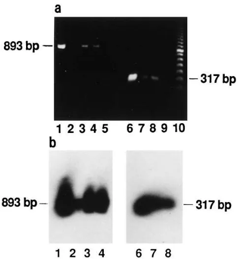
human prostate. The expression of mRNAs for the long and
short Prl receptor forms in human prostate was studied by RT-PCR. The expression of the long Prl receptor mRNA was shown by using primers 1 and 2 (Fig. 5). The sequences and the use of these primers have been published previously (30). Primers 1 and 2 were used for the amplification of an 893-bp cDNA fragment of the cytoplasmic domain of the long human Prl receptor form. Consequently, the amplification of a 320-bp cDNA fragment would have revealed the presence of the pu-tative intermediate Prl receptor (30) in human prostate, since these two receptor forms differ from each other in the length of their cytoplasmic domains (22, 25, 26). Only an 893-bp cDNA fragment was detected from reverse-transcribed total RNA of human prostates and the control samples (Fig. 6 a). This suggests that the long, but not the intermediate, Prl recep-tor mRNA is expressed in human prostate. Total RNA from T47D cells was used as positive control (29), and H2O and all
the samples without reverse transcriptase (data not shown) were used as negative controls (Fig. 6). The long Prl receptor RT-PCR products were identified by Southern blotting and hybridization with a HindIII cDNA fragment of the H1/H2
hu-man Prl receptor cDNA clone (29) (Fig. 6 b).
The expression of the short Prl receptor mRNA was stud-ied with primers 3 and 4 (Fig. 5) from the coding region of (62), and amplifying a cDNA fragment specific for, the short Prl receptor form (59, 62). The predicted (62) 317-bp cDNA fragment (Fig. 6 a) was amplified from reverse-transcribed hu-man prostatic total RNA. As a positive control for the short Prl receptor mRNA expression, RNA from the livers of estro-gen-treated female rats was used (22, 25, 59), and H2O and all
the samples without reverse transcriptase (data not shown) were used as negative controls (Fig. 6). The short form 317-bp Prl receptor RT-PCR products were shown to correspond to the short Prl receptor cDNA by Southern blotting and hybrid-ization with the AvaII/RsaI cDNA fragment (440 bp) from the coding region of the rat liver short Prl receptor cDNA clone F3
(59) (Fig. 6 b).
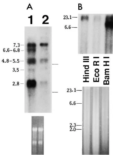
The sizes of mRNAs encoding the short Prl receptor in hu-man prostates were demonstrated by Northern blotting. We prepared and used a PstI/EcoRV fragment (243 bp) of the 39 untranslated region of the rat liver short Prl receptor cDNA clone F3 (22, 25, 59), which has been shown to recognize
selec-tively the short Prl receptor specific mRNAs (25) (Fig. 5). From total RNA of human prostatic tissues five mRNAs of 2.8, 3.5, 4.8–5.5, 6.6–6.8, and 7.3 kb were detected (Fig. 7 A). The sizes of mRNAs for the short Prl receptor in human
pros-Figure 3. Cross-linking of [125I]hPrl with membrane
preparations of human pros-tate. Membrane fractions (100 mg membrane proteins) of four separate human pros-tates were incubated in the presence (1) or in the ab-sence (2) of unlabeled hPrl (5 mg) at 208C for 16 h. Cross-linking was per-formed with 0.5 mM disuc-cimidylsuberate for 15 min, and the samples were elec-trophoresed on a 12% SDS-polyacrylamide gel under re-ducing conditions. A representative autoradiogram of dried gels is shown. The apparent Mr of protein markers are shown on the left,
[image:6.612.59.301.60.219.2]and the arrows indicate the major labeled species on the right. The experiment was repeated six times.
[image:6.612.57.367.617.741.2]tate were estimated by hybridizing the same blots with a HindIII fragment of the H1/H2 human long Prl receptor cDNA
(29). This probe detects 2.8-, 3.5-, and 7.3-kb mRNAs in hu-man tissues. The experiment was repeated five times. Further-more, we observed that the PstI/EcoRV fragment (243 bp) of the rat liver cDNA clone F3, which was used to detect the short
Prl receptor specific mRNAs (25), did not have any significant sequence similarity to other human genes described in the GenBank.
The presence of this short Prl receptor form specific se-quence in the genomic DNA of human prostate was demon-strated by Southern blotting (Fig. 7 B).
The demonstration of Prl synthesis in human prostatic
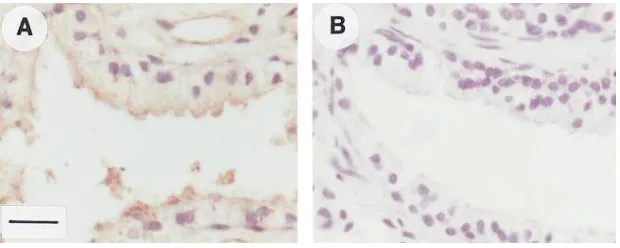
epi-thelium. The possible expression of Prl protein in human
pros-tatic tissue was studied immunohistochemically using a poly-clonal anti-hPrl antibody. The epithelium of human prostatic tissue showed an intensive cytoplasmic staining (Fig. 8 A), whereas prostatic stroma was negative. The stained cells were scattered as irregular areas within the secretory epithelium. Also, staining of the basal epithelial cells was noted, especially in the areas where the prostatic epithelium was stratified in several layers. There was no reaction in the sections stained with NRS (Fig. 8 B) or without the primary antibody. Human prolactinomas were used as positive control tissues.
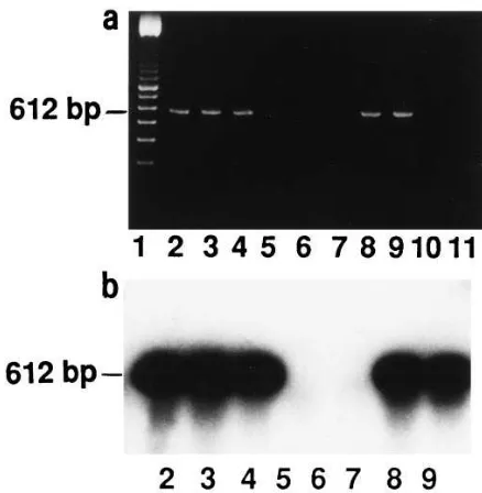
The expression of Prl in human prostate was examined also at the mRNA level by RT-PCR, to distinguish between endog-enous and exogendog-enous Prl within human prostatic tissue. The RT-PCR reactions of human prostates produced the expected (42, 63) 612-bp band by ethidium bromide staining (Fig. 9 a). The RT-PCR products were Southern blotted and hybridized with a specific hPrl cDNA probe (64) (Fig. 9 b). RNA from MCF-7 and T47D breast cancer cells were used as positive controls (30, 42) (Fig. 9), and H2O (Fig. 9) and all the samples
without reverse transcriptase (data not shown) were used as negative controls.
Discussion
In this work we demonstrate that Prl has direct effects on hu-man prostate. The expression of Prl receptors, by which the ef-fects of Prl are probably mediated, is also demonstrated and, furthermore, human prostatic epithelium is shown to
synthe-Figure 5. Schematic presenta-tion of the short and long Prl re-ceptor cDNAs. The coding se-quences are shown by boxes. The black areas are the regions encoding the transmembrane parts of the short and long Prl receptors. The dotted area is the region unique to the short form of the rat Prl receptor. The lines at the beginning and at the end of the coding regions represent the untranslated regions (UTR) contained in the cDNAs. The black box shows the location of the fragment specific for the short Prl receptor cDNA (PstI/EcoRV 243-bp fragment of the rat liver F3 Prl receptor clone) which was prepared
[image:7.612.59.484.58.201.2]and used as a cDNA probe in Northern and Southern blotting. The arrows marked with numbers indicate the positions, directions, and numbers of the primers for Prl receptor RT-PCR. Primers 1 and 2 yielded an 893-bp cDNA fragment deriving from long form Prl receptor mRNA, and primers 3 and 4 yielded a 317-bp cDNA fragment deriving from the short form Prl receptor mRNA.
Figure 6. Demonstration of the long and short Prl receptor mRNA expression in human prostate by RT-PCR method. (a) An 893-bp fragment from the cytoplasmic domain of the long hPrl receptor and a 317-bp fragment from the coding region of the short Prl receptor were both reverse transcribed and amplified from RNA of the human prostate tissue samples. RNA from T47D breast cancer cells and from livers of estrogen-treated female rats were used as positive con-trols for the expression of the long and short Prl receptor mRNA ex-pression, respectively. Lane 1, T47D cells; lanes 2–4, human pros-tates; lane 5, H2O; lane 6, liver of estrogen-treated female rat; lanes 7
and 8, human prostates; lane 9, H2O; lane 10, 100-bp DNA ladder. (b)
The 893-bp long Prl receptor and 317-bp short Prl receptor RT-PCR products were purified, separated on agarose gels, Southern blotted, and hybridized with a HindIII cDNA fragment of the H1/H2 human
Prl receptor cDNA clone and with an AvaII/RsaI cDNA fragment from the coding region of the rat liver Prl receptor cDNA clone F3,
[image:7.612.56.298.268.532.2]size Prl. The effects of Prl on the morphology and DNA syn-thesis of human prostates were investigated in organ cultures. This method was used because the function of differentiated epithelium and hormone responsiveness of both rat and hu-man prostatic tissues have been demonstrated to be well main-tained in this in vitro model (12, 36, 44–47).
Concentration of Prl in the culture medium was 45 nM, which has been shown to be suitable for Prl to elicit biological responses in prostate tissue in organ culture conditions (12, 46, 47, 65, 66). After a 7-d culture in the presence of Prl the epithe-lium was stratified, and round microlumina were formed irreg-ularly within the epithelium when compared with explants cul-tured in the basal medium. Furthermore, the epithelial cells seemed to have lost their polarized orientation in relation to the basement membrane around the acini. These morphologic features are strikingly different from those seen in explants cultured with DHT, which have been described earlier (44) and which were used as reference for the Prl effects. Interest-ingly, the appearance of prostate epithelium with microlumina
resembled a cribriform morphology, described as one pattern of prostatic intraepithelial neoplasia (67). It is also a common feature in in situ breast cancer (68). The level of DNA synthe-sis, measured by DNA labeling with tritiated thymidine, was increased on average 1.79-fold (P5 0.023) (95% confidence limit from 1.15 to 2.43) in Prl-treated explants. Although the basal level of DNA labeling was highly variable between the individuals, as also shown previously (44), the results suggest that Prl induces proliferation in human prostatic tissue. This is compatible with the results of Prl effects in rat prostate (12). The autoradiographic studies showed that the labeling of DNA occurred in the prostatic epithelium, in which also the Prl receptor proteins were shown to be located.
[image:8.612.56.255.56.329.2]The finding of the expression of both the short and long Prl receptors in human prostate was interesting, since the pres-ence of the short Prl receptor form in human tissues has not been demonstrated previously, although it has been suggested
Figure 7. Northern and Southern blot analyses showing the short Prl receptor expression in human prostate. (A) A representative North-ern blot hybridized with a PstI/EcoRV fragment (243 bp) of the rat liver Prl receptor cDNA clone F3 detecting selectively the short Prl
receptor mRNAs. 10 mg of total RNA from human prostates was used. The positions of rRNAs (18S, 28S) are marked, and the corre-sponding ethidium bromide staining of the blot is shown to verify the loading and transfer of RNA (bottom). The experiment was repeated six times. Lanes 1 and 2, human prostates. (B) 10 mg of DNA from human prostates was digested with HindIII, EcoRI, and BamHI, frac-tionated on agarose gel, transferred to a nitrocellulose membrane, and hybridized with the PstI/EcoRV 243-bp fragment of the rat liver F3 Prl receptor clone showing the presence of the short Prl receptor
[image:8.612.316.524.59.460.2]specific sequences from human prostatic DNA. Sizes of DNA (kb) standard are shown on the left. Ethidium bromide staining of the gel is shown (bottom).
indirectly (30, 31). In rat, three forms (the short, long, and the intermediate) of Prl receptor have been identified (22, 25, 26), whereas in human tissues only the cDNA for the long Prl re-ceptor has been cloned (29). However, in human tissues the expression of both the intermediate Prl receptor (30) and a novel splicing variant encoding only the extracellular domain of the Prl receptor protein (69) have been documented re-cently. We demonstrated the expression of the short and the long Prl receptors in human prostate by RT-PCR and by bind-ing assays and subsequent cross-linkbind-ing.
The Northern blot analysis of the mRNAs encoding the short Prl receptors in human prostates showed five transcripts of 2.8, 3.5, 4.8–5.5, 6.6–6.8, and 7.3 kb. Interestingly, three of them (2.8, 3.5, and 7.3 kb) are identical with those detected with the long Prl receptor form specific probe (HindIII frag-ment of H1/H2 human long Prl receptor cDNA) (29).
How-ever, all of the transcripts are not necessarily translated to pro-teins, since total RNA was used for the analysis of the different Prl receptor mRNAs and it contains also nuclear transcripts without the 59-cap-structures and poly-A-tails. In rat tissues there are several examples describing substantially small (0.6 kb) (59) and large (9.5 kb) (70) Prl receptor mRNA spe-cies that probably represent unprocessed mRNA or mRNA
splicing variants with insufficient sequences to encode a com-plete Prl receptor protein. The functions of these mRNA species are unknown, emphasizing the role of not only tran-scriptional and posttrantran-scriptional, but also translational, regu-lation in Prl receptor expression. A PstI/EcoRV fragment of the 39 untranslated region of the rat liver short Prl receptor cDNA clone F3 was used in Northern blotting as a probe. This
probe has been shown previously to specifically hybridize with the short Prl receptor specific mRNAs (25, 36), and we con-firmed that this fragment of the short Prl receptor cDNA has no significant sequence similarity to other human genes de-scribed in the GenBank.
A single gene for Prl receptor, composed of at least 11 ex-ons and spanning z 70 kb, has been identified in rat (22), and the 11th exon has been reported to encode the short form spe-cific sequences, whereas the 10th exon encodes the cytoplas-mic region of the long receptor form (22). In rat the different transcripts of the long and short Prl receptors are thought to derive from the same gene (22) either by use of different initia-tion or polyadenylainitia-tion sites or by alternative splicing of the common primary transcript (22, 25). So far, the structure of the human Prl receptor gene is not known, and the mechanism un-derlying the origin of the different transcripts for the short Prl receptor remains unclear. However, the presence of the short Prl receptor specific sequences in human prostatic genomic DNA was shown by Southern blotting with the short form spe-cific cDNA probe used in Northern blotting. In conclusion, the results suggest that both the short and long Prl receptor mRNAs are expressed in human prostate.
The localization of Prl receptors in human prostate tissue was studied by immunohistochemistry. We used a polyclonal anti-rPrl receptor antibody R120 (36), recognizing both the short and long Prl receptors. A strong degree of sequence identity between the human Prl receptor sequence and that of the rat, particularly in this extracellular domain, has been re-ported (29). In human prostate the staining of Prl receptors was localized mainly to the apical parts of the secretory epithe-lium. The nuclei remained unstained, but faint staining was also noted in prostatic stroma. The apical staining of the secre-tory epithelium of human prostate, as also seen in rat dorsolat-eral prostate (36), is surprising since the expected localization of Prl receptors, binding circulatory Prl, would be on the baso-lateral cell membranes of prostatic epithelial cells. However, the basolateral cell membranes in the positive control tissues showed clear staining, and the prostatic sections were pre-treated and pre-treated in the same way as the controls. Prostatic epithelial cells are joined to each other by tight junctions, and Prl receptors located to the apical parts of the prostatic epithe-lial cells are not accessible to circulatory Prl. Instead, they would be accessible to a secreted ligand.
[image:9.612.55.274.57.281.2]To test this hypothesis we investigated the presence of a possible autocrine and/or paracrine ligand available to these apically located Prl receptors. Indeed, a clear positive staining for Prl with the polyclonal anti-hPrl antibody was noted in pro-static epithelium. The stained cells were scattered irregularly within the prostatic epithelium, and mainly the secretory epi-thelial cells showed intensive staining. Conspicuously, how-ever, in the areas where the prostatic epithelium was stratified in several layers also staining of the prostatic basal cells was of-ten seen. The transcription of Prl gene was shown by RT-PCR using primers spanning all four introns of genomic Prl DNA and using MCF-7 and T47D cells as positive controls (30, 42).
Figure 9. Demonstration of Prl mRNA expression in human pros-tates by RT-PCR method. (a) A 612-bp fragment was reverse tran-scribed and amplified from the total RNA of the human prostate tis-sue samples. Total RNA from MCF-7 cells and T47D cells were used as positive controls, and H2O and all the samples without reverse
The correspondence of the expected sized (42, 63) 612-bp Prl RT-PCR products to the pituitary Prl was shown by Southern blotting and hybridizing the RT-PCR products with a specific hPrl cDNA probe (64).
Extrapituitary synthesis of Prl has been described in brain, placenta, and in lymphoid and mammary tissues (30, 37–42), where a local growth factor role for Prl has been suggested repeatedly (30, 37–39, 41, 42). In prostate tissue, neuroendo-crine-paracrine cells, differentiated from normal prostatic thelial cells, have been described as the third prostatic epi-thelial cell type and as an integral component of the normal prostatic acinar and ductal epithelium (71–74). They are known to produce a number of bioactive hormone-related substances including serotonin, chromogranin A, a thyroid-stimulating hormone–like peptide, calcitonin, calcitonin gene– related peptide, somatostatin, bombesin-like peptides, par-athyroid hormone–related protein, and cholecystokinin (71). Moreover, the neuroendocrine differentiation has been re-ported to be a common feature also in nearly all prostatic ade-nocarcinomas (71–75), where neuroendocrine-paracrine cells have been suggested to represent an androgen receptor–nega-tive cell reservoir harboring its own growth factor activity (71, 74). It is possible that the Prl synthesizing epithelial cells in hu-man prostates in our study represent these neuroendocrine-paracrine cells. However, the production of Prl by these cells has not been described previously (71). The local production of Prl in prostate tissue itself emphasizes the significance of Prl in the regulation of prostatic function in males. The locally produced Prl may act in an autocrine, paracrine, or even intra-crine way in human prostate tissue, and it may play an impor-tant role both during growth and differentiation of prostate as well as in the secretory process of the mature gland. It may also contribute to autocrine/paracrine prostatic tumor growth factor activity.
In conclusion, our results show that Prl has a characteristic effect on the prostatic morphology and that it increases DNA synthesis in human prostatic tissue in organ culture. Both the long and short Prl receptors were demonstrated in human prostate at protein and mRNA levels. Prl receptors were lo-cated to the prostatic epithelium and their possible ligand, Prl, was also shown to be produced by the same cells. Organ cul-tures of human prostates provide a useful model to study the regulation and growth factor activity of locally synthesized Prl in human prostate tissue.
Acknowledgments
The National Institute of Diabetes and Digestive and Kidney Dis-eases National Hormone (Pituitary) Program and Dr. Parlow are gratefully acknowledged for providing polypeptide hormones. We would like to thank Dr. P. Kelly who provided the cDNA clones for F3 rat short form and for H1/H2 human long form Prl receptor, and
Dr. R.A. Maurer (University of Iowa, Iowa City, IA) for permission to use the Prl cDNA as a probe. We want to thank Dr. T. Elsasser, Dr. H. Nikula, Dr. U. Petäjä-Repo, and Ms. A. Metsävuori for help and advice in binding assays. Also, we thank Dr. Hannu Kalimo for the control samples of Prl immunohistochemistry, Dr. Harry Kujari for the control samples of Prl receptor immunohistochemistry, Dr. Markku Kallajoki for helpful advice in Prl immunohistochemistry, and Juhani Tuominen (University of Turku), and Sakke Huhtala (Clinical Research Services, Turku, Finland) for the advice in the sta-tistical analysis of the data. Also, we thank Ms. Merja Tasanen, Ms. Pirkko Rauhamäki, and Ms. Leena Simola for technical assistance,
Ms. Arja Karppinen for the art work, and Mr. Henrik Wikgren for preparing the photographs.
This study was financially supported by the Academy of Finland, the Cancer Societies of Finland, the Finnish Medical Foundation and Duodecim, and Cancer Society of Southwestern Finland. Dr. P.M. Ingleton received financial support from the Yorkshire Cancer Re-search Campaign.
References
1. Sinha, Y.N. 1995. Structural variants of prolactin: occurrence and physio-logical significance. Endocr. Rev. 16:354–369.
2. Wallis, M. 1988. Mechanism of action of prolactin. In Hormones and Their Actions. Part II. B.A. Cooke, R.J.B. King, and H.J. van der Molen, edi-tors. Elsevier, Amsterdam. 295–319.
3. Guyette, W.A., R.A. Matusik, and J.M. Rosen. 1979. Prolactin-mediated transcriptional and post-transcriptional control of casein gene expression. Cell.
17:1013–1023.
4. Lesueur, L., M. Edery, S. Ali, J. Paly, P.A. Kelly, and J. Djiane. 1991. Comparison of long and short forms of the prolactin receptor on prolactin in-duced milk protein gene transcription. Proc. Natl. Acad. Sci. USA. 88:824–828.
5. Yu-Lee, L. 1990. Prolactin stimulates transcription of growth-related genes in Nb2 T lymphoma cells. Mol. Cell. Endocrinol. 68:21–28.
6. Stevens, A.M., and L. Yu-Lee. 1994. Multiple prolactin-responsive ele-ments mediate G1 and S phase expression of the interferon regulatory factor-1 gene. Mol. Endocrinol. 8:345–355.
7. Clevenger, C.V., A.L. Sillman, J. Hanley-Hyde, and M.B. Prystowsky. 1992. Requirement for prolactin during cell cycle regulated gene expression in cloned T-lymphocytes. Endocrinology. 130:3216–3222.
8. Costello, L.C., and R.B. Franklin. 1994. Effects of prolactin on the pros-tate. Prostate. 24:162–166.
9. Reiter, E., S. Lardinois, M. Klug, B. Sente, B. Hennuy, M. Bruyninx, J. Closset, and G. Hennen. 1995. Androgen-independent effects of prolactin on the different lobes of the immature rat prostate. Mol. Cell. Endocrinol. 112: 113–122.
10. Perez-Villamil, B., E. Bordiu, and M. Puente-Cueva. 1991. Involvement of physiological prolactin levels in growth and prolactin receptor content of prostate glands and testes in developing male rats. J. Endocrinol. 132:449–459.
11. Rui, H., and K. Purvis. 1987. Prolactin selectively stimulates ornithine decarboxylase in the lateral lobe of the rat prostate. Mol. Cell. Endocrinol. 50: 89–97.
12. Nevalainen, M.T., E.M. Valve, S.I. Mäkelä, M. Bläuer, P.J. Tuohimaa, and P.L. Härkönen. 1991. Estrogen and prolactin regulation of rat dorsal and lateral prostate in organ culture. Endocrinology. 129:612–622.
13. Price, D. 1963. Comparative aspects of development and structure in the prostate. Natl. Cancer Inst. Monogr. 12:351–369.
14. Wilson, J.D. 1980. The pathogenesis of benign prostatic hyperplasia.
Am. J. Med. 68:745–756.
15. Sissom, J.F., M.L. Eigenbrodt, and J.C. Porter. 1988. Anti-growth action on mouse mammary and prostate glands of a monoclonal antibody to prolactin receptor. Am. J. Pathol. 133:589–595.
16. Nakamura, A., T. Shirai, K. Ogawa, S. Wada, N.A. Fujimoto, A. Ito, and N. Ito. 1990. Promoting action of prolactin released from a grafted trans-plantable pituitary tumor (MtT/F84) on rat prostate carcinogenesis. Cancer Lett. 53:151–157.
17. Webber, M.M. 1981. Polypeptide hormones and the prostate. In The Prostatic Cell: Structure and Function. Part B. G.P. Murphy, A.A. Sandberg, and J.P. Karr, editors. Alan R. Liss, Inc., New York. 63–88.
18. Thomas, J.A., and M. Manadhar. 1975. Effects of prolactin and/or tes-tosterone on nucleic acid levels in prostate glands of normal and castrated rats.
J. Endocrinol. 65:149–150.
19. Walvoord, D.J., M.I. Resnick, and J.T. Grayhack. 1976. Effects of tes-tosterone, dihydrotestes-tosterone, estradiol, and prolactin on the weight and citric acid content of the lateral lobe of the rat prostate. Invest. Urol. 14:60–65.
20. Jones, R., P.R. Riding, and M.G. Parker. 1983. Effects of prolactin on testosterone-induced growth and protein synthesis in rat accessory sex glands.
J. Endocrinol. 96:407–416.
21. Prins, G.S. 1987. Prolactin influence cytosol and nuclear androgen re-ceptors in the ventral, dorsal, and lateral lobes of rat prostate. Endocrinology.
120:1457–1464.
22. Kelly, P.A., J. Djiane, M.C. Postel-Vinay, and M. Edery. 1991. The pro-lactin/growth hormone receptor family. Endocr. Rev. 12:235–251.
23. Bazan, J.F. 1990. Structural design and molecular evolution of a cyto-kine receptor superfamily. Proc. Natl. Acad. Sci. USA. 87:6934–6938.
24. Thoreau, E., B. Petridou, P. Kelly, J. Djiane, and J.P. Mornon. 1991. Structural symmetry of extracellular domain of the cytokine/growth hormone/ prolactin receptor family and interferon receptors revealed by hydrophobic cluster analysis. FEBS Lett. 282:26–31.
rat ovary and liver. Mol. Endocrinol. 4:1136–1143.
26. Ali, S., I. Pellegrini, and P.A. Kelly. 1991. A prolactin-dependent im-mune cell line (Nb2) expresses a mutant form of prolactin receptor. J. Biol.
Chem. 266:20110–20117.
27. Clevenger, C.V., and M.V. Medaglia. 1994. The protein tyrosine kinase p59fyn is associated with prolactin (Prl) receptor and is activated by Prl
stimula-tion of T-lymphocytes. Mol. Endocrinol. 8:674–681.
28. Clevenger, C.V., T. Torigoe, and J.C. Reed. 1994. Prolactin induces rapid phosphorylation and activation of prolactin receptor-associated RAF-1 kinase in a T-cell line. J. Biol. Chem. 269:5559–5565.
29. Boutin, J.M., M. Edery, M. Shirota, C. Jolicoeur, L. Lesueur, S. Ali, D. Gould, J. Djiane, and P.A. Kelly. 1989. Identification of a cDNA encoding a long form of prolactin receptor in human hepatoma and breast cancer cells.
Mol. Endocrinol. 3:1455–1461.
30. Clevenger, C.V., W.P. Chang, W. Ngo, T.L.M. Pasha, K.T. Montone, and J.E. Tomaszewski. 1995. Expression of prolactin and prolactin receptor in human breast carcinoma; evidence for an autocrine/paracrine loop. Am. J. Pathol. 146:695–705.
31. Das, R., and B.K. Vonderhaar. 1995. Transduction of prolactin’s growth signal through both long and short forms of the prolactin receptor. Mol. Endo-crinol. 9:1750–1759.
32. Nagano, M., and P.A. Kelly. 1994. Tissue distribution and regulation of rat prolactin receptor gene expression. J. Biol. Chem. 269:13337–13345.
33. Ouhtit, A., G. Morel, and P.A. Kelly. 1993. Visualization of gene ex-pression of short and long forms of prolactin receptor in rat reproductive tis-sues. Biol. Reprod. 49:528–536.
34. Ouhtit, A., P.A. Kelly, and G. Morel. 1994. Visualization of gene ex-pression of short and long forms of prolactin receptor in rat digestive tissues.
Am. J. Physiol. 266:G807–G815.
35. Lobie, P.E., J. Garcia-Aragon, and M.J. Waters. 1993. Prolactin recep-tor expression in the gastrointestinal tract: characterization of the prolactin re-ceptor of gastric mucosa. J. Endocrinol. 139:371–381.
36. Nevalainen, M.T., E.M. Valve, P.M. Ingleton, and P.L. Härkönen. 1996. Expression and hormone regulation of prolactin receptors in rat dorsal and lat-eral prostate. Endocrinology. 137:3078–3088.
37. Sabharwal, P., R. Glaser, W. Lafuse, S. Varma, Q. Liu, S. Arkins, R. Kooijman, L. Kutz, K.W. Kelley, and W.B. Malarkey. 1992. Prolactin synthe-sized and secreted by human peripheral mononuclear cells: an autocrine growth factor for lymphoproliferation. Proc. Natl. Acad. Sci. USA. 89:7713–7716.
38. Hooghe, R., M. Delhase, P. Vergani, A. Malur, and E.L. Hooghe-Peters. 1993. Growth hormone and prolactin are paracrine growth and differentiation factors in the haemopoietic system. Immunol. Today. 14:212–214.
39. Wu, H., R. Devi, and W. Malarkey. 1996. Expression and localization of prolactin messenger ribonucleic acid in the human immune system. Endocrinol-ogy. 137:349–353.
40. Handwerger, S., R.G. Richards, and E. Markoff. 1992. The physiology of decidual prolactin and other decidual protein hormones. Trends Endocrinol. Metab. 3:91–95.
41. Mershon, J., W. Sali, N. Mitchner, and N. Ben-Jonathan. 1995. Prolactin is a local growth factor in rat mammary tumors. Endocrinology. 136:3619–3623. 42. Ginsburg, E., and B.K. Vonderhaar. 1995. Prolactin synthesis and secre-tion by human breast cancer cells. Cancer Res. 55:2591–2595.
43. McKeehan, W.L., P.S. Adams, and D. Fast. 1987. Different hormonal requirements for androgen-independent growth of normal and tumor epithelial cells from rat prostate. In Vitro Cell. Dev. Biol. 23:147–152.
44. Nevalainen, M.T., E.M. Valve, W. Ping, M. Nurmi, P.L. Härkönen, and P.M. Martikainen. 1993. Hormone regulation of human prostate in organ cul-ture. Cancer Res. 53:5199–5207.
45. Martikainen, P.M., P.L. Härkönen, T. Vanhala, S.I. Mäkelä, M. Vil-janen, and J.J.O. Suominen. 1987. Multihormonal control of synthesis and se-cretion of prostatein in cultured rat ventral prostate. Endocrinology. 121:604– 611.
46. Martikainen, P. 1987. Maintenance of adult rat ventral prostate in organ culture. Anat. Rec. 218:166–174.
47. Johansson, R. 1975. RNA, protein and DNA synthesis stimulated by testosterone, insulin and prolactin in the rat ventral prostate cultured in chemi-cally defined medium. Acta Endocrinol. 80:761–764.
48. Chang, S.M., and L.W.K. Chung. 1989. Interaction between prostatic fi-broblasts and epithelial cells in culture: role of androgen. Endocrinology. 125: 2719–2727.
49. Cunha, G.R., N. Hayashi, and Y.C. Wong. 1991. Regulation of differen-tiation and growth of normal adult and neoplastic epithelia by inductive mesen-chyme. In Prostate Cancer. L.M. Franks and J.T. Isaacs, editors. Cold Spring Harbor Laboratory Press. Cancer Surveys. 11:73–89.
50. Burton, K. 1956. A study of the conditions and mechanisms of the
diphenylamine reaction for the colorimetric estimation of deoxyribonucleic acid. Biochem. J. 62:315–323.
51. SAS Institute Inc. 1990. SAS/STAT Users Guide, Version 6, 4th Edi-tion, Vols. 1 and 2. SAS Institute Inc., Cary, NC.
52. Sternberger, L.A. 1974. Immunocytochemistry. Prentice-Hall, Engle-wood Cliffs, NJ.
53. Danks, J.A., A.J. Devlin, P.M.W. Ho, H. Diefenbach-Jagger, D.M. Power, A.V.M. Canario, T.J. Martin, and P.M. Ingleton. 1993. Parathyroid hor-mone-related protein is a factor in normal fish pituitary. Gen. Comp. Endo-crinol. 92:201–212.
54. Ben-David, M., T. Kadar, and A.V. Schally. 1986. Micromethod for the determination of free and total prolactin receptors: measurement of receptor levels in normal and malignant mammary and prostate tissues. Proc. Natl. Acad. Sci. USA. 83:8375–8379.
55. Bradford, M.M. 1976. A rapid and sensitive method for the quantitation of microgram quantities of protein utilizing the principle of protein-dye bind-ing. Anal. Biochem. 72:248–254.
56. Salacinski, P.R.P., C. McLean, J.E.C. Sykes, V.V. Clement-Jones, and P.J. Lowry. 1981. Iodination of proteins, glycoproteins, and peptides using a solid-phase oxidizing agent, 1,3,4,6-tetrachloro-3a,6a-dipenyl glycoluril (IODO-GEN®). Anal. Biochem. 117:136–146.
57. Laemmli, U.K. 1970. Cleavage of structural proteins during assembly of the head of bacteriophage T4. Nature (Lond.). 227:680–685.
58. Chomczynski, P., and N. Sacchi. 1987. Single-step method of RNA isola-tion by acid guanidinium thiocyanate-phenol-chloroform extracisola-tion. Anal. Bio-chem. 162:156–159.
59. Boutin, J.M., C. Jolicoeur, H. Okamura, J. Gagnon, M. Edery, M. Shi-rota, D. Banville, I. Dusanter-Fourt, J. Djiane, and P.A. Kelly. 1988. Cloning and expression of the rat prolactin receptor, a member of the growth hormone/ prolactin receptor gene family. Cell. 53:69–77.
60. Feinberg, A.P., and B. Vogelstein. 1983. A technique for radiolabeling DNA restriction endonuclease fragments to high specific activity. Anal. Bio-chem. 132:6–13.
61. Mellanen, P., H. Minn, R. Grenman, and P. Härkönen. 1994. Expression of glucose transporters in head-and-neck tumors. Int. J. Cancer. 56:622–629.
62. Sakaguchi, K., T. Ohkubo, M. Tanaka, H. Ushiro, and K. Nakashima. 1994. Differential regulation of prolactin receptor mRNA expression in rat liver and kidney by testosterone and oestradiol. J. Endocrinol. 143:383–392.
63. Fields, K., E. Kulig, and R.V. Lloyd. 1993. Detection of prolactin mes-senger RNA in mammary and other normal and neoplastic tissues by poly-merase chain reaction. Lab. Invest. 68:354–359.
64. Gubbins, E.J., R.A. Maurer, M. Lagrimini, C.R. Erwin, and J.E. Donel-son. 1980. Structure of the rat prolactin gene. J. Biol. Chem. 255:8655–8662.
65. Rui, H., I. Brekke, P.A. Torjesen, and K. Purvis. 1986. Homologous up-regulation of the prolactin receptor in rat prostatic explants. Mol. Cell. Endo-crinol. 46:53–57.
66. Franklin, R.B., and L.C. Costello. 1990. Prolactin directly stimulates cit-rate production and mitochondrial aspartate aminotransferase of prostate epi-thelial cells. Prostate. 17:13–18.
67. Bostwick, D.G., M.B. Amin, P. Dundore, W. Marsh, and D.S. Schultz. 1993. Architectural patterns of high-grade prostatic intraepithelial neoplasia.
Hum. Pathol. 24:298–310.
68. Page, D.L., T.J. Anderson, and L.W. Rogers. 1987. Carcinoma in situ (CIS). In Diagnostic Histopathology of the Breast. D.L. Page and T.J. Ander-son, editors. Churchill Livingstone, Edinburgh-London-Melbourne-New York. 158–171.
69. Fuh, G., and J.A. Wells. 1995. Prolactin receptor antagonists that inhibit the growth of breast cancer cell lines. J. Biol. Chem. 270:13133–13137.
70. Hu, Z.Z., and M. Dufau. 1991. Multiple and differential regulation of ovarian prolactin receptor messenger RNAs and their expression. Biochem.
Biophys. Res. Commun. 181:219–225.
71. Abrahamssom, P.-A. 1996. Neuroendocrine differentiation and hor-mone-refractory prostate cancer. Prostate. 6:3–8.
72. Bonkhoff, H., U. Stein, and K. Remberger. 1995. Endocrine-paracrine cell types in the prostate and prostatic adenocarcinoma are postmitotic cells.
Hum. Pathol. 26:167–170.
73. Aprikian, A.G., C. Cordon-Cardo, W.R. Fair, and V.E. Reuter. 1993. Characterization of neuroendocrine differentiation in human benign prostate and prostatic adenocarcinoma. Cancer (Phila.). 71:3952–3965.
74. Bonkhoff, H., U. Stein, and K. Remberger. 1993. Androgen receptor status in endocrine-paracrine cell types of the normal, hyperplastic, and neo-plastic human prostate. Virchows Archiv. A Pathol. Anat. 423:291–294.
![Table I. The Level of [3DNA after a 7-d Organ Culture of Prostates from SevenH]Thymidine Incorporation intoIndividual Patients](https://thumb-us.123doks.com/thumbv2/123dok_us/8219703.821345/5.612.316.550.545.739/table-culture-prostates-sevenh-thymidine-incorporation-intoindividual-patients.webp)