Copyright © 1997, American Society for Microbiology
Simple Differential Detection of Entamoeba histolytica and
Entamoeba dispar in Fresh Stool Specimens by Sodium
Acetate-Acetic Acid-Formalin Concentration and PCR
HEIKE TROLL, HANSPETER MARTI,*ANDNIKLAUS WEISS
Swiss Tropical Institute, CH-4002 Basel, Switzerland
Received 4 December 1996/Returned for modification 15 January 1997/Accepted 26 March 1997
Amoebiasis is caused by two distinct species, a pathogenic form (Entamoeba histolytica) and a nonpathogenic form (Entamoeba dispar), which are morphologically identical. Although the distinction between these two species is of great clinical importance, the methods developed for this purpose either are very time-consuming or involve laborious procedures for isolation of the DNA. We report here a simple PCR method starting with fresh stool specimen that allows for the sensitive and reliable distinction between E. histolytica and E. dispar. After initial concentration by the sodium acetate-acetic acid-formalin (SAF) method and digestion with proteinase K, a 0.88-kb sequence of the multicopy 16S rRNA gene served as a target for PCR amplification. The method starting with unpreserved specimens proved to be very sensitive and was not influenced by the quick exposure to SAF fixative during the initial concentration step. However, storage in SAF fixative prior to testing resulted in a decreased sensitivity within 2 days. The detection limit of the method was as low as one copy of the 16S rRNA gene. No cross-reactivity was observed with other common intestinal protozoa. Mixed infections involving both E. histolytica and E. dispar could easily be detected at a ratio of 1:10,000 by agarose gel electrophoresis or a DNA hybridization immunoassay.
Amoebiasis is one of the most common parasitic infections worldwide. The disease affects about 500 million people, mostly in developing countries, where it is one of the major health problems. However, only 10% of the infections lead to severe disease like dysentery or amoebic liver abscess, whereas 90% of the carriers remain asymptomatic (30). As early as 1925, these clinical differences prompted a hypothesis that amoebic infections are caused by two species with the same morphology but with different pathogenicities (7). Five dec-ades later this hypothesis was revived when a correlation be-tween the presence or absence of symptoms with specific isoen-zyme patterns (zymodemes) was shown (23, 26). Yet the genetic relationship remained a subject of controversy for some time (11, 24). Today, DNA analysis leaves no doubt that amoebiasis is caused by two distinct species, Entamoeba
histo-lytica (pathogenic) and Entamoeba dispar (nonpathogenic),
re-spectively. Sequence analysis of the small subunit of the 16S rRNA gene revealed the genetic distance between E. histolytica and E. dispar to be 1.7%, nearly as much as that between the small-subunit rRNA of humans and mice (18). Despite these findings, in a clinical setting all infected persons are usually treated due to a lack of a simple, reliable diagnostic tool for discriminating between the two species. It was therefore our aim to develop a PCR method for the differential diagnosis of
E. histolytica and E. dispar starting directly with unpreserved
fresh fecal specimen.
MATERIALS AND METHODS
Parasites.Identification of parasite species was performed by microscopic
examination of stool specimens after concentration in sodium acetate-acetic acid-formalin (SAF) fixative (31). E. histolytica, E. dispar, Entamoeba hartmanni, Entamoeba coli, Dientamoeba fragilis, and Blastocystis hominis were isolated from stool samples by xenic cultivation in Robinson’s medium (20) in bijou bottles on an agar slope to a density of 2.03103to 2.03104cells/vial. Differentiation of
isolates as E. histolytica or E. dispar was done by isoenzyme electrophoresis by the protocol of Sargeaunt and Williams (25). E. histolytica HK-9 was grown axeni-cally in TYI-S-33 medium (10) in 12.0-ml glass tubes to a density of 23106to 33106cells. Entamoeba invadens, a species affecting reptiles, was cultivated on slopes of coagulated horse serum and horse serum diluted 1:4 with Ringer’s solution (103 mM NaCl, 1.34 mM KCl, 0.7 mM CaCl2z2H2O [pH 7.4]).
Stool specimens.The stool specimens used either were unpreserved (fresh
specimen stored in the refrigerator and in contact with SAF fixative only during concentration procedure) or were stored in SAF fixative for a period of up to 30 days.
DNA extraction.Trophozoites of strain HK-9 were harvested by centrifugation
at 3003g for 5 min after chilling on ice for 5 min. The pellet was washed twice in cold 0.02 M phosphate-buffered saline (pH 7.2). Parasites cultivated in Rob-inson’s medium and E. invadens were harvested by centrifugation at 3503g for 10 min in a 2.0-ml Eppendorff tube and were subsequently rinsed twice with phosphate-buffered saline. The final pellet was resuspended in 50ml of lysis buffer consisting of 1 mM EDTA, 1 mM dithiothreitol, and 1 mMε-amino capronic acid, and the mixture was centrifuged at 13,0003g for 20 s. The supernatant was diluted 1:10 in distilled water for PCR. For experiments with a defined number of cells, the parasites were counted in a counting chamber and were lysed in the appropriate volume of lysis buffer.
Stool samples were prepared for PCR as described by Acuna-Soto et al. (2), with minor modifications. In brief, the amoebae were concentrated by the SAF concentration technique. The resulting pellet was transferred to a 1.5-ml Eppen-dorf tube, washed three times with distilled water, and resuspended in 210ml of digestion buffer (100 mM Tris [pH 8], 25 mM EDTA). The tubes were then subjected to three cycles of freezing and thawing in ethanol-dry ice and short sonication in an ultrasonic cleaner. Finally, 20ml of 10% sodium dodecyl sulfate in digestion buffer and 20ml of 20 mg of proteinase K (Appligene Oncor, Basel, Switzerland) per ml in digestion buffer were added. The sample was then incu-bated at 50°C for 16 h. After inactivation by boiling for 10 min and centrifugation at 13,0003g for 30 s, the supernatant was transferred to an Eppendorf tube and diluted 1:100 in distilled water for PCR.
PCR.The target sequences of the 16S rRNA gene were amplified in a 50-ml reaction volume in 0.5-ml thin-walled reaction tubes (Axon Lab, Zu¨rich, Swit-zerland) according to the Taq polymerase manufacturer’s instructions (Life Technologies, Basel, Switzerland). Diluted cell lysate (1ml) was added to the reaction mixture, which consisted of 5ml of 103PCR buffer without MgCl2, 3 mM MgCl2, 0.05% W-1, 0.2 mM (each) dATP, dCTP, and dGTP (all by Life Technologies), 0.6 mM dUTP (Pharmacia, Du¨bendorf, Switzerland), 18 pmol of each primer (Eh-59and Eh-39) for E. histolytica or 18 pmol of each primer (Ed-59
and Ed-39) for E. dispar, 1.25 U of Taq DNA polymerase, and 0.5 U of uracil DNA glycosylase (UDG; Life Technologies). UDG and dUTP (instead of dTTP) were used to remove eventual (dUTP-containing) carryover contaminants from previous PCR amplifications (17). Samples were overlaid with 50ml of mineral oil to prevent evaporation. To degrade potential dUTP-containing contaminants * Corresponding author. Mailing address: Swiss Tropical Institute,
Socinstr. 57, 4002 Basel, Switzerland. Phone: 41 61 284 8252. Fax: 41 61 271 8654.
1701
on May 15, 2020 by guest
http://jcm.asm.org/
Downloaded from
on May 15, 2020 by guest
http://jcm.asm.org/
Downloaded from
on May 15, 2020 by guest
http://jcm.asm.org/
Downloaded from
on May 15, 2020 by guest
http://jcm.asm.org/
Downloaded from
on May 15, 2020 by guest
http://jcm.asm.org/
by UDG, the reaction mixture was incubated for 2.5 min at 50°C prior to PCR. Furthermore, to ensure double-stranded DNA denaturation, the samples were incubated at 94°C for 3 min. Subsequently, 40 amplification cycles were per-formed in a thermal cycler (Perkin-Elmer Cetus, Rotkreuz, Switzerland) by using the following cycle: 1 min of denaturation at 94°C, 1 min of annealing at 60°C, and 2 min of primer extension at 72°C. After the last cycle, primer extension was continued for 10 min at 72°C before 50ml of chloroform was added for inacti-vation of UDG.
Primers.The following pairs of primers were used for the study: Eh59(59-G
TAACTTACTTAACCGGTAAAACATG-39), Eh-39 (59-TCTCTTCGTAACA AAGATCTAGACTC-39), Ed-59(59-TGAATGTATTTAACCGGTGAAACAT G-39), and Ed-39(59-CTTCTTTGTAACAAAGATTTAGGTTCA-39).
Analysis of PCR products was by agarose gel electrophoresis (1% agarose) (22) in ethidium bromide solution (10 mg/ml). Furthermore, the amplification products were detected by an enzyme-linked immunosorbent assay (ELISA; Gen-etik-DEIA; Sorin Biomedica, Saluggia, Italy) according to the manufactur-er’s instructions. For this purpose, 1 ng of a 59 biotinylated oligonucleotide capture probe specific for E. histolytica (cpEh; 59-TAATGGACACAGTTGAT GGA-39) and E. dispar (cpEd; 59-ATGGACCCAGTTGAGTGAAA-39) was bound in microtiter plates to streptavidin-coated wells, and 5ml of the denatured PCR product was hybridized to the probe for 1.5 h at 45°C. The hybridized product was detected with a double strand-specific mouse monoclonal antibody and subsequently with anti-mouse immunoglobulin-horseradish peroxidase con-jugate. For restriction fragment analysis 10 PCRs for each species were per-formed with dTTP instead of dUTP and purified over Micro Spin S-400 columns (Pharmacia). DNA was concentrated by ethanol precipitation, and the resulting pellet was resuspended in 50ml of distilled water. Aliquots of 5ml were digested with the restriction endonucleases DraI (Appligene, Basel, Switzerland) and Sau96I (Promega, Wallisellen, Switzerland) under conditions recommended by the suppliers. Digested DNA was separated on a 2% agarose gel containing ethidium bromide.
RESULTS
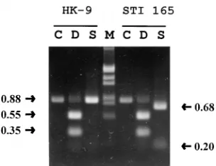
The chosen target for the PCR in the coding region of the 16S rRNA gene allowed for the amplification of a 0.88-kb gene fragment, which is revealed as a single band by agarose gel electrophoresis. Both PCR products vary by only one nucleo-tide in length but they differ in a Sau96I restriction site. In addition, the gene fragments contain a DraI restriction site which distinguishes both species from other protozoan para-sites. The amplified gene fragment could be cut with DraI, resulting in two fragments of 0.55 and 0.35 kb, as predicted from the nucleic acid sequence (Fig. 1). When the amplified DNA of E. dispar was digested with Sau96I, two fragments of the expected length (0.68 and 0.2 kb) were detected, whereas the DNA derived from E. histolytica was not cleaved by this enzyme (Fig. 1).
Specificities of the primers.The specificities of the primers for E. histolytica and E. dispar were verified by using cell lysates of different Entamoeba strains with known zymodeme patterns. In addition, cell lysates of E. invadens, a species infecting reptiles, and several human protozoan parasites were tested. PCR was performed with both primer sets in independent
reactions with each cell lysate. In all 14 cases tested, the result of the PCR corresponded to the one obtained by zymodeme analysis (Table 1). Additionally, no unspecific priming was seen with DNAs from E. coli, E. invadens, E. hartmanni, D.
fragilis, or B. hominis. Results for inhibition controls, carried
out to exclude the possibility that a negative PCR result was due to the failure of amplification, were negative for all reac-tions.
Sensitivity of the assay. The sensitivity of the PCR was assessed by using serial dilutions of lysates of 100 to 0.001 trophozoites of E. histolytica or E. dispar. After 40 PCR cycles, as little as one copy of the target gene was detected by both primer sets, as determined by agarose gel electrophoresis (Fig. 2). If amplification products were analyzed with the Gen-etik-DEIA system, the same sensitivity was obtained (Fig. 2). In order to determine the effect of feces on the assay sensitivity, we performed the SAF concentration procedure with a stool sample negative for parasites, resuspended the pellets in di-gestion buffer containing proteinase K, and added cell lysates corresponding to decreasing numbers of parasites. It was found that the PCR was completely inhibited by undiluted feces and was partially inhibited at a dilution of 1:10, resulting in de-creased sensitivity. However, at a dilution of 1:100, the detec-tion limit for the 16S rRNA gene is similar to that obtained with cell lysates in the absence of stool (data not shown). In order to estimate the sensitivity of the reaction for mixed infections, various numbers of E. histolytica trophozoites were mixed with a constant number of E. dispar trophozoites before lysis, and vice versa. One hundred cells of one species as the background still allowed for the detection of one copy of the target gene of the other species, i.e., a ratio of 1:104(Fig. 3).
Diagnosis of infections with E. histolytica and E. dispar in stool specimens. Target DNA in 19 stool samples stored in SAF for between 3 and 30 days and 18 unpreserved stool specimens, which all contained E. histolytica or E. dispar, as verified by microscopy, were amplified starting from a 1:100 dilution of the proteinase K-digested DNA extract. The target DNA was amplified in 25 cases, identifying 23 E. dispar and 2
E. histolytica infections. The PCR was falsely negative for 12
[image:2.612.102.254.68.185.2]specimens. As it turned out, the PCR was highly influenced by the starting material. By using unpreserved stool specimens, all 18 samples examined were positive by PCR, whereas by using samples stored in SAF for more than 2 days, only 7 of 19 samples (36.8%) were positive by PCR, accounting for all 12 false-negative results. Time course experiments with aliquots of SAF-fixed samples taken every 2 days over 2 weeks for PCR analysis revealed that the sensitivity of the PCR decreased strongly within 2 days, as reflected by the fact that the specific DNA band became more faint. In contrast, if unpreserved stool specimens stored in the refrigerator were used, the sen-sitivity of the PCR remained unchanged, even after a period of 2 weeks. Thus, the short time of contact of the specimen with SAF fixative during the concentration procedure had no influ-ence on the result of the PCR.
TABLE 1. Comparison of PCR results with the results of zymodeme analysis
Species Zymodeme isolatesNo. of
tested PCR result
Entamoeba dispar I, IV 10 E. dispar Entamoeba histolytica II, XIX 4 E. histolytica E. coli, E. hartmanni, E. invadens,
[image:2.612.315.556.652.727.2]B. hominis, D. fragilis 8 Negative for allorganisms FIG. 1. Restriction fragment analysis of PCR products of E. histolytica HK-9
and E. dispar STI-165. Undigested HK-9 and STI-165 DNAs were used as controls (lanes C). Lanes M, size markers (EcoRI-HindIII-cleavedlDNA). D, DraI; S, Sau96I. The sizes of the restriction fragments are indicated to the right and left (in kilobases).
1702 TROLL ET AL. J. CLIN. MICROBIOL.
on May 15, 2020 by guest
http://jcm.asm.org/
DISCUSSION
Amoebiasis affects about 500 million people worldwide, yet only about 10% of the infections are caused by E. histolytica, which leads to severe disease like dysentery or amoebic liver abscess (30). Considering the fact that most infections are due to the noninvasive species E. dispar, a rapid and sensitive diagnostic procedure for differentiating the two species would be of great medical importance. This report describes a very sensitive method which allows for the reliable distinction be-tween E. histolytica and E. dispar by a procedure that starts with unpreserved fresh fecal samples.
It is well established that fresh fecal material has a strong inhibitory effect on the PCR, resulting in a considerable loss of sensitivity (16). Concentration with SAF fixative as a first pre-paratory step is an elegant way to concentrate parasites and at the same time eliminate the inhibitory effect of the feces. The method does not require any further DNA purification or a time-consuming in vitro cultivation, which often ends up with a negative result. Furthermore, our PCR was very sensitive at detecting one copy of the target gene of one species in a background of 104copies of the other species when mimicking
mixed infections.
For optimal results, starting with unpreserved fecal material turned out to be crucial. By using unpreserved stool specimens, no false-negative results occurred when microscopy was used as a “gold standard,” even after storage of the fecal specimen in a refrigerator for 2 weeks. The very short time period during the concentration step during which the sample is in contact with SAF did not have an adverse effect on the PCR, because the preservative is removed immediately by the washing steps. However, by using feces stored in SAF fixative, the sensitivity of the PCR usually decreased within 2 days due to degradation of the target DNA, although exceptionally, a few samples
[image:3.612.132.486.69.329.2]per-formed well for up to 14 days. Since SAF itself showed no effect on DNA degradation, as verified by the addition of the amoebae DNA to the SAF fixative and subsequent PCR, we conclude that the DNA in SAF-preserved specimens is de-graded enzymatically. This is supported by the finding that the addition of 100 mM EDTA tolDNA incubated in SAF-stool specimens for 16 h prevents degradation. SAF does not inhibit
[image:3.612.358.512.487.655.2]FIG. 3. Detection of E. histolytica (A) and E. dispar (B) in mixed cell lysates. To 100 trophozoites of E. dispar STI-165 (A) or E. histolytica HK-9 (B), 100 cells (lane 1), 10 cells (lane 2), 1 cell (lane 3), 0.1 cell (lane 4), 0.01 cell (lane 5), and 0.001 cell (lane 6) of the other species were added. PCR was performed with the primer sets specific for E. histolytica (A) or E. dispar (B). For negative controls (lanes 7), amplification was done by using E. histolytica-specific primers with STI-165 DNA (A) and E. dispar-specific primers with HK-9 DNA (B), respec-tively. Lanes M, size markers (EcoRI/HindIII-cleavedlDNA).
FIG. 2. Sensitivity of PCR for detection of DNA of E. histolytica HK-9 and E. dispar STI-165. Lysed trophozoites were serially diluted to correspond to 100 cells (lane 1), 10 cells (lane 2), 1 cell (lane 3), 0.1 cell (lane 4), 0.01 cell (lane 5), and 0.001 cell (lane 6) and were subjected to PCR amplification. Amplified products were analyzed by agarose gel electrophoresis and DNA enzyme immunoassay. The sizes of the amplification products are indicated on the left (in kilobases). Lanes 7, negative control (PCR without DNA); lanes M, size markers (EcoRI-HindIII-cleavedlDNA); OD492, optical density at 492 nm.
on May 15, 2020 by guest
http://jcm.asm.org/
the DNA-degrading enzymes present in stool. After fixation, the passage of DNases through the wall of the cyst may be facilitated, while this process would not occur in unpreserved specimens. The first method described for the differentiation of E. histolytica and E. dispar was zymodeme analysis (26). However, this method requires cultivation of the amoebae starting with a fresh fecal sample, a lengthy procedure which is hampered by frequent failures. In recent years several investi-gators reported the use of monoclonal antibodies directed against the 170-kDa subunit of the galactose-specific adher-ence lectin for the differentiation of E. histolytica and E. dispar (1, 13, 14). However, since those investigators did not dispose of an antibody specific for E. dispar, the assays require two consecutive ELISAs. Furthermore, the sensitivity as well as specificity were not satisfactory. Alternatively, molecular DNA-based detection methods were developed. Several inves-tigators have described the use of DNA probes, e.g., by relying on intergenic repeated sequences of the rRNA gene circles specific for E. histolytica or E. dispar (6, 12, 21) or on a probe named IE-gen1, which is related to genomic sequences present only in E. histolytica (9). All of these techniques depend on either in vitro cultures or DNA purification and mostly use radioactively labelled probes, requiring special equipment and licensed laboratories.
In recent years PCR was established as a valuable tool for routine diagnosis of infectious diseases. On the basis of the genetic differences between E. histolytica and E. dispar, several groups developed PCR-based assays for the discrimination of the two species (2, 3, 8, 16, 18, 28, 29). Most of these assays used sequences of the extrachromosomal circular rRNA gene. Being present in about 200 copies in each cell (4, 5, 15, 19, 27), sequences of this rRNA gene are more easily detected than DNA fragments of a single-copy gene. Some investigators (2, 3) used differences in the highly repetitive sequences in the noncoding region of rDNA, whereas others (16, 18) based their test on the 16S rRNA genes of E. histolytica and E. dispar. Use of repeated sequences for PCR results in a smear or a ladder of amplified products in agarose gel electrophoresis, which can make interpretation of the results difficult and could reduce the sensitivity of the test. Therefore, we used a 0.88-kb sequence of the coding region of the 16S rRNA gene as the PCR target, leading to the amplification of a single band. In contrast to other investigators (16, 18) who used a single primer pair specific for both amoeba species and distinguished amplified DNA by restriction fragment analysis or nested PCR, we used two pairs of primers specific for either E.
histo-lytica or E. dispar. False-negative results were excluded by two
parallel reactions with both primer sets for each sample and a test for inhibition control for both primer pairs by adding E.
histolytica or E. dispar control DNA. In this way it was possible
to carry out the PCR in one step, minimizing the risk of carryover contaminations, a problem occurring in the second step of nested PCR, in which decontamination by using UDG degradation is not possible. On the other hand, a two-step approach (16, 18) offers additional specificity control, which in our case could be achieved by application of the Gen-etik-DEIA. This test, based on an ELISA system, is as sensitive as agarose gel electrophoresis and offers the advantage of the 96-well microtiter plate format, which makes it an ideal tool for large-scale analysis of PCR products.
Although microscopy remains the method of choice for ex-aminations for ova and parasites, the specific assay described here offers interesting perspectives as a complementary test to routine microscopy, even if an additional unpreserved speci-men must be ordered if the first one was submitted in SAF fixative. In patients requiring special attention like pregnant
women, human immunodeficiency virus-positive patients, or individuals with infections persisting after treatment, a differ-entiation between E. histolytica and E. dispar will greatly help a physician determine whether he or she must treat a poten-tially very dangerous infection or whether the patient is in-fected only with a nonpathogenic species.
REFERENCES
1. Abd-Alla, M. D., T. F. H. G. Jackson, V. Gathiram, A. M. El-Hawey, and J. I.
Ravdin.1993. Differentiation of pathogenic Entamoeba histolytica infections
from nonpathogenic infections by detection of galactose-inhibitable adher-ence protein antigen in sera and faeces. J. Clin. Microbiol. 31:2845–2850. 2. Acuna-Soto, R., J. Samuelson, P. de Girolami, L. Zarate, F. Millan-Velasco,
G. Schoolnick, and D. Wirth.1993. Application of the polymerase chain
reaction to the epidemiology of pathogenic and nonpathogenic Entamoeba histolytica. Am. J. Trop. Med. Hyg. 48:58–70.
3. Aguirre, A., D. C. Warhurst, F. Guhl, and I. A. Frame. 1995. Polymerase chain reaction-solution hybridization enzyme-linked immunoassay (PCR-SHELA) for the differential diagnosis of pathogenic and nonpathogenic Entamoeba histolytica. Trans. R. Soc. Trop. Med. Hyg. 89:187–188. 4. Bhattacharya, S., A. Bhattacharya, and L. S. Diamond. 1988. Comparison of
repeated DNA from strains of Entamoeba histolytica and other Entamoeba. Mol. Biochem. Parasitol. 27:257–262.
5. Bhattacharya, S., A. Bhattacharya, L. S. Diamond, and A. T. Soldo. 1989. Circular DNA of Entamoeba histolytica encodes ribosomal RNA. J. Proto-zool. 36:455–458.
6. Bracha, R., L. S. Diamond, J. P. Ackers, G. D. Burchard, and D. Mirelman. 1990. Differentiation of clinical isolates of Entamoeba histolytica by using specific DNA probes. J. Clin. Microbiol. 28:680–684.
7. Brumpt, E. 1925. Etude sommarie de l’ “Entamoeba dispar” n. sp. Amibe a` kystes quadrinucle´es, parasite de l’ homme. Bull. Acad. Med. Paris 94:943–952. 8. Clark, C. G., and L. S. Diamond. 1991. Ribosomal RNA genes of “patho-genic” and “nonpatho“patho-genic” Entamoeba histolytica are distinct. Mol. Bio-chem. Parasitol. 49:297–302.
9. Cruz-Reyes, J. A., and J. P. Ackers. 1992. A DNA probe specific to patho-genic Entamoeba histolytica. Arch. Med. Res. 23:271–275.
10. Diamond, L. S., D. R. Hariow, and C. C. Cunnick. 1978. A new medium for the axenic cultivation of Entamoeba histolytica and other Entamoeba. Trans. R. Soc. Trop. Med. Hyg. 72:431–432.
11. Diamond, L. S., and G. Clark. 1993. A redescription of Entamoeba histolytica Schaudinn, 1903 (Emended Walker, 1911) separating it from Entamoeba dispar Brumpt, 1925. J. Eukaryot. Microbiol. 40:340–344.
12. Garfinkel, L. I., M. Giladi, M. Huger, C. Gitler, D. Mirelman, M. Revel, and
S. Rozenblatt.1989. DNA probes specific for Entamoeba histolytica
possess-ing pathogenic and nonpathogenic zymodemes. Infect. Immun. 57:926–931. 13. Haque, R., L. M. Neville, S. Wood, and W. A. Petri, Jr. 1994. Detection of Entamoeba histolytica and E. dispar directly in stool. Am. J. Trop. Med. Hyg.
50:595–596.
14. Haque, R., L. M. Neville, P. Hahn, and W. A. Petri, Jr. 1995. Rapid diagnosis of Entamoeba infections by using Entamoeba and Entamoeba histolytica stool antigen detection kits. J. Clin. Microbiol. 33:2558–2561.
15. Huger, M., B. Koller, C. Gitler, D. Mirelman, M. Revel, S. Rozenblatt, and
L. Garfinkel.1989. Entamoeba histolytica ribosomal RNA genes are carried
on palindromic circular DNA molecules. Mol. Biochem. Parasitol. 32:285– 296.
16. Katzwinkel-Wladarsch, S., T. Lo¨scher, and H. Rinder. 1994. Direct ampli-fication and differentiation of pathogenic and nonpathogenic Entamoeba histolytica DNA from stool specimens. Am. J. Trop. Med. Hyg. 51:115–118. 17. Longo, M. C., M. S. Berninger, and J. L. Hartely. 1990. Use of uracil DNA glycosylase to control carry-over contamination in polymerase chain reac-tions. Gene 93:125–128.
18. Novati, S., M. Sironi, S. Granata, A. Bruno, S. Gatti, M. Scaglia, and C.
Brandi.1996. Direct sequencing of the PCR amplified SSU rRNA gene of
Entamoeba dispar and the design of primers for rapid differentiation from Entamoeba histolytica. Parasitology 112:363–369.
19. Ramachandran, S., A. Bhattacharya, and S. Bhattacharya. 1993. Nucleotide sequence analysis of the rRNA transcription unite of a pathogenic Entam-oeba histolytica strain HM-1: IMSS. Nucleic Acids Res. 21:2011.
20. Robinson, G. L. 1968. The laboratory diagnosis of human parasitic amoeba. Trans. R. Soc. Trop. Med. Hyg. 62:285–293.
21. Romero, J. L., S. Descoteaux, S. Reed, E. Orozco, J. Santos, and J.
Samuel-son. 1992. Use of polymerase chain reaction and nonradioactive DNA probes to diagnose Entamoeba histolytica in clinical samples. Arch. Med. Res. 23:277–279.
22. Sambrook, J., E. F. Fritsch, and T. Maniatis. 1989. Molecular cloning: a laboratory manual, 2nd ed. Cold Spring Harbor Laboratory Press, Cold Spring Harbor, N.Y.
23. Sargeaunt, P. G. 1988. Zymodemes of Entamoeba histolytica, p. 370–387. In J. I. Ravdin (ed.), Amoebiasis: human infection by Entamoeba histolytica. John Wiley & Sons, Inc., New York, N.Y.
1704 TROLL ET AL. J. CLIN. MICROBIOL.
on May 15, 2020 by guest
http://jcm.asm.org/
24. Sargeaunt, P. G. 1992. Entamoeba histolytica is a complex of two species. Aust. Microbiol. 13:359–361.
25. Sargeaunt, P. G., and J. E. Williams. 1978. Electrophoretic isoenzyme pat-terns of Entamoeba histolytica and E. coli. Trans. R. Soc. Trop. Med. Hyg.
72:164–166.
26. Sargeaunt, P. G., J. E. Williams, and J. D. Green. 1978. The differentiation of invasive and non-invasive Entamoeba histolytica isolates by isoenzyme electrophoresis. Trans. R. Soc. Trop. Med. Hyg. 72:519–521.
27. Sehgal, D., V. Mittal, S. Ramachandran, S. K. Dhar, A. Bhattacharya, and
S. Bhattacharya.1994. Nucleotide sequence organisation and analysis of the
nuclear ribosomal DNA circle of the protozoan parasite Entamoeba histo-lytica. Mol. Biochem. Parasitol. 67:205–214.
28. Tachibana, H., S. Ihara, S. Kobayashi, Y. Kaneda, T. Takeuchi, and Y.
Watanabe.1991. Differences in genomic DNA sequences between
patho-genic and nonpathopatho-genic isolates of Entamoeba histolytica identified by poly-merase chain reaction. J. Clin. Microbiol. 29:2234–2239.
29. Tannich, E., and G. D. Burchard. 1991. Differentiation of pathogenic from nonpathogenic Entamoeba histolytica by restriction fragment analysis of a single gene amplified in vitro. J. Clin. Microbiol. 29:250–255.
30. Walsh, J. A. 1986. Problems in recognition and diagnosis of amoebiasis. Estimates of the global magnitude of morbidity and mortality. Rev. Infect. Dis. 8:228–238.
31. Yang, J., and T. Scholten. 1977. A fixative for intestinal parasites permitting the use of concentration and permanent staining procedures. Am. J. Clin. Pathol. 300–304.
on May 15, 2020 by guest
http://jcm.asm.org/
ERRATA
Simple Differential Detection of
Entamoeba histolytica
and
Entamoeba dispar
in Fresh Stool Specimens by Sodium
Acetate-Acetic Acid-Formalin Concentration and PCR
HEIKE TROLL, HANSPETER MARTI,ANDNIKLAUS WEISS Swiss Tropical Institute, CH-4002 Basel, Switzerland
Volume 35, no. 7, p. 1701–1705, 1997. Page 1702, column 1, line 9 should read “Eh-5⬘(5⬘-GTACAAAATGGCCAATTCAT TCAATG-3⬘), Eh-3⬘ (5⬘-CTCAGATCTAGAAACAATGCTTCTCT-3⬘, Ed-5⬘ (5⬘-GTACAAAGTGGCCAATTTATGTAAGT-3⬘), and Ed-3⬘(5⬘-ACTTGGATTTAGAAACAATGTTTCTTC-3⬘).”
Genotyping of Rotaviruses in Environmental Water and Stool
Samples in Southern Switzerland by Nucleotide Sequence
Analysis of 189 Base Pairs at the 5
⬘
End of the VP7 Gene
FRANCA BAGGIANDRAFFAELE PEDUZZI Istituto Cantonale Batteriosierologico, Lugano, Switzerland
Volume 38, no. 10, p. 3681–3685, 2000. Page 3683, Fig. 2 legend: The last sentence, “Numbers indicate percent similarity,” should be deleted.
ERRATA
Simple Differential Detection of
Entamoeba histolytica
and
Entamoeba dispar
in Fresh Stool Specimens by Sodium
Acetate-Acetic Acid-Formalin Concentration and PCR
HEIKE TROLL, HANSPETER MARTI,ANDNIKLAUS WEISS Swiss Tropical Institute, CH-4002 Basel, Switzerland
Volume 35, no. 7, p. 1701–1705, 1997. Page 1702, column 1, line 9 should read “Eh-5⬘(5⬘-GTACAAAATGGCCAATTCAT TCAATG-3⬘), Eh-3⬘ (5⬘-CTCAGATCTAGAAACAATGCTTCTCT-3⬘, Ed-5⬘ (5⬘-GTACAAAGTGGCCAATTTATGTAAGT-3⬘), and Ed-3⬘(5⬘-ACTTGGATTTAGAAACAATGTTTCTTC-3⬘).”
Genotyping of Rotaviruses in Environmental Water and Stool
Samples in Southern Switzerland by Nucleotide Sequence
Analysis of 189 Base Pairs at the 5
⬘
End of the VP7 Gene
FRANCA BAGGIANDRAFFAELE PEDUZZI Istituto Cantonale Batteriosierologico, Lugano, Switzerland
Volume 38, no. 10, p. 3681–3685, 2000. Page 3683, Fig. 2 legend: The last sentence, “Numbers indicate percent similarity,” should be deleted.