R E S E A R C H A R T I C L E
Open Access
Kinematic characterization of clinically
observed aberrant movement patterns in
patients with non-specific low back pain: a
cross-sectional study
Peemongkon Wattananon
1*, David Ebaugh
2, Scott A. Biely
3, Susan S. Smith
4,5, Gregory E. Hicks
5and
Sheri P. Silfies
2Abstract
Background:Clinical observation of aberrant movement patterns during active forward bending is one criterion used to identify patients with non-specific low back pain suspected to have movement coordination impairment. The purpose of this study was to describe and quantify kinematic patterns of the pelvis and trunk using a dynamics systems approach, and determine agreement between clinical observation and kinematic classification.
Method:Ninety-eight subjects performed repeated forward bending with clinical observation and kinematic data simultaneously collected. Kinematic data were plotted using angle-angle, coupling-angle, and phase-plane diagrams. Accuracy statistics in conjunction with receiver operating characteristic curves were used to determine agreement between clinical observation and kinematic patterns.
Results:Kinematic patterns were consistent with clinical observation and definitions of typical and aberrant movement patterns with moderate agreement (kappa = 0.46–0.50; PABAK = 0.49–0.73). Early pelvic motion
dominance in lumbopelvic coupling-angle diagram≥59° within the first 38% of the movement represent observed
altered lumbopelvic rhythm. Frequent disruptions in lumbar spine velocity represented by phase-plane diagrams
with local minimum occurrences≥6 and sudden decoupling in lumbopelvic coupling-angle diagrams with sum of
local minimum and maximum occurrences≥15 represent observed judder.
Conclusion:These findings further define observations of movement coordination between the pelvis and lumbar spine for the presence of altered lumbopelvic rhythm and judder. Movement quality of the lumbar spine segment is key to identifying judder. This information will help clinicians better understand and identify aberrant movement patterns in patients with non-specific low back pain.
Keywords:Low back pain, Multi-segment kinematics, Clinical observation, Aberrant movement patterns
Background
Low back pain is one of the most common health prob-lems in the United States [1]. More importantly, high prevalence and recurrence rates increase the number of medical visits, hospitalization, and utilization of health care services, including physical therapy [1–3]. Low back
pain that is not attributable to a recognizable or known specific pathology is referred to as a non-specific low back pain (NSLBP). Non-specific low back pain is accountable for approximately 85% of all low back pain [4].
Current clinical research suggests patients with NSLBP demonstrate different clinical characteristics that may re-sult from different underlying contributory mechanisms [5–9]. Impairment in inter-segmental movement coordin-ation (e.g., coordincoordin-ation between the lumbar spine, pelvis and hip) has been proposed as one cause of NSLBP [8, 10]. Movement coordination impairment (MCI) is * Correspondence:[email protected]
1Motor Control and Neural Plasticity Laboratory, Faculty of Physical Therapy,
Mahidol University, 999 Phuttamonthon 4 Road, Salaya, Nakhon Pathom 73170, Thailand
Full list of author information is available at the end of the article
defined as poorly coordinated or controlled spine and pel-vis position and movement during functional tasks that places repeated abnormal stresses on musculoskeletal tissues eventually contributing to tissue injury and pain [11]. Clinicians have assumed that MCI is associ-ated with impaired neuromuscular control that can be identified by clinical observation of aberrant move-ment patterns [9, 11–17].
Evidence supports that aberrant movement patterns observed during an active forward bending task is one identifier of patients with MCI and that these patients benefit from exercises focused on trunk muscles and de-signed to improve coordination and control (e.g., core stabilization or lumbar stabilization) [7, 18]. Recent work has demonstrated that clinical observation of aberrant movement patterns during standing forward bend has fair to almost perfect (kappa = 0.35–0.89) inter-rater reli-ability when motion was observed simultaneously by two experienced clinicians [19]. Findings from this study also revealed that aberrant movement patterns were sig-nificantly associated with NSLBP providing construct val-idity for the association of aberrant movement patterns with current symptoms [19]. Furthermore, a greater fre-quency of aberrant patterns can be seen in patients with current NSLBP compared to healthy controls when per-forming multiple repetitions of forward bend [19].
Although evidence supports the use of clinical obser-vation for identifying patients with MCI, investigators have not systematically captured, described, or quanti-fied typical and aberrant movement patterns using con-tinuous kinematic data of multiple body segments (femur, pelvis, lumbar spine, and thoracic spine) during a forward bending motion. As a result, clinicians have lim-ited information about which segments and movement characteristics (range, velocity, and/or timing) significantly contribute to the observed aberrant movement patterns.
Kinematic data have been widely used for investigating the amount of trunk and pelvic motion during forward bending, with limited investigation into the movement patterns and underlying neuromuscular control [20–22]. Kinematics, in conjunction with a dynamic systems ap-proach, can be used to better understand movement pat-terns [22–25]. By plotting continuous angle changes between different body segments, or continuous angle changes against segmental instantaneous angular vel-ocity, kinematic data can be used to represent patterns of movement (inter-segment coordination, and movement control) during functional motions [26]. The purposes of this study were to 1) describe and quantify temporal and spatial 3-dimensional multi-segmental kinematics of the pelvis and trunk using a dynamics systems approach, and 2) determine agreement between clinical observation and kinematic classification of movement patterns. Detailed kinematic descriptions of these patterns should provide
clinicians with the ability to enhance their knowledge and understanding of inter-segment coordination and move-ment control associated with different aberrant movemove-ment patterns observed during forward bending. This could lead to better identification and treatment of MCI, and provide a significant step toward quantification of aberrant movement.
Methods
Subjects
Ninety-eight subjects with both clinical observation and kinematic data recorded simultaneously during a series of forward bending tasks were used in this secondary data analysis [19]. Subjects were between 18 and 65 years of age and took part in a study conducted within a university and private physical therapy clinic. This study was ap-proved by the university institutional review board, and all subjects provided written informed consent prior to par-ticipation. Thirty-five subjects had no history of LBP, 29 subjects were experiencing a current episode of LBP that started within the past 7 weeks, and 34 had a history of LBP but were currently pain free (Table 1). Exclusion cri-teria for all subjects consisted of: 1) clinical signs of sys-temic disease; 2) definitive neurologic signs including weakness or numbness in the lower extremity; 3) previous spinal operation; 4) diagnosed osteoporosis, severe spinal stenosis, and/or inflammatory joint disease; 5) pregnancy; 6) any lower extremity condition that would potentially alter trunk movement in standing; 7) vestibular dysfunc-tion; 8) extreme psychosocial involvement; or 9) active treatment of another medical condition that would pre-clude participation in any aspect of the study.
Procedures and kinematic instrumentation
Subjects performed 6 repetitions of an active forward bend task. Two experienced physical therapists observed the for-ward bend task while kinematic data was simultaneously collected. These therapists had at least 5 years of experience in spinal rehabilitation and completed a 2-h training session that standardized the definitions of aberrant patterns prior to data collection. For each subject, the therapists, who were blinded to the group assignment, independently rated the movement pattern as typical or aberrant. Table 2 pro-vides operational definitions of typical and aberrant move-ment patterns used by these clinicians to assess movemove-ment during standing forward bending [7, 12, 15, 17, 19, 27–29].
spinous process of S2), 3) lumbar spine (over the spinous process of L1), and 4) thoracic spine (over the spinous process of T3). Based upon the recommendations of the International Society of Biomechanics (ISB), the following anatomical landmarks were digitized to create a local ref-erence frame for each body segment: 1) thorax (sternal notch, xyphoid process, T8, and C7); 2) pelvis (right ASIS, left ASIS, right PSIS, and left PSIS); 3) lumbar spine (xyphoid process, T10, L3-L5); and 4) femur (medial epi-condyle, lateral epiepi-condyle, and femoral head) [30].
Preliminary work conducted in our lab established the intra- and inter-session coefficient of multiple correl-ation (CMC) for measuring movement patterns with the electromagnetic tracking system. The CMC was fair to excellent (intra-session CMC = 0.76–0.95 and inter-session CMC = 0.51–0.95) across segments demonstrat-ing consistency of the movement patterns in standdemonstrat-ing forward bend for time-series range of motion and angu-lar velocity. The lower CMC values were associated with movements in the frontal plane.
Data reduction
Mutual agreement on clinical observation by the two ex-perienced clinicians was used for stratification of individ-ual kinematic patterns derived from the forward bending task (98 subjects × 6 repetitions = 588 movement patterns) into typical or aberrant patterns of movement. This strati-fication was performed independent of the subject’s low back pain status.
Kinematic data reduction was completed using custom LabVIEW programs (National Instruments, Austin, TX.). Data were converted to segmental angular rotations using Euler angles following a Cardan sequence of x (flexion/extension), y (lateral bend to the right/left, and z (rotation to the right/left) (Fig. 1). Segmental rotations in-cluded: 1) total trunk motion (FT; thoracic spine motion with respect to the femur); 2) pelvic motion (FP; pelvic mo-tion with respect to the femur); 3) lumbar momo-tion (PL; lum-bar spine motion with respect to the pelvis); 4) thoracic motion (LT; thoracic spine motion with respect to the lum-bar spine); and 5) thoracolumlum-bar motion (PT; combined lumbar-thoracic spine motion with respect to the pelvis).
Based on total trunk motion, a LabVIEW program was used to determine the start and stop points for each repetition of the forward bend motion. Kinematic data were then filtered with a dual pass Butterworth filter (2nd order low pass frequency at 5 Hz) and time-normalized to 51 data points (0–50) to represent 100% of the forward bend motion. Typical and aberrant move-ment patterns were represented by the following kine-matic diagrams: 1) angle-angle, 2) coupling-angle, and 3) phase-plane diagrams [23, 31–33].
Kinematic representation and interpretation of movement patterns
Inter-segmental coordination
[image:3.595.57.538.99.182.2]Coordination of movement between the lumbar spine and pelvis is clinically referred to as lumbopelvic rhythm
Table 1Demographic data for control, current episode of LBP, and history of LBP subjects
N %Female Age ± SD (years) NPRS ± SD (score0–10) ODI ± SD (score0–100)
Control 35 57.1 40.9 ± 9.3 N/A N/A
Current episode of LBP 29 48.3 43.6 ± 12.3 4 ± 2.6 28 ± 14.1
History of LBP 34 50.0 46.7 ± 9.5 N/A N/A
Total 98 52.0 43.7 ± 10.5 N/A N/A
Group comparisonpvalue N/A 0.75a 0.07b N/A N/A
LBPLow back pain,NPRSNumeric pain rating scale,ODIOswestry disability index,SDStandard deviation
a
Group comparison using a chi-square test
b
Group comparison using a one-way analysis of variance (ANOVA)
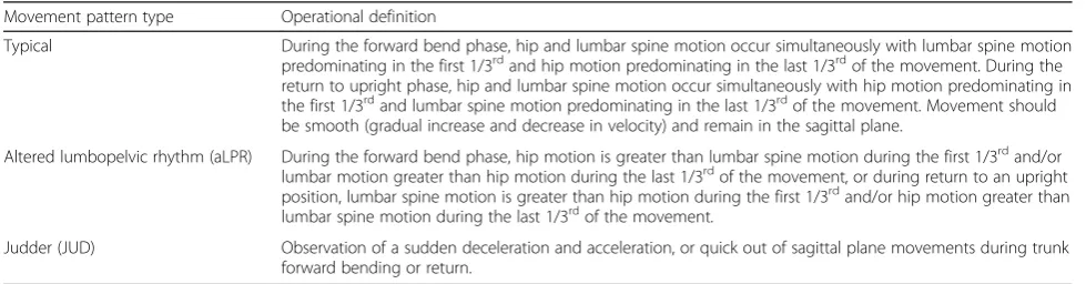
Table 2Operational definitions of clinically observed typical and aberrant movement patterns during a standing forward bend and return motion
Movement pattern type Operational definition
Typical During the forward bend phase, hip and lumbar spine motion occur simultaneously with lumbar spine motion predominating in the first 1/3rdand hip motion predominating in the last 1/3rdof the movement. During the
return to upright phase, hip and lumbar spine motion occur simultaneously with hip motion predominating in the first 1/3rdand lumbar spine motion predominating in the last 1/3rdof the movement. Movement should
be smooth (gradual increase and decrease in velocity) and remain in the sagittal plane.
Altered lumbopelvic rhythm (aLPR) During the forward bend phase, hip motion is greater than lumbar spine motion during the first 1/3rdand/or lumbar motion greater than hip motion during the last 1/3rdof the movement, or during return to an upright
position, lumbar spine motion is greater than hip motion during the first 1/3rdand/or hip motion greater than lumbar spine motion during the last 1/3rdof the movement.
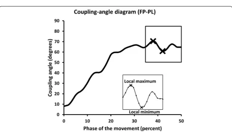
[image:3.595.55.545.603.731.2](LPR). This characteristic of movement can be captured and described using a segment angle-angle diagram (Fig. 2). The shape or trajectory of the diagram provides information regarding qualitative coordination between two segments. A diagonal straight line indicates that the two segments are moving at a constant ratio. Horizontal or vertical lines indicate that one segment is moving, whereas the other segment is not [33]. A limiting factor of using angle-angle diagrams to represent LPR arises when subjects move through different amounts of mo-tion. Vector coding (Fig. 2) can be used to address this limitation by standardizing a segment’s contribution by calculating a vector (coupling-angle) between two adja-cent points relative to the right horizontal [31, 34]. A coupling-angle diagram (Fig. 3) also represents coordin-ation between segments, and quantifies the shape or tra-jectory of movement coordination relative to the percent of movement. A coupling angle of 45° indicates 1:1 mo-tion between segments, greater than 45° indicates distal segment (pelvis) dominance; while less than 45° indicates proximal segment (lumbar spine) dominance [33]. These diagrams were also used to determine when, during the motion (% of movement), one segment dominated the motion relative to another segment.
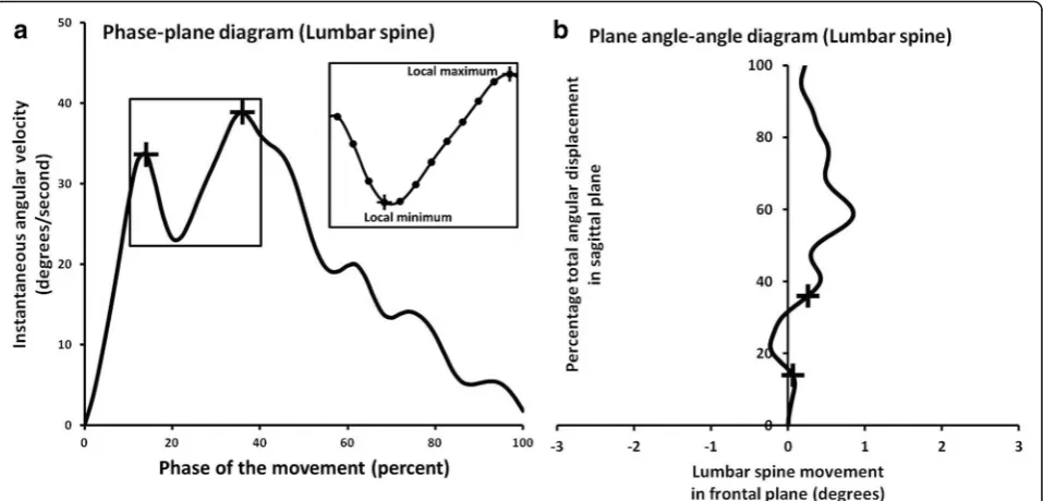
Movement control
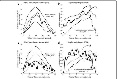
Movement control of a body segment was captured and described using phase-plane and plane angle-angle dia-grams (Fig. 4). Movement control is characterized by smoothness of the segment’s velocity. Disruptions in control can be identified by the number of local mini-mum (LMin) and maximini-mum (LMax) occurrences [23]. These occurrences represent sudden deceleration and acceleration during movement tasks that are clinically referred to as judder (JUD). Quick out of sagittal plane movement or off axis motion is another sign of impaired control that is another focus of clinical definition of jud-der. This presentation of poor movement control can also be captured and described using plane angle-angle and phase-plane diagrams. Additionally, changes in movement control of one segment might cause changes in the rela-tive coordination between segments. Therefore, phase-plane, plane angle-angle, and coupling-angle diagrams can be used to detect the segment responsible for coordin-ation changes identified in a coupling-angle diagram.
Statistical analysis
For the first purpose of this study, temporal and spatial 3-dimensional kinematics of the pelvis and trunk seg-ments (lumbar, thoracic) associated with both typical and aberrant forward bend movement patterns were de-scribed using means and standard deviations of derived kinematic variables (Table 3). The kinematic data were also graphed and additional descriptors were developed.
Individual kinematic variables were tested for normal-ity and homogenenormal-ity of variance assumptions. Independ-ent t-test (1-tailed) was used to test for differences between typical and aberrant movement patterns when those assumptions were met, while Mann-Whitney U test was used if those assumptions were violated. We intended to initially remove kinematic variables that did not differentiate between typical and aberrant movement patterns, but we did not wish to exclude any potentially useful kinematic variables. Therefore, we decided to use a liberal approach, in which individual kinematic vari-ables withp-value less than 0.10 (p< 0.10) were retained as potential key variables of segment and movement characteristics that would then be used to determine agreement between clinical observation and kinematic classification. Additionally, the mean and standard devi-ation for each kinematic data point from typical move-ment patterns were used to generate a mean typical movement pattern along with standard deviation bands that represented typical movement variability. Aberrant movement patterns were then plotted against these typical patterns to further describe differences in movement qual-ity. We found that the derived angle and velocity changes at the start and end of motion often caused errors in coupling-angle and phase-plane diagrams secondary to Fig. 1Location of kinematic sensors on the femur, pelvis, lumbar
[image:4.595.57.290.86.374.2]Fig. 3Example of a lumbopelvic (FP-PL) coupling-angle diagram that plots percentage total angular displacement during forward bending (x axis) versus coupling angles (y axis). Relative timing of a shift from lumbar domination to pelvic domination within the movement pattern is defined by % total angular displacement when the coupling angles are greater than 46°. Local minimum (LMin) or maximum (LMax) occurrences (insert) representing coordination changes in the coupling-angle diagrams was identified by the greatest (local maximum) or least (local minimum) values (X)
[image:5.595.55.538.87.349.2] [image:5.595.58.538.411.684.2]significant variability associated with values fluctuating around zero angular motion or velocity. Therefore, we used data between 5% and 95% of total trunk motion for further analysis.
The second purpose of this study was to determine the level of agreement between clinical observation and kinematic classification derived from kinematic diagrams that represent segments and movement characteristics contributing to the clinically observed aberrant move-ment patterns. An accuracy statistics approach in con-junction with receiver operating characteristic curves (ROC) was used for this analysis [35–38]. For altered lumbopelvic rhythm (aLPR, inter-segment coordination), we did not know at which point in the movement pat-tern, clinicians perceived onset of pelvic domination (pelvic-dominated angle). Therefore, we varied the lum-bopelvic coupling angle from 45°-90°, then derived a variable, called “timing” for each 1° pelvic-dominated angle increase, to determine when during the motion (%
of forwarding bending movement) the pelvis dominated the motion relative to the lumbar spine.
For judder (smoothness of movement), local minimum (LMin) and local maximum (LMax) occurrences in the phase-plane diagrams for each segment (FT, FP, PL, LT, and PT) and coupling-angle diagram for FP-PL were used to quantify movement control and inter-segment coordin-ation, respectively. LMin and LMax in the phase-plane and coupling-angle diagrams correspond to the two oper-ational definitions of judder (sudden deceleration and ac-celeration, and quick off axis or out of plane movement). However, clinical observation data did not indicate what type of judder had been identified. Therefore, after key segment and characteristics were identified, we further classified judder into quick out of plane movement based on corresponding plane angle-angle diagrams and calcu-lated prevalence of this type of judder.
[image:6.595.60.539.88.318.2]Contingency tables and receiver operating characteris-tic curves (ROC) were created using the total number of typical and each aberrant movement pattern based on clinical observation (reference standard) and the kine-matic variables derived from quantification of kinekine-matic diagrams. The pelvic-dominated angle and segment that generated the optimal area under the ROC curve (AUC) were then identified. The ROC of identified pelvic-dominated angle or segment and its kinematic variable was used to determine the cut-off point that maximized agree-ment on kinematic variables. Kappa values were used to Fig. 4aExample of lumbar spine phase-plane diagram representing percentage of total angular displacement (x axis) versus instantaneous angular velocity (y axis) during forward bending. Local minimum and maximum occurrence of the phase-plane diagram (+, insert) represent disruptions in angular velocity (sudden deceleration and acceleration) that are associated with judder. This pattern can also be characterized by quick out of plane deviation in the pelvis or lumbar spine plane angle-angle diagram (b) over a short period of time (as indicated by +, in both diagrams). These out of plane deviations are consistent with sudden decreases and increases in angular velocity in the pelvis or lumbar spine phase plane diagram
Table 3Kinematic variables used to describe aberrant movement pattern
Aberrant movement pattern Variable
aLPR Slope of AA (Mean CA), Timing
JUD LMin, LMax
aLPRAltered lumbopelvic rhythm,JUDJudder,AAAngle-angle diagram,CA
Coupling angle diagram,TimingWhen in the movement aLPR occurred,LMin
[image:6.595.57.292.665.705.2]assess the agreement between clinical observation and kinematic classification. Sensitivity, specificity, and positive and negative likelihood ratios (LR) were calculated for the cut point. Statistical analysis was performed using custom LabVIEW (National Instruments, Austin, TX) and SPSS (IBM SPSS Statistics for Windows, Version 21.0. Armonk, NY) software.
Results
Movement pattern classification: Clinical observation
Based upon clinical observation, two experienced clini-cians mutually agreed on 195 out of 588 movement trials (33%). One hundred and eight forward bend movement trials (18%) were classified as a typical, 57 trials (10%) were classified as demonstrating altered inter-segment coordin-ation (LPR), and 30 trials (5%) were classified as demon-strating poor movement control (JUD). In most trials, the thoracic segment’s movement pattern did not assist in the identification of any aberrant pattern. Therefore, this seg-ment was not included in statistical analyses related to our second purpose.
Kinematic classification Inter-segment coordination
The overall slope of the lumbopelvic (FP-PL) angle-angle diagram for those patterns with aLPR was significantly steeper than that of the typical movement pattern (Table 4). When broken down into specific ranges of the forward bend motion, the aLPR angle-angle slopes in the first and second third of the motion were significantly steeper than typical; however, in the last third of the mo-tion, aLPR slopes were significantly less steep than typical.
Analysis of the coupling-angle diagram revealed that the time (% of movement) when the pelvic contribution was greater than the lumbar contribution occurred sig-nificantly earlier in the aLPR patterns (21.9 ± 14.1) when compared with the typical pattern (30.6 ± 16.5). This variable was retained for analysis of agreement between clinical observation and kinematic classification of aLPR.
Movement control
The numbers of LMin, and LMax occurrences, as well as the sum of local minimum and maximum (LSum) occur-rences on the phase-plane and coupling-angle diagrams
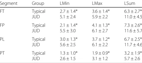
were significantly greater for JUD than the typical patterns (Tables 5 and 6). These kinematic variables were retained to determine the agreement between clinical observation and kinematic classification of judder.
Clinical and kinematic agreement
Accuracy statistics and ROC analysis revealed that lum-bar spine segment kinematics were the key for separat-ing typical from aberrant movement patterns. Table 7 shows the kinematic variables used for classification, AUC, kappa, and accuracy statistics at ROC cut-off point for each aberrant pattern. In addition, we found that 7 out of 30 judder patterns (23%) were further classified as quick out of plane movement based on lumbar spine plane angle-angle diagram. Figures 5 and 6 demonstrate examples of typical and aberrant patterns along with the kinematic variables used to identify the aberrant motion observed by the clinicians.
Discussion
[image:7.595.305.538.585.689.2]Our kinematic analysis of forward bend movement pat-terns provides information that enhances the under-standing of pelvis and trunk movement characteristics associated with typical and aberrant movement patterns. Additionally, our findings identified the key segments (pelvis and lumbar spine) and movement characteristics of these segments that best described aberrant move-ment patterns observed during clinical examination of forward bending. Kinematic classifications representing these segments (Table 7) demonstrated high specificity and positive likelihood ratio, which indicates that our kinematic variables have the ability to detect clinically observed aberrant movement patterns from the kine-matic data. Detailed kinekine-matic descriptions of typical and aberrant movement patterns are discussed in detail within the following paragraphs.
Table 4Mean and standard deviation of slope of typical and altered lumbopelvic rhythm (aLPR) angle-angle diagram (mean coupling angle) of lumbopelvic segments (FP-PL) for the first, second, and last 1/3rd of motion, and overall motion
Segment Group Slope
First 1/3 Second 1/3 Last 1/3 Overall
FP-PL Typical aLPR
32.0 ± 16.6* 44.7 ± 17.5
51.3 ± 11.0* 63.0 ± 10.4
69.17 ± 18.1* 58.91 ± 15.7
51.99 ± 7.0* 60.26 ± 7.4
*= statistical significance (p< 0.10)
Table 5Mean and standard deviation local minimum (LMin), local maximum (LMax), and sum of local minimum and maximum (LSum) occurrences of clinically observed typical and judder (JUD) patterns using phase-plane diagram for each segment
Segment Group LMin LMax LSum
FT Typical
JUD
2.7 ± 1.4* 5.1 ± 2.4
3.6 ± 1.4* 5.9 ± 2.2
6.3 ± 2.7* 11.0 ± 4.5
FP Typical
JUD
2.1 ± 1.4* 5.5 ± 3.0
4.1 ± 1.3* 6.1 ± 2.7
7.3 ± 2.6* 11.6 ± 5.7
PL Typical
JUD
3.0 ± 1.3* 5.6 ± 2.5
3.7 ± 1.2* 6.1 ± 2.2
6.7 ± 2.5* 11.7 ± 4.6
PT Typical
JUD
1.3 ± 1.0* 2.6 ± 1.5
1.9 ± 0.9* 3.1 ± 1.2
3.2 ± 1.9* 5.7 ± 2.6
FTTotal trunk (Thoracic spine (T3) with respect to right femur),FPPelvic
segment (Pelvis (S2) with respect to right femur),PLLumbar segment (Lumbar
spine (L1) with respect to pelvis (S2)),PTThoracolumbar segment (Thoracic
spine (T3) with respect to pelvis (S2))
[image:7.595.55.291.675.724.2]Typical forward bend movement pattern
Inter-segment coordination or lumbopelvic rhythm dur-ing a forward bend motion is described as smooth and continuous motion with the first third being dominated by lumbar spine motion, the second third shared motion between the lumbar and pelvic segments, and the last third being dominated by pelvic motion (Table 4). Typ-ical lumbopelvic coordination, represented in an angle-angle diagram, is characterized by a smooth concave line with gradual changes in segmental dominance (Fig. 5a). When plotted in a coupling-angle diagram, typical lum-bopelvic rhythm is represented by a diagonal line with a positive slope from lower left corner to upper right cor-ner (Fig. 5b). This kinematic description is consistent with the clinical definition of typical lumbopelvic rhythm described by Calliet and Farfan [27, 29].
The control of each segmental movement was demon-strated by a smooth gradual increase in velocity to mid-point of forward bend, and then a smooth gradual decrease in velocity to the end of the forward bend mo-tion. Overall the phase-plane diagram was bell shaped (Fig. 6a), with a minimal number of local minimum and maximum occurrences (Table 5). No quick out of sagit-tal plane movements were noticed during the forward bend motion. Inter-segmental movement coordination was also smooth and continuous with a minimal number of local minimum and maximum occurrences (Table 6 and Fig. 6b). These kinematic descriptions are also con-sistent with clinical definition of typical (normal) for-ward bending described by Paris [17].
Altered inter-segmental coordination
aLPR is characterized by either shared movement be-tween the pelvis and lumbar spine, or pelvic dominated motion during the first 1/3rd of the forward bend mo-tion. Motion continues with increased pelvis domination in the second 1/3rdof the motion. In the last 1/3rdof the motion the pattern is dominated by lumbar spine mo-tion (Table 4 and Fig. 5c). Overall this pattern is the re-verse of a typical forward bend pattern.
Coupling-angle diagrams revealed sharp increases in the coupling angle in the first 1/3rd of forward bend in-dicating shared motion between the pelvis and lumbar spine that occurs much earlier than the typical pattern. In the second 1/3rd of forward bend, the coupling angle increased indicating pelvis domination, which also ap-peared earlier than the typical pattern. In the last 1/3rd of forward bend, the coupling angle decreased indicating a reversed pattern (Fig. 5d).
Data suggested that patterns of lumbopelvic coupling angles are key for identifying aLPR (Table 7 and Fig. 5d). Clinicians seemed to perceive pelvic domination when coupling angle (pelvic-dominated angle) reached 59° (pelvic-lumbar ratio = 1.66:1). At a coupling angle of 59°, timing (relative to the % of motion during forward bend-ing task) that maximizes the agreement between clinical observation and kinematic classification derived from coupling-angle diagram demonstrated transition from lumbar spine domination to pelvis domination in the second 1/3rdof the movement (38%). This slight differ-ence between the clinical definition (shift within first 33% of motion) and kinematic cut point (38% of motion) is likely related to time normalization and averaging data across subjects. However, the lumbopelvic coupling-angle diagram represents what the clinicians observed as altered lumbopelvic rhythm with focus on the amount of pelvis contribution (pelvis domination) in the early phase (first 38%) of the movement.
[image:8.595.54.291.132.167.2]A limited number of studies exist that describe trunk and pelvic angular motion during a standing trunk for-ward bend task [20, 21, 39, 40]. In these studies, the
Table 6Mean and standard deviation of local minimum (LMin), local maximum (LMax), and sum of local minimum and maximum (LSum) occurrences of clinically observed typical and judder (JUD) using the lumbopelvic (FP-PL) coupling-angle diagram
Segment Group LMin LMax LSum
FP-PL Typical
JUD
3.6 ± 1.4* 5.5 ± 3.3
4.3 ± 1.3* 6.3 ± 3.3
8.0 ± 2.7* 11.8 ± 6.7
*= statistical significance (p< 0.10)
Table 7Agreement (95% CI) between clinical observation and kinematic lumbopelvic segment movement characteristics and accuracy statistics of the kinematic variables for predicting the observed movement pattern
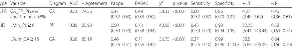
Type Variable Diagram AUC %Agreement Kappa PABAK χ2 pvalue Sensitivity Specificity +LR -LR aLPR CA_FP_PL@59
and Timing < 38%
CA 0.73 74.55 0.47
(0.32–0.60) 0.49 (0.33–0.62)
38.33 <0.001 0.60 (0.52–0.67)
0.86 (0.79–0.91)
4.27 (2.49–7.62)
0.46 (0.36–0.61)
JUD LMin_PL≥6 PP 0.85 85.93 0.50
(0.30–0.59) 0.73 (0.58–0.84)
40.55 <0.001 0.43 (0.30–0.49)
0.98 (0.94–0.99)
22.75 (5.44–143.44)
0.58 (0.51–0.74)
LSum_CA≥15 CA 0.66 85.19 0.46
(0.26–0.51) 0.71 (0.55–0.82)
36.75 <0.001 0.37 (0.25–0.40)
0.99 (0.96–0.1.00)
38.5 (5.69–796.05)
0.64 (0.60–0.79)
Typical (N= 108); Altered lumbopelvic rhythm (aLPR;N= 57); Judder (JUD;N= 30)
[image:8.595.61.540.608.689.2]researchers investigated ratios of pelvis to lumbar seg-ment motion at discrete points in the moveseg-ment, and reported means and standard deviations. Although they report differences between healthy and low back pain groups, this approach does not provide continuous in-formation about inter-segment coordination and control. Therefore, this existing body of work cannot fully de-scribe altered lumbopelvic rhythm and pinpoint when transition from lumbar domination to pelvic domination occurred. The only reported approach that focused on
[image:9.595.62.541.88.506.2]using lumbopelvic coupling-angle diagrams provides typical timing (relative to % of movement) for when the transition from lumbar to pelvic domination occurs. This spatial and temporal information can be used to further explain altered lumbopelvic rhythm.
Altered movement control
Qualitative assessment of segment control during for-ward bend suggests that the frequency of disruptions, or sudden decreases and increases in angular velocity, in the JUD group were significantly greater than the typical group (Table 5). Coupling-angle diagrams also revealed that the JUD group had a greater number of sudden de-coupling instances in inter-segmental coordination than the typical group (Table 6).
The data indicate that the kinematic patterns in phase-plane diagram considered as JUD are best defined as the number of local minimum occurrences equal to or greater than 6 in the lumbar spine segment. Clini-cians appear to focus on lumbar spine angular velocity or smoothness of the movement during the standing
trunk forward bend (Fig. 6a and c). This kinematic de-scription was consistent with clinical observation of JUD (a sudden deceleration and acceleration). Additionally, quick out of plane movement is best defined as when the pattern momentarily deviates away from sagittal plane in lumbar spine plane angle-angle diagram (Fig. 4). Quick out of plane movements were not frequently demonstrated in our dataset. We found that the occur-rence of quick out of plane movement was consistent with disruption in angular velocity in the lumbar spine phase-plane diagram. This suggests that clinical observa-tion of judder based on lumbar spine velocity may be sufficient and observation of quick out of plane motion might not be necessary for determination of judder. To date, no researcher has investigated the primary segment and movement control characteristics that represent JUD during standing trunk forward bend.
[image:10.595.57.541.87.413.2]using coupling-angle diagram was not matched with clinical observation of JUD, it seemed clinicians’ classifi-cation of this aberrant movement pattern is made through particular attention to changes in inter-segment coordination between the pelvis and lumbar spine. Po-tentially, this kinematic description of judder could be used to refine clinical observation of judder.
Collectively, the findings from our study provide de-tailed descriptions of temporal and spatial 3-dimensional multi-segmental kinematics of the pelvis and trunk seg-ments for typical and aberrant moveseg-ments during stand-ing forward bend motion. This information can be used to enhance knowledge and understanding of inter-segment coordination and movement control, and may help refine operational definitions that clinicians use to identify aberrant movement patterns. This information may also be useful for future studies that investigate typical and aberrant movement patterns or are designed to determine the ability of exercise and motor control based therapeutic interventions to alter these patterns.
The findings of this study should be considered in light of the following limitations. Our data interpretation was based on the observations of two experienced ortho-pedic physical therapists which limits generalizability. Data interpretation may also be influenced by their clin-ically imposed thresholds of aberrance. These thresholds directly affect the prevalence of typical and aberrant rat-ings. Our approach to analysis was from an accuracy sta-tistics perspective using maximum agreement to develop thresholds. It is possible that these thresholds or criteria are not the same as those used by other clinicians. We also had a relatively low percentage of mutual agreement between two experienced clinicians when we included only those repetitions where both raters indicated a typ-ical pattern or only one type of aberrant pattern on the same repetition. This was done to ensure that movement patterns we analyzed were clear representations of a typ-ical or aberrant pattern. Our prior work focusing on clinical agreement (clinician’s come to the same overall decision about typical or aberrant pattern for the sub-ject) demonstrated moderate to almost perfect agree-ment (kappa = 0.46–0.83) [18]. But this does suggest that multiple repetitions are likely necessary for clinical agreement on movement patterns. We also acknowledge the limitations associated with the use of the same data set to develop and test accuracy of the kinematic vari-ables. However, this works serves as a starting point for quantification of aberrant movement patterns and we recognize that further work and analysis is warranted.
Conclusion
Angle-angle, coupling-angle, and phase-plane diagrams can be used to qualitatively and quantitatively describe 3-dimensional multi-segmental kinematic patterns that
represent both typical and aberrant (altered lumbopelvic rhythm, or judder) movements during standing forward bend. Coordination of the movement between the pelvis and lumbar spine can be assessed for presence of altered lumbopelvic rhythm and judder. The lumbar spine seg-ment appears to be the key segseg-ment to observe judder. These detailed kinematic descriptions should provide cli-nicians with direction for identifying aberrant movement patterns. Collectively, these data can be used to help im-prove understanding of typical and aberrant movement patterns, train clinicians in their clinical observation of typical and aberrant movement patterns and to test the efficacy of interventions to change inter-segmental co-ordination and control.
Abbreviations
AA:Angle-angle diagram; aLPR: Altered lumbopelvic rhythm; ASIS: Anterior superior iliac spine; AUC: Area under the receiver operating characteristic curve; CA: Coupling angle diagram; CMC: Coefficient of multiple correlation; FP: Pelvic motion with respect to the femur; FT: Thoracic spine motion with respect to the femur; ISB: International Society of Biomechanics; JUD: Judder; LBP: Low back pain; LMax: Local maximum; LMin: Local minimum; LPR: Lumbopelvic rhythm; LR: Likelihood ratio; LSum: Sum of local minimum and maximum; LT: Thoracic spine motion with respect to the lumbar spine; MCI: Movement coordination impairment; NSLBP: Non-specific low back pain; PABAK: Prevalence-adjusted bias-adjusted kappa; PL: Lumbar spine motion with respect to the pelvis; PP: Phase-plane diagram; PSIS: Posterior superior iliac spine; PT: Combined lumbar-thoracic spine motion with respect to the pelvis; ROC: Receiver operating characteristic curve
Acknowledgements
We would like to thank the Rehabilitation Sciences Research Laboratories of Drexel University and Physiotherapy Associates Clinic for providing spaces for data collection. We would also like to thank all subjects who participated in this study.
Funding
This study was funded in part by the Orthopaedic Section of the American Physical Therapy Association.
Availability of data and materials
The datasets used and/or analyzed during this study would be available from corresponding author upon reasonable request.
Authors’contributions
SPS, SAB, SSS, and DE have contributed to the conception, research design, and manuscript preparation and edition. PW have substantially contributed to data collection, data analysis, and drafting and revising the manuscript. GEH has significantly contributed to data analysis, as well as editing and revising the manuscript. All authors read and approved the final manuscript.
Ethics approval and consent to participate
The study was approved by the Drexel University Institutional Review Board (project number: 1042185). All subjects provided written informed consent prior to participation.
Consent for publication
All subjects provided written informed consent. The consent stated that all data collected would be used for publication.
Competing interests
The authors declare that they have no competing interests.
Publisher’s Note
Author details
1Motor Control and Neural Plasticity Laboratory, Faculty of Physical Therapy,
Mahidol University, 999 Phuttamonthon 4 Road, Salaya, Nakhon Pathom 73170, Thailand.2Physical Therapy & Rehabilitation Sciences Department, Drexel University, 1601 Cherry Street, Philadelphia, PA 19102, USA.3Physical
Therapy Program, Neumann University, One Neumann Drive, Aston, PA 1901, USA.4College of Nursing and Health Professions, Drexel University, 245 N
15th St, Philadelphia, PA 19102, USA.5Department of Physical Therapy, University of Delaware, 540 S. College Ave, Suite 210E, Newark, DE 19713, USA.
Received: 26 April 2017 Accepted: 8 November 2017
References
1. Pengel LHM, Herbert RD, Maher CG, Refshauge KM. Acute low back pain: systematic review of its prognosis. BMJ. 2003;327:323.
2. Andersson GBJ. Epidemiological features of chronic low-back pain. Lancet. 1999;354:581–5.
3. Walker BF. The prevalence of low back pain: a systematic review of the literature from 1966 to 1998. J Spinal Disord Tech. 2000;13(3):205–17. 4. Carey TS, Garrett JM, Jackman AM. Beyond the good prognosis:
examination of an inception cohort of patients with chronic low back pain. Spine. 2000;25(1):115–20.
5. Dankaerts W, O'Sullivan PB, Burnett AF, Straker LM. The use of a
mechanism-based classification system to evaluate and direct management of a patient with non-specific chronic low back pain and motor control impairment–a case report. Manual Ther. 2007;12(2):181–91.
6. Flynn TW, Fritz JM, Whitman JM, Wainner RS, Magel JS, Rendeiro DG, et al. A clinical prediction rule for classifying patients with low back pain who demonstrate short-term improvement with spinal manipulation. Spine. 2002;27(24):2835–43.
7. Hicks GE, Fritz JM, Delitto A, McGill SM. Preliminary development of a clinical prediction rule for determining which patients with low back pain will respond to a stabilization exercise program. Arch Phys Med Rehab. 2005;86(9):1753–62.
8. O'Sullivan PB. Diagnosis and classification of chronic low back pain disorders. Maladaptive movement and motor control impairments as underlying mechanism. Manual Ther. 2005;10(4):242–55.
9. Van Dillen LR, Sahrmann SA, Norton BJ, Caldwell CA, McDonnell MK, Bloom NJ. Movement system impairment-based categories for low back pain: stage 1 validation. J Orthop Sport Phys. 2003;33(3):126–42.
10. O'Sullivan PB. Masterclass. Lumbar segmental 'instability': clinical presentation and specific stabilizing exercise management. Manual Ther. 2000;5(1):2–12. 11. Sahrmann SA. Diagnosis and treatment of movement impairment
syndromes. Saint Louis, MO: Mosby Publishers; 2001.
12. Delitto A, Erhard RE, Bowling RWA. Treatment-based classification approach to low back syndrome: identifying and staging patients for conservative treatment. Phys Ther. 1995;75(6):470–85.
13. van Dijk M, Smorenburg N, Visser B, Heerkens YF, Nijhuis-van der Sanden MWG. How clinicians analyze movement quality in patients with non-specific low back pain: a cross-sectional survey study with Dutch allied health care professionals. BMC Musculoskel Dis. 2017;18(1):288.
14. Delitto A, George SZ, Van Dillen LR, Whitman JM, Sowa G, Shekelle P, et al. Low back pain. J Orthop Sport Phys. 2012;42(4):A1–A57.
15. Farfan HF, Gracovetsky S. The nature of instability. Spine. 1984;9(7):714–9. 16. Kirkaldy-Willis WH, Farfan HF. Instability of the lumbar spine. Clin Orthop
Relat R. 1982;165:110–23.
17. Paris SV. Physical signs of instability. Spine. 1985;10(3):277–9.
18. Rabin A, Shashua A, Pizem K, Dickstein R, Dar G. A clinical prediction rule to identify patients with low back pain who are likely to experience short-term success following lumbar stabilization exercises: a randomized controlled validation study. J Orthrop Sport Phys. 2014;44(1):6–B13.
19. Biely SA, Silfies SP, Smith SS, Hicks GE. Clinical observation of standing trunk movements: what do the aberrant movement patterns tell us? J Orthop Sport Phys. 2014;44(4):262–72.
20. Esola MA, McClure PW, Fitzgerald GK, Siegler S. Analysis of lumbar spine and hip motion during forward bending in subjects with and without a history of low back pain. Spine. 1996;21(1):71–8.
21. McClure PW, Esola M, Schreier R, Siegler S. Kinematic analysis of lumbar and hip motion while rising from a forward, flexed position in patients with and without a history of low back pain. Spine. 1997;22(5):552–8.
22. Wong TKT, Lee RYW. Effects of low back pain on the relationship between the movements of the lumbar spine and hip. Hum Movement Sci. 2004;23(1):21–34. 23. Kurz MJ, Stergiou N. Applied dynamic system theory for the analysis of
movement. In: Innovative analyses of human movement: analytical tools for human movement research. Champaign: human kinetics; 2004. p. 93–119. 24. Silfies SP, Mehta R, Smith SS, Karduna AR. Differences in feedforward trunk
muscle activity in subgroups of patients with mechanical low back pain. Arch Phys Med Rehab. 2009;90(7):1159–69.
25. Stergiou N, Jensen JL, Bates BT, Scholten SD, Tzetzis G. A Dynamical systems investigation of lower extremity coordination during running over obstacles. Clin Biomech. 2001;16(3):213–21.
26. Spinelli BA, Wattananon P, Silfies S, Talaty M, Ebaugh D. Using kinematics and a dynamical systems approach to enhance understanding of clinically observed aberrant movement patterns. Manual Ther. 2015;20(1):221–6. 27. Cailliet R. Low back pain syndrome. Philadelphia: F.A. Davis Co; 1988. 28. Cook C, Brismee J-M, Sizer PS. Factors associated with physiotherapists'
confidence during assessment of clinical cervical and lumbar spine instability. Physiother Res Int. 2005;10(2):59–71.
29. Farfan HF. Muscular mechanism of the lumbar spine and the position of power and efficiency. Orthop Clin North Am. 1975;6(1):135–44. 30. Wu G, Siegler S, Allard P, Kirtley C, Leardini A, Rosenbaum D, et al. ISB
recommendation on definitions of joint coordinate system of various joints for the reporting of human joint motion—part I: ankle, hip, and spine. J Biomech. 2002;35(4):543–8.
31. Hamill J, Haddad JM, McDermott WJ. Issues in quantifying variability from a dynamical systems perspective. J Appl Biomech. 2000;16(4):407–18. 32. Miller RH, Chang R, Baird JL, Van Emmerik REA, Hamill J. Variability in
kinematic coupling assessed by vector coding and continuous relative phase. J Biomech. 2010;43(13):2554–60.
33. Winstein CJ, Garfinkel A. Qualitative dynamics of disordered human locomotion: a preliminary investigation. J Motor Behav. 1989;21(4):373–91. 34. Chang R, Van Emmerik R, Hamill J. Quantifying rearfoot-forefoot
coordination in human walking. J Biomech. 2008;41(14):3101–5. 35. Deyo RA, Centor RM. Assessing the responsiveness of functional scales to
clinical change: an analogy to diagnostic test performance. J Chron Dis. 1986;39(11):897–906.
36. Portney LG, Watkins MP. Foundations of clinical research: applications to practice. 3rd ed. Upper Saddle River, NJ: Pearson & Prentice Hall; 2009. 37. Sackett DLA. Primer on the precision and accuracy of the clinical
examination. JAMA. 1992;267(19):2638–44.
38. Sim J, Wright CC. The kappa statistic in reliability studies: use, interpretation, and sample size requirements. Phys Ther. 2005;85(3):257–68.
39. Mayer TG, Tencer AF, Kristoferson S, Mooney V. Use of noninvasive techniques for quantification of spinal range-of-motion in normal subjects and chronic low-back dysfunction patients. Spine. 1984;9(6):588–95. 40. Paquet N, Malouin F, Richards CL. Hip-spine movement interaction and
muscle activation patterns during sagittal trunk movements in low back pain patients. Spine. 1994;19(5):596–603.
41. Lee RY, Wong TK. Relationship between the movements of the lumbar spine and hip. Hum Movement Sci. 2002;21(4):481–94.
• We accept pre-submission inquiries
• Our selector tool helps you to find the most relevant journal
• We provide round the clock customer support
• Convenient online submission
• Thorough peer review
• Inclusion in PubMed and all major indexing services
• Maximum visibility for your research
Submit your manuscript at www.biomedcentral.com/submit