Transactions
PAPER
Cite this:Dalton Trans., 2014,43, 1125
Received 10th September 2013, Accepted 23rd October 2013
DOI: 10.1039/c3dt52480a
www.rsc.org/dalton
Synthesis, structure and photophysical properties
of [UO
2
X
2
(O
v
PPh
3
)
2
] (X = Cl, Br, I)
†
Emtithal Hashem,
aThomas McCabe,
aCarola Schulzke
band Robert J. Baker*
aThe synthesis of a series of uranyl compounds viaoxidation of [Li(THF)4][UX5(THF)] (X = Cl, Br, I) in the presence of Ph3PvO is described. The solid state structures of [Li(OvPPh3)(MeCN)2]2[UO2Cl4], [UO2Br2(OvPPh3)2] and [Li(OvPPh3)4][I3], formed as a by-product from the oxidation of [Li(THF)4 ]-[UI5(THF)], is reported. The electronic absorption spectra and photoluminescence spectra of [UO2X2(OvPPh3)2] (X = Cl, Br, I) have been measured and no significant changes in the position of the emission (515–530 nm) or the lifetimes (ca. 1μs) are observed in this series. However a bathochromic shift is observed in the U–X LMCT band in the electronic absorption spectrum.
Introduction
The chemistry of the actinides has undergone a resurgence of interest over the past decade, with small molecule activation at the forefront of this revolution.1Reactivity patterns have been developed that have no analogue in transition metal chemi-stry.2This has fuelled a deeper understanding of the bonding in these compounds, particularly the role the 5f and 6d orbi-tals play in metal–ligand interactions.3 One class of com-pounds that has known covalency is the uranyl ion, {UO2}2+.4
However, it has taken some time to fully elucidate the bonding in this species, which has immense importance as it is in the most stable oxidation state and prevalent in the environment and in spent nuclear fuels. A comprehensive understanding has come from both experiment and theory and an authorita-tive review by Denning summarises recent developments.5 Detailed studies of [UO2Cl4]2−have substantially aided in the
description of the bonding in the uranyl fragment. The photo-physical properties of the uranyl ion have been elucidated from these studies and the optical properties are due to a ligand-to-metal charge transfer (LMCT) transition involving promotion of an electron from a bonding–yl oxygen orbital (σu, σg, πu and πg) to a non-bonding uranium 5fδ and 5fϕ
orbital, centred atca. 420 nm. The characteristic green emis-sion arises from de-excitation of this3Πutriplet excited state.
Superimposed on the absorption and emission bands are‘hot
bands’ arising from strong coupling of the ground state Raman active symmetric vibrational OvUvO (ν1) mode with
the 3Πu electronic triplet excited state. The heavier halides
have been less well studied with few examples of structurally characterised [UO2Br4]2−.6 Due to the decrease in the U–X
bond energy, which has been documented for UX3 (U–F =
619 kJ mol−1, U–Cl = 495.4 kJ mol−1, U–Br = 424.3 kJ mol−1, and U–I = 343 kJ mol−1),7 uranyl iodides are rare. The first example unambiguously reported was the thermally unstable [UO2I2(H2O)2]·4Et2O,8 but since then further thermally stable
compounds have been prepared, such as [UO2I2( py)3],9
[UO2I2L2] (L = OvPPh3, OvP(NMe2)3)7 and [Ph4P]2[UO2I4].10
The bonding in [UO2X2(OvPPh3)2] has been explored
compu-tationally and found that as the size of the halogen increases, the covalent interactions between U and X increases.11To the best of our knowledge, the emissive properties of these heavier halides have not been reported in fluid solution.
We have an interest in uranyl chemistry and have recently reported upon the use of neutral uranyl halides and uranyl aryloxides as catalysts for the ring opening polymerisation of epoxides and lactones.12We also recently discussed the photo-luminescence properties of simple U(IV) compounds which was
observed for the first time in non-aqueous media.13We have begun to explore the synthetic utility of [Li(THF)4][UX5(THF)]
(X = Cl, Br, I) and herein we report on the oxidation of these species in the presence of Ph3PvO. A comparison of the
absorption and photoluminescence spectra on the series of compounds [UO2X2(OvPPh3)2] (X = Cl, Br, I) will be discussed.
Results and discussion
We recently showed that exposure of [Li(THF)4][UCl5(THF)] to
air in pyridine formed the uranyl complex [PyH]2[UO2Cl4] in
†Electronic supplementary information (ESI) available: UV-vis spectra and vibrational data of1,3 and4. CCDC 959953–959955. For ESI and crystallo-graphic data in CIF or other electronic format see DOI: 10.1039/c3dt52480a aSchool of Chemistry, University of Dublin, Trinity College, Dublin 2, Ireland.
E-mail: [email protected]; Fax: +353(0)1 6712826; Tel: +353-(0)1-8963501 bInstitut für Biochemie, Ernst-Moritz-Arndt Universität Greifswald,
Felix-Hausdorff-Straße 4, D-17487 Greifswald, Germany
Published on 24 October 2013. Downloaded by Trinity College Dublin on 28/05/2014 11:34:05.
good yield.13 Given that we had in hand the analogous [Li(THF)4][UX5(THF)] (X = Br, I) we sought to extend the
re-activity to prepare stable uranyl halide compounds and thus explore the photoluminescence properties of a series of uranyl compounds where the halide is systematically varied. We found that addition of Ph3PvO to the U(IV) species before air
oxidation afforded a suitable route to these species. Upon addition of a THF solution of Ph3PvO to the U(IV) compound
followed by exposure to air, a colour change to yellow was observed which indicates the formation of a uranyl moiety (Scheme 1). The products of this reaction depended upon the halide. When X = Cl two products were obtained that could be separated by fractional recrystallisation. The major product was the known compound [UO2Cl2(OvPPh3)2], 1,14 whilst ∼10% was [Li(OvPPh3)(MeCN)2]2[UO2Cl4],2. When X = Br the
compound [UO2Br2(OvPPh3)2], 3, was exclusively formed,
whilst when X = I [UO2I2(OvPPh3)2],4, was obtained, along
with [Li(OvPPh3)4][I3], 5, which could be readily separated
by hand. We have been unable to grow crystals of [UO2I2(OvPPh3)2]7 suitable for X-ray diffraction, but all other
compounds have been structurally characterised.
Solid-state structures
[Li(OvPPh3)(MeCN)2]2[UO2Cl4]·MeCN. The solid state
struc-ture of 2 is shown in Fig. 1. The bond lengths within the uranyl fragment are as expected based upon structurally characterised examples in the literature (UvO = 1.7730(14) Å, U–Cl = 2.6721(12) and 2.6762(9) Å),15whilst the [Li(OvPPh3
)-(solv)2]2 fragment, although quite rare, is also unremarkable
with Li–O bonds of 1.922(4) and 1.945(4) Å and PvO bonds of
1.5106(14) Å; the average distances are 1.984 Å and 1.512 Å respectively.16The supramolecular structure consists of layers of [UO2Cl4]2−anions and [Li(OvPPh3)(MeCN)2]2 cations held
together by extensive C–H⋯O hydrogen bonds17between the UvO and the acetonitrile (2.676 Å) or phenyl (2.575 and 2.452 Å) groups, and C–H⋯Cl hydrogen bonds between the acetonitrile of the anionic unit (2.937 and 2.911 Å) and phenyl groups (2.937 and 2.911 Å). Notably these C–H⋯Cl interactions are bifurcated hydrogen bonds, which are common in uranyl halide/pyridinium structures,18and in imidazolium type ionic liquids.19There are noπ–πinteractions between the aryl rings. The acetonitrile solvent molecules seemingly aid the assembly of this species.
[UO2Br2(OvPPh3)2]. 3 has been prepared previously,20but
the solid-state structure has not been reported; this is shown in Fig. 2. The uranium metal centre is six coordinate, with bromide and triphenylphosphine oxide ligands lying in a mutuallytranspositions about the uranium centre. The UvO bond length is 1.7511(3) Å and the U–Br bond length of 2.8373(7) Å is slightly longer than the U–Cl bond in [UO2Cl2(OvPPh3)2], (2.645(5) Å) but similar to the U–Br bond
in [UO2Br2(OvAsPh3)2] (2.828(1) Å).21 In the bis imido
analogue, [U(vNtBu)
2Br2(OvPPh3)2] the U–Br bond length is
slightly longer at 2.8677(14) Å.11 The U–O and PvO bond lengths of 2.3066(4) Å and 1.522(4) Å is essentially identical to that seen in [UO2Cl2(OvPPh3)2]. The supramolecular structure
consists of layers of [UO2Br2(OvPPh3)2] held together byπ–π
interactions and C–H⋯O hydrogen bonds between the uranyl oxygen and the phenyl rings (2.587 Å). Bifurcated C–H⋯Br hydrogen bonds are also found (3.103, 3.155 Å),17as observed in [C6mim]2[UO2Br4] (C6mim = 1-hexyl-3-methylimidazolium).6e
[Li(OvPPh3)4][I3]. The structure of 5is shown in Fig. 3. It
consists of layers of [Li(OvPPh3)4] cations and I3anions. The
lithium centre is tetrahedrally coordinated by four OvPPh3
molecules each. The average distance for PvO bonds is 1.487 Å and for Li–O is 1.879 Å, similar to those observed for [Li(OvPPh3)(MeCN)2]2[UO2Cl4]. The I–I–I bond is linear with
bond lengths of 2.9204(5) and 2.9094(5) Å.
Fig. 2 ORTEP diagram of the solid-state structure of [UO2Br2(OvPPh3)2],3. Ellipsoids are shown with 50% probability. Hydro-gen atoms are omitted for clarity.
Scheme 1
Fig. 1 Packing diagram of [Li(OvPPh3)(MeCN)2]2[UO2Cl4],2.
[image:2.595.47.292.51.155.2] [image:2.595.306.550.521.684.2]Spectroscopic characterisation
31P{1H} NMR spectroscopy confirms the structural formulation
with the phosphine oxide coordinated to the uranium appear-ing as a sappear-inglet at 28 and 31 ppm for 3 and 4 respectively which matches the literature values (Fig. 4), whilst the phos-phine oxide coordinated to the Li centre in2appear at lower fields (ca. 50 ppm). The uranyl moiety has pronounced vibrational modes that are active in both the IR (ν3
asym-metric) and Raman (ν1symmetric) spectra. For1these bands
are observed at 915 and 839 cm−1, for2at 928 and 847 cm−1 and for3at 920 and 839 cm−1 (Fig. S1 and S2†). In addition the PvO stretching frequencies for 3 and 4 are found at 1052 cm−1, identical to that found in1.
The electronic absorption spectra of pure (by31P{1H} NMR spectroscopic analysis) solutions of1,3and4in MeCN display three features: an intense band centred at λmax = 230 nm
assigned to a spin allowedπ–π* transition within the phenyl chromophore (Fig. S3†); a broad, featureless ligand-to-metal charge transfer (LMCT) transition from the equatorial bound halide to the uranium cation atca.λmax= 300 nm for complex1
which displays a bathochromic shift to 325 nm and 350 nm for3, and4respectively (Fig. 5); and weak transition centred at
ca.450–500 nm characteristic of the Laporte forbidden Oyl→U
LMCT transition with an extinction coefficient ofε= 50 M cm−1 for all samples (Fig. S4†). The observed red shift for the charge transfer transition resulting from the halide-to-uranium LMCT compares reasonably well with the reported theoretical values for the halide to uranyl charge transfer in [UO2X4]2−.22
Emission spectroscopy of the actinides has become an important technique for characterisation of the electronic structure,23 although emission spectra of uranyl in non-aqueous media have lagged behind that of uranyl compounds in aqueous media.241,3and4allow an opportunity to explore the influences of the halide on the photophysical properties of the uranyl ion, although it should be noted that 1 has pre-viously been reported.24 Also included in our study is the anionic species [PyH]2[UO2Cl4]·2Py. Excitation into either of
the absorption bands (between 230 and 460 nm) produces photoluminescence spectra dominated by the corresponding Oyl→U LMCT emission bands atca.515 nm to 520 nm.
Perti-nent values are recorded in Table 1 and the excitation and emission spectra are shown in Fig. 6. As seen in previously reported examples,24only the U–X and UvO LMCT bands are observed in the excitation spectra, suggesting that, in direct contrast to lanthanide emission spectroscopy, the ligandπ→π* is not sensitising the emission. It is worth noting that [UO2Br4]2−ions have been reported to be weakly emissive with
various pyridinium cations.6c
Fig. 3 Supramolecular structure of [Li(OvPPh3)4][I3],5.
Fig. 4 31P{1H} NMR spectra of1,3and4at 298 K in CD 3CN.
[image:3.595.50.288.49.204.2]Fig. 5 U–X LMCT region of the electronic absorption spectra of1,3 and4in MeCN.
Table 1 Photochemical properties of 1, 3, 4and [PyH]2[UO2Cl4]·2Py in MeCN
Compound
U–X CT (nm)
UvO CT (nm) λem(nm)
E0–0
(cm−1) τ(μs) χ2
1 300 440 515 20 325 1.08 1.07
3 325 445 520 19 267 1.04 1.18
4 350 445 517 20 920 1.04 1.78
1a 300 428 529 19 860 1.04 1.00 [PyH]2[UO2Cl4]·2Py 300 430 509 20 661 0.12 1.62
aIn DCM from ref. 24.
[image:3.595.314.542.53.210.2] [image:3.595.41.290.531.705.2] [image:3.595.302.553.626.707.2]As seen from Fig. 6, the maxima of the emission spectra for complexes1,3,4and [PyH]2[UO2Cl4]·2Py are almost identical
and no red shift matching that reported for complexes where the donor ligand was changed. For example, in the trans -[UO2Cl2L2] system, a red shift in the Oyl→U LMCT band in line
with the increased donor strength was observed and a shift from 517 nm to 529 nm to 531 nm for L = Ph3PvNH, Ph3PvO
and Ph3AsvO respectively was reported in DCM. This
observed red shift was postulated to signify a decrease in uranyl oxo bond order as a consequence of increased electron donation from the ancillary ligand in the equatorial plane to the uranium centre. Interestingly, for complexes1,3and4, the only difference observed is the small shift in the halide-to-uranium charge transfer transition in the absorption profile from 300 nm to 325 nm to 350 nm for Cl, Br and I respectively, in line with the increase in bond length. Notably, the emission maximum for1in MeCN is different to that reported in DCM (520 nm), so solvation effects are also important in determi-ning the position of the emission bands.
The fine vibrationally resolved structure of the Oyl→U
LMCT band is an indication that non-radiative back energy transfer quenching mechanisms to the aromatic electronic excited levels have been eliminated. The spectrum of4is par-ticularly well resolved and the vibronic progression that corres-pond to the ν1 and ν2 vibrational modes (828 cm−1) match
reasonably well with that determined from the Raman spectra
(839 cm−1). Similarly for 1, the vibronic progression is 833 cm−1matching previous reports.24The luminescence life-time of all complexes were determined by the correlated single photon counting on nanosecond scale for [PyH]2[UO2Cl4] and
microsecond scale for complexes 1, 3 and 4 following exci-tation at 294 nm with a nanoLED (Table 1). The kinetic decay profile of all complexes was fitted to a mono-exponential decay. The luminescence lifetimes for [PyH]2[UO2Cl4] in MeCN
was measured to be 120 ns whilst the luminescence life times for1,3and4were measured to be ca.1μs (cf.τ= 1.0μs for [UO2(NO3)2]·6H2O in H2O). No significant change in lifetime
was observed for the different halide system in1,3and4, in line with the fact that photoinduced electron transfer (PET) is the main quenching mechanism in uranyl halides.25 Consist-ent with this is when the number of halides is increased in [PyH]2[UO2Cl4]·2Py the lifetime is dramatically reduced. This
lifetime is much shorter than that measured for [UO2Cl4]2−
anions with different cations (for example MeBu3N in the
ionic liquid MeBu3N[Tf2N]τ= 0.7μs)26so further mechanisms
that increase the non-radiative deactivation of the excited state must occur in this system. The crystal structure of [PyH]2[UO2Cl4]·2Py has been reported by us13a and shows
extensive C–H⋯Cl, C–H⋯O and C–H⋯N hydrogen bonding. We therefore tentatively ascribe the short lifetime to a quench-ing mechanism by exchange of the solvated pyridine molecules.
Fig. 6 Excitation (red) and emission (black) spectra of1,3,4and [PyH]2[UO2Cl4]·2Py in MeCN at 298 K (λex= 230–350 nm,λem= 520 nm).
[image:4.595.64.534.47.359.2]Conclusions
The oxidation of [UX5(THF)]− in the presence of Ph3PvO
affords different products depending upon the halide, and these have been structurally characterised. For [Li(OvPPh3
)-(MeCN)]2[UO2Cl4], C–H⋯Cl hydrogen bonding is present, and
in the species [UO2Br2(OvPPh3)2] long C–H⋯Br hydrogen
bonds are observed. The isolation of [UO2X2(OvPPh3)2]
(X = Cl, Br, I) has allowed a comparison of the photophysical properties of the uranyl ion where the halide is systematically changed. The LMCT band originating from the halide shows a bathochromic shift, but the emission maxima and lifetimes of the Oyl→U LMCT band are not particularly sensitive to
variation of the halide.
Experimental section
Caution! Natural uranium was used during the course of these experiments. As well as the radiological hazards, uranium is a toxic metal and care should be taken with all manipulations.
All manipulations were carried out using standard Schlenk and glove box techniques under an atmosphere of high purity argon.1H,13C{1H} and31P{1H} NMR spectra were recorded on a Bruker AV400 spectrometer operating at 400.23 MHz, 155.54 MHz and 162 MHz respectively, and were referenced to the residual 1H resonances of the solvent used or external H3PO4. IR spectra were recorded on a Perkin Elmer Spectrum
One spectrometer with attenuated total reflectance (ATR) accessory. Raman spectra were obtained using 785 nm exci-tation on a Renishaw 1000 micro-Raman system in sealed capillaries. X-ray crystallography was measured on either a Rikagu Saturn or Bruker Apex Duo diffractometer. The struc-tures were solved by direct methods and refined onF2by full matrix least squares (SHELX97)27using all unique data. Crystal
data, details of data collections and refinement are given in Table 2. CCDC 959953–959955 contains the supplementary crystallographic data for this paper. UV-vis/NIR measurements were made on a Perkin Elmer Lambda 1050 spectrometer using fused silica cells with a path length of 1 cm. Emission spectra were recorded on a Horiba–Jobin–Yvon Fluorolog-3 spectrometer. THF was distilled over potassium, acetonitrile and d3-acetone were distilled over CaH2and degassed
immedi-ately prior to use. Spectroscopic measurements used spectro-scopic grade solvents which were purchased from commercial sources and dried over molecular sieves and thoroughly degassed before use. [Li(THF)4][UX5(THF)3] (X = Cl, Br, I) were
madeviathe literature procedure whilst all other reagents were obtained from commercial sources and dried appropriately.
[Li(Ph3PO)(MeCN)]2[UO2Cl4]
To a solution of [Li(THF)4][UCl5(THF)] (0.10 g, 0.129 mmol) in
THF was added two equivalents of triphenylphosphine oxide (72 mg, 0.257 mmol) in THF (10 cm3) and this was stirred for
24 hours at room temperature. The resulting yellow solution was filtered and the solvent was removed under high vacuum. Dissolution in acetonitrile and placement at−30 °C overnight yielded clear yellow crystals of 1 (0.056 g, 48%) whose NMR and infrared data are in accord with the literature. Concen-tration of the mother liquor and placement at −30 °C over-night yielded yellow crystals of 2 (0.02 g 11%). 1H NMR (CD3CN, 298 K): 7.55–7.67 (m, Ph);31P NMR (CD3CN, 298 K):
48.51 ppm (s, Li–OvPPh3); IR (cm−1): 1439, 1120, 1060, 1025,
995, 919 (UvO), 764, 753, 726, 690; Raman (cm−1): 1589, 1572, 1487, 1440, 1190, 1164, 1137, 1088, 1028, 1000, 929, 839 (OvU), 727, 687, 617, 417, 301. 256, 207, 193.
[UO2Br2(OvPPh3)2]
To a solution of [Li(THF)4][UBr5(THF)] (0.10 g, 0.10 mmol) in
[image:5.595.43.554.503.723.2]THF was added two equivalents of triphenylphosphine oxide
Table 2 Crystallographic data and structure refinement for complexes2,3and5
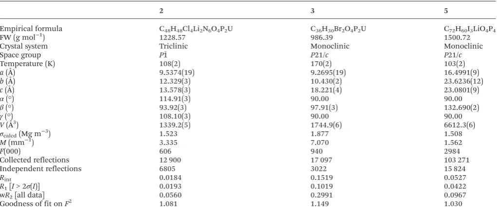
2 3 5
Empirical formula C48H48Cl4Li2N6O4P2U C36H30Br2O4P2U C72H60I3LiO4P4
FW (g mol−1) 1228.57 986.39 1500.72
Crystal system Triclinic Monoclinic Monoclinic
Space group P1ˉ P21/c P21/c
Temperature (K) 108(2) 170(2) 103(2)
a(Å) 9.5374(19) 9.2695(19) 16.4991(9)
b(Å) 12.329(3) 10.430(2) 23.6236(12)
c(Å) 13.578(3) 18.221(4) 23.0801(9)
α(°) 114.91(3) 90.00 90.00
β(°) 93.92(3) 97.91(3) 132.690(2)
γ(°) 108.10(3) 90.00 90.00
V(Å3) 1339.2(5) 1744.9(6) 6612.3(6)
σcalcd(Mg m−3) 1.523 1.877 1.508
M(mm−1) 3.335 7.070 1.562
F(000) 606 940 2984
Collected reflections 12 900 17 097 103 271
Independent reflections 6805 3022 15 824
Rint 0.0184 0.1519 0.0527
R1[I> 2σ(I)] 0.0193 0.1019 0.0422
wR2[all data] 0.0560 0.2991 0.0967
Goodness of fit onF2 1.081 1.149 1.030
(55.66 mg, 0.2 mmol) in THF (10 cm3) and this was stirred for 24 hours at room temperature. The resulting yellow solution was filtered and the solvent was removed under high vacuum. Dissolution in acetonitrile and placement at−30 °C overnight yielded dark yellow crystals suitable for X-ray diffraction (41 mg, 41.6%).1H NMR (CD3CN, 298 K): 7.67–7.57 ppm (m,
Ph); 31P NMR (CD3CN, 298 K): 28.46 ppm; IR (cm−1): 1439,
1120, 1050, 1025, 995, 928 (UvO), 764, 753, 726, 690; Raman (cm−1): 1590, 1672, 1487, 1440, 1190, 1164, 1137, 1088, 1028, 1000, 929, 847 (UvO), 727, 687, 617, 417, 301. 256, 207, 193.
[UO2I2(OvPPh3)2]
To a solution of [Li(THF)4][UCl5(THF)] (100 mg, 0.129 mmol)
in THF (5 cm3) was added Me3SiI in excess. The solution was
stirred for 24 hours to give a pale yellow solution. The solvent was removed under vacuum and the remaining orange powder was dissolved in acetonitrile (10 cm3). To this was added 2 equivalents of OvPPh3(72 mg, 0.257 mmol) in MeCN (5 cm3)
and the reaction was stirred for a further 24 hours. The result-ing orange solution was filtered and the solvent was reduced in volume. Placement at−30 °C overnight yielded dark orange crystals of 4 and orange powder of 5 (19 mg, 0.018 mmol, 13.6%) which were separated by hand. Spectroscopic data for4 were in accord with the literature data. 1H NMR (400 MHz, CD3CN, 298 K): δH = 7.55–7.69 (m, Ar–H) ppm; 31P NMR
(400 MHz, CD3CN, 298 K): 31.2 ppm (s, U–OvPPh3); IR (cm−1):
1439 (s), 1120 (m), 1052 (s, PvO), 1025 (m), 995 (m, OvU), 920 (m), 764 (m), 753 (m), 726 (s), 690 (s); Raman (cm−1): 1590 (w), 1568 (w), 1265 (w), 1229 (m), 1185 (m), 1156 (w), 1137 (w), 1090 (w), 1028 (w), 1000 (s), 839 (s, OvU), 687 (m), 617 (m). Anal. Calcd for C36H30O4P2I2U: C, 40.02; H, 2.79%. Found:
C, 38.83; H, 2.07.
Acknowledgements
We thank TCD for funding this work.
Notes and references
1 For leading references see: (a) V. Mougel, C. Camp, J. Pécaut, C. Copéret, L. Maron, C. E. Kefalidis and M. Mazzanti, Angew. Chem., Int. Ed., 2012, 51, 12280; (b) A.-C. Schmidt, A. V. Nizovtsev, A. Scheurer, F. W. Heinemann and K. Meyer,Chem. Commun., 2012,48, 8634; (c) D. M. King, F. Tuna, E. J. L. McInnes, J. McMaster, W. Lewis, A. J. Blake and S. T. Liddle, Science, 2012,337, 717; (d) B. M. Gardner, J. C. Stewart, A. L. Davis, J. McMaster, W. Lewis, A. J. Blake and S. T. Liddle, Proc. Natl. Acad. Sci. U. S. A., 2012,109, 9265; (e) O. P. Lamb and K. Meyer, Polyhedron, 2012, 32, 1; (f) O. P. Lam, F. W. Heinemann and K. Meyer,Chem. Sci., 2011,2, 1538; (g) P. L. Arnold, Chem. Commun., 2011, 47, 9005; (h) O. T. Summerscales and F. G. N. Cloke,Struct. Bonding, 2008, 127, 87; (i) O. P. Lam, C. Anthon and K. Meyer,
Dalton Trans., 2009, 9677; (j) A. J. Lewis, P. J. Carroll and E. J. Schelter,J. Am. Chem. Soc., 2013,135, 511.
2 For recent reviews see: (a) M. B. Jones and A. J. Gaunt,
Chem. Rev., 2013,113, 1137; (b) R. J. Baker,Coord. Chem. Rev., 2012,256, 2843; (c) T. W. Hayton,Dalton Trans., 2010, 39, 1145; (d) S. T. Liddle and D. P. Mills, Dalton Trans., 2009, 5592; (e) S. T. Liddle, Proc. R. Soc. London, Ser. A, 2009, 465, 1673; (f) I. Castro-Rodríguez and K. Meyer,
Chem. Commun., 2006, 1353; (g) M. Ephritikhine, Dalton Trans., 2006, 2501.
3 (a) M. L. Neidig, D. L. Clark and R. L. Martin,Coord. Chem. Rev., 2013, 257, 394; (b) N. Kaltsoyannis, Inorg. Chem., 2013,52, 3407.
4 R. J. Baker,Chem.–Eur. J., 2012,18, 16258. 5 R. G. Denning,J. Phys. Chem. A, 2007,111, 4125.
6 (a) M. B. Andrews and C. L. Cahill,Dalton Trans., 2012,41, 911; (b) R. E. Wilson, S. Skanthakumar, C. L. Cahill and L. Soderholm,Inorg. Chem., 2011,50, 10748; (c) N. P. Deifel and C. L. Cahill,C. R. Chim., 2010,13, 747; (d) M.-O. Sornein, M. Mendes, C. Cannes, C. Le Naour, P. Nockemann, K. Van Hecke, L. Van Meervelt, J.-C. Berthet and C. Hennig, Poly-hedron, 2009, 28, 1281; (e) P. Nockemann, K. Servaes, R. Van Deun, K. Van Hecke, L. Van Meervelt, K. Binnemans and C. Goerller-Walrand, Inorg. Chem., 2007, 46, 11335; (f) R. Bohrer, E. Conradi and U. Mueller, Z. Anorg. Allg. Chem., 1988,558, 119; (g) G. Van den Bossche, M. R. Spirlet, J. Rebizant and J. Goffart, Acta Crystallogr., Sect. C: Cryst. Struct. Commun., 1987, 43, 383; (h) E. Conradi, R. Bohrer and U. Mueller,Chem. Ber., 1986,119, 2582; (i) L. Di Sipio, E. Tondello, G. Pelizzi, G. Ingletto and A. Montenero,Cryst. Struct. Commun., 1977, 6, 723; (j) L. Di Sipio, E. Tondello, G. Pelizzi, G. Ingletto and A. Montenero, Cryst. Struct. Commun., 1974, 3, 301; (k) L. Di Sipio, E. Tondello, G. Pelizzi, G. Ingletto and A. Montenero, Cryst. Struct. Commun., 1974,3, 297; (l) Y. N. Mikhailov and V. G. Kuznetsov,
Zh. Neorg. Khim., 1971, 16, 2512; (m) Y. N. Mikhailov, V. G. Kuznetsov and E. S. Kovaleva,Zh. Neorg. Khim., 1965, 6, 787.
7 M.-J. Crawford, A. Ellern, K. Karaghiosoff, P. Mayer, H. Nöth and M. Suter,Inorg. Chem., 2004,43, 7120. 8 M.-J. Crawford, A. Ellern, H. Nöth and M. Suter, J. Am.
Chem. Soc., 2003,125, 11778.
9 J.-C. Berthet, M. Nierlich and M. Ephritikhine, Chem. Commun., 2004, 870.
10 M.-J. Crawford and P. Mayer,Inorg. Chem., 2005,44, 5547. 11 L. P. Spencer, P. Yang, B. L. Scott, E. R. Batista and
J. M. Boncella,C. R. Chim., 2010,13, 758.
12 (a) R. J. Baker and A. Walshe,Chem. Commun., 2012, 48, 985; (b) J. Fang, A. Walshe, L. Maron and R. J. Baker,Inorg. Chem., 2012,51, 9132; (c) A. Walshe, J. Fang, L. Maron and R. J. Baker,Inorg. Chem., 2013,52, 9077.
13 (a) E. Hashem, A. N. Swinburne, C. Schulzke, R. C. Evans, J. A. Platts, A. Kerridge, L. S. Natrajan and R. J. Baker,
RSC Adv., 2013, 3, 4350; (b) E. Hashem, G. Lorusso, M. Evangelisti, T. McCabe, C. Schulzke, J. A. Platts and R. J. Baker,Dalton Trans., 2013,42, 14677.
14 G. Bombieri, E. Forsellini, P. J. Day and W. I. Azeez,
J. Chem. Soc., Dalton Trans., 1978, 677.
15 (a) D. J. Watkin, R. G. Denning and K. Prout,Acta Crystal-logr., Sect. C: Cryst. Struct. Commun., 1991, 47, 2517; (b) R. J. Baker, E. Hashem, M. Motevalli, H. V. Ogilvie and A. Walshe,Z. Anorg. Allg. Chem., 2010,636, 443.
16 Determined from a survey of the Cambridge Structural Database.
17 The most recent IUPAC definition of a hydrogen bond states that“in most cases, the distance between H and Y are found to be less than the sum of their van der Waals radii”: E. Arunan, G. R. Desiraju, R. A. Klein, J. Sadlej, S. Scheiner, I. Alkorta, D. C. Clary, R. H. Crabtree, J. J. Dannenberg, P. Hobza, H. G. Kjaergaard, A. C. Legon, B. Mennucci and D. Nesbitt,Pure Appl. Chem., 2011,83, 1637. According to this criterion, H⋯Y distances of less than 2.72 Å for a C–H⋯O, 2.95 Å for C–H⋯Cl and 3.06 Å for C–H⋯Br are classed as hydrogen bonds. Van der Waals radii taken from S. Alvarez,Dalton Trans., 2013,42, 8617.
18 (a) N. P. Deifel and C. L. Cahill, CrystEngComm, 2009,11, 2739; (b) M. B. Andrews and C. L. Cahill, Dalton Trans.,
2012,41, 3911; (c) N. P. Deifel and C. L. Cahill,C. R. Chim., 2010,13, 747.
19 M. Deetlefs, C. L. Hussey, T. J. Mohammed, K. R. Seddon, J.-A. van den Berg and J. A. Zora, Dalton Trans., 2006, 2334.
20 J. P. Day and L. M. Venanzi,J. Chem. Soc. A, 1966, 1363. 21 F. J. Arnaiz, M. J. Miranda, R. Aguado, J. Mahia and
M. A. Maestro,Polyhedron, 2000,20, 3295.
22 F. Ruiperez and U. Wahlgren, J. Phys. Chem. A, 2010,114, 3615.
23 L. S. Natrajan,Coord. Chem. Rev., 2012,256, 1583.
24 M. P. Redmond, S. M. Cornet, S. D. Woodall, D. Whittaker, D. Collison, M. Helliwell and L. S. Natrajan,Dalton Trans., 2011,40, 3914.
25 Z. Fazekas, T. Yamamura and H. Tomiyasu, J. Alloys Compd., 1998,271–273, 756.
26 M.-O. Sornein, C. Cannes, C. Le Naour, G. Lagarde, E. Simoni and J.-C. Berthet, Inorg. Chem., 2006, 45, 10419.
27 G. M. Sheldrick, SHELXL-97, University of Göttingen, Göttingen, Germany, 1998.
![Fig. 1Packing diagram of [Li(OvPPh3)(MeCN)2]2[UO2Cl4], 2.](https://thumb-us.123doks.com/thumbv2/123dok_us/8802852.914856/2.595.306.550.521.684/fig-packing-diagram-li-ovpph-mecn-uo-cl.webp)

![Fig. 6Excitation (red) and emission (black) spectra of 1, 3, 4 and [PyH]2[UO2Cl4]·2Py in MeCN at 298 K (λex = 230–350 nm, λem = 520 nm).](https://thumb-us.123doks.com/thumbv2/123dok_us/8802852.914856/4.595.64.534.47.359/fig-excitation-emission-black-spectra-pyh-mecn-lem.webp)