
R E S E A R C H A R T I C L E
Open Access
B lymphocyte-typing for prediction of clinical
response to rituximab
Hans-Peter Brezinschek
1*, Franz Rainer
2, Kerstin Brickmann
1and Winfried B Graninger
1Abstract
Introduction:The prediction of therapeutic response to rituximab in rheumatoid arthritis is desirable. We
evaluated whether analysis of B lymphocyte subsets by flow cytometry would be useful to identify non-responders to rituximab ahead of time.
Methods:Fifty-two patients with active rheumatoid arthritis despite therapy with TNF-inhibitors were included in the national rituximab registry. DAS28 was determined before and 24 weeks after rituximab application. B cell subsets were analyzed by high-sensitive flow cytometry before and 2 weeks after rituximab administration. Complete depletion of B cells was defined as CD19-values below 0.0001 x10⁹cells/liter.
Results:At 6 months 19 patients had a good (37%), 23 a moderate (44%) and 10 (19%) had no EULAR-response. The extent of B lymphocyte depletion in peripheral blood did not predict the success of rituximab therapy. Incomplete depletion was found at almost the same frequency in EULAR responders and non-responders. In comparison to healthy controls, non-responders had elevated baseline CD95+pre-switch B cells, whereas responders had a lower frequency of plasmablasts.
Conclusions:The baseline enumeration of B lymphocyte subsets is still of limited clinical value for the prediction of response to anti-CD20 therapy. However, differences at the level of CD95+ pre switch B cells or plasmablasts were noticed with regard to treatment response. The criterion of complete depletion of peripheral B cells after rituximab administration did not predict the success of this therapy in rheumatoid arthritis.
Introduction
The use of monoclonal antibodies (mAbs) against cyto-kines or lymphocyte surface molecules has opened new therapeutic options for patients with rheumatoid arthritis (RA) [1]. By the prediction of a clinical response, these drugs, which are expensive and have the potential for ser-ious toxicity, could be allotted to those patients who would benefit most [2]. B-cell monitoring has been extensively used recently to assess the effect of B cell-directed therapies and the reconstitution of the periph-eral blood B-cell repertoire after treatment with the B cell-depleting mAb rituximab. Initially, the clinical response to this therapy was thought not to be correlated to B-cell subset distribution or depletion [3]. This view has been challenged by using high-sensitivity flow
cytometry, a technique originally developed to detect small numbers of residual malignant cells. Thus, com-plete depletion of B cells 2 weeks after the first infusion has been suggested to be an indicator for therapy respon-siveness [4-6]. Furthermore, subsequent articles indicated that complete depletion is also a prognostic factor for re-treatment [5] and efficacy of the rituximab therapy [6].
Several articles have analyzed the changes in B-cell sub-sets following depletion therapy with rituximab [7-9]. In most articles, B cells were characterized by the surface mar-kers IgD, CD27, CD38, and CD24, which allow separation of newly generated‘transitional’(IgD+, CD27-, CD24hi, and CD38hi) [10], naïve (IgD+and CD27-), pre-switch (IgD+ and CD27+) and post-switch (IgD-and CD27+) memory, and double-negative B (IgD-and CD27-) cells and plasma-blasts (IgD-and CD27++) [11-13] in the peripheral blood.
We set out to further delineate B-cell subsets by using high-sensitivity flow cytometry that might help to char-acterize RA patients who would benefit from rituximab therapy. We expanded our analysis to the co-stimulatory * Correspondence: [email protected]
1Department of Internal Medicine, Division of Rheumatology and Immunology, Medical University Graz, Auenbruggerplatz 15, Graz, A-8036, Austria
Full list of author information is available at the end of the article
marker CD80, which had been shown to be a potent regulator of IgG secretion by previously activated B cells [14], and CD95, which had been correlated with disease activity in systemic lupus erythematosus (SLE) [13].
Materials and methods Financial disclosure
This work was funded by an unrestricted grant from Roche (Vienna, Austria). The funders had no role in study design, data collection and analysis, decision to publish, or preparation of the manuscript.
Patients and controls
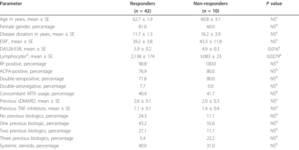
Fifty-two patients undergoingde novotreatment with ritux-imab for active RA were included in the national‘B Cell surveillance’registry. The participating clinical rheumatolo-gists from local and remote hospitals judged the need for the routine administration of rituximab. Informed consent was obtained from all patients before entering the study, in accordance with the protocol approved by the local ethics committee of the Medical University of Graz. All patients received two 1,000 mg infusions of rituximab preceded by the administration of 100 mg of prednisolone [15]. The characteristics of all patients are shown in Table 1. Disease activity score using 28 joint counts (DAS28) using the ery-throcyte sedimentation rate was determined before and 2 and 24 weeks after rituximab application in order to deter-mine the European League Against Rheumatism (EULAR) response. Peripheral blood samples from 17 healthy donors
(15 females and two males; mean age of 64 years) were used to determine the normal range for the different B-cell subsets.
Lymphocyte phenotyping
Peripheral blood samples were drawn before and 15 days after the first rituximab infusion. Peripheral mononuclear cells were prepared as described [4] and stained with the following antibodies: fluorescein isothiocyanate-labeled IgD, phycoerythrin (PE)-conjugated CD24, allophycocya-nin (APC)-conjugated CD27, PE-Cy7-labeled CD38, APC-H7-conjugated CD45, horizon Blue-labeled CD19, pyri-dine-chlorophyll-protein (PerCP)-conjugated CD3 and CD14, and PE-labeled CD80 and CD95 (all mAbs were obtained from BD Biosciences, Schwechat, Austria). Five
hundred thousand CD45+cells were acquired and
[image:2.595.57.538.463.704.2]ana-lyzed by using a seven-channel flow cytometry (BD Canto II cytometer, Software FACSDiva; BD Biosciences). According to their surface staining in the CD27/IgD blot, B cells were classified as naïve (CD19+, IgD+, and CD27-), pre-switch memory (CD19+, IgD+, and CD27+), post-switch memory (CD19+, IgD-, and CD27+), and double-negative (CD19+, IgD-, and CD27-) cells. Plasmablasts were classified as CD19+, IgD-, and CD27++[12,16]. In addition, the intensity of the CD38 expression was deter-mined in the post-switch memory and plasmablast popu-lations since this molecule was recently shown not to be constantly expressed within the populations [16,17]. The distribution of co-stimulatory molecules and the FAS
Table 1 Baseline characteristics of patients included in this study
Parameter Responders Non-responders Pvalue
(n= 42) (n= 10)
Age in years, mean ± SE 62.7 ± 1.9 60.8 ± 3.1 NSa
Female gender, percentage 81.0 60.0 NSb
Disease duration in years, mean ± SE 11.7 ± 1.3 16.2 ± 3.9 NSa
ESRc, mean ± SE 39.2 ± 3.8 43.3 ± 11.8 NSa
DAS28-ESR, mean ± SE 5.9 ± 0.2 4.9 ± 0.3 0.016a
Lymphocytesd, mean ± SE 2,138 ± 174 3,083 ± 23 0.0279a
RF-positive, percentage 90.8 100.0 NSb
ACPA-positive, percentage 76.9 80.0 NSb
Double-seropositive, percentage 71.8 80.0 NSb
Double-seronegative, percentage 7.7 0.0 NSb
Concomitant MTX usage, percentage 40.4 41.7 NSb
Previous sDMARD, mean ± SE 2.6 ± 0.1 2.0 ± 0.3 NSa
Previous TNF inhibitors, mean ± SE 1.1 ± 0.1 1.4 ± 0.4 NSa
No previous biologics, percentage 24.3 11.1 NSb
One previous biologic, percentage 43.2 55.6 NSb
Two previous biologics, percentage 27.1 11.1 NSb
Three previous biologics, percentage 5.4 22.2 NSb
Systemic steroids, percentage 40.0 31.0 NSb
a
Pvalues were calculated by using Mann-Whitney test;b
Pvalues were calculated by using the chi-squared test;c
mm per 1 hour;d
receptor on the different B-cell subsets was determined by replacing CD24 with CD80 or CD95. Complete depletion of B cells was defined as CD19 values of below 0.0001 × 10⁹cells per liter. The gating strategy and representative dot blots can be found in Figure S1 of Additional file 1.
Since 41% of the blood samples coming from peripheral hospitals had to be transported for more than 12 hours, we compared 15 samples of patients with RA in a pair-wise fashion (2 and 24 hours after blood letting) and found no significant change in absolute counts of B-lymphocyte subsets after this time.
Statistical analyses
Data were analyzed for normal distribution in order to decide whether to use parametric or non-parametric tests. Values are expressed as the mean ± standard error of the mean or as median (interquartile range) and were
calculated by using GraphPad Prism (version 5.0b; GraphPad Software, La Jolla, CA, USA). Baseline clinical variables and B-cell subsets as predictors of response to the first cycle of rituximab were compared with non-responders by using univariate logistic regression. Pvalues of less than 0.05 were considered significant.
Results
[image:3.595.59.539.310.693.2]Characterization of rheumatoid arthritis patients
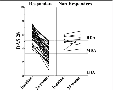
Table 1 summarizes the baseline demographics and clini-cal characteristics of the enrolled patients (n= 52), and Figure 1 shows the DAS28 values at baseline and 24 weeks after rituximab application. At baseline, EULAR respon-ders and non-responrespon-ders were significantly different with regard to the DAS28 only. Interestingly, this difference was due to elevated DAS28 values in RA patients with
moderate EULAR response (6.2 ± 0.2; P ≤ 0.0004),
whereas patients with good response were not statistically different from EULAR non-responders (5.7 ± 0.3 versus 4.9 ± 0.3, respectively). In our small cohort, the autoanti-body status had no influence on the effect of the treatment with rituximab. The few patients with double-seronegative RA were all in the responder group.
B-cell depletion and clinical response
Two weeks after the first rituximab infusion, there was a dramatic reduction in the number of B cells in all patients with RA. When high-sensitivity flow cytometry was used, the difference in the number of B cells 15 days after rituximab did not reach statistical significance between responders and non-responders. The median numbers (interquartile ranges) of B cells per 109/L were
0.022 (0.008 to 0.043) in RA patients with good EULAR response, 0.022 (0.011 to 0.059) in patients with a mod-erate response, and 0.008 (0.003 to 0.035) in patients with no response (Figure 2).
B-cell subpopulations and clinical response
[image:4.595.60.538.288.705.2]At baseline, the differences in the frequency of naïve, double-negative, pre-switch memory, and post-switch memory B cells between EULAR responders and non-responders did not reach significance. In addition, the expressions of CD80 or CD95 on the B-cell subsets were similar between RA patients and healthy controls. Only a few naïve B cells in patients with RA expressed the co-stimulation and activation markers (1.8% (1.1% to 3.5%) and 4.9% (3.0% to 8.5%), respectively), whereas the
highest frequency was found in post-switch memory B cells (58.0% (42.0% to 94.1%) and 65.8% (55.1% to 79.0%), respectively). In all populations analyzed, there was a significant correlation between the frequency of CD95+ and CD80+ B-cell subsets. In responders, this correlation was highest in the double-negative subset (R2 = 0.71; P≤ 0.0001) but was lowest in
non-respon-ders and controls (R2 = 0.520; P ≤ 0.0437 and R2 =
0.384;P≤0.0080, respectively).
When the frequencies of B cells in responders and non-responders at baseline were compared with those in
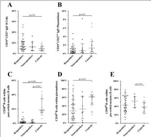
[image:5.595.56.538.235.672.2]healthy controls, significant dissimilarities were seen (Figure 3). Thus, responders had significantly more dou-ble-negative B cells (Figure 3a) and fewer plasmablasts (Figure 3b) in comparison with controls: 6.6% (4.1% to
11.1%) versus 4.4% (2.8% to 6.9%), P ≤ 0.02 and 0.6%
(0.3% to 1.3%) versus 1.3% (0.5% to 2.8%), P ≤ 0.03,
respectively. The frequencies of these B-cell subsets in non-responders were 6.2% (2.1% to 10.7%) and 0.9% (0.3% to 2.8%), respectively.
The total cohort of patients with RA had a
signifi-cantly lower frequency of CD38hi B cells within the
post-switch memory subset (P ≤ 0.0001). Thus, the median (interquartile range) frequencies of this B-cell subset were 0.9% (0.3% to 2.1%) in responders, 1.3% (0.3% to 3.9%) in non-responders, and 18.2% (14.7% to 34.5%) in controls (Figure 3c). Furthermore, the fre-quency of CD38hiB cells within plasmablasts was signifi-cantly diminished in EULAR responders compared with controls: 28.1% (9.6% to 54.3%) versus 61.1% (44.1% to 66.0%),P≤0.001 (Figure 3d). In contrast, only non-respon-ders had a significantly higher frequency of CD95+ pre-switch memory B cells than controls: 52.6% (37.9% to 62.6%) versus 37.9% (26.5% to 49.3%),P≤0.023 (Figure 3e). The frequency of CD95+cells in the other B-cell subsets is depicted in Figure S2 of Additional file 2. As indicated in Figure 3, no significant differences were found between EULAR responders and non-responders.
Prediction of first-cycle response
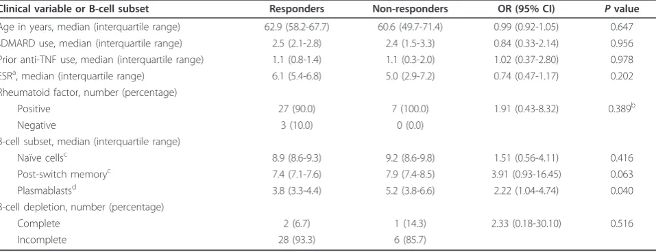
When univariate logistic regression analysis was used (Table 2), only a low frequency of plasmablasts was a valid predictor for EULAR responsiveness (odds ratio of 2.22;P≤ 0.04). In our small cohort, complete depletion on day 15 let us calculate an odds ratio of 2.33 for the lack of a therapeutic response to rituximab (P= 0.516).
Discussion
In our small registry cohort of patients with routinely treated RA, complete depletion of B cells 15 days after the first rituximab infusion was not a prognostic factor for clinical response. We did not find technical explana-tions for this discrepancy with previous reports [4-6]. The time lag between blood letting and analysis was proven not to be a valid explanation since no significant
difference was found between samples that were ana-lyzed within 6 or 24 hours (Figure S3 of Additional file 3). We cannot exclude biological differences between our cohort and the patients in previous reports [4-6].
Interestingly, in the regression analysis, the results for baseline distribution of B-cell subsets in responders and non-responders (Table 2) were similar to those reported by Vital and colleagues [5]. Thus, in both studies, fewer plasmablasts were significantly associated with response to rituximab. These B-cell populations were also signifi-cantly less frequent in responders than in healthy con-trols (Figure 3b, d).
Analyzing the frequency of the major B-cell subsets (that is, naïve, pre-switch and post-switch memory, and double-negative B cells and plasmablasts) in our RA patients before rituximab treatment, we and others did not find a significant difference with healthy age-matched controls [11]. Interestingly, separating the RA population in EULAR responders and non-responders revealed a signifi-cantly higher percentage of double-negative (IgD-/CD27-) B cells in the former group (Figure 3a). In a recent study in healthy older people, this population of B cells was shown to be enriched in exhausted cells [18]. In patients with RA, the humoral immune system already seems to be overstimulated, driving more B cells into the double-nega-tive subset in comparison with healthy controls (Figure 3). The significant result for EULAR responders might be related primarily to the higher number of patients in this group. In these patients (in contrast to patients with SLE [13]), disease activity did not correlate with CD95 expres-sion on double-negative B cells (data not shown).
[image:6.595.59.538.522.706.2]In all patients with RA, CD38hipost-switch memory B cells were significantly reduced in comparison with
Table 2 Baseline clinical variables and B-cell subsets in responders and non-responders, as determined by univariate logistic regression
Clinical variable or B-cell subset Responders Non-responders OR (95% CI) Pvalue Age in years, median (interquartile range) 62.9 (58.2-67.7) 60.6 (49.7-71.4) 0.99 (0.92-1.05) 0.647 sDMARD use, median (interquartile range) 2.5 (2.1-2.8) 2.4 (1.5-3.3) 0.84 (0.33-2.14) 0.956 Prior anti-TNF use, median (interquartile range) 1.1 (0.8-1.4) 1.1 (0.3-2.0) 1.02 (0.37-2.80) 0.978 ESRa, median (interquartile range) 6.1 (5.4-6.8) 5.0 (2.9-7.2) 0.74 (0.47-1.17) 0.202 Rheumatoid factor, number (percentage)
Positive 27 (90.0) 7 (100.0) 1.91 (0.43-8.32) 0.389b
Negative 3 (10.0) 0 (0.0)
B-cell subset, median (interquartile range)
Naïve cellsc 8.9 (8.6-9.3) 9.2 (8.6-9.8) 1.51 (0.56-4.11) 0.416
Post-switch memoryc 7.4 (7.1-7.6) 7.9 (7.4-8.5) 3.91 (0.93-16.45) 0.063
Plasmablastsd 3.8 (3.3-4.4) 5.2 (3.8-6.6) 2.22 (1.04-4.74) 0.040
B-cell depletion, number (percentage)
Complete 2 (6.7) 1 (14.3) 2.33 (0.18-30.10) 0.516
Incomplete 28 (93.3) 6 (85.7)
a
Square root values;b
calculated from contingency table, Pearson-Mantel-HaenszelPvalue;c
natural log (absolute number of cells);d
healthy age-matched controls (Figure 3c). CD38 is a 45-kDa transmembrane glycoprotein expressed on dif-ferent human cells, including T and B lymphocytes [17]. Activation of naïve B cells leads to an upregulation of this molecule, and transition of activated B lymphocytes into memory cells is characterized by loss of CD38 [19,20] but this molecule reappears when B cells develop into plasmablasts and plasma cells [21]. Plasmablasts can be separated further into CD38- early plasmablasts and CD38++ late plasmablasts [16]. Interestingly, in synovial tissue of patients with RA, CD38-B cells appear to serve as immunoglobulin-producing effector B cells [22]. Whether the CD38-/lowpost-switch memory B cells in the peripheral blood of patients with RA are similar to the cells described in the synovial tissue cannot be answered yet. Since the latter cells are enriched in the peripheral blood, it is now possible to perform
functional assays and determine whether CD38- B cells are similar to mouse B cells that lack RelB and that are defective in proliferative responses but are able to secrete immunoglobulins and undergo class switching [23].
[image:7.595.59.542.309.687.2]Recently, treatment with tumor necrosis factor (TNF) blockers was shown to alter the distribution of peripheral blood B cells [24]. Thus, infliximab therapy induces an increase in pre-switch memory B cells. Although the majority of our patients have received TNF inhibitors in the past, we did not find this difference. In contrast, our patients exhibited similarly low median percentages of IgD+CD27+memory B cells (8.6% and 6.8% for respon-ders and non-responrespon-ders, respectively) as described for patients with long-standing RA (10.4%). In addition, the frequencies of these B-cell subsets in the control groups in this study and in the article by Souto-Carneiro and
colleagues [24] were almost identical (14.9% and 15.1%, respectively). It is fascinating to speculate whether the missing increase in pre-switch memory B cells is a surro-gate marker for TNF failure, but further studies have to confirm these results.
Although pre-switch memory B cells are reduced in our RA cohort, this subset is significantly enriched in CD95+ cells in non-responders (Figure 3e). Similar results were found in patients with SLE [25]. It has been suggested that, in SLE, the pre-switch B-cell population is enriched in autoantibody-producing B cells that are constantly recruited into lymphoid tissue and that therefore are found less often in the peripheral blood. Under physiologi-cal conditions, expression of CD95 is important in main-taining peripheral self-tolerance by inducing apoptosis upon engagement with its ligand, CD178 [26]. In RA, defects of this pathway have been demonstrated [27], and accumulation of autoantibody-secreting plasma cells in synovium has been suggested to be the result of the lack of this regulatory mechanism [28]. Our results suggest that an increased frequency of activated (that is, CD95+ pre-switch memory) B cells is associated with poor response to rituximab. Figure 4 summarizes the alterations in the different B-cell subsets between EULAR responders and non-responders compared with healthy age-matched controls. Despite the great efforts made, neither certain cell populations nor serum markers that might help to choose a specific therapy have been found so far. The het-erogeneity of the patients with RA (for example, disease duration, previous medication, or co-medication or a com-bination of these) could skew the results.
Conclusions
In summary, even in this small cohort, the frequency of plasmablast seems to be the best predictor for response to a B cell-depleting therapy. Still, prospective studies have to confirm this finding. High-sensitivity flow cyto-metry appears to be very helpful to narrow further candidates.
Additional material
Additional file 1: Figure S1 Gating strategy. Mononuclear cells were incubated in 4 separate tubes containing the following monoclonal antibodies: a) Isotypen-specific control antibodies; b) IgD-FITC, CD27-APC, CD38-PE/Cy7, CD45-APC/H7, CD19-HorizonBlue, CD24-PE, CD3-/CD14-PerCP; c) IgD-FITC, CD27-APC, CD38-PE/Cy7, CD45-APC/H7, CD19-HorizonBlue, CD80-PE, CD3-/CD14-PerCP; d) IgD-FITC, CD27-APC, CD38-PE/Cy7, CD45-APC/H7, CD19-HorizonBlue, CD95-PE, CD3-/CD14-PerCP. CD45 positive leukocytes were gated and after eliminating PerCP positive T cells and monocytes, B cells were separated according to their CD27 and IgD expression. Thereafter the expression of CD38, CD80 or CD95 was analyzed in each B cell population.
Additional file 2: Figure S2 Effect of the time of preparation on the number of day 15 B cells in RA patients with good/moderate or no EULAR response.
Additional file 3: Figure S3 Frequency of CD95+cells in the naïve, post-switch and double negative B cell subset.
Abbreviations
APC: allophycocyanin; DAS28: disease activity score using 28 joint counts; EULAR: European League Against Rheumatism; mAb: monoclonal antibody; PE: phycoerythrin; RA: rheumatoid arthritis; SLE: systemic lupus
erythematosus; TNF: tumor necrosis factor.
Acknowledgements
We give special thanks to Irene Holzer, Carinna Köhler, and Veronika Krischan for their excellent technical work and Erich Kvas for the statistical analysis. We thank our study coordinator, Saelde Baumgartner, and Barbara Nussbaumer and all colleagues who participated in the study and provided the clinical data and samples.
Author details
1Department of Internal Medicine, Division of Rheumatology and Immunology, Medical University Graz, Auenbruggerplatz 15, Graz, A-8036, Austria.2Internal Medicine, Hospital Barmherzige Brueder, Bergstrasse 27, Graz-Eggenberg, A-8020, Austria.
Authors’contributions
HPB and WBG conceived the study and participated in its design and coordination and wrote the manuscript. FR and KB undertook recruitment of patients and collection of clinical data. All authors read and approved the final manuscript.
Competing interests
The authors declare that they have no competing interests.
Received: 10 February 2012 Revised: 17 June 2012 Accepted: 6 July 2012 Published: 6 July 2012
References
1. Furst DE, Keystone EC, Braun J, Breedveld FC, Burmester GR, De Benedetti F, Dorner T, Emery P, Fleischmann R, Gibofsky A, Kalden JR, Kavanaugh A, Kirkham B, Mease P, Sieper J, Singer NG, Smolen JS, Van Riel PL, Weisman MH, Winthrop K:Updated consensus statement on biological agents for the treatment of rheumatic diseases, 2010.Ann Rheum Dis
2011,70(Suppl 1):i2-36.
2. Tak PP, Kalden JR:Advances in rheumatology: new targeted therapeutics.
Arthritis Res Ther2011,13(Suppl 1):S5.
3. Breedveld F, Agarwal S, Yin M, Ren S, Li NF, Shaw TM, Davies BE:Rituximab pharmacokinetics in patients with rheumatoid arthritis: B-cell levels do not correlate with clinical response.J Clin Pharmacol2007,47:1119-1128. 4. Dass S, Rawstron AC, Vital EM, Henshaw K, McGonagle D, Emery P:Highly
sensitive B cell analysis predicts response to rituximab therapy in rheumatoid arthritis.Arthritis Rheum2008,58:2993-2999.
5. Vital EM, Dass S, Rawstron AC, Buch MH, Goeb V, Henshaw K, Ponchel F, Emery P:Management of nonresponse to rituximab in rheumatoid arthritis: predictors and outcome of re-treatment.Arthritis Rheum2010,
62:1273-1279.
6. Vital EM, Rawstron AC, Dass S, Henshaw K, Madden J, Emery P, McGonagle D:Reduced-dose rituximab in rheumatoid arthritis: efficacy depends on degree of B cell depletion.Arthritis Rheum2011,63:603-608. 7. Roll P, Palanichamy A, Kneitz C, Dorner T, Tony HP:Regeneration of B cell subsets after transient B cell depletion using anti-CD20 antibodies in rheumatoid arthritis.Arthritis Rheum2006,54:2377-2386.
8. Rehnberg M, Amu S, Tarkowski A, Bokarewa MI, Brisslert M:Short- and long-term effects of anti-CD20 treatment on B cell ontogeny in bone marrow of patients with rheumatoid arthritis.Arthritis Res Ther2009,11: R123.
9. Moller B, Aeberli D, Eggli S, Fuhrer M, Vajtai I, Vogelin E, Ziswiler HR, Dahinden CA, Villiger PM:Class-switched B cells display response to therapeutic B-cell depletion in rheumatoid arthritis.Arthritis Res Ther
10. Sims GP, Ettinger R, Shirota Y, Yarboro CH, Illei GG, Lipsky PE:Identification and characterization of circulating human transitional B cells.Blood2005,
105:4390-4398.
11. Anolik JH, Looney RJ, Lund FE, Randall TD, Sanz I:Insights into the heterogeneity of human B cells: diverse functions, roles in autoimmunity, and use as therapeutic targets.Immunol Res2009,
45:144-158.
12. Sanz I, Wei C, Lee FE, Anolik J:Phenotypic and functional heterogeneity of human memory B cells.Semin Immunol2008,20:67-82.
13. Jacobi AM, Reiter K, Mackay M, Aranow C, Hiepe F, Radbruch A, Hansen A, Burmester GR, Diamond B, Lipsky PE, Dorner T:Activated memory B cell subsets correlate with disease activity in systemic lupus erythematosus: delineation by expression of CD27, IgD, and CD95.Arthritis Rheum2008,
58:1762-1773.
14. Rau FC, Dieter J, Luo Z, Priest SO, Baumgarth N:B7-1/2 (CD80/CD86) direct signaling to B cells enhances IgG secretion.J Immunol2009,
183:7661-7671.
15. Edwards JC, Szczepanski L, Szechinski J, Filipowicz-Sosnowska A, Emery P, Close DR, Stevens RM, Shaw T:Efficacy of B-cell-targeted therapy with rituximab in patients with rheumatoid arthritis.N Engl J Med2004,
350:2572-2581.
16. Avery DT, Ellyard JI, Mackay F, Corcoran LM, Hodgkin PD, Tangye SG:
Increased expression of CD27 on activated human memory B cells correlates with their commitment to the plasma cell lineage.J Immunol
2005,174:4034-4042.
17. Shubinsky G, Schlesinger M:The CD38 lymphocyte differentiation marker: new insight into its ectoenzymatic activity and its role as a signal transducer.Immunity1997,7:315-324.
18. Buffa S, Bulati M, Pellicano M, Dunn-Walters DK, Wu YC, Candore G, Vitello S, Caruso C, Colonna-Romano G:B cell immunosenescence: different features of naive and memory B cells in elderly.Biogerontology
2011,12:473-483.
19. Lagresle C, Bella C, Defrance T:Phenotypic and functional heterogeneity of the IgD- B cell compartment: identification of two major tonsillar B cell subsets.Int Immunol1993,5:1259-1268.
20. Pascual V, Liu YJ, Magalski A, de Bouteiller O, Banchereau J, Capra JD:
Analysis of somatic mutation in five B cell subsets of human tonsil.J Exp Med1994,180:329-339.
21. Mei HE, Yoshida T, Muehlinghaus G, Hiepe F, Dorner T, Radbruch A, Hoyer BF:Phenotypic analysis of B-cells and plasma cells.Methods Mol Med2007,136:3-18.
22. Reparon-Schuijt CC, van Esch WJ, van Kooten C, Ezendam NP, Levarht EW, Breedveld FC, Verweij CL:Presence of a population of CD20+, CD38- B lymphocytes with defective proliferative responsiveness in the synovial compartment of patients with rheumatoid arthritis.Arthritis Rheum2001,
44:2029-2037.
23. Snapper CM, Rosas FR, Zelazowski P, Moorman MA, Kehry MR, Bravo R, Weih F:B cells lacking RelB are defective in proliferative responses, but undergo normal B cell maturation to Ig secretion and Ig class switching.
J Exp Med1996,184:1537-1541.
24. Souto-Carneiro MM, Mahadevan V, Takada K, Fritsch-Stork R, Nanki T, Brown M, Fleisher TA, Wilson M, Goldbach-Mansky R, Lipsky PE:Alterations in peripheral blood memory B cells in patients with active rheumatoid arthritis are dependent on the action of tumour necrosis factor.Arthritis Res Ther2009,11:R84.
25. Rodriguez-Bayona B, Ramos-Amaya A, Perez-Venegas JJ, Rodriguez C, Brieva JA:Decreased frequency and activated phenotype of blood CD27 IgD IgM B lymphocytes is a permanent abnormality in systemic lupus erythematosus patients.Arthritis Res Ther2010,12:R108.
26. Siegel RM, Chan FK, Chun HJ, Lenardo MJ:The multifaceted role of Fas signaling in immune cell homeostasis and autoimmunity.Nat Immunol
2000,1:469-474.
27. Peng SL:Fas (CD95)-related apoptosis and rheumatoid arthritis.
Rheumatology (Oxford)2006,45:26-30.
28. Rodriguez-Bayona B, Perez-Venegas JJ, Rodriguez C, Brieva JA: CD95-Mediated control of anti-citrullinated protein/peptides antibodies (ACPA)-producing plasma cells occurring in rheumatoid arthritis inflamed joints.Rheumatology (Oxford)2007,46:612-616.
29. Agematsu K, Nagumo H, Oguchi Y, Nakazawa T, Fukushima K, Yasui K, Ito S, Kobata T, Morimoto C, Komiyama A:Generation of plasma cells from
peripheral blood memory B cells: synergistic effect of interleukin-10 and CD27/CD70 interaction.Blood1998,91:173-180.
doi:10.1186/ar3901
Cite this article as:Brezinscheket al.:B lymphocyte-typing for prediction of clinical response to rituximab.Arthritis Research & Therapy201214: R161.
Submit your next manuscript to BioMed Central and take full advantage of:
• Convenient online submission
• Thorough peer review
• No space constraints or color figure charges
• Immediate publication on acceptance
• Inclusion in PubMed, CAS, Scopus and Google Scholar
• Research which is freely available for redistribution

![Figure 4 Model of B-cell differentiation (adapted from [10,12,16]). *All patients with rheumatoid arthritis (RA) had significantly fewer CD38+post-switch B cells in comparison with healthy age-matched controls](https://thumb-us.123doks.com/thumbv2/123dok_us/8833892.928246/7.595.59.542.309.687/differentiation-patients-rheumatoid-arthritis-significantly-comparison-matched-controls.webp)
