T cells in the control of organ-specific
autoimmunity
Jeffrey A. Bluestone, … , Mickie Cheng, Mark Anderson
J Clin Invest.
2015;
125(6)
:2250-2260.
https://doi.org/10.1172/JCI78089
.
Immune tolerance is critical to the avoidance of unwarranted immune responses against self
antigens. Multiple, non-redundant checkpoints are in place to prevent such potentially
deleterious autoimmune responses while preserving immunity integral to the fight against
foreign pathogens. Nevertheless, a large and growing segment of the population is
developing autoimmune diseases. Deciphering cellular and molecular pathways of immune
tolerance is an important goal, with the expectation that understanding these pathways will
lead to new clinical advances in the treatment of these devastating diseases. The vast
majority of autoimmune diseases develop as a consequence of complex mechanisms that
depend on genetic, epigenetic, molecular, cellular, and environmental elements and result
in alterations in many different checkpoints of tolerance and ultimately in the breakdown of
immune tolerance. The manifestations of this breakdown are harmful inflammatory
responses in peripheral tissues driven by innate immunity and self antigen–specific
pathogenic T and B cells. T cells play a central role in the regulation and initiation of these
responses. In this Review we summarize our current understanding of the mechanisms
involved in these fundamental checkpoints, the pathways that are defective in autoimmune
diseases, and the therapeutic strategies being developed with the goal of restoring immune
tolerance.
Review Series
Find the latest version:
Series Editor: Antonio La Cava
Introduction
Genetic predisposition for most autoimmune disorders is
polygenic and conferred by shared as well as disease-specific
alle-les. Genome-wide association studies have identified dozens of
genetic variants associated with autoimmunity (1). The MHC loci
confer the highest genetic risk in many autoimmune diseases,
pointing to a critical role for antigen T cell interactions in disease
pathogenesis. Additionally, many of the shared variants have
plei-otropic effects on pathways that are important for conventional
T cells (Tconvs) but are also critical for the homeostasis and/or
function of Tregs, such as IL-2, CD25, cytotoxic T lymphocyte–
associated protein 4 (CTLA4), and protein tyrosine phosphatase,
non-receptor type 22 (2–4). Conversely, disease-specific
associa-tions implicate variants for genes either encoding major
autoanti-gens or that are involved in their generation (5, 6). Although beyond
the scope of this Review, rare genetic variants have also been
crit-ically informative about the role of innate immunity or other arms
of the immune system in systemic autoimmune diseases such as
lupus (7), which are beyond the scope of this discussion. Taken
together, genetics studies point to the central role of pathways
involved in thymic T cell education and peripheral
immunoregula-tion by Tregs for the control of autoimmune diseases.
Immune tolerance stems from the control of autoreactive
T cells both in the thymus and the periphery, owing to mechanisms
known as central and peripheral tolerance, respectively. Central
tolerance eliminates potentially autoreactive lymphocytes that
develop in the thymus by subjecting thymocytes with high affinity
for self antigens to either clonal deletion (negative selection) or
selection into the Treg lineage. Many autoreactive T cells escape
this checkpoint and can be found in the peripheral blood of healthy
individuals; however, these self-reactive cells are not sufficient
to induce autoimmunity due to additional controls by peripheral
tolerance mechanisms (8–11). Peripheral tolerance is achieved
through T cell–intrinsic mechanisms that lead to clonal deletion,
anergy, or immunological ignorance as well as extrinsic control
by specialized populations of suppressor cells that regulate
poten-tially harmful responses of autoreactive T and B cells (12, 13). First
among these are CD4
+CD25
+Foxp3
+Tregs, a T cell population that
is essential for extrinsic control of peripheral tolerance (14, 15).
Tregs play a fundamental role in inhibiting self-reactivity and
maintaining immune tolerance (16). Several types of Tregs have
been described, including Foxp3
–IL-10–dependent Tr1 cells,
LAP
+TGF-
β
–dependent Th3 cells, and CD8
+Tregs; however, in
this Review we focus on Tregs that express the transcription
fac-tor Foxp3, a “master regulafac-tor” of this Treg lineage that is crucial
for their homeostasis and function. Loss-of-function mutations
in the FOXP3 gene are responsible for immune dysregulation,
polyendocrinopathy, enteropathy, X-linked (IPEX) syndrome,
which is characterized by widespread and often fatal
autoim-munity shortly after birth (17). Similarly, mice deficient in Foxp3
completely lack Tregs and rapidly develop lethal multi-organ
autoimmunity (18, 19). The requirement for Foxp3 expression in
Tregs is quantitative in nature and lifelong, as illustrated by the
development of lymphoproliferative disease within days of acute
depletion of Foxp3
+Tregs in adult mice (20, 21).
Central tolerance as a key checkpoint
The generation of an extremely diverse T cell repertoire in the
thymus through stochastic gene rearrangement of the TCR is
a powerful weapon in our immunity against pathogens. At the
same time, collateral damage can occur when autoreactive T cells
Immune tolerance is critical to the avoidance of unwarranted immune responses against self antigens. Multiple,
non-redundant checkpoints are in place to prevent such potentially deleterious autoimmune responses while preserving immunity
integral to the fight against foreign pathogens. Nevertheless, a large and growing segment of the population is developing
autoimmune diseases. Deciphering cellular and molecular pathways of immune tolerance is an important goal, with the
expectation that understanding these pathways will lead to new clinical advances in the treatment of these devastating
diseases. The vast majority of autoimmune diseases develop as a consequence of complex mechanisms that depend on
genetic, epigenetic, molecular, cellular, and environmental elements and result in alterations in many different checkpoints
of tolerance and ultimately in the breakdown of immune tolerance. The manifestations of this breakdown are harmful
inflammatory responses in peripheral tissues driven by innate immunity and self antigen–specific pathogenic
T and B cells. T cells play a central role in the regulation and initiation of these responses. In this Review we summarize our
current understanding of the mechanisms involved in these fundamental checkpoints, the pathways that are defective in
autoimmune diseases, and the therapeutic strategies being developed with the goal of restoring immune tolerance.
T cells in the control of organ-specific autoimmunity
Jeffrey A. Bluestone, Hélène Bour-Jordan, Mickie Cheng, and Mark Anderson
Diabetes Center, UCSF, San Francisco, California, USA.
Conflict of interest: Mark Anderson’s spouse is a shareholder of Medtronic and Merck.
Jeffrey A. Bluestone is a scientific advisor for Juno Therapeutics; a member of the scien-tific advisory boards of Flexus Biosciences, Kadmon Corporation, Pfizer, and NeoStem (Athelos Division); a shareholder of Macrogenics Inc.; and receives research support from NeoStem.
that insulin-specific CD4
+T cells in the NOD mouse model have
a propensity to react to a peptide of insulin that sits in the MHC
groove in the suboptimal of three binding registers (41, 42). The
affinity of this 15–amino acid peptide derived from the
insu-lin B chain (Ins9–23) for the NOD mouse MHC class II allele,
I-Ag7, is relatively weak. An I-Ag7/Ins9–23–optimized peptide
tetramer locked into the least favored register reacts with
mul-tiple T cells enriched in the islets of prediabetic mice. This work
has now been translated to human subjects with T1D that have
DQ8-restricted insulin-specific T cells (43). Recent work from
the Unanue group has suggested that this type of peptide-MHC
complex may involve insulin peptide–loading events that
uniquely occur in pancreatic islets but not in normal APCs (44,
45). In support of this hypothesis, when whole insulin protein is
provided to normal APCs, many Ins9–23–reactive T cells do not
respond; however, they can respond when peptide is presented
to them or antigen is provided from whole pancreatic islets
(44, 45). These studies suggest that T cells that recognize
insu-lin peptides in higher-affinity binding registers are efficiently
deleted in the thymus, perhaps in an Aire-dependent manner.
In addition to the example of insulin, there are also
docu-mented cases of myelin epitopes and other islet antigen epitopes,
in which the antigenic peptide recognized by autoreactive T cells is
sitting in the MHC binding groove in an unusual fashion (46–49).
Moreover, a recent study in humans suggests that epitope
presen-tation within the target tissue may affect the response of
patho-genic CD8
+T cells. Indeed, a 9-mer peptide of glutamate
carboxy-lase 65 is not generated by the endogenous pathway of antigen
processing in islets in normal conditions but is recognized by some
autoreactive CD8
+T cells and forms complexes with MHC class I
molecules that are more stable than those formed with the longer,
naturally presented 10-mer peptide (50). Likewise, the ability of
certain MHC types to accommodate posttranslationally modified
peptides may be part of the basis for T cell autoreactivity in RA and
celiac disease (51, 52). A potential key commonality to these
find-ings is the strong imposition of thymic tolerance on the immune
repertoire and autoreactive T cell specificities that often involve a
mismatch with the self antigens displayed in the thymus.
Thymic Treg selection
In addition to its role in promoting deletion of autoreactive T cells,
the thymus can also help to prevent autoimmunity by promoting
the positive selection of Foxp3
+Tregs (Figure 1). During thymic
selection the small subpopulation of CD4
+T cells that express
Foxp3 is selected on high-affinity peptides in a selective thymic
niche (53). Although multiple APC populations in the thymus
likely contribute to Treg selection (54), studies have suggested
a potential role for Aire in this process (55–57). Using a limited
T cell repertoire system in mice, the Hsieh group sequenced a large
number of TCR sequences in the presence or absence of Aire and
of individual APC populations in the thymus (55). Aire expression
controlled selective expression of certain Treg sequences through
the direct expression of self antigens by mTECs. However, a
sig-nificant fraction of Tregs develop in an Aire-independent manner,
which explains in part why Treg frequencies are relatively normal
in Aire-deficient mice (58). Looking forward, it will be interesting
to determine the actual antigen specificities of the Treg TCRs that
are generated through this stochastic process, which is a critical
challenge in immune tolerance. A key mechanism in
maintain-ing tolerance occurs in the thymic medulla, where self antigens
are presented to developing T cells by both medullary thymic
epithelial cells (mTECs) and resident bone marrow–derived APCs.
mTECs have the unusual property of expressing a wide array of
tissue-specific self antigens (TSAs), which shape the developing
T cell repertoire. This ectopic antigen expression relies on the
autoimmune regulator (Aire) gene (22–24); patients with defects in
Aire succumb to an autoimmune syndrome termed autoimmune
polyglandular syndrome type 1, which is characterized by multi-
organ immune infiltrates and autoantibodies (25). Tolerance
against TSAs, through clonal deletion and Treg development in
the thymus, appears to be remarkably efficient (Figure 1). Animal
models of Aire deficiency have shown that T cells specific for
thy-mic TSAs are virtually undetectable in the periphery when Aire is
functional (26). Additionally, the relevance of TSA display within
the thymus to autoimmune diseases is supported by genetic
stud-ies that have linked variants affecting thymic expression of human
insulin and the acetylcholine receptor to susceptibility for type 1
diabetes (T1D) and myasthenia gravis, respectively (5, 27, 28).
Interestingly, tolerance-inducing mTECs rapidly turn over,
with a half-life of 12–14 days in adult mice (29). After genetic cell
ablation, Aire
+mTECs recover within three to five days (30),
sug-gesting a significant reserve of resident thymic epithelial stem
cells, recently identified as a Sca1-expressing thymic epithelial
cell population (31, 32). Importantly, the RANK/RANKL signaling
pathway is a major factor in this process (33–35). Treatment with
anti-RANKL mAb results in deletion of Aire-expressing mTECs
and preserves the positively selecting cortical epithelial
compart-ment. Consistent with these results, negative selection of T cells
is perturbed and autoreactive T cells now escape thymic deletion.
The rapid turnover of mTECs likely represents an important
mech-anism to help ensure the continuous display of diverse self
anti-gens to developing T cells. While the human thymus undergoes
involution with age, increases in thymopoiesis can be seen
follow-ing immunodepletion (e.g., with HIV or cytotoxic therapies), and
new thymic emigrants can be observed in normal individuals even
late in life (36, 37), suggesting a contribution for thymic function
throughout life. This implies that approaches could be utilized to
either enhance or block the thymic negative selection process to
improve or break self-tolerance. Notably, these findings also
sug-gest that patients treated with denosumab (an anti-RANKL mAb)
should be closely examined for autoimmunity complications.
reach a consensus on the nature and extent
of Treg defects in T1D and other
autoim-mune diseases because of conflicting reports
on the number of circulating Tregs and their
in vitro suppressive ability in T1D patients
compared with healthy controls (15, 71–74).
These discrepancies may be related in part
to the absence of a unique Treg lineage
marker, the lack of in vitro assays that
faith-fully reflect in vivo suppressive function (75),
and, in most cases, the fact that the only cells
available from human subjects are PBMCs,
which may not always be appropriate to
reveal tissue-specific Treg defects (76).
Immunoregulation by distinct
subsets of Tregs
Tregs develop both as a specialized subset in
the thymus (tTregs) as described above (77,
78) and as a consequence of Foxp3
induc-tion in Tconvs upon exposure to antigens
in the periphery (pTregs), either in steady
state or following tolerogenic treatments
(79–83). The developmental pathways of
both tTregs and pTregs share requirements
for TCR stimulation and IL-2 signaling.
TGF-
β
and retinoic acid are critical for the
generation of pTregs and are likely involved
in the preferential induction of pTregs in
mucosal surfaces, notably the intestinal
mucosa (84–87). Commensal bacteria are
instrumental in the generation of large
numbers of colonic pTregs, as bacteria in
the gut provide a TGF-
β
–rich environment and produce
metabo-lites that induce epigenetic modifications that promote
differenti-ation of Tconvs into pTregs (88–91). It is unclear whether a specific
tissue niche is related to the unique features and requirements of
the gut or whether it results from tissue-specific mechanisms for
maintaining peripheral tolerance in distinct tissues.
While both tTregs and pTregs can efficiently suppress Tconv
responses in vitro, their respective roles in peripheral tolerance
remain controversial (92). Accumulating evidence suggests that
both tTregs and pTregs are required to prevent autoimmunity
under certain inflammatory conditions (93–95). A lack of pTregs
has been associated with inflammation at mucosal sites, even with
a normal tTreg compartment (94, 95). Conversely, pTregs are
capable of controlling islet-specific but not CNS-specific
autore-active T cells, while tTregs can control both (96). This raises the
possibility that pTregs and tTregs may play specialized and
com-plementary roles in peripheral tolerance (81). The TCR repertoire
of Tregs and Tconvs is largely distinct and overlaps primarily
between Tregs and autoreactive Tconvs (77, 97–99). We further
hypothesize that tTregs primarily maintain immune homeostasis
by continuously controlling T cell responses against shared and
ubiquitous self antigens, whereas pTregs are generated locally
after recognition of TSAs and, due to their limited stability,
tran-siently regulate autoreactive responses in tissues (Figure 2). The
are Aire dependent and why these cells adopt the Treg fate rather
than one of deletion.
Tregs in tolerance and autoimmunity
[image:4.585.38.367.53.267.2]Many studies have uncovered alterations in the Treg compartment
in autoimmune diseases. In mice, experimental manipulation of
Tregs has profound effects on the incidence, onset, and severity of
autoimmune diseases. Reducing the number or function of Tregs
results in exacerbation of disease, whereas replenishing defective
Treg populations is beneficial in mouse models of T1D, MS, SLE, or
inflammatory bowel disease (IBD) (59–61). Additionally, the
evo-lution of several autoimmune diseases mirrors qualitative changes
that occur over time in autoreactive effector T cells (Teffs) and that
affect their ability to be controlled by Tregs (62–64). Thus, the
devel-opment of autoimmunity reflects alterations in both pathogenic
T cells and Tregs, underlining the fact that a proper balance of Teffs
and Tregs is critical to achieve and maintain peripheral tolerance
(65). In humans, defects in the number, phenotype, and/or
func-tion of Tregs have been described in many autoimmune diseases,
including T1D and MS (15, 66, 67). Defective STAT5
phosphoryla-tion upon IL-2 signaling, which has been observed in patients with
T1D and MS, may alter Foxp3 expression and contribute to the loss
of peripheral tolerance and development of autoimmunity (refs.
68–70 and Figure 1). Of note, it has sometimes been challenging to
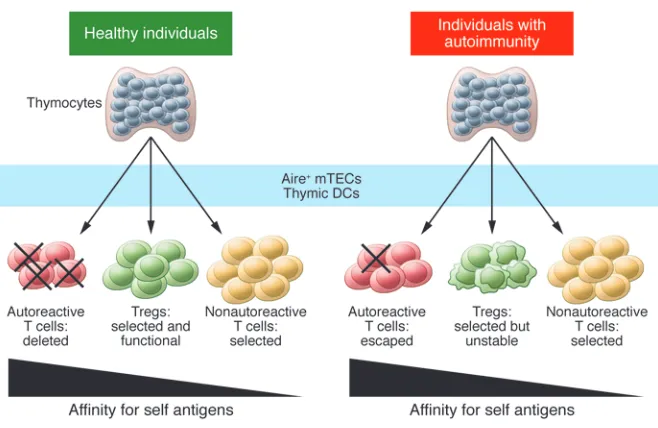
Figure 1. Defects in both central and peripheral tolerance contribute to the development of
autoim-munity. Left: In healthy individuals, most developing thymocytes with highly self-reactive TCRs are
deleted during negative selection, while nonautoreactive cells mature and leave the thymus. Tregs
are also selected on self antigens and express TCRs with higher affinity for self antigens than do
Tconvs. The presentation of self antigens to developing thymocytes by Aire
+mTECs and thymic DCs
function of pTregs in the inflammatory setting may contribute to
autoimmunity (Figure 2).
Stability of Tregs
Whereas the majority of Tregs remain Foxp3
+, a subset may become
unstable and lose Foxp3 expression in inflammatory or
lymp-hopenic conditions (102, 103). For example, CD4
+Foxp3
+Tregs can
be reprogrammed to produce IL-17 and IFN-
γ
in inflammatory
envi-ronments in mice and humans (76, 104, 105). These “ex-Foxp3”
cells can mediate autoimmunity in mouse models of autoimmune
diabetes and arthritis (102, 106). Of note, the stability of Tregs has
been a controversial topic, as other studies concluded that fully
differentiated Tregs were stable while ex-Foxp3 cells were derived
from loosely committed Tregs; however, these studies were not
per-formed in the setting of autoimmunity (107, 108). Conversely, in the
EAE model of MS, loss of Foxp3 and Treg instability were observed
in bona fide Tregs and occurred predominantly in autoreactive
Tregs in the context of self antigen–driven activation and
inflam-mation (109). Moreover, similar phenomena of Treg plasticity and
instability have been described in humans with T1D, RA, and MS
and correspond to distinct molecular stages with discrete epigenetic
and gene expression signatures (106, 110, 111).
functional differences and limited TCR overlap between tTregs
and pTregs expand the overall diversity of the Treg repertoire and
may be central to the requirement for both populations for proper
regulation of immune responses against diverse self antigens and
foreign antigens.
[image:5.585.45.548.57.300.2]Based on these studies, the overall emerging model
postu-lates that tTregs and pTregs synergize to prevent autoimmunity
in peripheral tissues of healthy individuals thanks to their
com-plementary repertoire and functional capabilities. In
individu-als prone to autoimmunity, as described above, the presence of
unique self-peptide/MHC complexes in tissues that are not
pres-ent in the thymus implies that tTregs may recognize a set of self
antigens in the thymus that is distinct from self antigens presented
in the periphery, thus affecting their ability to control
autoim-mune responses. While pTregs may in turn be able to recognize
this unique set of peripheral self antigens, the greater instability
of pTregs compared with tTregs may prevent the effective control
of autoreactive T cells in inflammatory settings. Thus, peripheral
TCR “reshaping” of Treg repertoires may play a role in the ability
of Tregs to recognize self antigens in a given target tissue to
pro-tect that tissue from autoimmunity (100, 101). However, an
inad-equate repertoire of tTregs combined with impaired stability and
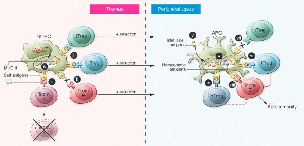
Figure 2. Model for self-peptide presentation in shaping T cell function and development of autoimmunity. Mounting data support a key role for
self-antigen presentation in T cell selection and autoimmunity. Left: In the thymus, CD4
+T cells with high affinity for self antigens undergo
based therapy, which raises concerns
about application of this approach to
dis-eases such as MS, in which the continued
destruction of vital tissue might lead to
increased morbidity. Thus, an important
goal for Treg-based therapy as well as
therapies designed to improve
Treg-medi-ated suppression will be to generate an
environment that may alter the Treg
tran-scriptome and possibly favor the stability
of the Treg lineage.
Therapeutic strategies to
restore tolerance
Many therapeutic approaches are aimed
at recalibrating pathogenic/regulatory
immune pathways to restore tolerance
without compromising anti-pathogen
defenses (Figure 3). To date, few
immuno-therapies have achieved immune tolerance,
i.e., non-responsiveness to self antigens,
without continuous immunosuppression.
Coupled with the difficulty in designing
effective clinical trials or testing of
unli-censed combination therapies in patients,
many barriers remain in realizing clinical
tolerance induction and merit more
criti-cal discussion in the future. Below, we
dis-cuss the strides in the last decade that are
leading to development of novel strategies
aimed at restoring tolerance (125).
Systemic, nonspecific immunotherapies
primarily targeting pathogenic autoreactive
Teffs. Immunomodulatory therapies that
target autoreactive Teffs are designed
to work in part by deleting pathogenic
cells, with the goal of “resetting” the immune system toward
a more balanced homeostasis (126). Given that Tregs work in
a dominant manner through bystander suppression and induce
“infectious tolerance” (127), many immunotherapies currently
under development target Treg defects identified in preclinical
studies, with the goal of restoring Treg function (15, 128).
Anti-thymocyte globulin (ATG) has shown promising results in NOD
mice and a small clinical trial in patients with T1D (129, 130)
with tolerogenic potential suggested by its favorable effect on
the Treg compartment in mice and humans (130–132). Recent
phase II trials in patients with new-onset T1D showed no clear
benefit for ATG monotherapy (133); however, a combination of
low-dose ATG and G-CSF tended to preserve
β
cell function at 12
months following initiation of therapy (134). The disappointing
outcome of ATG monotherapy may be due to ineffectual
deple-tion of effector memory T cells, which are particularly resistant
to deletion or suppression (135, 136), and suggests that
eliminat-ing these cells will be required for successful approaches.
Sim-ilarly, targeting of CD52 by alemtuzumab, recently approved
for relapsing-remitting MS, causes depletion of T and B cells,
with subsequent repopulation through preferential homeostatic
Mechanistically, the primary prerequisite for the maintenance
of the Treg population is stable Foxp3 expression (20, 112). The
requirements for Foxp3 expression in Tregs include signaling
through co-stimulatory and cytokine receptors (113–116). TCR/
CD28 and IL-2R signaling are not only required for Treg
devel-opment and homeostasis but are critical for their suppressive
function (117, 118). In particular, signaling through IL-2R is
criti-cal for maintaining Foxp3 expression and Treg homeostasis (119).
A Foxp3 intronic element known as the conserved noncoding
sequence 2 (CNS2; also referred to as Treg-specific demethylated
region) is highly demethylated in Tregs but completely methylated
in other T cell lineages (120–122). CNS2 is important to stabilize
Foxp3 expression upon Treg stimulation and division in
inflam-matory environments or conditions of limited IL-2 (123, 124).
Importantly, CNS2 ensures stable inheritance of Foxp3 expression
and maintains Treg lineage identity by acting as a sensor of TCR/
NFAT and IL-2/STAT5 signals.
Finally, the issues of Treg plasticity and stability have
important implications in the context of therapeutic approaches
in autoimmune diseases. The potential instability of a
frac-tion of Tregs may result in accelerafrac-tion of disease after
[image:6.585.38.367.56.321.2]proliferation of pTregs and effector memory T cells (137–139).
This selective expansion of T cell subsets may account for both
the drug’s efficacy and the high rate of other autoimmune
con-ditions seen in up to one-third of treated patients (140).
Like-wise, CD2 is expressed on almost all human T cells but is most
highly expressed in memory and pathogenic Teffs in
autoimmu-nity (141, 142). Treatment of psoriasis patients with a CD2
lig-and-specific Fc fusion protein (alefacept) resulted in sustained
remissions even after drug discontinuation in some patients
(143, 144). Mechanistically, alefacept preferentially depletes
effector memory T cells without eliminating Tregs (141, 145).
Recently, treatment of T1D with alefacept has shown promise,
with some evidence of efficacy in a phase II clinical trial (146).
Treatment with Fc receptor non-binding anti-CD3 mAbs
tepli-zumab or otelixitepli-zumab in patients with new-onset T1D
pre-served
β
cell function for up to two years; however, neither mAb
ultimately prevented the destruction of the remaining
β
cells
(147–150). The mechanisms of action of anti-CD3 mAbs remain
unclear but include the selective depletion of activated T cells
and induction or preferential retention of cells with regulatory
properties (151–155).
Antigen-specific tolerogenic therapies are expected to be safer
than nonspecific strategies due to a lower risk of global
immuno-suppression (125). Antigen therapy has successfully prevented or
reversed autoimmune diseases in the NOD and EAE mouse
mod-els (156–159), indicating that targeting responses against one or a
few self antigens can thwart polyclonal autoimmune responses.
Enrichment in Tregs has been observed after antigen
immuno-therapy in patients with RA and T1D (160–163). Administration
of antigen-coupled ethylene carbodiimide–fixed cells (as well as,
more recently, antigen-coated beads) has been extremely effective
at restoring tolerance in NOD mice and EAE (164–167).
Addition-ally, a recent phase I clinical trial in MS showed that this approach
reduced myelin-specific autoreactive T cell responses in humans
(168). Finally, recent studies in T1D have focused on oral antigen
delivery to promote tolerance due to postulated Treg induction
and clonal anergy/deletional mechanisms (169). Based on a
reduc-tion of diabetes incidence in a small set of higher-risk individuals
(170), a large-scale study of oral insulin for T1D prevention is
cur-rently ongoing (ClinicalTrials.gov identifier NCT00419562).
Immunotherapies aimed at restoring the control of autoimmune
responses by Tregs. The central role of IL-2 in Treg homeostasis and
function has led to therapeutic strategies that aim to improve IL-2
signaling in Tregs (171). In NOD and EAE mouse models,
treat-ment with low-dose IL-2 restored high levels of Foxp3 and CD25
expression, improved the stability of Tregs, and prevented or
restored the development of autoimmunity (76, 109, 172).
Admin-istration of low-dose IL-2 in new-onset T1D patients did not alter
glucose metabolism but did induce a dose-dependent increase in
the frequency of Tregs (173). However, dosage may significantly
affect the outcome, as low-dose versus high-dose IL-2 therapy
differentially promotes Tregs and Tconvs, respectively (174). In
fact, in contrast to low-dose IL-2, treatment of NOD mice with
high-dose IL-2 accelerated progression of diabetes (76, 175). Thus,
approaches aimed at boosting Tregs may need to combine IL-2
treatment with therapies targeting pathogenic Teffs. The mTOR
inhibitor rapamycin selectively inhibits the proliferation of Th1
and Th17 cells while enhancing Treg survival (176–178). Treatment
of patients with new-onset T1D with rapamycin plus low-dose IL-2
resulted in a transient increase in the frequency of Tregs and
sta-ble restoration of IL-2 signaling that persisted long after treatment
was discontinued (179). However, the combination therapy also
transiently impaired
β
cell function and dramatically increased
numbers of natural killer cells and eosinophils, which might have
adversely impacted pancreatic islet cells. Thus, IL-2 therapy alters
a complex cellular network and additional studies will be
neces-sary to design treatments specifically targeting Tregs. Improved
knowledge of the structural properties of IL-2 binding to its
recep-tors on different cell types and advances in protein bioengineering
may help solve this conundrum via generation of mutated forms of
IL-2 that selectively signal in Tregs (180, 181).
Cellular therapy to restore tolerance. The favorable
therapeu-tic profile of Tregs has led to strong interest in Treg-based
cellu-lar therapy in transplantation and autoimmune diseases (182).
Adoptive transfer of Tregs suppressed inflammation and disease
in EAE, NOD mice, and mouse models of IBD and SLE (59, 60,
183–185). Of note, Tregs expanded in vitro were more efficient
at controlling autoimmune responses than their freshly isolated
counterparts (184, 186). We have developed a clinically relevant
procedure for generating large numbers of CD4
+CD127
lo/–CD25
+Tregs without the need for additional selective agents (71, 72,
187) and used a current good manufacturing
practices–compli-ant method in a phase I clinical trial of Treg administration in
T1D patients. We found that autologous ex vivo–expanded Tregs
were well tolerated and long lived, and that average C-peptide
levels remained stable for up to two years after treatment
(Clin-icalTrials.gov identifier NCT01210664), consistent with the
one-year follow-up data of a small phase I study in children with
T1D (188). Future clinical applications may involve the use of
genetically modified Tregs that express genes that promote their
survival, stability, trafficking, or suppressive function. In models
of diabetes, tissue antigen–specific Tregs are more efficient than
polyclonal cells at suppressing autoimmunity (184, 189). However,
selective expansion of autoantigen-specific Tregs is challenging
because of their low precursor frequency and the uncertainty of
which antigens to target in most diseases. Redirecting polyclonal
Tregs by engineering expression of antigen-specific receptors may
help bridge this gap and may also circumvent Treg inefficiencies
related to expression of inadequate TCR repertoires, as we and
others have recently shown (190–193). These findings support
the notion that polyclonal human Tregs could be engineered to
express TCRs specific for self antigens in the target tissue in order
to improve the efficacy of Treg therapy at protecting this tissue
(15). Moreover, this approach could be combined with in vitro or
in vivo treatments aimed at correcting other Treg defects, such
as the long-term restoration of IL-2 signaling defects by low-dose
IL-2 therapy in vivo (179).
Other appealing approaches in cellular therapy involve the
use of tissues generated from human pluripotent stem cells
(hPSCs). Given that thymic transplantation offers the potential
to establish donor-specific tolerance, hPSCs could be
differen-tiated into both one organ for transplantation (e.g., pancreatic
immuno-suppression (194, 195). Coupled with such regenerative
strate-gies, advances in genetic modification of stem cells and iPSCs
may soon allow us to engineer thymus or correct defects in order
to modulate and enforce tolerance. Although not ready for prime
time, these therapeutic strategies have tremendous potential,
considering that human embryonic stem cells have recently been
used to generate both thymic epithelial progenitors (196, 197)
and islet-like structures (198, 199) that recapitulate the function
of their adult differentiated counterparts upon transplantation in
mice or humanized models.
Conclusion
Despite the wide swath of redundant mechanisms that control
central and peripheral tolerance, the high incidence of
autoim-mune diseases and difficulty restoring tolerance in humans reflect
the equally powerful mechanisms that ensure effective immune
responses against pathogens. Thus, multiple pathways will likely
need to be targeted to restore tolerance to self antigens without
compromising overall immunity (Figure 3). Tremendous progress
has been made in our understanding of the pathways that control
autoimmunity and defects associated with distinct autoimmune
diseases, leading to many novel therapeutic approaches
target-ing individual pathways. Combination therapies have been
intro-duced in clinical trials with mixed results, which emphasizes that
they could be more efficacious; however, combination treatments
might raise new safety challenges as well (200). In the future,
cus-tomized therapies and combination therapies should be informed
not only by pathways found to be important in each autoimmune
disease but also by the genetic and environmental influences in
each patient, as certain treatments may be predicted to have
greater efficacy depending on individual genetic susceptibility
and immunological history. This increased level of granularity will
undoubtedly reveal additional complexity in the molecular and
cellular interactions underlying autoimmunity, but it is also bound
to result in improved control of autoimmune diseases in individual
patients in the new era of precision medicine.
Acknowledgments
We thank members of the Bluestone and Anderson labs for
con-tributions to the science that drove much of the commentary
in this Review. This work was funded by the National Institute
of Allergy and Infectious Diseases (grants R01AI046643 and
R01AI097457); the National Institute of Diabetes, Digestive and
Kidney Diseases (grant R01DK101622); the Juvenile Diabetes
Research Foundation (grants 17-2011-661 and 17-2013-513); the
JDRF Collaborative Center for Treg Biology; and the California
Institute for Regeneration Medicine (grant RB5-07262).
Address correspondence to: Jeffrey Bluestone, Diabetes
Cen-ter, University of California San Francisco, HSW 1112 Box 0540,
513 Parnassus Ave., San Francisco, California 94143-0540, USA.
Phone: 415.514.0417; E-mail: [email protected].
1. Cotsapas C, Hafler DA. Immune-mediated dis-ease genetics: the shared basis of pathogenesis.
Trends Immunol. 2013;34(1):22–26.
2. Ueda H, et al. Association of the T-cell regulatory gene CTLA4 with susceptibility to autoimmune disease. Nature. 2003;423(6939):506–511. 3. Todd JA, et al. Robust associations of four new chromosome regions from genome-wide analyses of type 1 diabetes. Nat Genet. 2007;39(7):857–864.
4. Consortium WTCC. Genomewide associ-ation study of 14,000 cases of seven com-mon diseases and 3,000 controls. Nature. 2007;447(7145):661–683.
5. Pugliese A, et al. The insulin gene is transcribed in the human thymus and transcription levels correlated with allelic variation at the INS VNTR-IDDM2 susceptibility locus for type 1 diabetes.
Nat Genet. 1997;15(3):293–297.
6. Suzuki A. Functional haplotypes of PADI4, encoding citrullinating enzyme peptidylarginine deiminase 4, are associated with rheumatoid arthritis. Nat Genet. 2003;34(4):395–402. 7. Cheng MH, Anderson MS. Monogenic
autoim-munity. Annu Rev Immunol. 2012;30:393–427. 8. Wucherpfennig KW, et al. Clonal expansion
and persistence of human T cells specific for an immunodominant myelin basic protein peptide.
J Immunol. 1994;152(11):5581–5592.
9. Zhang L, Nakayama M, Eisenbarth GS. Insulin as an autoantigen in NOD/human diabetes. Curr
Opin Immunol. 2008;20(1):111–118.
10. Reijonen H, et al. Detection of GAD65-specific T-cells by major histocompatibility complex class II tetramers in type 1 diabetic patients and at-risk
subjects. Diabetes. 2002;51(5):1375–1382. 11. Su LF, Kidd BA, Han A, Kotzin JJ, Davis MM.
Virus-specific CD4(+) memory-phenotype T cells are abundant in unexposed adults. Immunity. 2013;38(2):373–383.
12. Bour-Jordan H, Esensten JH, Martinez-Llordella M, Penaranda C, Stumpf M, Bluestone JA. Intrin-sic and extrinIntrin-sic control of peripheral T-cell toler-ance by costimulatory molecules of the CD28/B7 family. Immunol Rev. 2011;241(1):180–205. 13. Bluestone JA. Mechanisms of tolerance. Immunol
Rev. 2011;241(1):5–19.
14. Wing K, Sakaguchi S. Regulatory T cells exert checks and balances on self tolerance and auto-immunity. Nat Immunol. 2010;11(1):7–13. 15. Brusko TM, Putnam AL, Bluestone JA. Human
regulatory T cells: role in autoimmune disease and therapeutic opportunities. Immunol Rev. 2008;223:371–390.
16. Sakaguchi S, Miyara M, Costantino CM, Hafler DA. FOXP3+ regulatory T cells in the human immune system. Nat Rev Immunol. 2010;10(7):490–500.
17. Bennett CL, et al. The immune dysregulation, polyendocrinopathy, enteropathy, X-linked syn-drome (IPEX) is caused by mutations of FOXP3.
Nat Genet. 2001;27(1):20–21.
18. Fontenot JD, Gavin MA, Rudensky AY. Foxp3 programs the development and function of CD4(+)CD25(+) regulatory T cells. Nat Immunol. 2003;4(4):330–336.
19. Hori S, Nomura T, Sakaguchi S. Control of regula-tory T cell development by the transcription fac-tor Foxp3. Science. 2003;299(5609):1057–1061. 20. Wan YY, Flavell RA. Regulatory T-cell
func-tions are subverted and converted owing to attenuated Foxp3 expression. Nature. 2007;445(7129):766–770.
21. Kim JM, Rasmussen JP, Rudensky AY. Regula-tory T cells prevent catastrophic autoimmunity throughout the lifespan of mice. Nat Immunol. 2007;8(2):191–197.
22. Anderson MS, et al. Projection of an immuno-logical self shadow within the thymus by the aire protein. Science. 2002;298(5597):1395–1401. 23. Liston A, Lesage S, Wilson J, Peltonen L,
Goodnow CC. Aire regulates negative selec-tion of organ-specific T cells. Nat Immunol. 2003;4(4):350–354.
24. Anderson MS, Su MA. Aire and T cell develop-ment. Curr Opin Immunol. 2011;23(2):198–206. 25. Perheentupa J. Autoimmune
polyendocrinop-athy-candidiasis-ectodermal dystrophy. J Clin
Endocrinol Metab. 2006;91(8):2843–2850.
26. Taniguchi RT, et al. Detection of an autoreactive T-cell population within the polyclonal repertoire that undergoes distinct autoimmune regulator (Aire)-mediated selection. Proc Natl Acad Sci U S A. 2012;109(20):7847–7852.
27. Giraud M, et al. An IRF8-binding promoter variant and AIRE control CHRNA1 pro-miscuous expression in thymus. Nature. 2007;448(7156):934–937.
30. Metzger TC, et al. Lineage tracing and cell ablation identify a post-Aire-expressing thymic epithelial cell population. Cell Rep. 2013; 5(1):166–179.
31. Ucar A, et al. Adult thymus contains FoxN1(–) epithelial stem cells that are bipotent for med-ullary and cortical thymic epithelial lineages.
Immunity. 2014;41(2):257–269.
32. Wong K, et al. Multilineage potential and self-renewal define an epithelial progenitor cell population in the adult thymus. Cell Rep. 2014;8(4):1198–1209.
33. Roberts NA, et al. Rank signaling links the devel-opment of invariant gammadelta T cell progeni-tors and Aire(+) medullary epithelium. Immunity. 2012;36(3):427–437.
34. Rossi SW, et al. RANK signals from CD4(+)3(–) inducer cells regulate development of Aire-expressing epithelial cells in the thymic medulla.
J Exp Med. 2007;204(6):1267–1272.
35. White AJ, et al. Sequential phases in the devel-opment of Aire-expressing medullary thymic epithelial cells involve distinct cellular input.
Eur J Immunol. 2008;38(4):942–947.
36. Jamieson BD, et al. Generation of functional thymocytes in the human adult. Immunity. 1999;10(5):569–575.
37. McFarland RD, Douek DC, Koup RA, Picker LJ. Identification of a human recent thymic emigrant phenotype. Proc Natl Acad Sci U S A. 2000;97(8):4215–4220.
38. Wucherpfennig KW, Call MJ, Deng L, Mariuzza R. Structural alterations in peptide–MHC recog-nition by self-reactive T cell receptors. Curr Opin
Immunol. 2009;21(6):590–595.
39. Hahn M, Nicholson MJ, Pyrdol J, Wucherpfennig KW. Unconventional topology of self peptide– major histocompatibility complex binding by a human autoimmune T cell receptor. Nat Immunol. 2005;6(5):490–496.
40. Bulek AM, et al. Structural basis for the killing of human β cells by CD8+ T cells in type 1 diabetes.
Nat Immunol. 2012;13(3):283–289.
41. Crawford F, et al. Specificity and detection of insulin-reactive CD4+ T cells in type 1 diabetes in the nonobese diabetic (NOD) mouse. Proc Natl
Acad Sci U S A. 2011;108(40):16729–16734.
42. Stadinski BD, Zhang L, Crawford F, Marrack P, Eisenbarth GS, Kappler JW. Diabetogenic T cells recognize insulin bound to IAg7 in an unex-pected, weakly binding register. Proc Natl Acad
Sci U S A. 2010;107(24):10978–10983.
43. Yang J, et al. Autoreactive T cells specific for insulin B:11-23 recognize a low-affinity peptide register in human subjects with auto-immune diabetes. Proc Natl Acad Sci U S A. 2014;111(41):14840–14845.
44. Mohan JF, Petzold SJ, Unanue ER. Register shift-ing of an insulin peptide-MHC complex allows diabetogenic T cells to escape thymic deletion.
J Exp Med. 2011;208(12):2375–2383.
45. Mohan JF, Unanue ER. A novel pathway of pre-sentation by class II-MHC molecules involving peptides or denatured proteins important in autoimmunity. Mol Immunol. 2013; 55(2):166–168.
46. Stadinski BD, et al. Chromogranin A is an autoantigen in type 1 diabetes. Nat Immunol.
2010;11(3):225–231.
47. Li Y, Huang Y, Lue J, Quandt JA, Martin R, Mar-iuzza RA. Structure of a human autoimmune TCR bound to a myelin basic protein self-peptide and a multiple sclerosis-associated MHC class II molecule. EMBO J. 2005;24(17):2968–2979. 48. He XL, Radu C, Sidney J, Sette A, Ward ES,
Gar-cia KC. Structural snapshot of aberrant antigen presentation linked to autoimmunity: the immu-nodominant epitope of MBP complexed with I-Au. Immunity. 2002;17(1):83–94. 49. McGinty JW, Chow IT, Greenbaum C,
Ode-gard J, Kwok WW, James EA. Recognition of posttranslationally modified GAD65 epitopes in subjects with type 1 diabetes. Diabetes. 2014;63(9):3033–3040.
50. Knight RR, et al. A distinct immunogenic region of glutamic acid decarboxylase 65 is naturally processed and presented by human islet cells to cytotoxic CD8 T cells. Clin Exp Immunol. 2015;179(1):100–107.
51. Wegner N. Autoimmunity to specific citru-llinated proteins gives the first clues to the etiology of rheumatoid arthritis. Immunol Rev. 2010;233(1):34–54.
52. Busch R, De Riva A, Hadjinicolaou AV, Jiang W, Hou T, Mellins ED. On the perils of poor editing: regulation of peptide loading by HLA-DQ and H2-A molecules associated with celiac disease and type 1 diabetes. Expert Rev Mol Med. 2012;14:e15.
53. Bautista JL, et al. Intraclonal competition limits the fate determination of regulatory T cells in the thymus. Nat Immunol. 2009;10(6):610–617. 54. Klein L, Kyewski B, Allen PM, Hogquist KA. Positive and negative selection of the T cell repertoire: what thymocytes see (and don’t see).
Nat Rev Immunol. 2014;14(6):377–391.
55. Perry JS, et al. Distinct contributions of Aire and antigen-presenting-cell subsets to the genera-tion of self-tolerance in the thymus. Immunity. 2014;41(3):414–426.
56. Aschenbrenner K, et al. Selection of Foxp3+ regula-tory T cells specific for self antigen expressed and presented by Aire+ medullary thymic epithelial cells. Nat Immunol. 2007;8(4):351–358. 57. Malchow S, et al. Aire-dependent thymic
devel-opment of tumor-associated regulatory T cells.
Science. 2013;339(6124):1219–1224.
58. Anderson MS, Venanzi ES, Chen Z, Berzins SP, Benoist C, Mathis D. The cellular mechanism of Aire control of T cell tolerance. Immunity. 2005;23(2):227–239.
59. Kohm AP, Carpentier PA, Anger HA, Miller SD. Cutting edge: CD4+CD25+ regulatory T cells suppress antigen-specific autoreactive immune responses and central nervous sys-tem inflammation during active experimental autoimmune encephalomyelitis. J Immunol. 2002;169(9):4712–4716.
60. Mottet C, Uhlig HH, Powrie F. Cutting edge: cure of colitis by CD4+CD25+ regulatory T cells.
J Immunol. 2003;170(8):3939–3943.
61. Salomon B, et al. B7/CD28 costimulation is essential for the homeostasis of the CD4+CD25+ immunoregulatory T cells that control autoim-mune diabetes. Immunity. 2000;12(4):431–440. 62. You S, et al. Autoimmune diabetes onset results
from qualitative rather than quantitative age-de-pendent changes in pathogenic T-cells. Diabetes. 2005;54(5):1415–1422.
63. Schneider A, et al. In active relapsing-remitting multiple sclerosis, effector T cell resistance to adaptive T(regs) involves IL-6-mediated signaling.
Sci Transl Med. 2013;5(170):170ra115.
64. Schenten D, et al. Signaling through the adaptor molecule MyD88 in CD4+ T cells is required to overcome suppression by regulatory T cells.
Immunity. 2014;40(1):78–90.
65. Bour-Jordan H, Salomon BL, Thompson HL, Szot GL, Bernhard MR, Bluestone JA. Costimulation controls diabetes by altering the balance of pathogenic and regulatory T cells. J Clin Invest. 2004;114(7):979–987.
66. Cvetanovich GL, Hafler DA. Human regulatory T cells in autoimmune diseases. Curr Opin Immunol. 2010;22(6):753–760.
67. Buckner JH. Mechanisms of impaired regulation by CD4+CD25+FOXP3+ regulatory T cells in human autoimmune diseases. Nat Rev Immunol. 2010;10(12):849–859.
68. Long SA, et al. Defects in IL-2R signaling con-tribute to diminished maintenance of FOXP3 expression in CD4(+)CD25(+) regulatory T-cells of type 1 diabetic subjects. Diabetes. 2010;59(2):407–415.
69. Long SA, et al. An autoimmune-associated variant in PTPN2 reveals an impairment of IL-2R signaling in CD4(+) T cells. Genes Immun. 2011;12(2):116–125.
70. Cerosaletti K, et al. Multiple autoimmune-asso-ciated variants confer decreased IL-2R signaling in CD4+ CD25(hi) T cells of type 1 diabetic and multiple sclerosis patients. PLoS One. 2013;8(12):e83811.
71. Liu W, et al. CD127 expression inversely correlates with FoxP3 and suppressive func-tion of human CD4+ T reg cells. J Exp Med. 2006;203(7):1701–1711.
72. Putnam AL, et al. Expansion of human regulatory T-cells from patients with type 1 diabetes. Diabetes. 2009;58(3):652–662.
73. Garg G, et al. Type 1 diabetes-associated IL2RA variation lowers IL-2 signaling and contributes to diminished CD4+CD25+ regulatory T cell func-tion. J Immunol. 2012;188(9):4644–4653. 74. Lindley S, Dayan CM, Bishop A, Roep BO,
Peak-man M, Tree TI. Defective suppressor function in CD4(+)CD25(+) T-cells from patients with type 1 diabetes. Diabetes. 2005;54(1):92–99.
75. Zhou X, et al. Selective miRNA disruption in T reg cells leads to uncontrolled autoimmunity. J Exp
Med. 2008;205(9):1983–1991.
76. Tang Q, et al. Central role of defective inter-leukin-2 production in the triggering of islet autoimmune destruction. Immunity. 2008;28(5):687–697.
77. Hsieh CS, Liang Y, Tyznik AJ, Self SG, Liggitt D, Rudensky AY. Recognition of the peripheral self by naturally arising CD25+CD4+ T cell receptors.
Immunity. 2004;21(2):267–277.
78. Jordan MS, et al. Thymic selection of CD4+CD25+ regulatory T cells induced by an agonist self- peptide. Nat Immunol. 2001;2(4):301–306. 79. Abbas AK, et al. Regulatory T cells:
2013;14(4):307–308.
80. Bluestone JA, Abbas AK. Natural versus adap-tive regulatory T cells. Nat Rev Immunol. 2003;3(3):253–257.
81. Yadav M, Stephan S, Bluestone JA. Peripherally induced tregs — role in immune homeostasis and autoimmunity. Front Immunol. 2013;4:232. 82. Apostolou I, Sarukhan A, Klein L, Von
Boeh-mer H. Origin of regulatory T cells with known specificity for antigen. Nat Immunol. 2002;3(8):756–763.
83. Corse E, Gottschalk RA, Allison JP. Strength of TCR-peptide/MHC interactions and in vivo T cell responses. J Immunol. 2011;186(9):5039–5045. 84. Coombes JL, et al. A functionally specialized
population of mucosal CD103+ DCs induces Foxp3+ regulatory T cells via a TGF-β and retin-oic acid-dependent mechanism. J Exp Med. 2007;204(8):1757–1764.
85. Li MO, Sanjabi S, Flavell RA. Transforming growth factor-beta controls development, homeostasis, and tolerance of T cells by regula-tory T cell-dependent and -independent mecha-nisms. Immunity. 2006;25(3):455–471. 86. Sun CM, et al. Small intestine lamina propria
dendritic cells promote de novo generation of Foxp3 T reg cells via retinoic acid. J Exp Med. 2007;204(8):1775–1785.
87. Mucida D, et al. Reciprocal TH17 and regulatory T cell differentiation mediated by retinoic acid.
Science. 2007;317(5835):256–260.
88. Lathrop SK, et al. Peripheral education of the immune system by colonic commensal microbiota.
Nature. 2011;478(7368):250–254.
89. Atarashi K, et al. Treg induction by a ratio-nally selected mixture of Clostridia strains from the human microbiota. Nature. 2013;500(7461):232–236.
90. Arpaia N, et al. Metabolites produced by commen-sal bacteria promote peripheral regulatory T-cell generation. Nature. 2013;504(7480):451–455. 91. Furusawa Y, et al. Commensal microbe-derived
butyrate induces the differentiation of colonic reg-ulatory T cells. Nature. 2013;504(7480):446–450. 92. Yadav M, Stephan S, Bluestone JA. Peripherally
induced tregs — role in immune homeostasis and autoimmunity. Front Immunol. 2013;4:232. 93. Haribhai D, et al. A central role for induced
regu-latory T cells in tolerance induction in experimen-tal colitis. J Immunol. 2009;182(6):3461–3468. 94. Haribhai D, et al. A requisite role for induced
regulatory T cells in tolerance based on expand-ing antigen receptor diversity. Immunity. 2011;35(1):109–122.
95. Josefowicz SZ, et al. Extrathymically generated regulatory T cells control mucosal TH2 inflam-mation. Nature. 2012;482(7385):395–399. 96. Yadav M, et al. Neuropilin-1 distinguishes
nat-ural and inducible regulatory T cells among regulatory T cell subsets in vivo. J Exp Med. 2012;209(10):1713–1722.
97. Pacholczyk R, Ignatowicz H, Kraj P, Ignatowicz L. Origin and T cell receptor diversity of Foxp3+ C-D4+CD25+ T cells. Immunity. 2006;25(2):249–259. 98. Hsieh CS, Zheng Y, Liang Y, Fontenot JD, Ruden-sky AY. An intersection between the self-reactive regulatory and nonregulatory T cell receptor repertoires. Nat Immunol. 2006;7(4):401–410.
99. Wong J, Obst R, Correia-Neves M, Losyev G, Mathis D, Benoist C. Adaptation of TCR repertoires to self-peptides in regulatory and nonregulatory CD4+ T cells. J Immunol. 2007;178(11):7032–7041.
100. Samy ET, Parker LA, Sharp CP, Tung KS. Con-tinuous control of autoimmune disease by anti-gen-dependent polyclonal CD4+CD25+ regulatory T cells in the regional lymph node. J Exp Med. 2005;202(6):771–781.
101. Seddon B, Mason D. Peripheral autoantigen induces regulatory T cells that prevent autoimmu-nity. J Exp Med. 1999;189(5):877–882.
102. Zhou X, et al. Instability of the transcription factor Foxp3 leads to the generation of patho-genic memory T cells in vivo. Nat Immunol. 2009;10(9):1000–1007.
103. Bailey-Bucktrout SL, Bluestone JA. Regulatory T cells: stability revisited. Trends Immunol. 2011;32(7):301–306.
104. Beriou G, et al. IL-17-producing human periph-eral regulatory T cells retain suppressive func-tion. Blood. 2009;113(18):4240–4249. 105. Voo KS, et al. Identification of IL-17-producing
FOXP3+ regulatory T cells in humans. Proc Natl
Acad Sci U S A. 2009;106(12):4793–4798.
106. Komatsu N, et al. Pathogenic conversion of Foxp3+ T cells into TH17 cells in autoimmune arthritis. Nat Med. 2014;20(1):62–68. 107. Rubtsov YP, et al. Stability of the reg-ulatory T cell lineage in vivo. Science. 2010;329(5999):1667–1671.
108. Miyao T, et al. Plasticity of Foxp3(+) T cells reflects promiscuous Foxp3 expression in con-ventional T cells but not reprogramming of regu-latory T cells. Immunity. 2012;36(2):262–275. 109. Bailey-Bucktrout SL, et al. Self-antigen-driven
activation induces instability of regulatory T cells during an inflammatory autoimmune response.
Immunity. 2013;39(5):949–962.
110. McClymont SA, et al. Plasticity of human regula-tory T cells in healthy subjects and patients with type 1 diabetes. J Immunol. 2011;186(7):3918–3926. 111. Dominguez-Villar M, Baecher-Allan CM, Hafler
DA. Identification of T helper type 1-like, Foxp3+ regulatory T cells in human autoimmune disease.
Nat Med. 2011;17(6):673–675.
112. Gavin MA, et al. Foxp3-dependent programme of regulatory T-cell differentiation. Nature. 2007;445(7129):771–775.
113. Bour-Jordan H, Bluestone JA. Regulating the regu-lators: costimulatory signals control the homeosta-sis and function of regulatory T cells. Immunol Rev. 2009;229(1):41–66.
114. Tang Q, et al. Cutting edge: CD28 controls peripheral homeostasis of CD4+CD25+ regula-tory T cells. J Immunol. 2003;171(7):3348. 115. Josefowicz SZ, Rudensky A. Control of regulatory
T cell lineage commitment and maintenance.
Immunity. 2009;30(5):616–625.
116. Rudensky AY. Regulatory T cells and Foxp3.
Immunol Rev. 2011;241(1):260–268.
117. Barron L, et al. Cutting edge: mechanisms of IL-2-dependent maintenance of functional regula-tory T cells. J Immunol. 2010;185(11):6426–6430. 118. Levine AG, Arvey A, Jin W, Rudensky AY.
Contin-uous requirement for the TCR in regulatory T cell function. Nat Immunol. 2014;15(11):1070–1078.
119. Cheng G, Yu A, Malek TR. T-cell tolerance and the multi-functional role of IL-2R signaling in T-regu-latory cells. Immunol Rev. 2011;241(1):63–76. 120. Baron U, et al. DNA demethylation in the human
FOXP3 locus discriminates regulatory T cells from activated FOXP3(+) conventional T cells.
Eur J Immunol. 2007;37(9):2378–2389.
121. Floess S, et al. Epigenetic control of the foxp3 locus in regulatory T cells. PLoS Biol. 2007;5(2):e38.
122. Polansky JK, et al. DNA methylation con-trols Foxp3 gene expression. Eur J Immunol. 2008;38(6):1654–1663.
123. Feng Y, Arvey A, Chinen T, van der Veeken J, Gasteiger G, Rudensky AY. Control of the inheri-tance of regulatory T cell identity by a cis element in the Foxp3 locus. Cell. 2014;158(4):749–763. 124. Li X, Liang Y, LeBlanc M, Benner C, Zheng Y.
Function of a Foxp3 cis-element in protecting reg-ulatory T cell identity. Cell. 2014;158(4):734–748. 125. Bluestone JA, Bour-Jordan H. Current and future
immunomodulation strategies to restore toler-ance in autoimmune diseases. Cold Spring Harb
Perspect Biol. 2012;4(11):a007542.
126. van de Linde P, et al. Mechanisms of antibody immunotherapy on clonal islet reactive T cells.
Hum Immunol. 2006;67(4):264–273.
127. Tang Q, Bluestone JA. The Foxp3+ regulatory T cell: a jack of all trades, master of regulation.
Nat Immunol. 2008;9(3):239–244.
128. Herold KC, Vignali DA, Cooke A, Bluestone JA. Type 1 diabetes: translating mechanistic obser-vations into effective clinical outcomes. Nat Rev
Immunol. 2013;13(4):243–256.
129. Saudek F, Havrdova T, Boucek P, Karasova L, Novota P, Skibova J. Polyclonal anti-T-cell ther-apy for type 1 diabetes mellitus of recent onset.
Rev Diabet Stud. 2004;1(2):80–88.
130. Simon G, et al. Murine antithymocyte globulin therapy alters disease progression in NOD mice by a time-dependent induction of immunoregulation.
Diabetes. 2008;57(2):405–414.
131. Feng X, et al. Rabbit ATG but not horse ATG promotes expansion of functional CD4+CD25highFOXP3+ regulatory T cells in vitro.
Blood. 2008;111(7):3675–3683.
132. Lopez M, Clarkson MR, Albin M, Sayegh MH, Najafian N. A novel mechanism of action for anti-thymocyte globulin: induction of CD4+CD25+Foxp3+ regulatory T cells. J Am Soc
Nephrol. 2006;17(10):2844–2853.
133. Gitelman SE, et al. Antithymocyte globulin treatment for patients with recent-onset type 1 diabetes: 12-month results of a randomised, placebo-controlled, phase 2 trial. Lancet Diabetes
Endocrinol. 2013;1(4):306–316.
134. Haller MJ, et al. Anti-thymocyte globulin/G-CSF treatment preserves β cell function in patients with established type 1 diabetes. J Clin Invest. 2015;125(1):448–455.
135. Laughlin E, Burke G, Pugliese A, Falk B, Nepom G. Recurrence of autoreactive antigen-specific CD4+ T cells in autoimmune diabetes after pancreas transplantation. Clin Immunol. 2008;128(1):23–30. 136. Vendrame F, et al. Recurrence of type 1 diabetes
CD4 T-cells. Diabetes. 2010;59(4):947–957. 137. Jones JL, et al. Human autoimmunity after
lymphocyte depletion is caused by homeostatic T-cell proliferation. Proc Natl Acad Sci U S A. 2013;110(50):20200–20205.
138. Turner MJ, et al. Immune status following alemtuzumab treatment in human CD52 trans-genic mice. J Neuroimmunol. 2013;261(1):29–36. 139. Zhang X, et al. Differential reconstitution of T cell
subsets following immunodepleting treatment with alemtuzumab (CD52 monoclonal anti-body) in patients with relapsing-remitting multi-ple sclerosis. J Immunol. 2013;191(12):5867–5874. 140. Havrdova E, Horakova D, Kovarova I.
Alemtu-zumab in the treatment of multiple sclerosis: key clinical trial results and considerations for use.
Ther Adv Neurol Disord. 2015;8(1):31–45.
141. Chamian F, et al. Alefacept (anti-CD2) causes a selective reduction in circulating effector memory T cells (Tem) and relative preservation of central memory T cells (Tcm) in psoriasis. J Transl Med. 2007;5:27.
142. Weaver TA, et al. Alefacept promotes co-stimula-tion blockade based allograft survival in nonhu-man primates. Nat Med. 2009;15(7):746–749. 143. Gottlieb AB, et al. CD4+ T-cell-directed antibody
responses are maintained in patients with psori-asis receiving alefacept: results of a randomized study. J Am Acad Dermatol. 2003;49(5):816–825. 144. Krueger GG, Ellis CN. Alefacept therapy
pro-duces remission for patients with chronic plaque psoriasis. Br J Dermatol. 2003;148(4):784–788. 145. Ellis CN, Krueger GG, Alefacept Clinical Study G.
Treatment of chronic plaque psoriasis by selec-tive targeting of memory effector T lymphocytes.
N Engl J Med. 2001;345(4):248–255.
146. Rigby MR, et al. Targeting of memory T cells with alefacept in new-onset type 1 diabetes (T1DAL study): 12 month results of a randomised, dou-ble-blind, placebo-controlled phase 2 trial. Lancet
Diabetes Endocrinol. 2013;1(4):284–294.
147. Herold KC, et al. Treatment of patients with new onset Type 1 diabetes with a single course of anti-CD3 mAb Teplizumab preserves insulin production for up to 5 years. Clin Immunol. 2009;132(2):166–173.
148. Herold KC, et al. Teplizumab (anti-CD3 mAb) treatment preserves C-peptide responses in patients with new-onset type 1 diabetes in a ran-domized controlled trial: metabolic and immu-nologic features at baseline identify a subgroup of responders. Diabetes. 2013;62(11):3766–3774. 149. Herold KC, et al. Anti-CD3 monoclonal antibody in new-onset type 1 diabetes mellitus. N Engl J Med. 2002;346(22):1692–1698.
150. Keymeulen B, et al. Insulin needs after CD3-antibody therapy in new-onset type 1 dia-betes. N Engl J Med. 2005;352(25):2598–2608. 151. Belghith M, Bluestone JA, Barriot S, Megret J,
Bach JF, Chatenoud L. TGF-β-dependent mech-anisms mediate restoration of self-tolerance induced by antibodies to CD3 in overt autoim-mune diabetes. Nat Med. 2003;9(9):1202–1208. 152. Herold KC, Burton JB, Francois F,
Poumi-an-Ruiz E, Glandt M, Bluestone JA. Activation of human T cells by FcR nonbinding anti-CD3 mAb, hOKT3γ1(Ala-Ala). J Clin Invest. 2003;111(3):409–418.
153. Penaranda C, Tang Q, Bluestone JA. Anti-CD3 therapy promotes tolerance by selectively deplet-ing pathogenic cells while preservdeplet-ing regulatory T cells. J Immunol. 2011;187(4):2015–2022. 154. Ochi H, et al. Oral CD3-specific antibody
sup-presses autoimmune encephalomyelitis by inducing CD4+CD25– LAP+ T cells. Nat Med. 2006;12(6):627–635.
155. Waldron-Lynch F, et al. Teplizumab induces human gut-tropic regulatory cells in human-ized mice and patients. Sci Transl Med. 2012;4(118):118ra112.
156. Samson MF, Smilek DE. Reversal of acute exper-imental autoimmune encephalomyelitis and prevention of relapses by treatment with a myelin basic protein peptide analogue modified to form long-lived peptide-MHC complexes. J Immunol. 1995;155(5):2737–2746.
157. Smilek DE, Wraith DC, Hodgkinson S, Dwivedy S, Steinman L, McDevitt HO. A single amino acid change in a myelin basic protein peptide confers the capacity to prevent rather than induce exper-imental autoimmune encephalomyelitis. Proc
Natl Acad Sci U S A. 1991;88(21):9633–9637.
158. Tian J, et al. Modulating autoimmune responses to GAD inhibits disease progression and prolongs islet graft survival in diabetes-prone mice. Nat Med. 1996;2(12):1348–1353.
159. Zhang ZJ, Davidson L, Eisenbarth G, Weiner HL. Suppression of diabetes in nonobese diabetic mice by oral administration of porcine insulin. Proc Natl
Acad Sci U S A. 1991;88(22):10252–10256.
160. Hjorth M, Axelsson S, Ryden A, Faresjo M, Ludvigsson J, Casas R. GAD-alum treatment induces GAD65-specific CD4+CD25highFOXP3+ cells in type 1 diabetic patients. Clin Immunol. 2011;138(1):117–126.
161. Orban T, et al. Autoantigen-specific regulatory T cells induced in patients with type 1 diabetes mellitus by insulin B-chain immunotherapy.
J Autoimmun. 2010;34(4):408–415.
162. Prakken BJ, et al. Epitope-specific immunother-apy induces immune deviation of proinflamma-tory T cells in rheumatoid arthritis. Proc Natl Acad
Sci U S A. 2004;101(12):4228–4233.
163. Thrower SL, et al. Proinsulin peptide immuno-therapy in type 1 diabetes: report of a first- in-man Phase I safety study. Clin Exp Immunol. 2009;155(2):156–165.
164. Vandenbark AA, Vainiene M, Ariail K, Miller SD, Offner H. Prevention and treatment of relapsing autoimmune encephalomyelitis with myelin peptide-coupled splenocytes. J Neurosci Res. 1996;45(4):430–438.
165. Fife BT, et al. Insulin-induced remission in new-onset NOD mice is maintained by the PD-1-PD-L1 pathway. J Exp Med. 2006;203(12):2737–2747.
166. Smith CE, Miller SD. Multi-peptide coupled-cell tolerance ameliorates ongoing relapsing EAE asso-ciated with multiple pathogenic autoreactivities.
J Autoimmunity. 2006;27(4):218–231.
167. Niens M, Grier AE, Marron M, Kay TW, Greiner DL, Serreze DV. Prevention of “Humanized” diabetogenic CD8 T-cell responses in HLA-trans-genic NOD mice by a multipeptide coupled-cell approach. Diabetes. 2011;60(4):1229–1236. 168. Lutterotti A, et al. Antigen-specific tolerance
by autologous myelin peptide-coupled cells: a phase 1 trial in multiple sclerosis. Sci Transl Med. 2013;5(188):188ra175.
169. Wang X, et al. Mechanism of oral tolerance induction to therapeutic proteins. Adv Drug Deliv
Rev. 2013;65(6):759–773.
170. Skyler JS, et al. Effects of oral insulin in relatives of patients with type 1 diabetes: The Diabe-tes Prevention Trial — Type 1. DiabeDiabe-tes care. 2005;28(5):1068–1076.
171. Bayer AL, Pugliese A, Malek TR. The IL-2/IL-2R system: from basic science to therapeutic appli-cations to enhance immune regulation. Immunol
Res. 2013;57(1):197–209.
172. Grinberg-Bleyer Y, et al. IL-2 reverses established type 1 diabetes in NOD mice by a local effect on pancreatic regulatory T cells. J Exp Med. 2010;207(9):1871.
173. Hartemann A, et al. Low-dose interleukin 2 in patients with type 1 diabetes: a phase 1/2 ran-domised, double-blind, placebo-controlled trial.
Lancet Diabetes Endocrinol. 2013;1(4):295–305.
174. Boyman O, Kovar M, Rubinstein MP, Surh CD, Sprent J. Selective stimulation of T cell subsets with antibody-cytokine immune complexes. Science. 2006;311(5769):1924–1927.
175. Baeyens A, et al. Limitations of IL-2 and rapamy-cin in immunotherapy of type 1 diabetes. Diabetes. 2013;62(9):3120–3131.
176. Battaglia M, Stabilini A, Roncarolo MG. Rapamy-cin selectively expands CD4+CD25+FoxP3+ regu-latory T cells. Blood. 2005;105(12):4743–4748. 177. Delgoffe GM, et al. The mTOR kinase differentially
regulates effector and regulatory T cell lineage commitment. Immunity. 2009;30(6):832–844. 178. Delgoffe GM, et al. The kinase mTOR regulates
the differentiation of helper T cells through the selective activation of signaling by mTORC1 and mTORC2. Nat Immunol. 2011;12(4):295–303. 179. Long SA, et al. Rapamycin/IL-2 combination
therapy in patients with type 1 diabetes augments Tregs yet transiently impairs beta-cell function.
Diabetes. 2012;61(9):2340–2348.
180. Levin AM, et al. Exploiting a natural confor-mational switch to engineer an interleukin-2 ‘superkine’. Nature. 2012;484(7395):529–533. 181. Wang X, Rickert M, Garcia KC. Structure of the
qua-ternary complex of interleukin-2 with its α, β, and γc receptors. Science. 2005;310(5751):1159–1163. 182. Tang Q, Bluestone JA. Regulatory T-cell therapy
in transplantation: moving to the clinic. Cold
Spring Harb Perspect Med. 2013;3(11):a015552.
183. Bluestone JA, Tang Q. Therapeutic vacci-nation using CD4+CD25+ antigen-specific regulatory T cells. Proc Natl Acad Sci U S A. 2004;101(suppl 2):14622–14626.
184. Tang Q, et al. In vitro-expanded antigen-specific regulatory T cells suppress autoimmune diabetes.
J Exp Med. 2004;199(11):1455–1465.
185. Scalapino KJ, Tang Q, Bluestone JA, Bonyhadi ML, Daikh DI. Suppression of disease in New Zealand Black/New Zealand White lupus-prone mice by adoptive transfer of ex vivo expanded regulatory T cells. J Immunol. 2006;177(3):1451–1459. 186. Chai JG, Coe D, Chen D, Simpson E, Dyson J,
Scott D. In vitro expansion improves in vivo regu-lation by CD4+CD25+ regulatory T cells.