aa
v Integrin expression by DCs is required for
Th17 cell differentiation and development of
experimental autoimmune encephalomyelitis in
mice
Mridu Acharya, … , Richard O. Hynes, Adam Lacy-Hulbert
J Clin Invest.
2010;
120(12)
:4445-4452.
https://doi.org/10.1172/JCI43796
.
Th17 cells are a distinct lineage of T helper cells that protect the body from bacterial and
fungal infection. However, Th17 cells also contribute to inflammatory and autoimmune
disorders such as multiple sclerosis. Th17 cell generation requires exposure of naive T
cells to the cytokine TGF-
b
in combination with proinflammatory cytokines. Here we show
that differentiation of Th17 cells is also critically dependent on
a
v integrins. In mice, lack of
integrin
a
v in the immune system resulted in loss of Th17 cells in the intestine and lymphoid
tissues. It also led to protection from experimental autoimmune encephalomyelitis (EAE).
Further analysis indicated that
a
v integrins on DCs activated latent TGF-
b
during T cell
stimulation and thereby promoted differentiation of Th17 cells. Furthermore, pharmacologic
inhibition of
a
v integrins using cyclic RGD peptides blocked TGF-
b
activation and Th17 cell
generation in vitro and protected mice from EAE. These data demonstrate that activation of
TGF-
b
by
a
v-expressing myeloid cells may be a critical step in the generation of Th17 cells
and suggest that
a
v integrins could be therapeutic targets in autoimmune disease.
Research Article
Immunology
Find the latest version:
α
v Integrin
expression by DCs is required
for Th17 cell differentiation and
development of experimental autoimmune
encephalomyelitis in mice
Mridu Acharya,1 Subhankar Mukhopadhyay,1,2 Helena Païdassi,1,3 Tahseen Jamil,1 Camille Chow,1 Stephan Kissler,4 Lynda M. Stuart,1 Richard O. Hynes,4,5 and Adam Lacy-Hulbert1
1Program of Developmental Immunology, Department of Pediatrics, Massachusetts General Hospital and Harvard Medical School,
Boston, Massachusetts, USA. 2Sir William Dunn School of Pathology, University of Oxford, Oxford, United Kingdom. 3Queen’s Medical Research Institute and MRC Centre for Inflammation Research, University of Edinburgh, Edinburgh, United Kingdom.
4Koch Institute for Integrated Cancer Research, Massachusetts Institute of Technology, Cambridge, Massachusetts, USA. 5Howard Hughes Medical Institute, Chevy Chase, Maryland, USA.
Th17 cells are a distinct lineage of T helper cells that protect the body from bacterial and fungal infection.
However, Th17 cells also contribute to inflammatory and autoimmune disorders such as multiple sclerosis.
Th17 cell generation requires exposure of naive T cells to the cytokine TGF-
β
in combination with
proinflam-matory cytokines. Here we show that differentiation of Th17 cells is also critically dependent on
α
v integrins.
In mice, lack of integrin
α
v in the immune system resulted in loss of Th17 cells in the intestine and lymphoid
tissues. It also led to protection from experimental autoimmune encephalomyelitis (EAE). Further analysis
indicated that
α
v integrins on DCs activated latent TGF-
β
during T cell stimulation and thereby promoted
dif-ferentiation of Th17 cells. Furthermore, pharmacologic inhibition of
α
v integrins using cyclic RGD peptides
blocked TGF-
β
activation and Th17 cell generation in vitro and protected mice from EAE. These data
demon-strate that activation of TGF-
β
by
α
v-expressing myeloid cells may be a critical step in the generation of Th17
cells and suggest that
α
v integrins could be therapeutic targets in autoimmune disease.
Introduction
Th17 cells are a recently described subset of T helper cells distinct from Th1 and Th2 cells (1–4). They were initially characterized by expression of IL-17A and IL-17F, but also express IL-21 and IL-22 in addition to other cytokines, and are defined by expression of the transcription factor ROR-γ T (5). Th17 cells are an important com-ponent of adaptive immune responses to extracellular bacteria and fungi at mucosal surfaces and are most prevalent in the intestinal lamina propria (LP) (3), where they are generated in response to colo-nization by microbes such as segmented filamentous bacteria (6, 7). In the intestine, Th17 cells protect against infection and also mediate intestinal homeostasis though expression of IL-17A and IL-22 (8, 9). In contrast, Th17 cells also act as pathogenic effectors in several mouse models of autoimmunity, most notably in experimental auto- immune encephalomyelitis (EAE), the mouse model of multiple scle-rosis (10). Recent cellular and genetic association studies have also linked Th17 cells to a wide range of human chronic inflammatory and autoimmune disorders, including multiple sclerosis, rheumatoid arthritis, and Crohn disease (4, 11, 12). However, progress in under-standing the role of Th17 cells in human disease is complicated due to their apparent plasticity (13) and overlapping patterns of cytokine expression between Th17 and other immune cell populations, and additional tools to selectively target Th17-responses are needed.
Th17 differentiation is critically dependent on TGF-β , in com-bination with IL-6 or IL-21 (14–16). TGF-β also promotes dif-ferentiation of adaptive Tregs (aTregs), and Th17 cells and Tregs
share a common precursor that expresses both ROR-γT and the Treg-specific transcription factor FoxP3 (17). TGF-β is synthe-sized as an inactive latent precursor that requires cleavage and/or dissociation from the latency-associated peptide (LAP) to engage the TGF-β receptor and signal. α v Integrins are important physi-ological regulators of TGF-β activation, and deletion of α v inte-grins or disruption of the αv-binding site in TGF-β causes failure of effective TGF-β signaling in vivo (18–20). We have previously shown that deletion of αv from myeloid cells leads to loss of intestinal Tregs and development of spontaneous colitis, which we attribute to failure of TGF-β activation by DCs and loss of TGF-β signaling to T cells (21). Considering this observation and the common requirement for TGF-β in early commitment of both Tregs and Th17 cells, we set out to determine whether Th17 cell generation may also be regulated by αv integrins.
Results
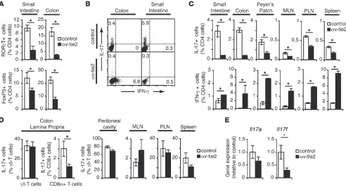
αv-Deficient mice lack intestinal Th17 cells due to loss of αv from myeloid cells. We first analyzed T cells isolated from the LP of αv-tie2 mice, which lack α v integrins in all hematopoietic cells (21). The propor- tion of Th17 cells (determined either by high expression of the tran-scription factor ROR-γT or by production of IL-17) was significantly reduced in the intestines of αv-tie2 mice, consistent with a role for αv integrins in Th17 cell development. Indeed, deletion of αv integrins had a more significant effect on Th17 cells (7-fold reduc-tion) than on FoxP3+ Tregs (3-fold reduction; Figure 1A). Similar
reductions in the proportions of Th17 cells were seen in lymphoid tissues, and in all cases the absolute numbers of Th17 cells were also reduced (data not shown). In contrast, IFN-γ–producing Th1
Conflict of interest: The authors have declared that no conflict of interest exists.
research article
cells were expanded in the intestine and lymphoid organs (Figure 1, B and C). Furthermore, other IL-17–producing lymphocyte popula-tions appeared to be largely unaffected by deletion of α v. In particu-lar, γδ T cells, a major source of IL-17 in vivo, were present in similar numbers in control and αv-tie2 mice (data not shown) and showed equivalent levels of IL-17 production (Figure 1D). Consequently, expression of Il17a was not significantly decreased in the intes-tine of αv-tie2 mice, although Il17f was reduced 3-fold, suggesting that Th17 cells contribute significantly to the production of this cytokine in the intestine (Figure 1E). We therefore concluded that deletion of αv integrins from immune cells led to loss of Th17 cells but did not affect other IL-17–producing cells.
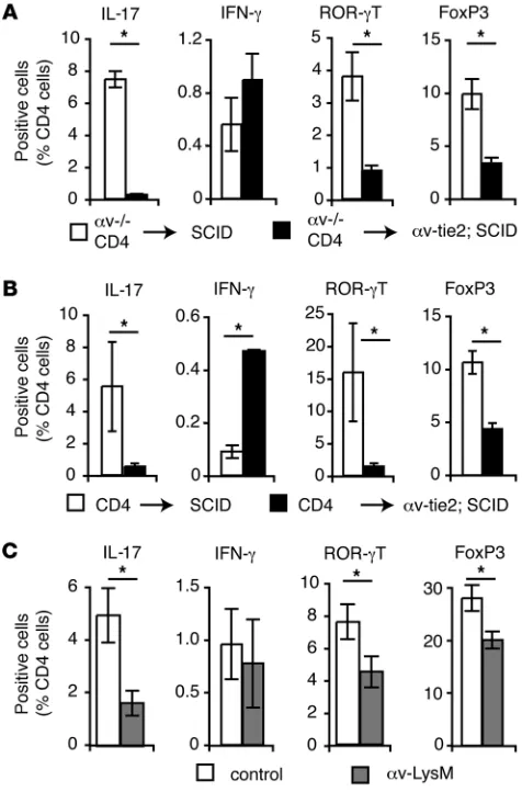
Loss of Th17 cells was not due to intrinsic defects in αv-deficient T cells, as CD4+ T cells from αv-tie2 mice differentiated normally
into Th17 cells when adoptively transferred into αv-expressing SCID mice (Figure 2A). Instead, αv expression by innate immune cells was required for differentiation of naive cells to Th17 cells, as T cells from wild-type mice did not become Th17 cells when trans-ferred to αv-tie2/ SCID recipients (Figure 2B). To determine whether this was due to deletion of αv from myeloid cells, we analyzed Th17 cells in αv-LysM mice, in which α v is deleted specifically from macro-phages, DCs, and neutrophils. Th17 cell numbers were significantly decreased in α v-LysM mice (Figure 2C), similar to our previous obser-vations that the generation of intestinal Tregs required αv expression by DCs (21). In addition, the proportion of IFN-γ–producing cells
was not markedly increased in these mice, suggesting that the loss of Th17 cells was not due to expansion of Th1 cells, as has been pro-posed in other studies (22). Together, these data show that Th17 cells were reduced in αv-deficient mice and that αv expression by myeloid cells was required to establish the intestinal Th17 population.
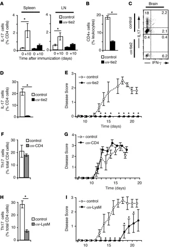
αv-Deficient mice do not develop pathological Th17 cells and are protected from EAE. In contrast to their protective role in mucosal immunity (8, 9), Th17 cells are implicated in immune-mediated diseases and are critical for development of EAE (10). To establish whether αv integrins were involved in generation of pathological Th17 cells, we tested whether α v-knockout mice were protected from EAE. Induc- tion of EAE by immunization with myelin oligodendrocyte glyco-protein–derived (MOG-derived) peptide in control mice resulted in generation of Th17 cells in the LNs and spleen (Figure 3A) with sub-sequent infiltration into the brain and associated paralysis (Figure 3, B–E). In contrast, immunization of α v-tie2 mice induced neither sig-nificant numbers of Th17 cells nor clinical signs of EAE (Figure 3). Thus, we concluded that αv-tie2 mice fail to generate pathological Th17 cells, resulting in complete protection from EAE.
[image:3.585.45.537.83.355.2]Building on these observations, we next assessed whether protec-tion from EAE was due to loss of αv from T cells or from myeloid cells. Mice in which αv was deleted from T cells (α v-CD4 mice) devel-oped brain-infiltrating Th17 cells and paralysis to the same degree as littermate controls, demonstrating that αv was not required on T cells for pathological Th17 development (Figure 3, F and G). Figure 1
Lack of intestinal Th17 cells in αv-tie2 mice. (A) Proportion of ROR-γT+ and FoxP3+ CD4+ cells in LP of control and αv-tie2 mice. (B and C)
Proportion of IL-17–producing (Th17 cells) and IFN-γ–producing (Th1 cells) CD4+ T cells isolated from the LP of the small intestine and colon,
Peyer’s patch, MLN, peripheral LN (PLN), and spleen of control and αv-tie2 mice. (B) Representative FACS data from one experiment. The plots are gated on CD4+ cells, and the numbers represent the percentage of CD4+ cells that stained for IL-17 or IFN-γ. (C) Mean ± SEM from at least
3 mice/group. (D) Proportion of IL-17–producing γδ and CD8α+ T cells isolated from the LP of the colon, and IL-17–producing γδ T cells from
In contrast, αv-LysM mice were protected from disease, with delayed development of paralysis (18 days post-immunization in αv-LysM mice compared with 12 days in controls) and reduced disease sever-ity when compared with controls (mean disease score at sacrifice of 1.4 ± 0.8 vs. 2.6 ± 0.3 in α v-LysM and controls, respectively; Fig-ure 3, H and I). Furthermore, Th17 cells were reduced 3-fold in the brain of αv-LysM mice with EAE, confirming that αv expression by myeloid cells was essential for generation of pathological Th17 cells. The protection was less than that seen with αv-tie2 mice, probably due to incomplete deletion of αv from myeloid cells in αv-LysM mice (21). These results demonstrate that generation of pathologi-cal Th17 cells and susceptibility to Th17-mediated disease required
αv expression by myeloid cells but not T cells.
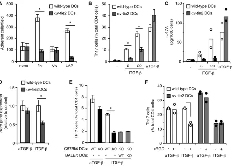
DC αv integrins activate latent TGF-β for generation of Th17 cells. We next set out to determine the mechanism by which myeloid cell αv integrins promoted Th17 cell generation. As we have reported previously, DCs appear to develop normally in αv-tie2 mice and migrate to tissues and LNs (ref. 21 and Supplemen-tal Figure 1; supplemental material available online with this article; doi:10.1172/JCI43796DS1). Furthermore, αv-deficient DCs produced normal levels of the Th17-promoting proinflam-matory cytokines IL-6 and IL-23 when stimulated in vitro, and these cytokines were expressed in the intestine of αv-tie2 mice at similar or higher levels than those in control mice (Supplemental Figures 1 and 2). It was therefore unlikely that loss of Th17 cells
was due to defects in antigen presentation or proinflammatory cytokine production by DCs. Instead, the defects in generation of both Th17 cells and Tregs in αv-tie2 mice implicated defects in TGF-β signaling. An important and nonredundant function of αv integrins in vivo is to bind latent TGF-β and catalyze con-version to an active form (18–20). Our previous work, and that of others, has suggested that DCs express αvβ8, which allows them to bind and activate TGF-β, which is then available for paracrine signaling to cognate T cells (21, 23). α v Integrin–mediated bind-ing to TGF-β occurs through an Arg-Gly-Asp (RGD) tripeptide in the LAP (24). We found that DCs were able to bind both LAP and the RGD-containing integrin ligand fibronectin, and this was dependent on αv expression (Figure 4A). To test whether DCs could activate TGF-β for signaling to T cells and promotion of Th17 differentiation, naive CD4+ T cells were co-cultured with
DCs in the presence of IL-6 with active or latent TGF-β . DCs cul-tured with T cells alone or in the presence of IL-6 alone did not generate significant numbers of Th17 cells, whereas addition of active TGF-β stimulated generation of Th17 cells by both wild-type and αv-deficient DCs to an equivalent degree, as assessed by IL-17 production and expression of rorc (Figure 4, B–D). In contrast, increasing concentrations of latent TGF-β stimulated significantly higher proportions of Th17 cells in cultures of wild-type DCs than those with αv-deficient DCs (Figure 4, B–D). Generation of Th17 cells was independent of T cell expression of αv, as αv-deficient T cells became Th17 cells in response to both active and latent TGF-β when co-cultured with wild-type DCs (data not shown). We next tested whether there needed to be a cognate interaction between the αv-expressing DC and the T cell, or whether bystander DCs that could not present antigen could nonetheless activate latent TGF-β and rescue the defect in αv knockouts. Using OT-2 TCR transgenic T cells, which respond to ovalbumin-derived peptide in the context of I-Ab, we
showed that DCs of C57BL/6 background (which express I-Ab)
could generate Th17 cells in the presence of latent TGF-β, and that this required αv expression by the DCs. This could not be rescued by addition of wild-type DCs of BALB/c background, which expressed αv but could not present antigen to OT-2 T cells (Figure 4E). Together these data indicate that αv expression by DCs is required for interacting T cells to respond to latent, but not active, TGF-β . We conclude that, during antigen presenta-tion, DC αv activates latent TGF-β, which can then signal to the interacting T cells and promote Th17 differentiation.
[image:4.585.42.280.83.444.2]RGD peptide mimetics inhibit Th17 differentiation and protect from EAE. Small molecule mimetics of RGD have been used clinically to inhibit integrin-mediated adhesion and signaling in tumors, principally glioblastoma (25). Using our in vitro co-culture sys-tem and a cyclic RGD (cRGD) peptide selective for αv integrins (26), we tested whether RGD mimetics might also block the abil-ity of αv expressed by DCs to bind and activate TGF-β, and hence Figure 2
αv Expression by myeloid cells is required for Th17 cell differentiation. (A and B) Tregs and Th17 and Th1 cells in the colonic LP of SCID control and αv-tie2/ SCID mice 6 weeks following adoptive transfer with splenic CD4+ T cells from αv-tie2 (A) or wild-type control (B) mice. Data
research article
prevent Th17 differentiation. cRGD had no effect on Th17 cell differentiation in the presence of active TGF-β in both wild-type and αv-deficient DCs. In contrast, wild-type DCs treated with cRGD no longer responded to latent TGF-β , reducing Th17 gen-eration to the levels seen when αv-deficient DCs were used (Figure 4F). cRGD had no effect on co-cultures of T cells and αv-deficient DCs, confirming that the cRGD was mediating its effects by inhi-bition of αv integrins on DCs. These results support a role for DC αv in TGF-β activation. Moreover, they demonstrate that the activation of TGF-β by DCs could be manipulated pharmacologi-cally by the addition of cRGD peptides.
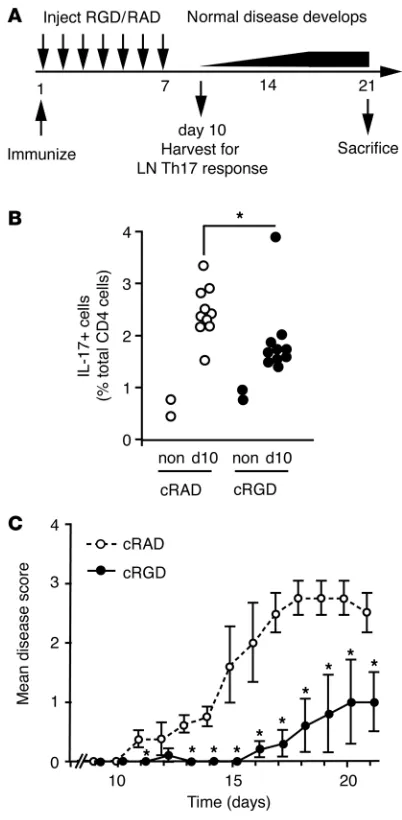
[image:5.585.43.392.81.591.2]To establish whether this pharmacological approach could be used therapeutically, we tested whether cRGD could block development of Th17-mediated pathology in vivo. Wild-type mice treated with cRGD for 1 week following immunization with MOG peptide generated fewer Th17 cells than mice treated with an inactive control peptide (Figure 5, A and B). Furthermore, cRGD-treated mice were protected from EAE; over half of the mice (3/5) developed no major signs of EAE, and the remaining mice developed disease that started later and was significantly less severe than in control mice (Figure 5C). Hence, cRGD administered during T cell differentiation
Figure 3
αv-tie2 mice are protected from EAE. (A) Percentage of CD4+ T cells that
express IL-17 in spleen and LNs of αv-tie2 and control mice before immu-nization (day 0) and 10 days after immunization with MOG peptide in CFA (+10). (B–D) Percentage of CD4+
cells (B) or IL-17– and IFN-γ –produc-ing cells (C and D) in leukocytes iso-lated from brain 21 days after immuni-zation. (C) representative FACS data gated on CD4+ cells. (D) Percentage
of CD4+ cells that expressed IL-17.
(E) Progression of EAE in control and αv-tie2 mice. Similar data were seen in 3 independent experiments. (F and
H) IL-17–producing T cells in the brain of αv-CD4 mice (F) and αv-LysM mice (H) 21 days after induction of EAE. (G
blocked Th17 differentiation and prevented later development of disease, consistent with inhibition of TGF-β activation by cRGD during initiation of Th17 responses.
Discussion
In summary, our data demonstrate a critical role for DC α v integ-rins in generating Th17 cells in normal homeostatic immunity or in pathological settings. We propose that this is due to the failure of αv-deficient DCs to activate latent TGF-β, which is required for T cell differentiation into Th17 cells and aTregs. Consequently, mice in which αv is deleted from myeloid cells did not develop Th17 cells in the intestine or in the periphery following immunization and were protected from Th17-mediated inflammatory disease. Furthermore, pharmacological inhibition of αv integrins impaired Th17 differentiation in vivo and ameliorated Th17-mediated pathology in a mouse model.
These studies complement and build on the work of others (23) and on our previous work (21), in which we demonstrated that αv integrins were required for generation of aTregs in the intestine. We previously postulated that α v integrins on myeloid cells are essen-tial for T cell responses to TGF-β by facilitating activation of latent TGF-β. Here we provide direct in vitro evidence for this hypothesis and demonstrate that this mechanism underlies DC control of gen-eration of both Tregs and Th17 cells. Th17 cells and aTregs appear to share a common precursor and a requirement for TGF-β signal-ing during early development, and the loss of both populations of cells in αv-deficient mice is consistent with this. However, our data show that αv deletion causes a greater reduction in Th17 cells than in FoxP3+ Tregs (Figures 1 and 2), which are only depleted in the
[image:6.585.46.516.81.418.2]intestine and are actually found at higher numbers in mesenteric LNs (MLNs) of αv-tie2 mice (21). This probably reflects the existence of a separate population of FoxP3+ Tregs (innate or natural Tregs)
Figure 4
research article
that develop independently of TGF-β signaling (27). However, it is also possible that aTregs are less dependent on DC-mediated TGF-β activation than Th17 cells due to greater sensitivity to TGF-β, as was proposed by Littman and colleagues (17), or to alternative mechanisms of TGF-β activation.
Th17 cells are increasingly implicated as pathogenic effectors in inflammatory disorders in humans, including rheumatoid arthritis, psoriasis, and multiple sclerosis, and our data support such a role, as αv-tie2 mice lacking Th17 cells are protected in an auto-inflammatory disease model, EAE. Th17 cells have also been implicated in inflammatory bowel disease (28, 29), and high levels of IL-17A and other Th17-derived cytokines are found in inflamed mucosa of patients with either Crohn disease or ulcerative coli-tis (30). Genome-wide association studies have also highlighted Th17-conditioning cytokine IL-23 and other genes in the Th17 pathway as potential contributors to inflammatory bowel disease (12, 31–34). However, αv-tie2 mice develop spontaneous colitis, as we described previously (21), and our data therefore show that Th17 cells are not required for intestinal inflammation in mice. Furthermore, a number of recent studies in mouse models sug-gest that Th17 cells may actually be protective against colitis and
act to maintain epithelial integrity during infection and immunity (7, 8, 35). It is possible, therefore, that the loss of Th17 cells in
αv-deficient mice contributes to the development of spontaneous colitis, which we had previously attributed principally to the loss of aTregs. In this regard, it is important to recognize that despite the lack of Th17 cells in αv-tie2 mice, other lymphocyte sources of IL-17 were largely unaffected by αv deletion, and IL-17A expression in the colon was not significantly changed. Hence, associations of inflammatory bowel disease with increased IL-17 do not neces-sarily implicate Th17 cells, and further studies with mice such as
αv-tie2, which lack Th17 cells rather than Th17-derived cytokines, will be important to understand the function of this complex T cell subset in disease.
In this study, we have identified αv integrin as a new component of the mechanism by which DCs generate Th17 cells. Although we have not directly addressed the identity of the β subunit that is required for generation of Th17 cells, conditional deletion of β8 in DCs similarly is associated with colitis and loss of intestinal Tregs (23), strongly implicating αvβ8 in activation of TGF-β and associ-ated Th17 cell generation by DCs. Our data further suggest that pharmacological disruption of this DC/αvβ8/TGF-β dialogue is a potential therapeutic option in Th17-mediated diseases.
Methods
Mice. αv-tie2 and αv-LysM mouse strains (21) were both backcrossed to C57BL/6 mice for at least 9 generations. αv-CD4 mice were generated by crossing C57BL/6 background (9 generations) αv-knockout and αv-floxed mice with C57BL/6 CD4 Cre transgenic mice (36). In all cases, experimen-tal mice carried 1 floxed and 1 knockout allele of Itgav with the respective CRE transgene; littermate mice with 1 floxed, 1 wild-type Itgav allele, and the CRE transgene were used as controls. For adoptive transfer experi-ments, αv-tie2 mice backcrossed 10 generations to BALB/c background were crossed into BALB/c SCID mice to generate αv-tie2; SCID mice and control SCID mice. CD4+ T cells were purified from spleens of control 6- to 10-week-old BALB/c mice by MACS, and 3 × 10⁶ CD4+ cells were injected intravenously into 6-week-old αv-tie2 SCID and littermate control mice. Mice were sacrificed 6 weeks later and intestines analyzed for T cell devel- opment. All animals were housed under specific pathogen–free condi-tions at Massachusetts General Hospital and Massachusetts Institute of Technology. Animal experiments were approved by the Subcommittee on Research Animal Care at Massachusetts General Hospital or the Division of Comparative Medicine at Massachusetts Institute of Technology.
Lymphocyte isolation. Lymphocytes were harvested from lymphoid tissue by physical disruption and filtering through 40-μm strainer in PBS/2% BSA/2 mM EDTA (MACS buffer). Splenocytes were depleted of rbc (rbc
lysis buffer; Sigma-Aldrich). For isolation of LP lymphocytes, small intes-Figure 5
αv Blockade inhibits Th17 cell differentiation and protects from EAE in vivo. (A) Schematic of experiments to assess effects of cRGD on EAE progression. Mice were immunized (day 1) and given daily injections of cRGD or control peptide for 7 days, and their immune response was assessed at day 10, with paralysis monitored until day 21. (B) Percentage of LN CD4+ T cells that expressed IL-17 ten days after
[image:7.585.60.261.81.491.2]tine and colon were dissected from fat and Peyer’s patches and washed to remove fecal content. Tissue was shaken three times for 15 minutes each time in HBSS/2 mM EDTA at 37°C to remove intraepithelial lymphocytes, digested in HBSS containing DNaseI (20 mg/ml) and Liberase (0.33 mg/ml; both from Roche) for 30 minutes, and incubated in PBS/2% BSA/10 mM EDTA. Leukocytes were purified using a 40%/80% percoll gradient (GE Healthcare), washed, and resuspended in MACS buffer or T cell medium for experiments. EAE . For induction of EAE, mice of more than 12 weeks age were immu-nized subcutaneously with 200 μ g MOG35–55 peptide (Hooke Labora-tories) emulsified in CFA (containing 400 μg Mycobacterium tuberculosis
H37Ra). Mice also received 250 ng of pertussis toxin (Hooke Laboratories) intraperitoneally at the time of immunization and 1 day later. For RGD blockade of αv integrins, 50 μg cRGD peptide in PBS (Enzo Life Sciences) was injected subcutaneously daily from immunization until day 7. The con-trol group received 50 μg of cRAD peptide instead. Mice were monitored daily for clinical signs of paralysis and scored as follows: 0, no disease; 1, limp tail; 2, weak/partially paralyzed hind legs; 3, limp tail and complete paralysis of hind legs; 4, complete hind and partial front leg paralysis; 5, complete paralysis/death. Female mice were used for EAE studies, except in experiments with αv-CD4 CRE, in which predominantly male mice were used, which resulted in a slightly accelerated and more variable disease than with females.
Isolation of brain- infiltrating cells . Brains from mice with EAE were collect-ed in ice-cold HBSS/10% fetal bovine serum, disrupted through a 100-μm strainer, and washed, and leukocytes were purified using a 37%/70% per-coll gradient. Mononuclear cells collected from the 37%/70% interface were washed with HBSS/serum and used immediately for experiments. Cells from individual brains were processed separately.
In vitro T cell differentiation. Naive CD4+CD25– T cells were sorted by mag-netic separation using CD4+CD25+ regulatory T cell selection kit (Stem Cell Technologies). Purity of more than 95% was routinely achieved. T cells were cultured in X-VIVO15 medium (Lonza) supplemented with 100 U/ml penicillin, 100 μg/ml streptomycin, 50 μM 2-β-mercaptoethanol for all experiments except RGD blockade, which was carried out in RPMI 1640 supplemented with 10% fetal bovine serum, 2 mM L-glutamine, 100 U/ml penicillin, 100 μg/ml streptomycin, and 50 μM 2-β-mercaptoethanol. Splenic DCs were sorted to more than 95% purity using CD11c microbeads (Miltenyi Biotech). Cells were cultured in 2 ml volume containing 1 × 10⁵ DCs and 2.5 × 10⁵ naive CD4+ T cells in the presence of 0.5 μg/ml anti-CD3 (145-2C11; BD Biosciences). Additional cytokines used were recombinant active TGF-β1 (1 ng/ml), purified human latent TGF-β (20 ng/ml), and IL-6 (20 ng/ml) (all R&D Systems). For antigen-specific T cell stimulation, 1 × 10⁵ DCs from mice on a C57BL/6 background were incubated with 2.5 × 10⁵ naive CD4+ T cells from OT-2 TCR transgenic mice, with 100 nM OVA323–339 peptide and cytokines, in the presence or absence of 1 × 10⁵ DCs from BALB/c-background mice. For RGD blockade experiments, cRGD peptide was added at 2 μg/ml. Cells were cultured for 4 or 5 days before analysis of Th17 differentiation. IL-17A production was measured by intracellular cytokine staining, as described below, or by IL-17A release
into the culture supernatant, measured using cytokine bead array (BD Biosciences). Expression of the Th17-specific transcription factor was measured by quantitative real-time PCR (QRT-PCR), as described below.
Flow cytometry and intracellular cytokine staining. For intracellular cytokine staining, cells obtained from dissection of LP, mesenteric and peripheral LNs, Peyer’s patches, spleens, in vitro cultures, or brains were resuspended in T cell medium and stimulated for 5 hours with 10 ng/ml PMA/750 ng/ml ionomycin in the presence of intracellular transport inhibitors (1 μg/ml GolgiPlug; BD Biosciences). Cells were treated with Fc-blocking mAbs and surface stained with anti-CD4–APC (RM4-5), anti-CD8α–PerCP (53-6.7), or anti–TCR-γ–APC (GL3), followed by intracellular cytokine staining with anti–IL-17–PE (TC11-18H10), anti–IFN-γ–FITC (XMG1.2), anti-RORγ–PE (AFKJS-9), and anti-Foxp3–FITC (FJK-16s) according to the manufactur-er’s instructions (BD Biosciences or eBioscience). Data were acquired using FACSCalibur instrument (BD Biosciences) and analyzed using FlowJo soft- ware (Tree Star Inc.). Percentage and absolute numbers of cytokine-express-ing cells within the CD4+, CD8+, or TCR-γ+ populations were determined.
QRT-PCR. RNA was prepared from 0.5 cm of the proximal colon of αv-tie2 mice and littermate controls of 6–8 weeks of age or from cultured T cells at day 4 of culture in Th17-generating conditions using Trizol (Invitrogen). cDNA was synthesized using reverse transcription reagent (Applied Bio-systems). cDNA was analyzed by Fast start SYBR Green reagents (Roche) using a RealPlex Mastercycler (Eppendorf) and the inflammatory cytokine and receptors rtPCR array (SA Biosciences) or the following primers: Il17a
5′-GAAGATGCTGGTGGGTGTGG-3′ and 5′ AGCCGCGGGTCTCT-GTTTAG-3′; Il17f 5′-TGAATTCCAGAACGCTCCA-3′ and 5′ -TCTTCCT-GACCCTGGGCATT-3′; Rorc 5′-CCGCTGAGAGGGCTTCAC-3′ and 5′-TGCAGGAGTAGGCCACATTACA-3′. Expression levels of each gene were normalized to β-actin expression.
Acknowledgments This work was supported by grants from the Hood Pediatric Research Foundation (to A. Lacy-Hulbert) and the Howard Hughes Medical Institute (R.O. Hynes) and by a Wellcome Trust Travelling Fellowship (to S. Mukhopadhyay). Helena Païdassi was supported by MRC grant G0802069 to John Savill, University of Edinburgh, who we thank for support and helpful discussion. Received for publication May 24, 2010, and accepted in revised form September 29, 2010.
Address correspondence to: Adam Lacy-Hulbert, Program of Developmental Immunology, Massachusetts General Hospital GRJ1402, 55 Fruit Street, Boston, Massachusetts 02114, USA. Phone: 617.643.5346; Fax: 617.724.3248; E-mail: alacy-hulbert@ partners.org. Stephan Kissler’s present address is: Rudolf Virchow Center, DFG Center for Experimental Biomedicine, University of Würzburg, Würzburg, Germany. 1. Harrington LE, et al. Interleukin 17-producing CD4+ effector T cells develop via a lineage distinct from the T helper type 1 and 2 lineages. Nat Immu-nol. 2005;6(11):1123–1132.
2. Park H, et al. A distinct lineage of CD4 T cells regu-lates tissue inflammation by producing interleukin 17. Nat Immunol. 2005;6(11):1133–1141. 3. Korn T, Bettelli E, Oukka M, Kuchroo VK. IL-17 and
Th17 cells. Annu Rev Immunol. 2009;27:485–517. 4. Steinman L. A brief history of T(H)17, the first
major revision in the T(H)1/T(H)2 hypothesis of T cell-mediated tissue damage. Nat Med. 2007;
13(2):139–145.
5. Ivanov II, et al. The orphan nuclear receptor RORgammat directs the differentiation program of proinflammatory IL-17+ T helper cells. Cell. 2006;126(6):1121–1133.
6. Ivanov II, et al. Specific microbiota direct the dif-ferentiation of IL-17-producing T-helper cells in the mucosa of the small intestine. Cell Host Microbe. 2008;4(4):337–349.
7. Ivanov II, et al. Induction of intestinal Th17 cells by segmented filamentous bacteria. Cell. 2009;139(3):485–498.
8. Zenewicz LA, Yancopoulos GD, Valenzuela DM, Murphy AJ, Stevens S, Flavell RA. Innate and adaptive interleukin-22 protects mice from inflammatory bowel disease. Immunity. 2008; 29(6):947–957.
9. O’Connor W Jr, et al. A protective function for interleukin 17A in T cell-mediated intestinal inflammation. Nat Immunol. 2009;10(6):603–609.
11. Lock C, et al. Gene-microarray analysis of mul-research article
tiple sclerosis lesions yields new targets validated in autoimmune encephalomyelitis. Nat Med. 2002;8(5):500–508.
12. Duerr RH, et al. A genome-wide association study identifies IL23R as an inflammatory bowel disease gene. Science. 2006;314(5804):1461–1463. 13. Lee YK, et al. Late developmental plasticity in the T
helper 17 lineage. Immunity. 2009;30(1):92–107. 14. Veldhoen M, Hocking RJ, Atkins CJ, Locksley RM,
Stockinger B. TGFbeta in the context of an inflam- matory cytokine milieu supports de novo differen-tiation of IL-17-producing T cells. Immunity. 2006; 24(2):179–189.
15. Mangan PR, et al. Transforming growth factor-beta induces development of the T(H)17 lineage. Nature. 2006;441(7090):231–234.
16. Bettelli E, et al. Reciprocal developmental pathways for the generation of pathogenic effector TH17 and regulatory T cells. Nature. 2006;441(7090):235–238. 17. Zhou L, et al. TGF-beta-induced Foxp3 inhibits
T(H)17 cell differentiation by antagonizing ROR-gammat function. Nature. 2008;453(7192):236–240. 18. Munger JS, et al. The integrin alpha v beta 6 binds
and activates latent TGF beta 1: a mechanism for regulating pulmonary inflammation and fibrosis.
Cell. 1999;96(3):319–328.
19. Aluwihare P, et al. Mice that lack activity of {alpha}-v{beta}6- and {alpha}v{beta}8-integrins reproduce the abnormalities of Tgfb1- and Tgfb3-null mice.
J Cell Sci. 2009;122(pt 2):227–232.
20. Yang Z, et al. Absence of integrin-mediated TGFbeta1 activation in vivo recapitulates the phenotype of TGF-beta1-null mice. J Cell Biol. 2007;176(6):787–793.
21. Lacy-Hulbert A, et al. Ulcerative colitis and autoim-munity induced by loss of myeloid alpha (v) integrins.
Proc Natl Acad Sci U S A. 2007;104(40):15823–15828.
22. Das J, et al. Transforming growth factor beta is dis-pensable for the molecular orchestration of Th17 cell differentiation. J Exp Med. 2009;206(11):2407–2416. 23. Travis MA, et al. Loss of integrin alpha(v)beta(8) on
dendritic cells causes autoimmunity and colitis in mice. Nature. 2007;449(7160):361–365.
24. Munger JS, Harpel JG, Giancotti FG, Rifkin DB. Interactions between growth factors and integrins: latent forms of transforming growth factor-beta are ligands for the integrin alphavbeta1. Mol Biol Cell. 1998;9(9):2627–2638.
25. Reardon DA, Nabors LB, Stupp R, Mikkelsen T. Cilengitide: an integrin-targeting arginine-glycine-aspartic acid peptide with promising activity for glioblastoma multiforme. Expert Opin Investig Drugs. 2008;17(8):1225–1235.
26. Kumagai H, Tajima M, Ueno Y, Giga-Hama Y, Ohba M. Effect of cyclic RGD peptide on cell adhesion and tumor metastasis. Biochem Biophys Res Commun. 1991; 177(1):74–82.
27. Bluestone JA, Abbas AK. Natural versus adaptive reg-ulatory T cells. Nat Rev Immunol. 2003;3(3):253–257.
28. Liu ZJ, Yadav PK, Su JL, Wang JS, Fei K. Potential role of Th17 cells in the pathogenesis of inflam-matory bowel disease. World J Gastroenterol. 2009; 15(46):5784–5788.
29. Abraham C, Cho J. Interleukin-23/Th17 pathways and inflammatory bowel disease. Inflamm Bowel Dis. 2009;15(7):1090–1100.
30. Fujino S, et al. Increased expression of interleu-kin 17 in inflammatory bowel disease. Gut. 2003; 52(1):65–70.
31. Barrett JC, et al. Genome-wide association defines more than 30 distinct susceptibility loci for Crohn’s disease. Nat Genet. 2008;40(8):955–962.
32. Fisher SA, et al. Genetic determinants of ulcerative colitis include the ECM1 locus and five loci implicat-ed in Crohn’s disease. Nat Genet. 2008;40(6):710–712. 33. Franke A, et al. Sequence variants in IL10, ARPC2 and multiple other loci contribute to ulcerative coli-tis susceptibility. Nat Genet. 2008;40(11):1319–1323. 34. Franke A, et al. Replication of signals from recent
studies of Crohn’s disease identifies previously unknown disease loci for ulcerative colitis. Nat Genet. 2008;40(6):713–715.
35. Ahern PP, Izcue A, Maloy KJ, Powrie F. The inter-leukin-23 axis in intestinal inflammation. Immunol Rev. 2008;226:147–159.