GABA interneurons mediate the rapid
antidepressant-like effects of scopolamine
Eric S. Wohleb, … , Meenakshi Alreja, Ronald S. Duman
J Clin Invest.
2016;
126(7)
:2482-2494.
https://doi.org/10.1172/JCI85033
.
Major depressive disorder (MDD) is a recurring psychiatric illness that causes substantial
health and socioeconomic burdens. Clinical reports have revealed that scopolamine, a
nonselective muscarinic acetylcholine receptor antagonist, produces rapid antidepressant
effects in individuals with MDD. Preclinical models suggest that these rapid antidepressant
effects can be recapitulated with blockade of M1-type muscarinic acetylcholine receptors
(M1-AChR); however, the cellular mechanisms underlying activity-dependent synaptic and
behavioral responses to scopolamine have not been determined. Here, we demonstrate that
the antidepressant-like effects of scopolamine are mediated by GABA interneurons in the
medial prefrontal cortex (mPFC). Both GABAergic (GAD67
+) interneurons and
glutamatergic (CaMKII
+) interneurons in the mPFC expressed M1-AChR. In mice,
viral-mediated knockdown of M1-AChR specifically in GABAergic neurons, but not glutamatergic
neurons, in the mPFC attenuated the antidepressant-like effects of scopolamine.
Immunohistology and electrophysiology showed that somatostatin (SST) interneurons in the
mPFC express M1-AChR at higher levels than parvalbumin interneurons. Moreover,
knockdown of M1-AChR in SST interneurons in the mPFC demonstrated that M1-AChR
expression in these neurons is required for the rapid antidepressant-like effects of
scopolamine. These data indicate that SST interneurons in the mPFC are a promising
pharmacological target for developing rapid-acting antidepressant therapies.
Research Article
Neuroscience
Find the latest version:
Introduction
Major depressive disorder (MDD) is a recurring neuropsychiatric ill-ness that affects up to 17% of the population and causes substantial social and economic burdens (1–4). While some patients respond to existing medications, currently available antidepressants take weeks to months to have an effect, and many patients are consid-ered treatment resistant because they fail to respond to 2 or more antidepressants (5). Recent studies demonstrate that scopolamine, a nonselective muscarinic acetylcholine (ACh) receptor (mAChR) antagonist, and ketamine, an NMDA receptor antagonist, produce rapid antidepressant actions (within hours) and are effective even in treatment-resistant MDD patients (6–8). The rapid antidepres-sant actions of scopolamine and ketamine are dependent on glu-tamate release and induction of new spine synapses in the medial prefrontal cortex (mPFC) (9–11), effects that directly target the syn-aptic pathophysiology of MDD and chronic stress (12–19).
However, a major question in the field is, what are the initial cellular targets that mediate the increase in glutamate transmis-sion, leading to increased synapse formation and rapid antide-pressant behavior (10, 20, 21)? One hypothesis is that scopol-amine blocks muscarinic receptors on GABAergic interneurons, resulting in disinhibition of pyramidal neurons and increased glu-tamate transmission. Alternatively, scopolamine may act directly on pyramidal neurons to enhance synaptic plasticity and produce antidepressant behavioral responses. Recent studies suggest that the M1-type muscarinic ACh receptor (M1-AChR) subtype may mediate the antidepressant actions of scopolamine (9, 22, 23).
M1-AChRs are expressed on both GABAergic interneurons and glutamatergic pyramidal neurons in the mPFC and regulate the activity of both cell types (24–26).
In the present study, we used transgenic mice and viral-mediated gene transfer to drive Cre-dependent expression of M1-AChR shRNA in different subtypes of GABA interneurons or glutamate neurons in the mPFC. The results demonstrate that M1-AChR knockdown in GABA interneurons, but not pyramidal neurons, blocks the antidepressant-like response to scopolamine in mouse behavioral models. In addition, we show that knock-down of M1-AChR in somatostatin (SST), but not parvalbumin (PV), interneurons, blocks the actions of scopolamine. These findings reveal that M1-AChRs on SST interneurons in the mPFC are a critical cellular target underlying the rapid antidepressant effects of scopolamine.
Results
CaMKII+ and GAD+ cell type–specific knockdown of M1-AChR in the
mPFC. To determine whether glutamatergic pyramidal neurons
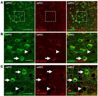
and GABAergic interneurons in the mPFC express M1-AChR, dou-ble immunohistology was conducted with an M1-AChR antibody and Ca2+/calmodulin-dependent kinase II (CaMKII; pyramidal cell) or glutamate decarboxylase-67 (GAD67; GABA interneuron) (25). The results show that CaMKII+ pyramidal neurons display punctate M1-AChR labeling in the mPFC, and higher magnification shows the presence of M1-AChR+ neurons that lack CaMKII labeling (Fig-ure 1, A and B). Double labeling with GAD67 shows that many of the CaMKII– neurons were GABA interneurons (Figure 1C).
For cell-specific M1-AChR knockdown, we used a construct that expresses shRNA targeting M1-AChR (M1shRNA) in a Cre-recombinase–dependent manner (Supplemental Figure 1A). Both
Major depressive disorder (MDD) is a recurring psychiatric illness that causes substantial health and socioeconomic burdens. Clinical reports have revealed that scopolamine, a nonselective muscarinic acetylcholine receptor antagonist, produces rapid antidepressant effects in individuals with MDD. Preclinical models suggest that these rapid antidepressant effects can be recapitulated with blockade of M1-type muscarinic acetylcholine receptors (M1-AChR); however, the cellular mechanisms underlying activity-dependent synaptic and behavioral responses to scopolamine have not been determined. Here, we demonstrate that the antidepressant-like effects of scopolamine are mediated by GABA interneurons in the medial prefrontal cortex (mPFC). Both GABAergic (GAD67+) interneurons and glutamatergic (CaMKII+) interneurons in the mPFC expressed
M1-AChR. In mice, viral-mediated knockdown of M1-AChR specifically in GABAergic neurons, but not glutamatergic neurons, in the mPFC attenuated the antidepressant-like effects of scopolamine. Immunohistology and electrophysiology showed that somatostatin (SST) interneurons in the mPFC express M1-AChR at higher levels than parvalbumin interneurons. Moreover, knockdown of M1-AChR in SST interneurons in the mPFC demonstrated that M1-AChR expression in these neurons is required for the rapid antidepressant-like effects of scopolamine. These data indicate that SST interneurons in the mPFC are a promising pharmacological target for developing rapid-acting antidepressant therapies.
GABA interneurons mediate the rapid
antidepressant-like effects of scopolamine
Eric S. Wohleb,1 Min Wu,1 Danielle M. Gerhard,2 Seth R. Taylor,1 Marina R. Picciotto,1 Meenakshi Alreja,1 and Ronald S. Duman1
1Department of Psychiatry, Yale University School of Medicine, New Haven, Connecticut, USA. 2Department of Psychology, Yale University, New Haven, Connecticut, USA.
Conflict of interest: The authors have declared that no conflict of interest exists. Submitted: October 8, 2015; Accepted: April 14, 2016.
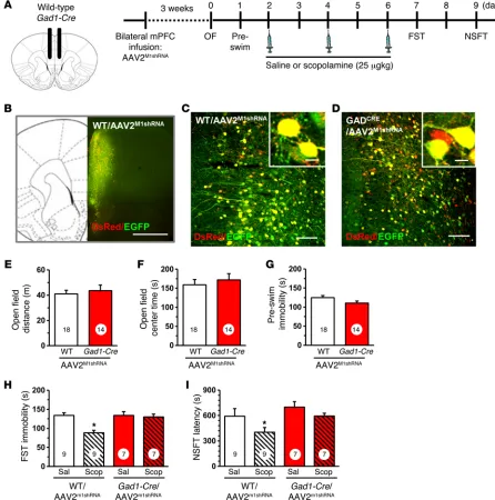
Behavioral studies using the same design demonstrate that infusions of AAV2M1shRNA into Gad1-Cre mice had no significant effects on baseline behaviors in the open field test (total dis-tance and center time, anxiety measure) or forced swim test (FST) (immobility, behavioral despair measure) compared with those of WT controls receiving the same viral infusions (Figure 2, E–G). Mice then received 3 doses of scopolamine (25 μg/kg, i.p.) every other day, a dosing regimen based on the schedule used in depressed human subjects and shown to produce robust signaling and behavioral responses in rodents (23). In the FST, scopolamine significantly decreased immobility time in WT mice infused with AAV2M1shRNA, but these effects were completely blocked in
Gad1-Cre mice receiving AAV2M1shRNA infusions into the mPFC (interac-tion, F1,28 = 8.17, P < 0.0008; Figure 2H). In the novelty suppressed feeding test (NSFT), scopolamine decreased latency to feed in WT mice infused with AAV2M1shRNA, but again had no effect in
Gad1-Cre mice receiving the same viral infusions (main effect of
scopol-amine, F1,28 = 6.44, P < 0.02; main effect of genotype, F1,28 = 3.88,
P < 0.06; interaction, F1,28 = 2.71, P < 0.11; Figure 2I).
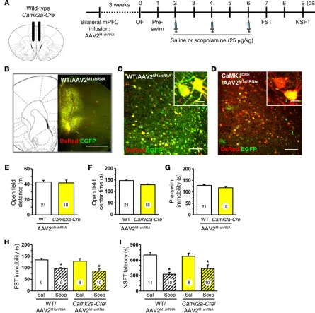
Similar studies were conducted in Camk2a-Cre mice with infu-sions of AAV2M1shRNA in the mPFC (Figure 3, A and B). There was no recombination observed in WT mice infused with AAV2M1shRNA (Figure 3, B and C), but in Camk2a-Cre mice, the majority of neu-rons showed recombination (DsRed+ only) due to the high density of glutamatergic pyramidal neurons in mPFC (Figure 3D). Behav-ioral testing showed that AAV2M1shRNA had no effect on baseline behavior in the open field test (distance or center time) or immobil-ity in the FST (Figure 3, E–G). Scopolamine administration (3-dose regimen) significantly decreased immobility in the FST in both WT DsRed and EGFP are expressed unless recombination deletes
EGFP and allows expression of only DsRed and M1shRNA. To test this construct, N2a cells were cotransfected with Cre-expressing (CAG-Cre) and M1shRNA plasmids (pM1shRNA). Transfection with pM1shRNA alone resulted in EGFP and DsRed expression (Supplemental Figure 1B), while cotransfection with CAG-Cre and pM1shRNA resulted in recombination, shown by reduced EGFP expression and retained DsRed expression (Supplemental Figure 1C). Cotransfection with CAG-Cre decreased EGFP mRNA lev-els in control and pM1shRNA cultures (main effect, F1,14 = 38.86,
P < 0.0001, Supplemental Figure 1D) and, importantly, resulted in
robust knockdown of M1-AChR mRNA (interaction, F1,22 = 16.07,
P < 0.0006, Supplemental Figure 1E). There was no significant
effect on mAChR subtypes (mAChR2–5), demonstrating specific-ity of the M1shRNA (Supplemental Figure 1F).
Knockdown of M1-AChR in GAD+, but not CaMKII+, neurons
blocks the antidepressant effects of scopolamine. pM1shRNA was
[image:3.585.40.357.54.366.2]packaged into an adeno-associated virus-2 (AAV2M1shRNA) and infused bilaterally into the mPFC of Gad1-Cre and Camk2a-Cre mice or their WT littermate controls. The mPFC was targeted, as prior studies show direct infusion of scopolamine into this region caused antidepressant actions. Initial studies were conducted in Gad1-Cre mice, and 3 weeks after viral infusion, brains were collected for histology (Figure 2A). Figure 2B shows the spread of AAV2M1shRNA infusion in the mPFC, and higher magnification shows that there was no observed recombination in WT mice (Fig-ure 2C). However, infusion with AAV2M1shRNA into Gad1-Cre mice resulted in a subset of cells displaying DsRed only (Figure 2C), demonstrating cell-specific expression of the M1shRNA construct.
Figure 1. M1-AChR colocalized with CaMKII+ and GAD67+ neurons in the mPFC. WT mice were perfused,
M1-AChR knockdown in GAD+ interneurons blocks
scopol-amine stimulation of FosB. Prior studies indicate that scopolscopol-amine
increases Fos labeling in the mPFC, further evidence of increased glutamate neurotransmission and neuronal activation (23). To determine whether M1-AChR knockdown blocks scopolamine and Camk2a-Cre mice infused with AAV2M1shRNA (main effect, F
[image:4.585.65.511.58.508.2]1,32 = 18.58, P < 0.0001, Figure 3H). Similar results were observed in the NSFT, where scopolamine administration reduced the latency to feed in both WT and Camk2a-Cre mice receiving AAV2M1shRNA infu-sion (main effect, F1,35 = 27.28, P < 0.0001, Figure 3I).
Figure 2. Infusion of AAV2M1shRNA in mPFC of Gad1-Cre mice blocked the antidepressant effects of scopolamine. WT or Gad1-Cre mice received bilateral
infusion of AAV2M1shRNA in the mPFC. After 3 weeks, mice were tested for baseline behavioral changes, then received saline or scopolamine, followed by
additional behavioral tests. (A) Schematic showing experimental approach and time line. A subset of mice were perfused, and brains were processed for histology. Representative confocal images of fluorescence in the mPFC are shown. OF, open field. (B) Low magnification image in WT/AAV2M1shRNA mouse
to show representative infusion in mPFC. Original magnification, ×4. Scale bar: 1 mm. (C) Representative image of AAV2M1shRNA in prelimbic mPFC of WT
mouse. Original magnification, ×20. Inset shows all neurons have colocalization of eGFP (green) and DsRed (red) fluorescence. Scale bars: 100 μm; 10 μm (inset). (D) Representative image of AAV2M1shRNA in prelimbic mPFC of Gad1-Cre mouse. Original magnification, ×20. Scale bar: 100 μm. Inset shows some
neurons was observed in both the infralimbic and prelimbic cor-tices of WT/AAV2M1shRNA, but not in Gad1-Cre/AAV2M1shRNA mice (infralimbic, interaction, F1,14 = 17.51, P < 0.0009; prelimbic, inter-action, F1,14 = 4.22, P < 0.06; Figure 4, B and C). Studies of
Camk2a-Cre mice showed that scopolamine induction of FosB+ neurons was observed in both WT/AAV2M1shRNA and Camk2a-Cre/AAV2M1shRNA mice (infralimbic, main effect, F1,12 = 83.09, P < 0.0001; prelim-induction of neuronal activation in the mPFC, we examined
FosB immunohistology (27). WT and Gad1-Cre mice infused with AAV2M1shRNA received a final injection of scopolamine (25 μg/kg), and then 1 hour later, brains were collected and processed for FosB
immunohistology. Scopolamine increased the number of FosB+
[image:5.585.69.514.57.500.2]neurons in the mPFC of WT, but not Gad1-Cre, mice following AAV2M1shRNA infusions (Figure 4A). Scopolamine induction of FosB+
Figure 3. Infusion of AAV2M1shRNA in mPFC of Camk2a-Cre mice did not influence the antidepressant effects of scopolamine. WT or Camk2a-Cre mice
received bilateral infusion of AAV2M1shRNA in the mPFC. After 3 weeks, mice were tested for baseline behavioral changes, then received saline or
scopol-amine, followed by additional behavioral tests. (A) Schematic showing experimental approach and time line. A subset of mice were perfused, and brains were processed for histology. Representative confocal images of fluorescence in the mPFC are shown. (B) Low-magnification image in WT/AAV2M1shRNA
mouse to show representative infusion in mPFC. Original magnification, ×4. Scale bar: 1 mm. (C) Representative image of AAV2M1shRNA in prelimbic mPFC of
WT mouse. Original magnification, ×20. Inset shows all neurons have colocalization of EGFP (green) and DsRed (red) fluorescence. Scale bars: 100 μm; 10 μm (inset). (D) Representative image of AAV2M1shRNA in prelimbic mPFC of Camk2a-Cre mouse. Original magnification, ×20. Inset shows that many
tdTomato interneurons showed inward current responses. We also found that SST-tdTomato interneurons displayed increased excit-atory postsynaptic currents (EPSCs) after muscarine application, as a result of excitatory inputs from pyramidal neurons (Figure 6B). Prior application of the mACh receptor antagonist telenzepine blocked both muscarine-induced inward current (musc: mean = 40 ± 9.6 pA; telenzepine + musc: mean = 0 ± 0 pA, Figure 6B) and EPSCs in SST-tdTomato interneurons (Figure 6C).
To confirm neuron subtype-specific knockdown of M1-AChR, SST interneurons from Sst-Cre/AAV2M1shRNA mice were recorded (Figure 6D). DsRed+ SST interneurons in these mice had rest-ing membrane potentials equal to –77.3 ± 2.8 mV, comparable to SST-tdTomato interneurons shown above. In Sst-Cre/AAV2M1shRNA mice, a minimal muscarine-induced inward current was observed in 1 out of 6 (17%) interneurons tested. Muscarine-induced EPSCs were observed in 5 out of 6 (83%) interneurons tested (Figure 6E), indicating that synaptic inputs onto SST interneurons were not affected in Sst-Cre/AAV2M1shRNA mice. These EPSCs were blocked when telenzepine was applied prior to muscarine (Figure 6E). Collectively, these data show that cholinergic stimulation of SST interneurons in the mPFC is mediated by M1-AChR and that AAV2M1shRNA effectively blocked muscarinic stimulation of SST interneurons. Knockdown of M1-AChR was confirmed by immu-nolabeling studies, which showed that infusion of AAV2M1shRNA into Sst-Cre mice significantly reduced M1-AChR immunolabel-ing compared with that of Sst-Cre mice infused with a scrambled shRNA control (AAV2SCR) (P < 0.02, Supplemental Figure 2).
Knockdown of M1-AChR in SST, but not PV, interneurons pre-vents the antidepressant-like effects of scopolamine. To determine
the GABA ergic interneuron subtype that mediates the behav-ioral responses to scopolamine, WT, Sst-Cre, and PV-Cre mice received bilateral infusion of AAV2M1shRNA into the mPFC and were analyzed 3 weeks later (Figure 7A). Infusion of AAV2M1shRNA into the mPFC of Sst-Cre mice resulted in recombination in a subset of neurons, shown by expression of only DsRed (Figure 7, B and C). AAV2M1shRNA infusion in Sst-Cre mice produced no significant bic, main effect, F1,12 = 67.52, P < 0.0001; Supplemental Figure
3). Knockdown of M1-AChR was confirmed by immunolabeling studies that showed that Camk2a-Cre/AAV2M1shRNA significantly reduced M1-AChR immunolabeling in pyramidal neurons com-pared with WT/AAV2M1shRNA mice (P < 0.02, Supplemental Figure 2). These findings provide further evidence for functional knock-down of M1-AChR in Gad1-Cre mice and blockade of scopolamine-induced neuronal activity.
PV and SST interneurons show varied distribution and expression of M1-AChR in the mPFC. GABAergic interneurons in the PFC have
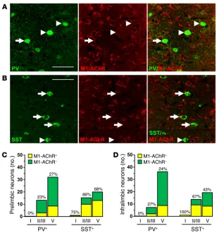
diverse physiological properties and can be categorized into sub-types based on expression of calcium-binding proteins, such as PV, and neuropeptides, such as SST. Each interneuron subtype has a dynamic role in modulating the activity of glutamatergic pyrami-dal cells based on firing rates and synaptic connections (28, 29). To further characterize the role of GABA interneurons in the mPFC, we used double immunohistology to determine distribution and colocalize expression of M1-AChR on PV and SST interneuron subtypes (Figure 5, A and B). PV interneurons were concentrated in deeper cortical layers (III–V), while SST interneurons were dis-persed evenly across cortical layers (Supplemental Figure 4 and Figure 5D). Further analyses showed that M1-AChR is more fre-quently colocalized with SST compared with PV interneurons, with approximately 62% of SST and 24% of PV interneurons colocalized with M1-AChR across both mPFC regions (Figure 5, C and D).
M1-AChR mediates cholinergic stimulation of SST interneurons in the mPFC. To determine the role of M1-AChR in SST interneuron
responses to cholinergic stimulation, brain slice electrophysiology was performed with SST-tdTomato mice. ACh or muscarine was applied to mPFC SST interneurons in mPFC slices under whole-cell voltage clamp (layer II/III and V). The SST-tdTomato interneu-rons had a mean resting membrane potential equal to –75.4 ± 1.4 mV, and ACh induced an inward current (mean = 85.7 ± 17.8 pA). Muscarine application also produced an inward current (mean = 43.3 ± 9.9 pA), while nicotine application caused no change in tdTomato interneuron currents (Figure 6A). In all, 92% of
SST-Figure 4. M1-AChR knockdown in Gad1-Cre
mice decreased FosB activation fol-lowing scopolamine treatment. WT or Gad1-Cre mice received bilateral infusion of AAV2M1shRNA in the mPFC. Following
behavioral tests, mice received an acute scopolamine injection (25 μg/kg) and were perfused 1 hour later. Brains were collected and processed for immunohistology. (A) Representative images of FosB labeling in the prelimbic mPFC of WT/AAV2M1shRNA
and Gad1-Cre/AAV2M1shRNA mice treated with
saline or scopolamine. Scale bar: 100 μm. (B) Quantification of FosB+ neurons in the
[image:6.585.35.383.55.277.2]changes in baseline behavior other than a small increase in pre-swim immobility compared with that of WT mice (P < 0.003; Supplemental Figure 5, B–D). In littermate WT/AAV2M1shRNA mice, scopolamine reduced immobility in the FST as expected, but this effect was completely absent in Sst-Cre/AAV2M1shRNA mice (interaction, F1,27 = 7.95, P < 0.009; Figure 7E). In the NSFT, sco-polamine decreased latency to feed in WT/AAV2M1shRNA mice, but had no effect in Sst-Cre/AAV2M1shRNA mice (interaction, F
1,26 = 3.75,
P = 0.06, Figure 7F). In PV-Cre mice, infusion of AAV2M1shRNA resulted in recombination in a subset of neurons, shown by expres-sion of DsRed only (Figure 7D). M1-AChR knockdown in PV-Cre mice caused no baseline behavioral effects in either the open field test or FST (Supplemental Figure 5, E–G) and had no effect on the antidepressant-like actions of scopolamine in the FST (main effect, F1,23 = 22.35, P < 0.0001, Figure 7G) or NSFT (main effect, F1,24 = 13.12, P < 0.001, Figure 7H). Following behavioral testing, studies were conducted to determine whether M1-AChR knock-down in SST interneurons blocks scopolamine induction of FosB. In WT/AAV2M1shRNA mice, scopolamine increased FosB+ staining in the mPFC, but there were no significant differences in Sst-Cre/ AAV2M1shRNA mice (Figure 8A). Scopolamine increased the number of FosB+ neurons in both the prelimbic (interaction, F
1,14 = 21.64, P < 0.004; Figure 8B) and infralimbic (interaction, F1,14 = 7.68, P < 0.01; Figure 8C) cortices of WT/AAV2M1shRNA mice, but there were no sig-nificant effects in Sst-Cre/AAV2M1shRNA mice (Figure 8, B and C).
To test the possibility that the effects of AAV2M1shRNA knock-down might occur via nonspecific actions of shRNA expression, we examined the effects of a scrambled shRNA control (AAV2SCR)
in the mPFC of Sst-Cre mice. Immunohistology showed that
M1-AChR knockdown occurred in Sst-Cre/AAV2M1shRNA mice
(P < 0.003, Supplemental Figure 2). Similarly to what occurred in prior studies (Figure 7, E and F), Sst-Cre/AAV2M1shRNA mice showed no antidepressant-like response in the FST following scopolamine treatment (Figure 9, A–C). In contrast, Sst-Cre/AAV2SCR mice showed antidepressant-like responses comparable to those of WT/AAV2M1shRNA mice, with significant reductions in FST immo-bility (interaction, F2,27 = 5.15, P < 0.01, Figure 9B). In the NSFT, no significant interaction was observed; however, there was a trend for an effect of scopolamine across groups (main effect, F1,27 = 3.29,
P = 0.08, Supplemental Figure 6). Ad hoc analyses showed that
WT/AAV2M1shRNA and Sst-Cre/AAV2SCR mice treated with scopol-amine exhibited a decrease in latency to feed (P < 0.02 for both, Supplemental Figure 6).
We have also conducted an additional test that examines the antidepressant-like effects of scopolamine in a rodent model of anhedonia, a core symptom of depression. The female urine–sniff-ing test (FUST) is based on the attraction of male rodents to plea-surable pheromones in urine and serves as a measure of reward-seeking behavior (30). For FUST, the mice are first exposed to a cotton tip dipped in water and then exposed to another cotton tip with fresh urine collected from same-strain females. The results demonstrate that scopolamine administration to WT/AAV2M1shRNA and Sst-Cre/AAV2SCR mice results in a significant increase in inter-action time spent with the female urine (interinter-action, F2,26 = 3.92, P < 0.03, Figure 9C); there was no effect on time spent with the water (data not shown). In contrast, Sst-Cre/AAV2M1shRNA mice
adminis-Figure 5. Parvalbumin and SST interneurons have varied expression of M1-AChR in the mPFC. WT mice were perfused, and brains were processed for histology. Representative confocal images of immuno-fluorescent labeling in the mPFC are shown. (A) Images of PV (green) and M1-AChR (red) in the mPFC are shown. Original magnification, ×40, zoom 2. (B) Images of SST (green) and M1-AChR (red) in the mPFC are shown. Original magnification, ×40, zoom 2. Scale bars: 50 μm. Arrows show interneurons colabeled for specific marker and M1-AChR, while arrowheads show interneurons that do not colabel with M1-AChR. (C and D) Number and proportion of PV and SST interneurons that colocalized with M1-AChR in layers I, II/III, and V of the (C) prelimbic or (D) infralimbic mPFC. Proportions represent the percentage of M1-AChR+ interneurons in each
[image:7.585.40.348.52.382.2]tered scopolamine showed no significant effect on interaction time, providing further evidence that the antidepressant-like actions of scopolamine are mediated by M1-AChR on SST interneurons.
Discussion
Preclinical studies have started to unravel the molecular signaling pathways underlying the actions of scopolamine, but the initial cellular trigger by which scopolamine produces an antidepressant behavioral response has not been identified. In the present study, we determine whether the antidepressant effects of scopolamine are mediated through M1-AChRs on inhibitory interneurons or on
glutamatergic pyramidal neurons in the mPFC, thereby testing the indirect or direct hypothesis of action (refs. 10, 20, 21, and Figure 10). Toward this objective, we characterized M1-AChR expression in subtypes of neurons in the mPFC and developed molecular tools to selectively knock down M1-AChR levels in a neuron subtype– and brain region–specific manner. We tested the role of M1-AChR knockdown in 4 different neuron subtypes in the mPFC
(Gad1-Cre, Camk2a-(Gad1-Cre, Sst-(Gad1-Cre, PV-Cre) with an innovative molecular
[image:8.585.84.491.58.478.2]approach that provides increased temporal and spatial control compared with constitutive or conditional knockout approaches that are not brain region specific.
Figure 6. M1-AChR mediates cholinergic stimulation of SST interneurons in the mPFC. SST-tdTomato and Sst-Cre/AAV2M1shRNA mice were used for brain
slice electrophysiology. (A) Representative electrophysiology traces from SST-tdTomato interneurons in the mPFC following application of ACh, muscarine, or nicotine. Consecutive traces shown in the lower panel depict the spontaneously occurring excitatory, inward synaptic currents recorded at –70 mV in the absence and presence of agonists. Representative image of SST-tdTomato neuron with whole cell patch clamp. Scale bar: 10 μm. (B) Representative electrophysiology traces from SST-tdTomato interneurons in the mPFC after application of ACh alone or telenzepine followed by ACh. Lower traces show SST-tdTomato interneurons in the mPFC after application of muscarine alone or telenzepine followed by muscarine. (C) Proportion of SST-tdTomato inter-neurons that exhibited inward current and EPSCs following stimulation with muscarine (n = 14) or telenzepine followed by muscarine (n = 6). (D) Schematic and representative electrophysiology traces from DsRed+ interneurons in the mPFC of Sst-Cre/AAV2M1shRNA mice following application of muscarine alone
or telenzepine followed by muscarine. (E) Proportion of DsRed+ interneurons that exhibited inward current and EPSCs after stimulation with muscarine
mPFC is the cellular trigger for the antidepressant behavioral actions of scopolamine, supporting the indirect mechanism. These results also confirm that M1-AChR is the critical receptor subtype underlying the antidepressant actions of scopolamine (9, 23). It is important to note that different doses of scopolamine along with varied behavioral testing were used in the present stud-ies compared with prior studstud-ies focused on muscarininc recep-tors in models of learning and memory. First, the dose (25 μg/ kg) is lower than in studies that report sensorimotor and cognitive Using this brain region– and neuron-specific approach, we
[image:9.585.72.497.58.502.2]found that M1-AChR knockdown in mPFC of Gad1-Cre mice prevents antidepressant behavioral responses to scopolamine in the FST, NSFT, and FUST. In contrast, Camk2a-Cre mice with M1-AChR knockdown continued to show an antidepressant response to scopolamine in the FST and NSFT. M1-AChR knock-down in Gad1-Cre or Camk2a-Cre mice had no effect on baseline behaviors in the preswim test or in overall activity. These results indicate that blockade of M1-AChR on GAD+ interneurons in the
Figure 7. Infusion of AAV2M1shRNA in mPFC of Sst-Cre mice blocked the antidepressant effects of scopolamine. WT or Sst-Cre mice received bilateral
infusion of AAV2M1shRNA in the mPFC. After 3 weeks, mice were tested for baseline behavioral changes, then received saline or scopolamine, followed by
additional behavioral tests. (A) Schematic showing experimental approach and time line. A subset of mice were perfused, and brains were processed for histology. (B) Representative image of AAV2M1shRNA in prelimbic mPFC of WT mouse. Inset shows all neurons have colocalization of EGFP (green) and DsRed
(red) fluorescence. Scale bars: 100 μm; 10 μm (inset). (C and D) Representative image of AAV2M1shRNA in prelimbic mPFC of (C) Sst-Cre or (D) PV-Cre mouse.
nal activity in the mPFC is required for the behavioral response to scopolamine (9, 23). Consistent with these data, we found that scopolamine increases FosB immunolabeling in the mPFC of WT mice infused with AAV2M1shRNA and that this response is absent in Gad1-Cre mice with M1-AChR knockdown. These data support previous studies showing that the behavioral actions of scopolamine are dependent on neuronal activation in the mPFC and, importantly, show that neuronal activation and subsequent behavioral responses are prevented by M1-AChR knockdown on GAD+ interneurons.
impairments, which typically use 100 μg/kg to 3 mg/kg doses of scopolamine (31–33). Second, the antidepressant-like effects were observed 24 hours after the final 25 μg/kg dose of scopolamine, at a time when the drug had largely been cleared.
Prior results showed that administration of scopolamine increased neuronal cFos expression in the prelimbic and infralimbic mPFC. In addition, behavioral actions of
sco-polamine are blocked by α
-amino-3-hydroxy-5-methyl-4-isoxazolepropionic acid (AMPA) receptor antagonism or GABAA receptor agonist pretreatment, providing evidence that
neuro-Figure 8. M1-AChR knockdown in Sst-Cre mice decreased FosB activation following scopolamine treatment.
WT or Sst-Cre mice received bilateral infusion of AAV2M1shRNA in the mPFC.
Following behavioral tests, mice received an acute scopolamine injec-tion (25 μg/kg) and were perfused 1 hour later. Brains were collected and processed for immunohistology. (A) Representative images of FosB labeling in the prelimbic mPFC of WT/ AAV2M1shRNA and Sst-Cre/AAV2M1shRNA
mice treated with saline or scopol-amine. Scale bar: 100 μm. Quanti-fication of FosB+ neurons in the (B)
[image:10.585.37.409.55.291.2]prelimbic or (C) infralimbic mPFC. Bars represent the mean ± SEM, n = 4–5/ group. *P < 0.05, means significantly different from respective saline group based on ANOVA.
Figure 9. Infusion of AAV2M1shRNA in mPFC of Sst-Cre mice blocked the antidepressant-like effects of scopolamine compared with scrambled control.
Sst-Cre mice received bilateral infusion of AAV2M1shRNA or AAV2SCR in the mPFC. After 3 weeks, mice were tested for baseline behavioral changes, then
[image:10.585.103.479.466.670.2]similar to those of WT mice infused with AAV2M1shRNA. It is notable that M1-AChR knockdown in Sst-Cre mice did not cause baseline behavioral changes, since receptor knockdown might be expected to produce effects similar to those of scopolamine blockade of M1-AChR. However, viral-mediated shRNA knockdown reduces M1-AChR expression by approximately 50% (Supplemental Figure 2), and this decrease occurs gradually, potentially allowing for compensatory neuronal adaptations. These results further reinforce that
acute M1-AChR antagonism of SST
interneurons is an important cellular trigger leading to antidepressant-like effects. Consistent with this idea, M1-AChR knockdown in Sst-Cre mice blocked scopolamine-induced increases of FosB+ neurons in the mPFC, indicating that neuronal activation is associated with antide-pressant-like behavioral responses to scopolamine and that this neuronal activation is dependent on M1-AChR expression in mPFC SST interneurons (Figure 10).
SST interneurons are altered following chronic stress expo-sure, and dysfunction of these neurons is thought to contribute to depressive symptoms (39, 40). Postmortem studies have reported a reduced number of calbindin+ (putative SST) interneurons and decreased expression of SST interneuron markers in the PFC (41, 42), demonstrating that altered SST/GABA function is associated with depressive symptoms (43). Our prior studies indicate that scopolamine can reverse anhedonia in a chronic unpredictable stress (CUS) model of depression. In line with these studies, we provide evidence that M1-AChR on SST interneurons is necessary for increased reward-seeking behaviors in the FUST after scopol-amine. Despite these findings, it will be important in future stud-ies to determine whether M1-AChR on SST interneurons mediates antidepressant-like actions in a pathological stress model, such as CUS. Collectively, these studies indicate that mPFC interneu-rons have disparate expression of M1-AChR, which leads to varied responses to cholinergic stimulation. The differential response of mPFC interneurons to cholinergic stimulation is an important determinant of cortical network activity and can therefore influ-ence behavioral states (44, 45).
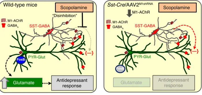
In summary, the current results indicate that the rapid antide-pressant responses to scopolamine are mediated by M1-AChRs on SST interneurons in the mPFC, demonstrating that scopolamine acts via an indirect, disinhibition mechanism to regulate pyra-midal neuron activity and subsequent behavioral responses. It is possible that SST interneurons also play a critical role in the rapid antidepressant-like effects of the noncompetitive NMDA receptor antagonist ketamine, as prior reports suggest that convergent cel-lular and molecular mechanisms underlie the behavioral respons-es to scopolamine and ketamine (21, 46). Enriched exprrespons-ession GAD-expressing interneurons in the mPFC are diverse and
display different synaptic connections, physiological properties, and molecular signaling (28). Immunohistology shows that SST interneurons have enriched expression of M1-AChR compared with PV interneurons, and electrophysiological recordings show that cholinergic stimulation of SST-tdTomato interneurons is mediated by M1-AChR. These findings correspond with previous reports showing that SST interneurons exhibit inward currents following cholinergic stimulation, while PV interneurons show minimal responses (34, 35). In addition, the mAChR antagonist pirenzepine blocked the ACh-induced inhibitory responses in the cortex, suggesting that these effects are mediated by M1-AChRs (36). SST interneurons, such as bitufted and Martinotti cells, send projections to the dendritic tufts of cortical pyramidal cells (37) and can inhibit pyramidal neuron activity directly (38). In this context, baseline cholinergic stimulation of SST interneurons through M1-AChRs leads to increased activity that would inhibit pyramidal neurons. Thus, scopolamine antagonism of M1-AChR on SST interneurons would reduce inhibitory activity, leading to disinhibition of proximal pyramidal neurons. Studies that exam-ine cholexam-inergic innervation of SST interneurons in the mPFC may provide additional insight into their role in the antidepressant-like effects of scopolamine.
Based on these data, we infused AAV2M1shRNA into Sst-Cre and
PV-Cre mice to identify the GABAergic interneuron subtype that
mediates the effects of scopolamine. AAV2M1shRNA infusion into the mPFC of Sst-Cre mice reduced M1-AChR immunolabeling and prevented muscarine-induced inward currents in recombined DsRed+ SST interneurons. In addition, M1-AChR knockdown in
Sst-Cre mice blocked the antidepressant-like behavioral response
to scopolamine in the FST and NSFT. In contrast, PV-Cre mice with M1-AChR knockdown displayed antidepressant-like responses
[image:11.585.46.396.58.224.2]FST. FST was conducted as previously described (11). Mice were
placed for 10 minutes in a clear cylinder filled with water (24 ± 1 °C, 18 cm depth). Sessions were video recorded and scored for total immo-bility time by a blinded experimenter. Time immobile during the 2- to 6-minute block is reported.
NSFT. NSFT was conducted as previously described (52). Mice were
food deprived for 18 hours and placed in a dimly lit, novel environment (24 × 40 × 14 cm, fresh bedding) with food in the center. The latency to feed was recorded. Home cage food intake was measured as a control.
FUST. FUST is a validated test of reward-seeking behavior in
rodents and was performed similarly to the way it was performed in prior reports (53). Mice were acclimated to a novel, single-housed cage for 1 hour. Following the acclimation period, mice were habituated to a water-soaked cotton-tipped applicator for 1 hour. Subsequently, a fresh water-soaked cotton-tipped applicator was inserted, and inter-action time was taped for 5 minutes. The cotton-tipped applicator was removed, and mice were left undisturbed for 45 minutes. Following this time, a cotton-tipped applicator with female urine was inserted into the cage, and interaction time was taped for 5 minutes. Time spent interacting with the urine cotton-tipped applicator is presented.
Immunohistology. Brains were collected from mice after
trans-cardiac perfusion with sterile PBS and 4% paraformaldehyde (PFA). Brains were post-fixed in 4% PFA for 24 hours and incubated in 30% sucrose for an additional 24 hours. Fixed brains were frozen and sectioned using a Microm HM550 cryostat. Free-floating sections were washed, then blocked for 1 hour at room temperature. Sections were washed, then incubated with the following primary antibod-ies (1:1,000 dilution) overnight at 4°C: rabbit anti–M1-AChR (Fron-tier Institute; mAChR-M1- RB-Af340), rat anti–Camk2a-Cre (Enzo Life Sciences; KAM-CA002-D), mouse anti–GAD67 (Sigma-Aldrich; G5419), mouse anti-PV (Millipore; MAB1572), rat anti-SST (Milli-pore; MAB354), and rabbit anti-FosB (Santa Cruz Biotechnology Inc.; sc-48). Sections were then washed and incubated with conjugated sec-ondary antibody (1:1,000 dilution) overnight at 4°C. Immunofluores-cence was visualized using an Olympus BX61WI confocal microscope. Images were captured with Fluoview (FV1000) and a Hamamatsu high-resolution digital camera (ORCA-ER).
Quantitative immunofluorescence. For quantification of FosB, 2 to
3 mPFC brain slices (25 μm) were selected and bilateral images were obtained for each sample (all experimental groups included). Cell counts were quantified with ImageJ software (NIH). Images were transformed to 8-bit format, then threshold, particle size, and circularity were set with constant parameters used across experimental groups. Blinded counts were recorded and averaged in each cortical region, and the average across samples was used for statistical analyses (n = 4–5). To quantify M1-AChR colocalization with PV and SST interneurons in the mPFC, 2 to 3 mPFC brain slices (25 μm) were selected and bilateral images were obtained. The total number of PV and SST interneurons in each cortical layer was quantified along with the proportion of each subtype that colo-calized with M1-AChR (n = 2–3). To quantify knockdown of M1-AChR through immunohistology, 2 to 3 mPFC brain slices (25 μm) were selected and immunolabeling was performed. Bilateral confocal images were collected and analyzed in ImageJ. In brief, neurons infected with AAV2M1shRNA or AAV2SCR that underwent Cre recombination were identi-fied by DsRed expression and M1-AChR levels were measured by rela-tive fluorescent intensity. For each sample, 6 to 8 neurons were quanti-fied and sample averages were compiled for statistical analyses.
of M1-AChR on SST interneurons and disruption of SST in MDD indicate that these neurons have an important functional role in the transduction of cholinergic signaling in the cortex that influ-ences mood and emotion as well as cognition. Additional molecu-lar and cellumolecu-lar studies to examine the neurotransmitter receptor signaling profile of SST interneurons can build on these findings and provide insight into novel pharmacological targets for rapid antidepressant development.
Methods
Animals. Male transgenic mice and WT C57BL/6 littermates were
obtained from in-house breeders. Gad1-Cre mice were originally obtained from Marina Picciotto (47), and Camk2a-Cre mice were obtained from Günter Schütz (German Cancer Research Center, Hei-delberg, Germany) (48). PV-Cre (catalog 008069) and Sst-Cre (cata-log 013044) mice were obtained from Jackson Laboratories. To obtain SST-tdTomato mice, Sst-Cre mice were bred with Ai9(RCL-tdT) mice (catalog 007909), also from Jackson Laboratories. All studies were performed with 6- to 12-week-old mice. Mice were housed in standard ventilated under a 12-hour light/12-hour dark cycle with ad libitum access to water and rodent chow.
shRNA and viral preparation. For knockdown of M1-AChR in
spe-cific neuronal cell types, an shRNA sequence was designed targeting the M1-AChR (49). M1-AChR shRNA (5′-TGCAACGC CTCTGTC ATGAATCTTCTTCAAGAGAGAAGATTCATGACAGAGGCGT TGCTTTTTTC-3′) or scrambled control (5′ -TATCTATAGCGAC- GCTCCTTGTCCATTCAAGAGATGGACAAGGAGCGTCGCTATA-GATTTTTTTC-3′) (Integrated DNA Technologies) was ligated into a plasmid (pM1shRNA) designed to restrict shRNA expression to cells that express Cre recombinase (50). The TATAloxP-flanked CMV-EGFP cassette was subcloned from pSico (from Tyler Jacks, Massachu-setts Institute of Technology, Boston, MassachuMassachu-setts, USA; Addgene plasmid catalog 11578) into pAAV-DsRed (from Ralph DiLeone, Yale University School of Medicine) using HindIII and ApaI. This plasmid allows ubiquitous DsRed expression and conditional EGFP expres-sion driven by floxed CMV cassette that prevents shRNA expresexpres-sion with U6 promoter disruption (Supplemental Figure 1A). This construct and scrambled control were packaged into AAV2M1shRNA, as previously described (19). Neuro-(N)2a cells obtained from ATCC were used to test the efficacy of pM1shRNA.
Scopolamine administration. Mice received 3 injections of vehicle
or scopolamine (25 μg/kg, i.p.) every 48 hours, and behavioral test-ing was started 24 hours later (23). Followtest-ing completion of behav-ioral testing, mice received a final injection of vehicle or scopolamine 1 hour prior to sacrifice.
Surgery and cortical infusion. Mice were anesthetized with a
solu-tion of ketamine/xylazine (100/10 mg/kg). Bilateral viral infusions into the mPFC (1 μl; 0.1 μl/minute) were performed with coordinates (from bregma) as follows: +2.0 mm anterior-posterior, ±0.2 mm medial-lateral, and –2.8 mm dorsal-ventral (51). Incisions were closed with sutures, and an antibiotic was applied to prevent infection. Mice received i.p. injection of carprofen (5 mg/kg) immediately after sur-gery and daily for the next 2 days.
Anxiety-like behavior. For open-field activity, mice were placed in
uated with Fisher’s least significant difference (LSD) test. Significant group differences were only reported if protected by significant main effects or interactions with ANOVA.
Study approval. Animal use and procedures were in accordance
with NIH guidelines and approved by the Yale University Animal Care and Use Committees.
Author contributions
ESW, SRT, MRP, and RSD designed research studies. ESW, MW, and MA conducted experiments and acquired data. ESW, MW, DMG, SRT, MRP, MA, and RSD provided resources and materials. ESW, MW, MA, and RSD analyzed data and wrote the manuscript.
Acknowledgments
This research was supported by National Institute of Mental Health grants MH045481 and MH093897 (to R.S. Duman) and the state of Connecticut. We would also like to thank Riley Rich-mond and Xiao Li for their technical assistance.
Address correspondence to: Ronald S. Duman, 34 Park Street, New Haven, Connecticut 06520, USA. Phone: 203.974.7726; E-mail: [email protected].
RNA isolation and real-time PCR. RNA was collected from N2a
cells with TRIzol reagent based on the manufacturer’s protocol (54) (Life Technologies). RNA (1 μg) was reverse transcribed to cDNA using the High Capacity cDNA Reverse Transcription Kit (Applied Biosys-tems). Primers were designed with Primer3 (Supplemental Table 1). cDNA was amplified on an ABI PRISM 7500-sequence detection sys-tem (Applied Biosyssys-tems) by real-time PCR and normalized based on reference gene expression (Gapdh). Data were analyzed with the com-parative threshold cycle method.
Brain slice electrophysiology. Brain slices containing the mPFC
were prepared from male and female SST-tdTomato or male Sst-Cre/ AAV2M1shRNA mice (8 to 16 weeks old). Brains were placed in artificial cere-brospinal fluid (aCSF) (pH 7.35–7.38) equilibrated with 95% O2/5% CO2. Coronal slices of 300-μm thickness containing the mPFC were trans-ferred to the fixed stage of an Olympus BX50WI scope for whole-cell recording. The chamber was continuously perfused with normal aCSF at a rate of 2 to 3 ml/min and its temperature maintained at 33 ± 0.5°C.
Statistics. Data were subjected to statistical analyses with
Graph-Pad Prism 6. Significant main effects and interactions were deter-mined using 1-way (genotype, treatment) or 2-way (genotype × treat-ment) ANOVA. Grouped analyses were completed without repeated measures, and multiple comparisons between group means were
1. Kessler RC. The costs of depression. Psychiatr
Clin North Am. 2012;35(1):1–14.
2. Kessler RC, Chiu WT, Demler O, Merikangas KR, Walters EE. Prevalence, severity, and comorbid-ity of 12-month DSM-IV disorders in the National Comorbidity Survey Replication. Arch Gen
Psy-chiatry. 2005;62(6):617–627.
3. Murray CJ, et al. The state of US health, 1990– 2010: burden of diseases, injuries, and risk fac-tors. JAMA. 2013;310(6):591–608.
4. Simon GE. Social and economic burden of mood disorders. Biol Psychiatry. 2003;54(3):208–215. 5. Souery D, Papakostas GI, Trivedi MH.
Treat-ment-resistant depression. J Clin Psychiatry. 2006;67(suppl 6):16–22.
6. Drevets WC, Zarate CA Jr, Furey ML. Antide-pressant effects of the muscarinic cholinergic receptor antagonist scopolamine: a review. Biol
Psychiatry. 2013;73(12):1156–1163.
7. Furey ML, Khanna A, Hoffman EM, Drevets WC. Scopolamine produces larger antidepressant and antianxiety effects in women than in men.
Neuro-psychopharmacology. 2010;35(12):2479–2488.
8. Furey ML, Drevets WC. Antidepressant efficacy of the antimuscarinic drug scopolamine: a ran-domized, placebo-controlled clinical trial. Arch
Gen Psychiatry. 2006;63(10):1121–1129.
9. Voleti B, et al. Scopolamine rapidly increases mammalian target of rapamycin complex 1 signal-ing, synaptogenesis, and antidepressant behavior-al responses. Biol Psychiatry. 2013;74(10):742–749. 10. Duman RS, Aghajanian GK. Synaptic dysfunction
in depression: potential therapeutic targets.
Sci-ence. 2012;338(6103):68–72.
11. Li N, et al. mTOR-dependent synapse formation underlies the rapid antidepres-sant effects of NMDA antagonists. Science. 2010;329(5994):959–964.
12. Rajkowska G. Postmortem studies in mood dis-orders indicate altered numbers of neurons and
glial cells. Biol Psychiatry. 2000;48(8):766–777. 13. Rajkowska G, et al. Morphometric evidence
for neuronal and glial prefrontal cell pathol-ogy in major depression. Biol Psychiatry. 1999;45(9):1085–1098.
14. Drevets WC. Functional anatomical abnormalities in limbic and prefrontal cortical structures in major depression. Prog Brain Res. 2000;126:413–431. 15. Radley JJ, et al. Repeated stress alters dendritic
spine morphology in the rat medial prefrontal cortex. J Comp Neurol. 2008;507(1):1141–1150. 16. Radley JJ, et al. Repeated stress induces dendritic
spine loss in the rat medial prefrontal cortex.
Cereb Cortex. 2006;16(3):313–320.
17. Krishnan V, Nestler EJ. The molecular neurobiology of depression. Nature. 2008;455(7215):894–902. 18. Kang HJ, et al. Decreased expression of synapse-related genes and loss of synapses in major depres-sive disorder. Nat Med. 2012;18(9):1413–1417. 19. Ota KT, et al. REDD1 is essential for
stress-induced synaptic loss and depressive behavior.
Nat Med. 2014;20(5):531–535.
20. Miller OH, Moran JT, Hall BJ. Two cellular hypotheses explaining the initiation of ket-amine’s antidepressant actions: Direct inhibi-tion and disinhibiinhibi-tion. Neuropharmacology. 2016;100:17–26.
21. Wohleb ES, Gerhard D, Thomas A, Duman RS. Molecular cellular mechanisms of rapid-acting antidepressants ketamine scopolamine. Curr
Neuropharmacol. 2016;21(3):454–464.
22. Witkin JM, et al. M1 and m2 muscarinic receptor subtypes regulate antidepressant-like effects of the rapidly acting antidepressant scopolamine.
J Pharmacol Exp Ther. 2014;351(2):448–456.
23. Navarria A, et al. Rapid antidepressant actions of scopolamine: role of medial prefrontal cortex and M1-subtype muscarinic acetylcholine recep-tors. Neurobiol Dis. 2015;82:254–261.
24. Disney AA, Reynolds JH. Expression of m1-type
muscarinic acetylcholine receptors by parval-bumin-immunoreactive neurons in the primary visual cortex: a comparative study of rat, guinea pig, ferret, macaque, and human. J Comp Neurol. 2014;522(5):986–1003.
25. Yamasaki M, Matsui M, Watanabe M. Preferential localization of muscarinic M1 receptor on den-dritic shaft and spine of cortical pyramidal cells and its anatomical evidence for volume transmis-sion. J Neurosci. 2010;30(12):4408–4418. 26. Amar M, Lucas-Meunier E, Baux G, Fossier P.
Blockade of different muscarinic receptor sub-types changes the equilibrium between excita-tion and inhibiexcita-tion in rat visual cortex.
Neurosci-ence. 2010;169(4):1610–1620.
27. Perrotti LI, et al. Induction of deltaFosB in reward-related brain structures after chronic stress. J Neurosci. 2004;24(47):10594–10602. 28. Markram H, Toledo-Rodriguez M, Wang Y,
Gupta A, Silberberg G, Wu C. Interneurons of the neocortical inhibitory system. Nat Rev Neurosci. 2004;5(10):793–807.
29. Gupta A, Wang Y, Markram H. Organizing principles for a diversity of GABAergic inter-neurons and synapses in the neocortex. Science. 2000;287(5451):273–278.
30. Malkesman O, et al. The female urine sniffing test: a novel approach for assessing reward-seeking behavior in rodents. Biol Psychiatry. 2010;67(9):864–871.
31. Lieben CK, Blokland A, Sik A, Sung E, van Nieu-wenhuizen P, Schreiber R. The selective 5-HT6 receptor antagonist Ro4368554 restores memory performance in cholinergic and serotonergic models of memory deficiency in the rat.
Neuro-psychopharmacology. 2005;30(12):2169–2179.
32. Falsafi SK, Deli A, Höger H, Pollak A, Lubec G. Scopolamine administration modulates mus-carinic, nicotinic and NMDA receptor systems.
33. Singh P, Konar A, Kumar A, Srivas S, Thakur MK. Hippocampal chromatin-modifying enzymes are pivotal for scopolamine-induced synaptic plastic-ity gene expression changes and memory impair-ment. J Neurochem. 2015;134(4):642–651. 34. Kawaguchi Y. Selective cholinergic modulation of
cortical GABAergic cell subtypes. J Neurophysiol. 1997;78(3):1743–1747.
35. Gulledge AT, Park SB, Kawaguchi Y, Stuart GJ. Heterogeneity of phasic cholinergic signal-ing in neocortical neurons. J Neurophysiol. 2007;97(3):2215–2229.
36. McCormick DA, Prince DA. Two types of mus-carinic response to acetylcholine in mamma-lian cortical neurons. Proc Natl Acad Sci U S A. 1985;82(18):6344–6348.
37. Neske GT, Patrick SL, Connors BW. Contribu-tions of diverse excitatory and inhibitory neurons to recurrent network activity in cerebral cortex.
J Neurosci. 2015;35(3):1089–1105.
38. Silberberg G, Markram H. Disynaptic inhibition between neocortical pyramidal cells mediated by Martinotti cells. Neuron. 2007;53(5):735–746. 39. Lin LC, Sibille E. Somatostatin, neuronal
vulner-ability and behavioral emotionality. Mol
Psychia-try. 2015;20(3):377–387.
40. Soumier A, Sibille E. Opposing effects of acute versus chronic blockade of frontal cortex somatostatin-positive inhibitory neurons on behavioral emotionality in mice.
Neuropsycho-pharmacology. 2014;39(9):2252–2262.
41. Sibille E, Morris HM, Kota RS, Lewis DA. GABA-related transcripts in the dorsolateral prefrontal cortex in mood disorders. Int J
Neuropsychophar-macol. 2011;14(6):721–734.
42. Karolewicz B, Maciag D, O’Dwyer G, Stockmeier CA, Feyissa AM, Rajkowska G. Reduced level of glutamic acid decarboxylase-67 kDa in the pre-frontal cortex in major depression. Int J
Neuropsy-chopharmacol. 2010;13(4):411–420.
43. Northoff G, Sibille E. Why are cortical GABA neurons relevant to internal focus in depression? A cross-level model linking cellular, biochemi-cal and neural network findings. Mol Psychiatry. 2014;19(9):966–977.
44. Picciotto MR, Higley MJ, Mineur YS. Acetylcho-line as a neuromodulator: choAcetylcho-linergic signaling shapes nervous system function and behavior.
Neuron. 2012;76(1):116–129.
45. Lawrence JJ. Cholinergic control of GABA release: emerging parallels between neo-cortex and hippocampus. Trends Neurosci. 2008;31(7):317–327.
46. Monteggia LM, Kavalali ET. Scopolamine and ketamine: evidence of convergence?. Biol
Psy-chiatry. 2013;74(10):712–713.
47. Taylor SR, Badurek S, Dileone RJ, Nashmi R, Minichiello L, Picciotto MR. GABAergic and glu-tamatergic efferents of the mouse ventral tegmen-tal area. J Comp Neurol. 2014;522(14):3308–3334.
48. Casanova E, et al. A CamKIIα iCre BAC allows
brain-specific gene inactivation. Genesis. 2001;31(1):37–42.
49. Reynolds A, Leake D, Boese Q, Scaringe S, Marshall WS, Khvorova A. Rational siRNA design for RNA interference. Nat Biotechnol. 2004;22(3):326–330.
50. Ventura A, et al. Cre-lox-regulated conditional RNA interference from transgenes. Proc Natl
Acad Sci U S A. 2004;101(28):10380–10385.
51. Paxinos G, Franklin KBJ. The Mouse Brain In
Ste-reotaxic Coordinates. 2nd ed. San Diego,
Califor-nia, USA: Academic Press; 2001.
52. Elsayed M, Banasr M, Duric V, Fournier NM, Licznerski P, Duman RS. Antidepressant effects of fibroblast growth factor-2 in behavioral and cellular models of depression. Biol Psychiatry. 2012;72(4):258–265.
53. Dutheil S, Ota KT, Wohleb ES, Rasmussen K, Duman RS. High-fat diet induced anxiety and anhedonia: impact on brain homeostasis and inflammation [published online ahead of print December 14, 2015]. Neuropsychopharmacology. doi: 10.1038/npp.2015.357.