Pruning the ricket thicket
Valentin David, Myles Wolf
J Clin Invest.
2016;
126(2)
:473-476.
https://doi.org/10.1172/JCI85005
.
Overexpression of FGF23 results in hypophosphatemic rickets, which is characterized by
renal phosphate wasting, inappropriately low circulating levels of the active form of vitamin
D, and skeletal abnormalities. The precise mechanisms of how excess FGF23 leads to
hypophosphatemic rickets are not clear. In this issue of the
JCI
, Bai and colleagues
demonstrate that deletion or inhibition of CYP24A1, which initiates degradation of the active
form of vitamin D, ameliorates skeletal abnormalities in two mouse models of
hypophosphatemic rickets. While this work supports an important role for excess CYP24A1
activity in the pathogenesis of FGF23-mediated hypophosphatemic rickets, more work will
need to be done before CYP24A1 inhibition can be integrated into the management of
patients living with these diseases.
Commentary
Find the latest version:
Pruning the ricket thicket
Valentin David and Myles Wolf
Division of Nephrology and Hypertension, Department of Medicine, Center for Translational Metabolism and Health, Institute for Public Health and Medicine, Northwestern University, Chicago, Illinois, USA.
Precise regulation of FGF23 is
required for bone health
Just the right amount of FGF23 is required to maintain healthy bone and mineral metabolism. FGF23 deficiency results in tumoral calcinosis, because loss of this factor impedes renal phosphate excre-tion and enables unopposed parathyroid hormone–dependent (PTH-dependent) stimulation of 1,25-dihydroxyvitamin D [1,25(OH)2D] production, which together culminate in hyperphosphatemia and metastatic calcification (1). In contrast, FGF23 overload results in hypophosphate-mic rickets, which is characterized by inappropriately low levels of 1,25(OH)2D (2, 3). Yet, for all we have learned about FGF23 in the past decade, the fundamen-tal mechanisms of how FGF23 overload leads to rickets is still unclear. Is the pri-mary driver FGF23 excess, phosphate depletion, 1,25(OH)2D depletion, second-ary calcium depletion, a combination of these, or other factors altogether?
Targeting CYP24A1 ameliorates
skeletal abnormalities
In this issue, Bai et al. shed important new light on this controversy (4). Specif-ically, the authors investigated the role of CYP24A1, the cytochromal enzyme that catalyzes the first step in vitamin D degradation, in the pathogenesis of two prototypical disorders of primary FGF23 excess: X-linked hypophosphatemic rick-ets (XLH), which is caused by mutations in PHEX that lead to elevated FGF23 through unknown mechanisms, and autosomal dominant hypophosphate-mic rickets (ADHR), which results from activating mutations in FGF23 itself. Bai and colleagues crossed Hyp mice, which are the murine homolog of human XLH and FGF23 transgenic mice, which over-express the cleavage-resistant form of FGF23 (FGF23R176Q) produced in human
ADHR, with Cyp24a1-null mice. As a sec-ondary approach to evaluate the involve-ment of CYP24A1 in these diseases, Hyp
and ADHR mice were also treated with a pharmacological CYP24A1 inhibitor (4).
Using this experimental approach, Bai and colleagues demonstrated that either genetic deletion or pharmaco-logical inhibition of CYP24A1 induces near-complete healing of rickets in Hyp and ADHR mice. Amelioration of the animals’ skeletal defects occurred in the absence of correcting severe hypophos-phatemia or altering serum calcium lev-els. On the basis of these results, Bai et al. conclude that inactivation of CYP24A1 heals the skeleton by prolonging the local half-life of 1,25(OH)2D in bone, such that even low levels of circulating and locally produced 1,25(OH)2D have pro-tracted effects (4). The stable serum min-eral levels observed in these models are especially noteworthy, because a large amount of calcium and phosphate must have been deposited in bone to achieve such substantial remineralization. Most likely, Cyp24a1 deletion induced a major increase in gastrointestinal absorption of calcium and phosphate due to pro-longed effects of 1,25(OH)2D in entero-cytes. Although Bai and colleagues did not directly explore this mechanism by performing metabolic balance stud-ies, their results indirectly support this hypothesis, as circulating levels of PTH and 1,25(OH)2D were substantially decreased, and expression of vitamin D–dependent calcium transporters in the gut of the compound mutant mice was increased (4). Direct suppression of PTH by prolonged 1,25(OH)2D activity in the parathyroid glands is another pos-sible mechanism to explain the low PTH and thus low systemic 1,25(OH)2D lev-els; however, this scenario is less likely, because primary suppression of PTH in the setting of massive bone calcium deposition would have resulted in severe hypocalcemia, unlike what Bai et al. report. Moreover, FGF23 levels were further increased in the compound Hyp
Cyp24a1–null mice compared with levels
Related Article: p. 667
Conflict of interest: V. David reports receiving research support from Keryx Biopharmaceuticals. M. Wolf reports receiving
consulting fees from or has an ownership interest in Amgen, Keryx Biopharmaceuticals, Pfizer, and Ultragenyx and is an inventor on a patent application focused on targeting FGF receptor 4 to attenuate cardiovascular disease.
Reference information: J Clin Invest. 2016;126(2):473–476. doi:10.1172/JCI85005.
Roles of circulating and local
vitamin D metabolites
It should be noted that Bai et al. did not study the possible effects of other vitamin D metabolites. By catalyzing hydroxyla-tion of 25-hydroxyvitamin D [25(OH)D] to 24,25-dihydroxyvitamin D [24,25(OH)2D], CYP24A1 reduces the amount of 25(OH) D that CYP27B1 can convert into the more potent 1,25(OH)2D (5, 6). CYP24A1 also reduces the half-life of previously synthe-sized 1,25(OH)2D by converting 1,25(OH)2D into 1,24,25(OH)3D, which is further metabolized into calcitroic acid (1-hydroxy- 23-carboxyvitamin D) or 1α,25-(OH)2 D3-26,23-lactone (7, 8). Consistently, previous reports have demonstrated a total absence of calcitroic acid and 1α,25-(OH)2 D3-26,23-deletion and administration of a
sys-temically active pharmacological inhib-itor of CYP24A1. Bone-specific Cyp24a1 deletion will be needed to definitively show that it is CYP24A1 in this organ that mediates the restoration of normal skele-tal structure in their models. Regardless of the precise mechanism, by demon-strating that the skeletal abnormalities of hypophosphatemic rickets can be cor-rected without lowering elevated FGF23 or raising depressed serum phosphate levels, Bai and colleagues have provided important new evidence against these factors as direct mechanistic contribu-tors to bone disease in XLH, ADHR, and other related conditions mediated by pri-mary increases in FGF23 (4).
detected in the Hyp mice, perhaps reflect-ing a consequence of positive phosphate and calcium balance induced by the prolonged effects of 1,25(OH)2D locally in enterocytes and/or the further stim-ulation of Fgf23 transcription by the prolonged local effects of 1,25(OH)2D in osteocytes.
[image:3.585.38.370.62.507.2]The totality of the data generally support the hypothesis that there is a direct and beneficial effect of suppress-ing bone CYP24A1 on the skeletal defects in hypophosphatemic rickets. However, an important limitation of the Bai et al. study is the inability to definitively implicate bone CYP24A1 as the cause of the phenotype as opposed to second-ary effects conferred by global Cyp24a1
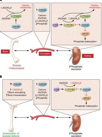
Figure 1. Vitamin D functions are regulated by a balance of 1,25(OH)2D synthesis and
degradation, both systemically and locally, in vitamin D target tissues. (A) In syndromes of FGF23-dependent hypophosphatemic rickets, excess FGF23 production by bone enters the circulation and reduces systemic vitamin D levels through two independent mechanisms in the kidney. FGF23 induces CYP24A1 activity, which converts vitamin D storage [25(OH)D] and active [1,25(OH)2D] forms to inactive metabolites, and it inhibits CYP27B1, which is responsible for converting 25(OH)D to 1,25(OH)2D. These effects of FGF23 are counter-regulated by the opposing effects of PTH. FGF23 also promotes renal phos-phate wasting by reducing the expression of sodium-dependent phosphate cotransporters (NPT2) in the kidney and thereby reducing phosphate reabsorption. Hypophosphatemic rickets results as a consequence of these met-abolic perturbations, but the primary mediator of the skeletal disease is unclear. (B) Inhibition
of Cyp24a1 in murine models of FGF23-
patients with nutritional vitamin D supple-mentation or exogenous 25(OH)D may be only of marginal benefit, because exces-sive local CYP24A1 activity would degrade most of the 25(OH)D and 1,25(OH)2D that managed to make it to target cells. Like-wise, exogenous 1,25(OH)2D that raises systemic 1,25(OH)2D levels might only serve to further upregulate CYP24A1 at the tissue level, resulting in minimal changes in end-organ effects of 1,25(OH)2D. It is into this gap that CYP24 inhibitors may ultimately prove to have a unique and potentially exciting profile to boost end-organ 1,25(OH)2D effects, without increasing serum phosphate or calcium levels. Stay tuned for more progress in a dynamic and rapidly evolving field.
Acknowledgments
V. David is supported by NIH grant R01DK102815, and M. Wolf is sup-ported by NIH grants R01DK076116 and K24DK093723.
Address correspondence to: Myles Wolf, 633 N. St. Clair St., Suite 18-089, Chicago, Illinois 60611, USA. Phone: 312.503.8013; E-mail: [email protected].
1. Benet-Pages A, Orlik P, Strom TM, Lorenz- Depiereux B. An FGF23 missense mutation causes familial tumoral calcinosis with hyperphos-phatemia. Hum Mol Genet. 2005;14(3):385–390. 2. ADHR-Consortium. Autosomal dominant
hypophosphataemic rickets is associ-ated with mutations in FGF23. Nat Genet. 2000;26(3):345–348.
3. Shimada T, et al. Targeted ablation of Fgf23 demonstrates an essential physiological role of FGF23 in phosphate and vitamin D metabolism.
J Clin Invest. 2004;113(4):561–568.
4. Bai X, et al. CYP24 inhibition as a therapeutic target in FGF23-mediated renal phosphate wasting disorders. J Clin Invest.
2016;126(2):667–680.
5. Okuda K, Usui E, Ohyama Y. Recent progress in enzymology and molecular biology of enzymes involved in vitamin D metabolism. J Lipid Res. 1995;36(8):1641–1652.
6. Ohyama Y, et al. Structural characterization of the gene encoding rat 25-hydroxyvitamin D3 24-hydroxylase. Biochemistry. 1993;32(1):76–82. 7. Ishizuka S, Norman AW. Metabolic pathways
from 1 alpha,25-dihydroxyvitamin D3 to 1 alpha,25-dihydroxyvitamin D3-26,23-lactone. Stereo-retained and stereo-selective lactoniza-tion. J Biol Chem. 1987;262(15):7165–7170. 8. Reddy GS, Tserng KY. Calcitroic acid, end product
of renal metabolism of 1,25-dihydroxyvitamin D3 through C-24 oxidation pathway. Biochemistry. 1989;28(4):1763–1769.
demonstrate the dichotomy between local and systemic vitamin D regulation and the potential pitfalls of interpreting end-organ effects of vitamin D on the basis of circu-lating 1,25(OH)2D levels. Furthermore, regulation of 1,25(OH)2D synthesis has his-torically garnered far more attention than its degradation. The recent description of human mutations in CYP24A1 as the cause of previously idiopathic cases of hypercal-cemia in newborns and adults has cast new light on the importance of the degradation pathway in health and disease that is ele-gantly emphasized by Bai et al. (14, 15).
Conclusions and future
directions
The current standard of care for XLH and ADHR includes the administration of oral 1,25(OH)2D and phosphate supple-ments; however, these treatments, which address downstream consequences of the diseases rather than the root causes, are only partially effective and often poorly tolerated (16, 17). Novel strategies for these diseases are currently under devel-opment. For example, neutralizing anti-bodies against FGF23 directly target the underlying molecular mechanism of dis-ease (18), and FGF receptor antagonists inhibit the end-organ effects of FGF23 (19). While the results of the current study by Bai and colleagues suggest that target-ing CYP24A1 may be another viable ther-apeutic strategy, especially to correct the underlying skeletal defects, the inability to correct hypophosphatemia suggests that CYP24A1 antagonists may need to be rel-egated to an adjunctive role. Furthermore, the finding that circulating 1,25(OH)2D levels increased significantly in Hyp mice that were treated with the CYP24A1 inhibi-tor raises the question as to whether exoge-nous calcitriol treatment, which was not tested by Bai et al., could have achieved similar skeletal success. More in-depth research will be needed to define a new and improved standard of care for XLH, ADHR, and related conditions.
Beyond those orphan diseases of pri-mary FGF23 excess, could there be a role for CYP24A1 inhibition in diseases of secondary FGF23 excess, such as chronic kidney disease? If elevated FGF23 accel-erates 1,25(OH)2D degradation by stimu-lating CYP24A1 in extra-renal tissues, as it appears to in bone, treating kidney disease lactone formation in Cyp24a1-KO mice (9).
Since 1α,25-(OH)2D3-26,23-lactone can act as a vitamin D receptor antagonist and thus inhibit osteoclastogenesis and bone resorption (10), the extent to which rescue of the bone phenotype observed by Bai and colleagues can be explained entirely by the increase in the half-life of local 1,25(OH)2D or by an additional contribution of the con-comitant absence of 1α,25-(OH)2D3-26, 23-lactone is not certain.
As Cyp24a1-null mice are unable to degrade 1,25(OH)2D, it is somewhat sur-prising that 1,25(OH)2D levels are low and hypercalcemia is absent in these animals. Indeed, approximately 50% of Cyp24a1-KO mice die in the early postnatal period due to endogenous 1,25(OH)2D intoxica-tion and consequent hypercalcemia (11). For reasons that are not clear, the other 50% are capable of lowering their cir-culating 1,25(OH)2D levels to the point that hypercalcemia can be avoided and prolonged survival is possible (11). Thus, another important limitation of the study by Bai and colleagues is that only the prog-eny of Cyp24a1-KO mice that successfully adapted their vitamin D homeostasis to survive can be studied. Is it appropriate to proclaim skeletal success when only the hypercalcemia-resistant half of the progeny were analyzed? As a corollary, do the results understate the risks related to hypercalcemia in the event that all-comer human hypophosphatemic rickets patients are treated with CYP24A1 inhibitors?
of calcitriol on renal handling of phosphate, serum phosphate, and bone mineralization. J Clin
Endocrinol Metab. 1981;52(3):463–472.
17. Carpenter TO, Imel EA, Holm IA, Jan de Beur SM, Insogna KL. A clinician’s guide to X-linked hypophosphatemia. J Bone Miner Res. 2011;26(7):1381–1388.
18. Carpenter TO, et al. Randomized trial of the anti-FGF23 antibody KRN23 in X-linked hypophosphatemia. J Clin Invest. 2014;124(4):1587–1597.
19. Wohrle S, et al. Pharmacological inhibition of fibroblast growth factor (FGF) receptor signaling ameliorates FGF23-mediated hypophosphatemic rickets. J Bone Miner Res. 2013;28(4):899–911.
2000;141(7):2658–2666.
12. Martin A, David V, Quarles LD. Regulation and function of the FGF23/klotho endocrine path-ways. Physiol Rev. 2012;92(1):131–155. 13. Zehnder D, et al. Extrarenal expression of
25-hydroxyvitamin d(3)-1α-hydroxylase.
J Clin Endocrinol Metab. 2001;86(2):888–894.
14. Schlingmann KP, et al. Mutations in CYP24A1 and idiopathic infantile hypercalcemia. N Engl J Med. 2011;365(5):410–421.
15. Tebben PJ, et al. Hypercalcemia, hypercalciuria, and elevated calcitriol concentrations with auto-somal dominant transmission due to CYP24A1 mutations: effects of ketoconazole therapy. J Clin
Endocrinol Metab. 2012;97(3):E423–E427.
16. Costa T, et al. X-linked hypophosphatemia: effect 9. Masuda S, et al. Altered pharmacokinetics of
1α,25-dihydroxyvitamin D3 and 25-hydroxyvi-tamin D3 in the blood and tissues of the 25-hydroxyvitamin D-24-hydroxylase (Cyp24a1) null mouse. Endocrinology. 2005;146(2):825–34.
10. Ishizuka S, et al. 1α,25-Dihydroxyvitamin D3[1 alpha,25-(OH)2D3]-26,23-lactone inhibits 1,25-(OH)2D3-mediated fusion of mouse bone marrow mononuclear cells. Endocrinology. 1988;123(2):781–786.