INTRODUCTION
The development of the vertebrate limb has long served as an experimental and conceptual model system with which to study a variety of biological processes (reviewed by Capdevila and Izpisua Belmonte, 2001; Niswander, 2003; Tabin and Wolpert, 2007). Numerous studies have identified several secreted cell-cell signaling molecules, such as members of the Wnt, fibroblast growth factor (Fgf) and hedgehog gene families, responsible for a variety of processes during limb development. For example, embryonic and genetic studies have demonstrated the role of the Wntfamily in the initiation of limb development (Kawakami et al., 2001), the role of Fgf genes as an instructive factor for proximal-distal patterning (Mariani et al., 2008), and the role of sonic hedgehog (Shh) in anterior-posterior patterning (Riddle et al., 1993).
These signaling pathways cooperate with one another to sustain limb outgrowth. For instance, Shh, produced in the zone of the polarizing activity (ZPA), regulates gremlin 1 (Grem1) expression in the posterior mesenchyme (Capdevila et al., 1999; Panman et al., 2006). Grem1-mediated BMP antagonism is crucial for maintenance
of the expression of Fgfs in the apical ectodermal ridge (AER) (Khokha et al., 2003; Michos et al., 2004). FGF proteins, secreted from the AER, act on underlying mesenchyme to promote cell survival (Dudley et al., 2002), and in the posterior mesenchyme to maintain Shhexpression (Laufer et al., 1994). Such a feedback loop mediates distal outgrowth, and, thus, proper formation of the autopod (Scherz et al., 2004). Shh-mediated counteraction of the Gli3 repressor also regulates the anterior-posterior patterning of digits (Litingtung et al., 2002; te Welscher et al., 2002). Although numerous studies have focused on the role of such signaling pathways and their interactions, the specific mechanisms that regulate region-specific morphogenesis, which leads to a stereotyped morphology of each limb skeletal element, remain elusive.
Human syndromes provide an opportunity to identify novel genes involved in limb development and have indeed given us invaluable clues in understanding the molecular and genetic bases of limb development (Wilkie, 2003). One such gene identified from human diseases is the SALL1gene, one of the four SALL genes in humans and mice, which are related to the Drosophila spalt gene. SALL1 encodes a multi-zinc finger domain transcription factor (Nishinakamura and Osafune, 2006; Sweetman and Munsterberg, 2006), and mutations in the SALL1gene cause Townes-Brocks syndrome (TBS) (Kohlhase et al., 1998). Individuals with TBS exhibit multiple defects, including limb alterations. The human TBS disorders are mainly due to a dominant-negative action of the truncated SALL1 protein, though milder phenotypes have been reported from haploinsufficiency of SALL1(Borozdin et al., 2006; Kohlhase, 2000). In mice, no limb defects are reported in Sall1 knockout or heterozygous mice (Nishinakamura et al., 2001). Conversely, two mouse models producing truncated Sall1 protein have shown some TBS-like phenotypes in the limb (Kiefer et al., 2003; Kiefer et al., 2008). These reports, together with a biochemical analysis demonstrating
Sall genes regulate region-specific morphogenesis in the
mouse limb by modulating Hox activities
Yasuhiko Kawakami1,2,3,4, Yukako Uchiyama5, Concepcion Rodriguez Esteban1, Toshiaki Inenaga5,
Naoko Koyano-Nakagawa2,4,6, Hiroko Kawakami1,2,3, Merce Marti7, Marie Kmita8, Paula Monaghan-Nichols9, Ryuichi Nishinakamura5and Juan Carlos Izpisua Belmonte1,7,*
The genetic mechanisms that regulate the complex morphogenesis of generating cartilage elements in correct positions with precise shapes during organogenesis, fundamental issues in developmental biology, are still not well understood. By focusing on the developing mouse limb, we confirm the importance of transcription factors encoded by the Sall gene family in proper limb morphogenesis, and further show that they have overlapping activities in regulating regional morphogenesis in the autopod. Sall1/Sall3double null mutants exhibit a loss of digit1 as well as a loss or fusion of digit2 and digit3, metacarpals and carpals in the autopod. We show that Sall activity affects different pathways, including the Shh signaling pathway, as well as the Hox network. Shh signaling in the mesenchyme is partially impaired in the Sallmutant limbs. Additionally, our data suggest an antagonism between Sall1-Sall3 and Hoxa13-Hoxd13. We demonstrate that expression of Epha3and Epha4is downregulated in the Sall1/Sall3 double null mutants, and, conversely, is upregulated in Hoxa13and Hoxd13mutants. Moreover, the expression of Sall1and Sall3is upregulated in Hoxa13and Hoxd13mutants. Furthermore, by using DNA-binding assays, we show that Sall and Hox compete for a target sequence in the Epha4upstream region. In conjunction with the Shh pathway, the antagonistic interaction between Hoxa13-Hoxd13and Sall1-Sall3in the developing limb may contribute to the fine-tuning of local Hox activity that leads to proper
morphogenesis of each cartilage element of the vertebrate autopod.
KEY WORDS: Sall, Townes-Brocks syndrome, Hox, Limb development, Shh, Eph, Mouse Development 136, 585-594 (2009) doi:10.1242/dev.027748
1Gene Expression Laboratory, The Salk Institute for Biological Studies, 10010 N.
Torrey Pines Road, La Jolla, CA 92037, USA. 2Stem Cell Institute, University of
Minnesota, 2001 6th SE, Minneapolis, MN. 55455, USA. 3Department of Genetics,
Cell Biology and Development, 6-160 Jackson Hall, 321 Church St. SE, Minneapolis, MN 55455, USA. 4Developmental Biology Center, University of Minnesota, 321
Church St. SE, Minneapolis, MN 55455, USA. 5Division of Integrative Cell Biology,
Institute of Molecular Embryology and Genetics, Global COE ‘Cell Fate Regulation Research and Education Unit’, Kumamoto University, Kumamoto, Japan 860-0811.
6Department of Neuroscience, University of Minnesota, 321 Church St. SE,
Minneapolis, MN 55455, USA.7Center of Regenerative Medicine in Barcelona,
Doctor Aiguader, 88, 08003 Barcelona, Spain. 8Laboratory of Genetics and
Development, Institut de Recherches Cliniques de Montréal (IRCM), Université de Montréal, 110 avenue des Pins Ouest, H2W 1R7, Montréal, Quebec, Canada.
9Department of Neurobiology, University of Pittsburgh, Pittsburgh, PA 15261, USA.
*Author for correspondence (e-mail: [email protected])
Accepted 8 December 2008
D
E
V
E
LO
P
M
E
N
that truncated Sall1 protein can form complexes with all Sall proteins (Kiefer et al., 2003), suggest that the truncated Sall1 protein inhibits other Sall family proteins, leading to the TBS phenotypes. However, no limb phenotype has been reported to date in mice lacking other Sall genes by conventional knockout approaches. Sall2is dispensable for embryonic development (Sato et al., 2003). Sall3-null mice exhibit a cleft palate, but no limb defects are observed (Parrish et al., 2004). Sall4mutants die at the peri-implantation stage, making it difficult to evaluate its role in organogenesis (Elling et al., 2006; Sakaki-Yumoto et al., 2006; Warren et al., 2007). Interestingly, a genetic interaction between Sall1and Sall4is needed for the proper development of several organs (Sakaki-Yumoto et al., 2006), suggesting functional redundancy between Sall genes.
Recent reports suggest a functional interaction between Sall and Hox genes during development in invertebrates. For example, spalt, the invertebrate homolog of Sall, acts in combination with Hox genes in Drosophilaembryos to specify segmental identities (Copf et al., 2006). During wing/haltere development, spaltis regulated by a Hox gene, Ubx (Galant et al., 2002). In C. elegans, a spalt homolog, sem-4, directly regulates the expression of Hox genes, elg-5and lin-39, in touch receptor specification and vulval development, respectively (Grant et al., 2000; Toker et al., 2003). In the crustacean Artemia, spaltrepresses a Hox gene during the morphogenesis of trunk segments (Copf et al., 2006). Therefore, functional interactions between spaltand Hox genes have important roles in many aspects of invertebrate development.
In vertebrates, Hox proteins are crucial for limb development (reviewed by Zakany and Duboule, 2007). Hox genes encode transcription factors and in the mammalian genome the 39 genes are organized as 13 paralogs into four clusters (Hoxa, Hoxb, Hoxcand Hoxd) (Pearson et al., 2005), of which Hoxaand Hoxdare crucial for proper limb development (Kmita et al., 2005). Hoxaand Hoxd genes, which are located at the 5⬘extremity of their respective clusters (so called 5⬘ Hox genes) are necessary for proper development of digits (Zakany et al., 1997). Human mutations have also highlighted the importance of HOX genes in human limb development. Hand-foot-genital syndrome is caused by mutations in the HOXA13gene, and synpolydactyly type II is caused by mutations in the HOXD13gene (Goodman, 2002; Lappin et al., 2006). In gene targeting experiments, Hoxa13and Hoxd13mutant mice each exhibit distinct phenotypes affecting autopod development (Fromental-Ramain et al., 1996). Mice with compound mutations in the Hoxa13and Hoxd13genes exhibit complex and more severe phenotypes, suggesting distinct and redundant functions of these two crucial Hox13 paralogous genes. Furthermore, misexpression experiments in chick and mouse embryos have demonstrated that Hoxa13 and Hoxd13 regulate region-specific morphogenesis of cartilage elements in the autopod (Goff and Tabin, 1997; Williams et al., 2006; Yokouchi et al., 1995). Despite all of these advances, our understanding of how Hox genes specifically control region-specific morphogenesis in the limb is still unclear.
Based on the human TBS limb phenotypes and on the lack of limb defects in mice with individual Sall knockouts, we speculated that during limb development, Sall genes might have redundant activities that can only be identified by the study of compound mutants. We have analyzed Sall1;Sall3allelic series, and demonstrate that Sall1 and Sall3have partially redundant activity. Our analyses suggest that Sall genes are involved in the Shh signaling, as well as in Shh-independent processes. We further show evidence that Sall and Hox activities are mutually antagonistic in the autopod, and that this
antagonism may contribute to a fine-tuning of local Hox activity that leads to proper morphogenesis of each cartilage element of the vertebrate autopod.
MATERIALS AND METHODS Mouse mutants
Sall1and Sall3mutants have been described previously (Nishinakamura et
al., 2001; Parrish et al., 2004). As Sall1/Sall3double heterozygous mice were almost infertile when assessed by natural mating; all of the progeny were obtained by in vitro fertilization, which lead to the efficient production of compound mutant embryos. Hoxa13and Hoxd13mutants have been described previously (Fromental-Ramain et al., 1996; Kmita et al., 2000).
Shhmutants (Chiang et al., 1996), Gli3mutants (Buscher et al., 1998) and
Tbx5mutants (Bruneau et al., 2001) have been previously described. Skeletal samples were examined as described (McLeod, 1980).
In situ hybridization
Whole-mount in situ hybridization was performed following a standard protocol (Wilkinson, 1993).
Electrophoretic mobility shift assay (EMSA)
HA-Sall1, Flag-Hoxa13and Flag-Hoxd13were cloned into pcDNA3.1
(Invitrogen, Carlsbad, CA) and translated in vitro by using the TNT T7 system (Promega, Madison, WI.) according the manufacturers’ instruction. The double-strand probes corresponding to –2028 to –2001 (transcription starting site as 1 with the NM_007936 as cDNA sequence) of the mouse
Epha4 gene contains the following sequences: wt probe,
CGCGGT-TATTTTTAATAATTTATGCACA; mutant 1, CGCGGTTATTTTTAAT-cATTgATGCACA; mutant 2, CGCGGggcgTTTTAATAATTTATGCA-CA; mutant 3, CGCGGggcgTTccgATcAcTgATGCACA. (Lower case letters indicate mutations.)
EMSA was performed following a standard protocol (Yoh and Privalsky, 2001). Anti-HA (Covance, 16B12, Emeryville, CA) and anti-Flag (Sigma, M2, St Louis, MO) antibodies were used.
Luciferase reporter assay
A mouse Epha4 upstream region (–2110 to –1980) that contains the sequence analyzed in the EMSA assay was subcloned into pGL3 (Promega) with the thymidine kinase promoter (TK). pRL-TK (Promega) was used as an internal control. NIH3T3 cells were transfected with the Epha4-TK-Luciferase, pRL-TK and various combinations of expression plasmids carrying Sall1, Hoxa13or Hoxd13by using Fugene6 (Roche, Indianapolis, IN), according to the manufacturer’s instructions. Forty-eight hours after transfection, cells were subjected to analysis using the Dual-Luciferase Reporter Assay System (Promega). Results were expressed as fold increase compared with samples with an empty vector. Experiments were performed in triplicate, and statistical significance is analyzed by ANOVA followed by Tukey’s comparison.
RESULTS
Combined activity of Sall1and Sall3contributes to the development of the autopod
SALL1mutations in humans cause TBS, which results in limb defects (Kohlhase et al., 1998). The fact that no limb defects are reported in mice lacking Sall1, Sall2, Sall3 orSall4suggested a functional redundancy between Sall genes (Nishinakamura et al., 2001; Parrish et al., 2004; Sato et al., 2003). Among those Sall genes expressed in limb buds (Buck et al., 2001; Kohlhase et al., 2002; Ott et al., 2001), we focused on Sall1and Sall3 double mutants, as early lethality of Sall4–/– embryos prevented the analysis of limb
development in absence of Sall4function.
To gain insights into the respective contribution of Sall1and Sall3 genes, we generated Sall1/Sall3 allelic series and analyzed skeletons at E15.5. We did not obtain Sall1–/–;Sall3–/–embryos at older stages,
probably because loss of both Sall1 andSall3leads to lethality.The reason for the lethality is unknown at this point; however, E15.5
D
E
V
E
LO
P
M
E
N
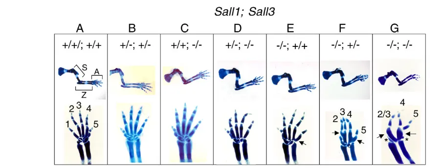
skeletons provided us with information to investigate the requirement of Sall1and Sall3during mouse limb development. Although the stylopod and zeugopod of all Sall1;Sall3mutants appear normal, we observed defects in the autopod both in the forelimb and hindlimb at E15.5 (Fig. 1; data not shown). We observed a fusion or lack of carpal elements, as well as fusion of metacarpal elements. Sall1–/–;Sall3+/+mutants show a mild fusion phenotype between metacarpal elements for digit4 and digit5 at a very proximal region (Fig. 1E). Sall1–/–;Sall3+/–mutants exhibit a more severe phenotype, as shown by the gross fusion in the metacarpal elements for digit2 and digit3, and those for digit4 and digit5, in addition to a loss of digit1 (Fig. 1F). Finally, Sall1–/–;Sall3–/–mutants exhibit further small carpal elements, more severe metacarpal fusion and a loss of digit1, and loss or fusion of digit2 and digit3 (Fig. 1G). Conversely, in the Sall1+/–;Sall3+/–and Sall1+/–;Sall3–/–mutants, we did not observe these defects (Fig.
1B,D), indicating that a single allele of the Sall1gene is sufficient for proper limb development.
These results indicate that Sall1and Sall3are partially redundant, but not equivalent. Sall1can compensate for the loss of Sall3, whereas Sall3can only partially compensate for the loss of Sall1 based on minor defects in the carpal elements observed inSall1–/–
autopods. Together, our data indicate that a combined activity of Sall1and Sall3contributes to the proper formation of the autopod.
Expression of Sall1and Sall3is regulated by the Shh-Gli3 pathway in the developing limb
The Sall1–/–;Sall3–/–mutant limb exhibited defects in the autopod.
Progression of limb development and formation of the autopod requires Shh-mediated counteraction of Gli3 (Litingtung et al., 2002; te Welscher et al., 2002). Previous experiments in chicks suggested that Sall1might be involved in distal limb patterning and that this putative function involves Shh signaling (Capdevila et al., 1999; Farrell and Munsterberg, 2000). Furthermore, it has been recently shown that reduced Shhsignaling preferentially affects the formation of digit3 (Scherz et al., 2007; Zhu et al., 2008). In our study, we also observed that Sall1;Sall3inactivation predominantly disrupted the formation of digit3 (Fig. 1), consistent with the
possibility that the function of Sall1and Sall3is linked to Shh signaling. As several factors involved in the Shh pathway are regulated by Shh signaling itself (Zuniga et al., 1999), we first analyzed whether Sall1and Sall3expression is regulated by Shh and Gli3. The endogenous expression of Sall1and Sall3at E10.5 is restricted to the distal mesenchyme and is posteriorly biased (Fig. 2A,D). In the Shh–/–limb, both genes are severely downregulated (Fig. 2B,E), indicating that Shh signaling is required for expression of Sall1and Sall3. By contrast, expression of both genes is expanded towards the anterior in the Gli3–/–limb (Fig. 2C,F), indicating that
Gli3 signaling negatively regulates expression of Sall1and Sall3. These results suggest that the Shh-Gli3 pathway impacts upon Sall1 and Sall3expression at early stages of limb development.
Reduced Shh signaling in the Sall1;Sall3mutant limb
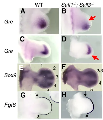
To further examine whether Sall activity is involved in Shh signaling, we monitored the expression of several key genes downstream of Shhthat are required for normal limb outgrowth. Although Shhexpression in the ZPA and Fgf8expression in the AER appear normal at E10.5 (see Fig. S1 in the supplementary material), we observed an alteration in Grem1expression (Fig. 3A,B), which is known to be regulated by the Shh-BMP pathway in the posterior mesenchyme (Capdevila et al., 1999; Merino et al., 1999; Nissim et al., 2006; Panman et al., 2006). We detected wild-type Grem1 expression in a wide region of the posterior mesenchyme. By contrast, Grem1expression was weaker and restricted to a smaller region in the Sall1–/–;Sall3–/–limb (Fig. 3A,B). This was also evident
[image:3.612.52.503.497.669.2]in the E11.5 mutant limb (Fig. 3C,D). These results suggest reduced Shh signaling in the absence of Sall1 and Sall3. Despite these changes, it seems that Sall activity is not required in the entire limb mesenchyme, as the skeletal phenotype is restricted to the autopod (Fig. 1), consistent with Sox9expression at E11.5 (Fig. 3E,F). Reflecting the defects at E15.5, we observed loss of digit1 and fusion of digit2 and digit3 primordia at E11.5 (Fig. 3E,F). Segregation of digit4 and digit5 primordia was also delayed. Correlating with the defects, the anterior and posterior margin of the Fgf8expression domain in the AER is shorter in the mutant than in
Fig. 1. Combined activity of Sall1and Sall3contributes to the development of the autopod. Alcian Blue-stained E15.5 forelimbs of
Sall1;Sall3mutants are shown. Genotypes of Sall1;Sall3are indicated on the top: (A) +/+;+/+, (B) +/–;+/–, (C) +/+;–/–, (D) +/–;–/–, (E) –/–;+/+, (F) –/–;+/– and (G) –/–;–/–. Middle panels show lateral views of entire forelimb skeletons, and the bottom panels show dorsal views of the autopod. In A, the stylopod, zeugopod and autopod are indicated as S, Z and A, and digits are indicated with 1-5. Metacarpal fusions in the Sall1–/–;Sall3+/+
(E), Sall1–/–;Sall3+/–(F) and Sall1–/–;Sall3–/–(G) mutants are indicated by arrows. Two small carpal elements left in the Sall1–/–;Sall3–/–(G) mutant are
indicated by asterisks. Skeletal phenotypes become more severe from the left (Sall1+/+;Sall3+/+; A) to the right (Sall1–/–;Sall3–/–; G).
D
E
V
E
LO
P
M
E
N
the control limb at E11.5 (Fig. 3G,H), which is associated with a smaller autopod area. Given that digit1 develops in the absence of Shh(Chiang et al., 2001; Kraus et al., 2001), these results suggest that, in addition to reduced Shh signaling, other mechanisms also contribute to the Sall1–/–;Sall3–/–limb phenotype.
Relationship between Sall4-Tbx5and Sall1-Sall3 A recent report using a Sall4-gene trap line that would generate a truncated Sall4 protein, similar to the truncated SALL1 in individuals with TBS, suggested that a genetic interaction between Sall4 and Tbx5regulates the development of digit1 (Koshiba-Takeuchi et al., 2006). As digit1 is also affected in the Sall1–/–;Sall3–/– autopod, this raised the possibility that the phenotype observed in the Sall1–/–;Sall3–/–autopod could be due to
the altered expression of Sall4, Tbx5(or Tbx4). However, we did not observe a significant alteration in the expression of these genes in the Sall1–/–;Sall3–/–limb bud (see Fig. S2 in the supplementary material). These results indicate that Sall1and Sall3do not regulate the expression of Sall4, Tbx5and Tbx4.
Conversely, we examined the possibility that Sall1and Sall3act downstream of Tbx5, similar to the case of Sall4in the forelimb bud (Harvey and Logan, 2006; Koshiba-Takeuchi et al., 2006). Although a clear downregulation of Sall4is reported in Tbx5+/–limb buds, we did not observe a significant alteration of Sall1and Sall3expression in the limb buds between Tbx5+/–and wild-type littermates at E11.0 and E11.5 (see Fig. S3 in the supplementary material; data not shown). These results indicate that the expression of Sall1and Sall3 is not regulated by Tbx5 function. As it has been recently demonstrated that Tbx5is required for forelimb initiation, but not for skeletal patterning (Hasson et al., 2007), our data collectively suggest that anterior autopod defects in the Sall1–/–;Sall3–/–limb are not directly linked to the function of the Tbx5-Sall4 interaction.
Normal expression of region-specific Hox genes in the absence of Sall1and Sall3
Studies in invertebrates have suggested that the function of spaltis closely associated with that of Hox genes in several developmental contexts (Copf et al., 2006; Galant et al., 2002; Toker et al., 2003). In vertebrates, Hox genes play crucial roles during limb development (reviewed by Zakany and Duboule, 2007). Specifically, Hoxa13and Hoxd13are required for proper autopod development in mice (Fromental-Ramain et al., 1996). Other Hox
genes also cooperate with these Hox13paralogous genes (Kmita et al., 2002; Tarchini et al., 2006). Thus, it is possible that altered Hox expression may account for the Sall1–/–;Sall3–/–limb phenotype. To examine this possibility, we analyzed the expression of Hoxa and Hoxd genes, which are known to be important for the development of the autopod. We observed similar expression of Hoxa11, Hoxa13, Hoxd11, Hoxd12and Hoxd13 in the control and the Sall1–/–;Sall3–/– limbs (see Fig. S4 in the supplementary material). Slightly smaller expression domains of Hoxa13, Hoxd12 and Hoxd13 were detectable in Sall1–/–;Sall3–/– mutant limbs. However, as
morphological alterations are visible at E11.5 (Fig. 3E,F), the minimal changes observed in Hox gene expression are likely to be the consequence, but not the cause, of the morphological alterations. These results indicate that abrogating Sall activity does not affect the regulation of 5⬘Hoxa and Hoxd genes during autopod development.
Expression of the Hox target Epha3and Epha4is altered in the absence of Sall1and Sall3
Although the expression pattern of Hox genes does not change in the Sall1–/–;Sall3–/–mutant limbs, it remains possible that the function of
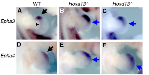
[image:4.612.52.302.58.167.2]Hox proteins is altered. To examine this possibility, we first sought to identify an in vivo readout of Hox activity. A previous comprehensive study has identified several genes regulated by Hoxd13(Cobb and Duboule, 2005). Epha3is one of the genes characterized as a downstream target of Hoxd13. It is not known, however, whether Epha3expression is also regulated by Hoxa13.We demonstrate, by in situ hybridization analysis, that Epha3expression is altered in both Hoxa13–/–and Hoxd13–/–mutant limbs (Fig. 4A-C).
Fig. 2. Expression of Sall1and Sall3is regulated by Shh-Gli3.
Dorsal views of E10.5 forelimbs stained with Sall1(A-C) and Sall3(D-F) with the anterior towards the top. Wild-type (WT; A,D), Shh–/–(B,E) and Gli3–/–(C,F) limbs are shown. Normal expression of Sall1and Sall3is
restricted to the distal-posterior mesenchyme (A,D; black arrows). Both
Sall1and Sall3are downregulated in Shh–/–limbs (B,E; red arrows), and
are ectopically expressed in the anterior mesenchyme in the Gli3–/–
limbs (C,F; blue arrows).
Fig. 3. Reduced Shh signaling in Sall1–/–
;Sall3–/–mutant limbs. Dorsal views of E10.5 (A,B) and E11.5 (C-H) limb buds stained with
Grem1(A-D), Sox9(E,F) and Fgf8(G,H), with the anterior towards the top. Wild-type (WT; A,C,E,G) and Sall1–/–;Sall3–/–(B,D,F,H) limbs are
shown. (A-D)Grem1expression is downregulated in the mutant limb (B,D; arrows), compared with control limbs (A,C). (E,F) Morphological alteration was visible at E11.5 by Sox9in situ hybridization. The control limb has primordia for digit1-digit5 (E). The mutant limb lacks digit1 primordia, exhibits fused digit2 and digit3 primordia, and has delayed separation of digit4 and digit5 primordia (F). (G,H) The Fgf8expression domain is shorter along the anterior-posterior axis in the AER in the mutant (H), compared with the control limb (G). The anterior and posterior margins of Fgf8expression domain are indicated by arrows.
D
E
V
E
LO
P
M
E
N
[image:4.612.343.522.59.267.2]This change includes not only an upregulation of expression but is also an expansion of the expression domain from the anterior edge to the distal middle region. Furthermore, we found that Epha4was also mis-expressed in Hox13mutants, making this gene a likely Hox gene target. Similar to the case of Epha3, Epha4expression is upregulated in both Hoxa13–/–and Hoxd13–/–mutant limbs. These results indicate that Hoxa13and Hoxd13repress Epha3and Epha4expression, and that the expression of Epha3and Epha4is a bona fide indicator of Hoxa13 and Hoxd13 activity in the limb bud.
In order to examine the possibility that Hox gene function is altered in the absence of Sall1and Sall3, we analyzed the expression of the Hox target genes Epha3and Epha4in the Sallmutant limbs. In this analysis, we compared controls with Sall1–/–;Sall3+/–and
Sall1–/–;Sall3–/–mutant limbs in order to clarify whether elimination of more Sall gene alleles has a more severe effect on the expression of Hox targets, as we showed above for limb skeletal elements. In situ hybridization of Epha3and Epha4demonstrated that the extent of mis-expression was directly correlated with Sall gene dose (Fig. 5). In the Sall1–/–;Sall3+/–mutant limb, expression of Epha3and
Epha4is slightly, but clearly, reduced; expression of both genes in the prospective carpal element-forming region is downregulated, and anterior expression of Epha4is also downregulated (Fig. 5B,E). In Sall1–/–;Sall3–/–mutant limbs, the expression of Epha3and Epha4
is more significantly reduced, and the anterior mesenchyme expression of both genes is severely downregulated (Fig. 5C,F). Our results also suggest that Sall1and Sall3regulate expression of Epha3and Epha4. As Hoxa13and Hoxd13repress expression of Epha3and Epha4, these results suggest a possible gain of Hox gene function in Sall1;Sall3mutant limbs.
Hox represses Sall expression
Our results suggest a relationship between Hox activity and Sall activity. We hypothesized that Hox activity represses the expression of Sall1and Sall3, resulting in downregulation of Epha3and Epha4 expression. To test this possibility, we analyzed the expression of Sall1and Sall3in Hoxmutants. Normal expression of Sall1and Sall3starts to regress from the most distal mesenchyme in the E11.5
hindlimb (Fig. 6A,E) (Buck et al., 2001; Ott et al., 2001). Expression of Sall1and Sall3in the Hoxa13–/–mutant limb is slightly stronger than that of a wild-type E11.5 littermate hindlimbs (Fig. 6B,F). In the Hoxd13–/–mutant limb, the expression of Sall1and Sall3is upregulated, and the expression was prolonged in the most distal region when compared with a wild-type littermate (Fig. 6C,G). In the Hoxa13–/–;Hoxd13–/–mutant limb, the expression of Sall1and
Sall3is stronger and more expanded in the large region of the distal mesenchyme when compared with single Hoxa13or Hoxd13mutant limbs (Fig. 6D,H). These results indicate a synergistic activity of Hoxa13 and Hoxd13 in repressing Sall1and Sall3expression.
Sall and Hox compete for a target sequence
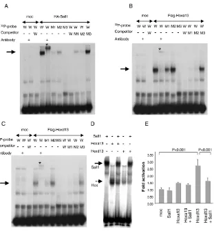
As Hoxexpression is not affected in the absence of Sall1 and Sall3(see Fig. S4 in the supplementary material), the possible gain of Hoxfunction in Sall1–/–;Sall3–/–limbs might be by post-transcriptional regulation. A possible mechanism for such regulation could be that Sall and Hox compete for regulatory elements of common target genes such as Epha3and Epha4. In an effort to address this, we found that the mouse Epha4gene has an AT-rich stretch in the upstream region. At –2028 bp from the transcription start position, we found two recently identified, tandemly positioned, AT-rich Sall1 consensus sequences (Lauberth et al., 2007; Yamashita et al., 2007).
[image:5.612.322.549.59.173.2]As Hox proteins have preferential binding to AT-rich sequences (Pearson et al., 2005), these proteins may act antagonistically in the upstream region for the transcriptional regulation of Epha4. Therefore, we analyzed whether Sall1, Hoxa13 and Hoxd13 can recognize the AT-rich Sall1 consensus sequence upstream of the Epha4gene by EMSA. As shown in Fig. 7A, in vitro translated HA-tagged Sall1 binds to the wild-type probe (arrow). The specificity is confirmed by the supershift induced by the anti-HA antibody (asterisk). With a probe carrying two types of mutations in distinct domains, the binding clearly became weaker. The binding was more affected by introducing two-point mutations on the 3⬘side (M1 probe) than four-point mutations on the 5⬘side (M2 probe). With a probe containing multiple mutations that disrupted the AT-rich Fig. 4. Expression of Epha3and Epha4is upregulated in the
Hoxa13and Hoxd13mutants. Dorsal views of E11.5 forelimbs stained with Epha3(A-C) and Epha4(D-F) with the anterior towards the top. Wild-type (WT; A,D), Hoxa13–/–(B,E) and Hoxd13–/–(C,F) limbs
are shown. Normal Epha3expression in the anterior edge (arrow) and prospective wrist region (arrowhead) (A) is upregulated and expanded posteriorly in the Hoxa13–/–(B, arrow) and Hoxd13–/–(C, arrow) limbs.
Normal Epha4expression in the distal-anterior mesenchyme (D, arrow) is upregulated and expanded distal-posteriorly in the Hoxa13–/–
[image:5.612.52.293.60.182.2](E, arrow) and Hoxd13–/–(F, arrow) limbs.
Fig. 5. Expression of Epha3and Epha4is downregulated in
Sall1;Sall3mutant limbs. Dorsal views of E11.5 forelimbs stained with Epha3(A-C) and Epha4(D-F) with the anterior towards the top. Wild-type (A,D), Sall1–/–;Sall3+/–(B,E) and Sall1–/–;Sall3–/–(C,F) limbs are
shown. (A) Normal expression of Epha3is detected in the anterior edge (arrow) and prospective wrist region (arrowhead). (B)Epha3expression is downregulated in the Sall1–/–;Sall3+/–limb. (C) The anterior edge
expression is more severely downregulated and the prospective wrist region expression is undetectable in the Sall1–/–;Sall3–/–limb. (D) Normal Epha4expression in detected the distal anterior mesenchyme (arrow). (E,F)Epha4expression is downregulated in the Sall1–/–;Sall3+/–limb (E),
and is more severely downregulated in the Sall1–/–;Sall3–/–limb (F).
D
E
V
E
LO
P
M
E
N
sequence (M3 probe), the binding was completely abolished. Conversely, when these wild-type and mutant probes are used as excess amount of cold competitors, we observed complementary results. These results demonstrate that Sall1 binds to the AT-rich sequence in the upstream region of the Epha4gene.
Next, we performed similar experiments with in vitro translated Flag-tagged Hoxa13 and Flag-tagged Hoxd13 (Fig. 7B,C). Both Hoxa13 and Hoxd13 bound the wild-type probe (arrows), and the specificity was confirmed by the supershift induced by the anti-Flag antibody (asterisks). The M1 mutant probe showed reduced binding, although the M2 mutant probe had little effect on binding. Introducing multiple mutations (M3) abolished binding. A complementary result was also observed by using these probes as cold competitors. These results demonstrate that Hoxa13 and Hoxd13 also recognize the upstream sequence of the Epha4gene that is recognized by Sall1.
The results obtained from DNA-binding assays suggest that the competition for a common binding sequence could be one of the mechanisms for the antagonistic function between Sall and Hox. We tested this possibility by examining the relationship between Sall1 and Hox13 for a common binding sequence in vitro. Sall1, Hoxa13 and Hoxd13 bind to the wild-type probe (Fig. 7A-C), and when Sall1 was present together with Hox13, the binding of Hox13 to the probe was reduced (Fig. 7D). The Sall1-DNA complex also became weaker. This suggests that Sall1 and Hoxa13 (or Hoxd13) compete for the target sequence and that such a mechanism could contribute to the mutual antagonistic function between Sall and Hox proteins.
We further examined whether such a competition could functionally contribute to the regulation of Hox activity. For this purpose, we set up a luciferase reporter assay by using an Epha4 upstream region that contains the Sall-Hox binding site. Hoxd13 activated reporter activity, whereas Hoxa13and Sall1 did not activate this element. Importantly, co-expression of Sall1 significantly reduced Hoxd13-dependent reporter activation. Although Hoxa13 and Hoxd13 show different functional contributions to this specific upstream element, similar to the autopod development in vivo (Fromental-Ramain et al., 1996), our data support the idea that DNA binding competition could contribute to the functional antagonism between Sall and Hox13.
DISCUSSION
Sall genes regulate autopod development
Most of the human TBS defects seem to involve the dominant-negative action of a truncated SALL1 protein. Indeed, defects in the anterior part of hands and feet, renal agenesis and anal deformities were observed in a mutant mouse line carrying a truncated Sall1 form that can interact with all Sall proteins (Kiefer et al., 2003). The absence of limb phenotypes in mice mutant for individual Sallgene is most likely due to functional redundancy between the different members of the Sall gene family. The autopod phenotype of Sall1–/–;Sall3–/–mutant confirmed that functional redundancy exists between Sall1and Sall3, though their functional activity is not equivalent. Based on the skeletal phenotypes of the Sall1;Sall3 allelic series, Sall1appears to have a more important contribution than does Sall3. Indeed, Sall1–/–;Sall3–/– mutants showed the strongest phenotype, and milder phenotypes were apparent with the addition of Sall3functional alleles (Sall1–/–;Sall3+/+mutants exhibit a milder phenotype than do Sall1–/–;Sall3+/–mutants). No limb
alterations were observed in mice with other genotypes such as Sall1+/–;Sall3–/–, Sall1+/–;Sall3+/– (Fig. 1). Given that mice
expressing truncated Sall1exhibit loss of digit1 and several carpal elements (Kiefer et al., 2003), our data indicate that the dominant-negative action of a truncated Sall1 might inhibit not only Sall1 but also Sall3 function. Overall, the findings described here support the concept that Sall1and Sall3have partially redundant activity. Such redundant activity among members of the Sallgene family is also observed in Drosophila, where loss of spaltand spalt-relatedgenes cause defects in multiple organs (Dong et al., 2003). Thus, the idea that Sall genes function cooperatively in organogenesis seems to be a conserved feature in vertebrates and invertebrates.
[image:6.612.52.466.59.184.2]Mouse genetic studies shown here, as well as previous studies by others, suggest an organ-specific requirement for different SALL genes in relation to the TBS. The kidney agenesis appears to be caused mainly by a loss of Sall1(Nishinakamura et al., 2001), the anal and heart phenotypes are probably due to inhibition of both Sall4and Sall1 function (Sakaki-Yumoto et al., 2006), and the limb phenotype seems to be caused by a reduction of Sall1and Sall3 function (this study). Sall2 appears to be dispensable for limb development, as the Sall1–/–;Sall2–/–;Sall3–/–triple mutant limb was indistinguishable from that of Sall1–/–;Sall3–/–mutant limb (data not
Fig. 6. Hoxrepresses the expression of Sall1and Sall3. Dorsal views of E11.5 hindlimbs stained with Sall1(A-D) and Sall3(E-H) with anterior towards the top. Wild-type (A,E), Hoxa13–/–(B,F), Hoxd13–/–(C,G) and Hoxa13–/–;Hoxd13–/–(D,H) limbs are shown. (A) Normal Sall1expression
starts to regress from the most distal mesenchyme (arrow). (B) In the Hoxa13–/–limb, the Sall1expression domain became larger and the signal
stronger. (C) In the Hoxd13–/–limb, the Sall1signal is detected in the distal region (arrow) and is stronger than that in the wild type. (D) In the Hoxa13–/–;Hoxd13–/–limb, a large domain in the distal mesenchyme expresses significantly high levels of Sall1(arrow). (E) Normal Sall3expression
also starts to regress from the most distal mesenchyme (arrow). (F) In the Hoxa13–/–limb, higher level of Sall3expression is detected in the anterior
mesenchyme (arrow). (G) In the Hoxd13–/–limb, higher level of Sall3expression is detected in the distal-middle region (arrow). (H) In the Hoxa13–/–;Hoxd13–/–limb, strong expression of Sall3is detected in the wide region of the distal mesenchyme.
D
E
V
E
LO
P
M
E
N
shown). As Sall4is also expressed in the limb mesenchyme, it is possible that Sall4acts together with Sall1and Sall3. As Sall4–/– embryos die soon after implantation (Elling et al., 2006; Sakaki-Yumoto et al., 2006; Warren et al., 2007), the generation of a Sall4 conditional allele is necessary to investigate this issue.
Relationship between Sall4 and Sall1-Sall3
A recent report with a Sall4gene trap line suggests that Sall4is involved in anterior autopod patterning through genetic interaction with Tbx5(Koshiba-Takeuchi et al., 2006). Our analyses suggest that the expression of Sall1and Sall3is not regulated by Tbx5, and that the Sall1–/–;Sall3–/–autopod phenotype is unlikely to be linked to Sall4and Tbx5(see Fig. S2 in the supplementary material). The Sall4gene trap allele would generate a truncated form of Sall4 that might act as a dominant negative, similar to the truncated SALL1 in individuals with TBS. Thus, the observed phenotype in the Sall4GT/+;Tbx5+/–limb might involve reduced activity of Sall1 and
Sall3 by a dominant-negative action of the truncated Sall4, in addition to Sall4haploinsufficiency. Further investigation of the Sall4loss-of-function phenotype, alone and in combination with Sall1and Sall3, will be of particular interest for a comprehensive analysis of the function and contribution of the Sall gene family during limb development.
Sall and Shh signaling
Sall activity appears to be part of Shh pathway. Both Shhand Gli3 signaling have an impact on Sall1and Sall3expression (Fig. 2). Experiments in chicks have demonstrated that Sall1expression in limb buds is regulated by Shh and Fgf, which suggested a possible
involvement of Sall1in distal limb bud patterning (Farrell and Munsterberg, 2000). The reduced expression of Grem1, a Shh-signaling target gene in distal/posterior mesenchyme suggest that Sall function acts to maintain proper levels of Shh signaling in the limb mesenchyme (Fig. 3). Abrogating Shhfunction at various time points during limb development revealed that digit3 formation is the most sensitive to the loss of Shh(Scherz et al., 2007; Zhu et al., 2008). Interestingly, the Sall1;Sall3 mutation affects primarily the formation of digit3 (Fig. 1). Such similarity further supports the idea that Sall1-Sall3 contribution to Shh signaling. However, the fact that digit5, a second digit sensitive to the loss of Shh activity, developed in the Sall1–/–;Sall3–/–mutant limb suggests that Sall1-Sall3 activity is not a major player in Shh signaling events. Our observation that Grem1expression is not abolished but is partially downregulated (Fig. 3) also supports this idea. It is conceivable that Sall4, which is expressed in the distal mesenchyme of the developing limb (Kohlhase et al., 2002), partially compensates for the loss of Sall1 and Sall3. Besides a possible redundancy between Sall1,Sall3and Sall4, the loss of digit1, a Shh-independent digit, in the Sall1–/–;Sall3–/–mutant suggests that Sall1–/–;Sall3–/–phenotype is not exclusively due to reduced Shh signaling.
Epha3and Epha4as targets of Hox activity
Genetic lineage tracing experiments have demonstrated that digit1, which is missing in the Sall1–/–;Sall3–/– mutant autopod, is
[image:7.612.55.366.56.398.2]developed independently of Shh activity (Ahn and Joyner, 2004; Harfe et al., 2004). Furthermore, digit1 develops in the absence of Shhfunction (Chiang et al., 2001; Kraus et al., 2001). Thus, the loss of digit 1 in the Sall1–/–;Sall3–/–mutant limb most probably involves
Fig. 7. Sall1 modulates Hox activity post-transcriptionally. (A) EMSA assay with the HA-Sall1 protein. Sall1 recognizes the Epha4upstream element (arrow). The specificity was confirmed by supershift induced by anti-HA antibody (asterisk). The Sall1-probe complex became weaker by introducing mutations in the probe (M1, M2), and is abolished by introducing multiple mutations (M3). (B,C) EMSA assay with Flag-Hoxa13 protein (B) and Flag-Hoxd13 protein (C). Both Hoxa13 and Hoxd13 recognize the Epha4upstream element (arrows). The specificity was confirmed by supershift induced by anti-Flag antibody (asterisks). The Hox13-probe complex was weaker with the M1 mutant probe, but was not severely affected with the M2 mutant probe. The binding was abolished with the M3 mutant probe containing multiple mutations. M1, mutant1; M2, mutant2; M3, mutant3 (see Materials and methods). (D) Sall1 and Hox13 compete for an
Epha4upstream element. Specific bands formed between 32P-labeled wild-type probe and Sall1
(arrow), and between 32P-labeled wild-type probe
and Hox13 (broken arrow) were detected. By co-incubating with Sall1, the Hoxa13-DNA complex and the Hoxd13-DNA complex became weaker. (E) Luciferase-reporter assay showing Hox-activity modulation by Sall1. The reporter construct was co-transfected with 100 ng of Hoxa13, Hoxd13 and/or Sall1 expression constructs, together with 20 ng pRL-TK. Data are shown as mean±s.d. Significant differences between mock transfected (moc), and Hoxd13, Hoxd13 and Hoxd13+Sall1 are detected (P<0.001).
D
E
V
E
LO
P
M
E
N
a Shh-independent process. Several lines of evidence led us to examine the possible role of Hox genes in this phenotype: (1) in invertebrates, the function of spaltgene has been closely associated with the functions of Hox genes in several developmental contexts (Copf et al., 2006); (2) mice with compound mutations in the 5⬘ Hoxd genes, such as Hoxd11, Hoxd12and Hoxd13, show defects in carpal and metacarpal elements (Davis and Capecchi, 1996), which are also observed in the Sall1–/–;Sall3–/– limb; (3) mice with
compound mutations in Hoxa13 and Hoxd13 exhibit complex autopod phenotypes, such as abnormal condensation and fusion of cartilage elements, demonstrating that correct levels of Hoxa13and Hoxd13function is important for autopod development (Fromental-Ramain et al., 1996); (4) a recent analysis demonstrated that differences in the local level of Hox transcripts specifically regulates digit1 morphogenesis (Montavon et al., 2008). Thus, given that Sall genes encode transcription factors, they might be part of the mechanisms that control Hox gene expression in limbs, which would explain, at least in part, the Sall mutant phenotype. However, we did not observe alteration in the expression of 5⬘Hox genes in Sall1/Sall3 mutants (see Fig. S4 in the supplementary material). An alternative possibility is that Sall proteins have an impact on Hox function at a post-transcriptional level. Recent analyses have identified several genes regulated by 5⬘Hoxd genes (Cobb and Duboule, 2005). One such gene is Epha3, the expression of which is negatively regulated by 5⬘Hoxd genes. Our genetic analyses further uncovered that Epha4 is also regulated by Hoxa13 and Hoxd13(Fig. 4). As such, Epha3and Epha4can be used as bona fide indicators of Hoxa13 and Hoxd13 activity. Even though Hox mutant phenotypes appear to involve complex processes and numerous target genes, analysis of Epha3 and Epha4expression allowed us to evaluate the activities of Hoxa13 and Hoxd13, and revealed that Hox13 and Sall proteins compete for binding on common target sequences.
It is not completely understood how Hox regulates limb morphogenesis. Studies have suggested that Hoxa13regulates cell surface affinity, which affects region-specific cell-cell aggregation and segregation (Stadler et al., 2001; Yokouchi et al., 1995). In these studies, it is suggested that Hoxa13-mediated boundary formation may be an important process for morphogenesis of cartilage elements, and further suggested that the Eph-ephrin system might be involved in the regulation of cell surface affinity and morphogenesis. Eph encodes a receptor tyrosine kinase and ephrin encodes a transmembrane or glycosylphosphatidylinositol-anchored membrane protein. Their interaction is known to regulate cell-cell repulsion as well as attraction, and discrete spatial expression of Ephs and ephrins is known to be important for boundary formation during tissue morphogenesis (Holder and Klein, 1999; Klein, 2004; Poliakov et al., 2004). Interestingly, ectopic expression of ephrin A2 in the developing chick limb, which caused the formation of abnormal ephrin A2 expression boundaries, resulted in abnormal chondrogenic progenitor segregation, leading to a disruption of cartilage morphology (Wada et al., 2003). Furthermore, inactivation of ephrin B1, which causes mosaic expression of the X-linked ephrin B1 in heterozygous mice, generated ectopic ephrin B1-EphB interactions and abnormal digit formation (Compagni et al., 2003). These studies link Hox activity and cell-cell interaction in the control of skeletal elements formation. Hox genes regulate the Eph-ephrin system in other organs (Bruhl et al., 2004; Shaut et al., 2007) and might be important during development of other organs. Thus, studying compound mutants with Eph and ephrin genes in the future could contribute to understanding the role of cell-cell interaction for cartilage morphogenesis.
Sall modulates Hox activity in the limb
Our analysis suggests that Salland Hoxhave antagonistic functions during autopod development. By using the expression of Epha3and Epha4as a marker of Hoxfunction, we have found that Hox and Sall have an opposite impact on their expression. Furthermore, our genetic analysis clearly demonstrates that Hoxa13 and Hoxd13 repress expression of Sall1and Sall3in the autopod. In turn, Sall proteins antagonize Hox function at a post-transcriptional level. Our EMSA assays demonstrated that Sall1, Hoxa13 and Hoxd13 bind to a sequence upstream of the Epha4gene, suggesting that they might directly regulate Epha4expression. Moreover, when co-incubated together, Sall1 competes with Hox13 for binding on the target DNA sequence (Fig. 7D). Luciferase-reporter assay experiments further supported that such competition could contribute to modulating transcriptional activity (Fig. 7E). Hoxd13activated the reporter with the Epha4-upstream element, whereas genetic evidence has demonstrated Hoxd13 as a repressor. Such a context-dependent activator/repressor conversion has been known to occur with several transcription factors, including Hox (Fry and Farnham, 1999; Svingen and Tonissen, 2006). Importantly, co-expressed Sall1 repressed Hoxd13-dependent reporter activation, whereas Sall1 alone did not show an effect on this reporter (Fig. 7E). The reason that Hoxa13did not show significant activation of this reporter is unclear. As the expression of Epha4 is more affected in the Hoxd13–/–limb than in the Hoxa13–/–limb (Fig. 4), the contribution of Hoxd13to the regulation of Epha4expression might be more significant than that by Hoxa13, and reporter activation in vitro might reflect such a difference. Alternatively, such a reporter assay might not completely recapitulate in vivo functions. Nonetheless, our data demonstrate that Sall and Hox can compete for a common target sequence and such competition could contribute to functional modulation. Such competition might, at least in part, contribute to their possible antagonistic function.
Target recognition by Hox proteins is not very strict, favoring a four-base AT-rich core sequence (Pearson et al., 2005). Therefore, depending on the molecular partners that might affect stringency and affinity to target sequences (Svingen and Tonissen, 2006), Hox proteins might bind to a variety of regulatory elements. Contrary to Hox, target recognition by Sall1 is rather stringent (Lauberth et al., 2007; Yamashita et al., 2007). Thus, antagonism by Sall proteins might serve to add local and developmentally timed specific modulation of Hox activity in the autopod. In turn, such antagonistic interactions between Hox and Sall in the autopod might contribute to fine-tuning local cell-cell affinity, leading to segregation or aggregation of chondrogenic progenitors, and thus contribute to generating the complex cartilage architecture of the vertebrate autopod.
The authors are grateful to Dr Naoyuki Wada for helpful discussions, to Dr Suk-Hyun Hong for help and suggestions for the EMSA assay, and to members of the Izpisua Belmonte laboratory for helpful discussions. They also thank Drs Anna Petryk, Atsushi Kuroiwa, Benoit Bruneau, Denis Duboule, Gail Martin, Laura Gammil, Matthew Scott, Pascal Dolle, Pierre Chambon, Richard Maas, Vincenzo Zappavigna and Yasushi Nakagawa for sharing materials. This work is supported by grants from the G. Harold and Leila Y. Mathers Charitable Foundation, Fundacion Cellex, MEC and Marato to J.C.I.B.; by a grant from the Ministry of Education, Culture, Sports, Science and Technology, Japan to R.N.; and by a grant from the Canadian Health Research Institute to M.K.
Supplementary material
Supplementary material for this article is available at http://dev.biologists.org/cgi/content/full/136/4/585/DC1
References
Ahn, S. and Joyner, A. L.(2004). Dynamic changes in the response of cells to
positive hedgehog signaling during mouse limb patterning. Cell118, 505-516.
D
E
V
E
LO
P
M
E
N
Borozdin, W., Steinmann, K., Albrecht, B., Bottani, A., Devriendt, K., Leipoldt, M. and Kohlhase, J.(2006). Detection of heterozygous SALL1 deletions by quantitative real time PCR proves the contribution of a SALL1 dosage effect in the pathogenesis of Townes-Brocks syndrome. Hum. Mutat.27, 211-212.
Bruhl, T., Urbich, C., Aicher, D., Acker-Palmer, A., Zeiher, A. M. and Dimmeler, S.(2004). Homeobox A9 transcriptionally regulates the EphB4 receptor to modulate endothelial cell migration and tube formation. Circ. Res. 94, 743-751.
Bruneau, B. G., Nemer, G., Schmitt, J. P., Charron, F., Robitaille, L., Caron, S., Conner, D. A., Gessler, M., Nemer, M., Seidman, C. E. et al.(2001). A murine model of Holt-Oram syndrome defines roles of the T-box transcription factor Tbx5 in cardiogenesis and disease. Cell106, 709-721.
Buck, A., Kispert, A. and Kohlhase, J.(2001). Embryonic expression of the murine homologue of SALL1, the gene mutated in Townes-Brocks syndrome.
Mech. Dev.104, 143-146.
Buscher, D., Grotewold, L. and Ruther, U.(1998). The XtJ allele generates a Gli3 fusion transcript. Mamm. Genome9, 676-678.
Capdevila, J. and Izpisua Belmonte, J. C.(2001). Patterning mechanisms controlling vertebrate limb development. Annu. Rev. Cell Dev. Biol. 17, 87-132.
Capdevila, J., Tsukui, T., Rodriquez Esteban, C., Zappavigna, V. and Izpisua Belmonte, J. C.(1999). Control of vertebrate limb outgrowth by the proximal factor Meis2 and distal antagonism of BMPs by Gremlin. Mol. Cell 4, 839-849.
Chiang, C., Litingtung, Y., Lee, E., Young, K. E., Corden, J. L., Westphal, H. and Beachy, P. A.(1996). Cyclopia and defective axial patterning in mice lacking Sonic hedgehog gene function. Nature383, 407-413.
Chiang, C., Litingtung, Y., Harris, M. P., Simandl, B. K., Li, Y., Beachy, P. A. and Fallon, J. F.(2001). Manifestation of the limb prepattern: limb development in the absence of sonic hedgehog function. Dev. Biol. 236, 421-435.
Cobb, J. and Duboule, D.(2005). Comparative analysis of genes downstream of the Hoxd cluster in developing digits and external genitalia. Development132, 3055-3067.
Compagni, A., Logan, M., Klein, R. and Adams, R. H.(2003). Control of skeletal patterning by ephrinB1-EphB interactions. Dev. Cell5, 217-230.
Copf, T., Rabet, N. and Averof, M.(2006). Knockdown of spalt function by RNAi causes de-repression of Hox genes and homeotic transformations in the crustacean Artemia franciscana. Dev. Biol. 298, 87-94.
Davis, A. P. and Capecchi, M. R.(1996). A mutational analysis of the 5⬘HoxD genes: dissection of genetic interactions during limb development in the mouse.
Development122, 1175-1185.
Dong, P. D., Todi, S. V., Eberl, D. F. and Boekhoff-Falk, G.(2003). Drosophila spalt/spalt-related mutants exhibit Townes-Brocks’ syndrome phenotypes. Proc. Natl. Acad. Sci. USA100, 10293-10298.
Dudley, A. T., Ros, M. A. and Tabin, C. J.(2002). A re-examination of proximodistal patterning during vertebrate limb development. Nature418, 539-544.
Elling, U., Klasen, C., Eisenberger, T., Anlag, K. and Treier, M.(2006). Murine inner cell mass-derived lineages depend on Sall4 function. Proc. Natl. Acad. Sci. USA103, 16319-16324.
Farrell, E. R. and Munsterberg, A. E.(2000). csal1 is controlled by a combination of FGF and Wnt signals in developing limb buds. Dev. Biol. 225, 447-458.
Fromental-Ramain, C., Warot, X., Messadecq, N., LeMeur, M., Dolle, P. and Chambon, P.(1996). Hoxa-13 and Hoxd-13 play a crucial role in the patterning of the limb autopod. Development122, 2997-3011.
Fry, C. J. and Farnham, P. J.(1999). Context-dependent transcriptional regulation. J. Biol. Chem.274, 29583-29586.
Galant, R., Walsh, C. M. and Carroll, S. B.(2002). Hox repression of a target gene: extradenticle-independent, additive action through multiple monomer binding sites. Development129, 3115-3126.
Goff, D. J. and Tabin, C. J.(1997). Analysis of Hoxd-13 and Hoxd-11 misexpression in chick limb buds reveals that Hox genes affect both bone condensation and growth. Development124, 627-636.
Goodman, F. R.(2002). Limb malformations and the human HOX genes. Am. J. Med. Genet.112, 256-265.
Grant, K., Hanna-Rose, W. and Han, M.(2000). sem-4 promotes vulval cell-fate determination in Caenorhabditis elegans through regulation of lin-39 Hox. Dev. Biol. 224, 496-506.
Harfe, B. D., Scherz, P. J., Nissim, S., Tian, H., McMahon, A. P. and Tabin, C. J.
(2004). Evidence for an expansion-based temporal Shh gradient in specifying vertebrate digit identities. Cell118, 517-528.
Harvey, S. A. and Logan, M. P.(2006). sall4 acts downstream of tbx5 and is required for pectoral fin outgrowth. Development133, 1165-1173.
Hasson, P., Del Buono, J. and Logan, M. P.(2007). Tbx5 is dispensable for forelimb outgrowth. Development134, 85-92.
Holder, N. and Klein, R.(1999). Eph receptors and ephrins: effectors of morphogenesis. Development126, 2033-2044.
Kawakami, Y., Capdevila, J., Buscher, D., Itoh, T., Rodriguez Esteban, C. and Izpisua Belmonte, J. C.(2001). WNT signals control FGF-dependent limb initiation and AER induction in the chick embryo. Cell104, 891-900.
Khokha, M. K., Hsu, D., Brunet, L. J., Dionne, M. S. and Harland, R. M.
(2003). Gremlin is the BMP antagonist required for maintenance of Shh and Fgf signals during limb patterning. Nat. Genet. 34, 303-307.
Kiefer, S. M., Ohlemiller, K. K., Yang, J., McDill, B. W., Kohlhase, J. and Rauchman, M.(2003). Expression of a truncated Sall1 transcriptional repressor is responsible for Townes-Brocks syndrome birth defects. Hum. Mol. Genet.12, 2221-2227.
Kiefer, S. M., Robbins, L., Barina, A., Zhang, Z. and Rauchman, M.(2008). SALL1 truncated protein expression in Townes-Brocks syndrome leads to ectopic expression of downstream genes. Hum. Mutat. 29, 1133-1140.
Klein, R.(2004). Eph/ephrin signaling in morphogenesis, neural development and plasticity.Curr. Opin. Cell Biol. 16, 580-589.
Kmita, M., Kondo, T. and Duboule, D.(2000). Targeted inversion of a polar silencer within the HoxD complex re-allocates domains of enhancer sharing. Nat. Genet. 26, 451-454.
Kmita, M., Fraudeau, N., Herault, Y. and Duboule, D.(2002). Serial deletions and duplications suggest a mechanism for the collinearity of Hoxd genes in limbs. Nature420, 145-150.
Kmita, M., Tarchini, B., Zakany, J., Logan, M., Tabin, C. J. and Duboule, D.
(2005). Early developmental arrest of mammalian limbs lacking HoxA/HoxD gene function. Nature435, 1113-1116.
Kohlhase, J.(2000). SALL1 mutations in Townes-Brocks syndrome and related disorders. Hum. Mutat.16, 460-466.
Kohlhase, J., Wischermann, A., Reichenbach, H., Froster, U. and Engel, W.
(1998). Mutations in the SALL1 putative transcription factor gene cause Townes-Brocks syndrome. Nat. Genet. 18, 81-83.
Kohlhase, J., Heinrich, M., Liebers, M., Frohlich Archangelo, L., Reardon, W. and Kispert, A.(2002). Cloning and expression analysis of SALL4, the murine homologue of the gene mutated in Okihiro syndrome. Cytogenet. Genome Res. 98, 274-277.
Koshiba-Takeuchi, K., Takeuchi, J. K., Arruda, E. P., Kathiriya, I. S., Mo, R., Hui, C. C., Srivastava, D. and Bruneau, B. G.(2006). Cooperative and antagonistic interactions between Sall4 and Tbx5 pattern the mouse limb and heart. Nat. Genet. 38, 175-183.
Kraus, P., Fraidenraich, D. and Loomis, C. A.(2001). Some distal limb structures develop in mice lacking Sonic hedgehog signaling. Mech. Dev.100, 45-58.
Lappin, T. R., Grier, D. G., Thompson, A. and Halliday, H. L.(2006). HOX genes: seductive science, mysterious mechanisms. Ulster Med. J.75, 23-31.
Lauberth, S. M., Bilyeu, A. C., Firulli, B. A., Kroll, K. L. and Rauchman, M.
(2007). A phosphomimetic mutation in the Sall1 repression motif disrupts recruitment of the nucleosome remodeling and deacetylase complex and repression of Gbx2. J. Biol. Chem.282, 34858-34868.
Laufer, E., Nelson, C. E., Johnson, R. L., Morgan, B. A. and Tabin, C.(1994). Sonic hedgehog and Fgf-4 act through a signaling cascade and feedback loop to integrate growth and patterning of the developing limb bud. Cell79, 993-1003.
Litingtung, Y., Dahn, R. D., Li, Y., Fallon, J. F. and Chiang, C.(2002). Shh and Gli3 are dispensable for limb skeleton formation but regulate digit number and identity. Nature418, 979-983.
Mariani, F. V., Ahn, C. P. and Martin, G. R.(2008). Genetic evidence that FGFs have an instructive role in limb proximal-distal patterning. Nature453, 401-405.
McLeod, M. J.(1980). Differential staining of cartilage and bone in whole mouse fetuses by alcian blue and alizarin red S. Teratology22, 299-301.
Merino, R., Rodriguez-Leon, J., Macias, D., Ganan, Y., Economides, A. N. and Hurle, J. M.(1999). The BMP antagonist Gremlin regulates outgrowth, chondrogenesis and programmed cell death in the developing limb.
Development126, 5515-5522.
Michos, O., Panman, L., Vintersten, K., Beier, K., Zeller, R. and Zuniga, A.
(2004). Gremlin-mediated BMP antagonism induces the epithelial-mesenchymal feedback signaling controlling metanephric kidney and limb organogenesis.
Development131, 3401-3410.
Montavon, T., Le Garrec, J. F., Kerszberg, M. and Duboule, D.(2008). Modeling Hox gene regulation in digits: reverse collinearity and the molecular origin of thumbness. Genes Dev.22, 346-359.
Nishinakamura, R. and Osafune, K.(2006). Essential roles of Sall family genes in kidney development. J. Physiol. Sci.56, 131-136.
Nishinakamura, R., Matsumoto, Y., Nakao, K., Nakamura, K., Sato, A., Copeland, N. G., Gilbert, D. J., Jenkins, N. A., Scully, S., Lacey, D. L. et al.
(2001). Murine homolog of SALL1 is essential for ureteric bud invasion in kidney development. Development128, 3105-3115.
Nissim, S., Hasso, S. M., Fallon, J. F. and Tabin, C. J.(2006). Regulation of Gremlin expression in the posterior limb bud. Dev. Biol. 299, 12-21.
Niswander, L.(2003). Pattern formation: old models out on a limb.Nat. Rev. Genet.4, 133-143.
Ott, T., Parrish, M., Bond, K., Schwaeger-Nickolenko, A. and Monaghan, A. P.(2001). A new member of the spalt like zinc finger protein family, Msal-3, is expressed in the CNS and sites of epithelial/mesenchymal interaction. Mech. Dev.101, 203-207.
Panman, L., Galli, A., Lagarde, N., Michos, O., Soete, G., Zuniga, A. and
Zeller, R.(2006). Differential regulation of gene expression in the digit forming
D
area of the mouse limb bud by SHH and gremlin 1/FGF-mediated epithelial-mesenchymal signalling. Development133, 3419-3428.
Parrish, M., Ott, T., Lance-Jones, C., Schuetz, G., Schwaeger-Nickolenko, A. and Monaghan, A. P.(2004). Loss of the Sall3 gene leads to palate deficiency, abnormalities in cranial nerves, and perinatal lethality. Mol. Cell. Biol. 24, 7102-7112.
Pearson, J. C., Lemons, D. and McGinnis, W.(2005). Modulating Hox gene functions during animal body patterning.Nat. Rev. Genet.6, 893-904.
Poliakov, A., Cotrina, M. and Wilkinson, D. G.(2004). Diverse roles of eph receptors and ephrins in the regulation of cell migration and tissue assembly.
Dev. Cell7, 465-480.
Riddle, R. D., Johnson, R. L., Laufer, E. and Tabin, C.(1993). Sonic hedgehog mediates the polarizing activity of the ZPA. Cell75, 1401-1416.
Sakaki-Yumoto, M., Kobayashi, C., Sato, A., Fujimura, S., Matsumoto, Y., Takasato, M., Kodama, T., Aburatani, H., Asashima, M., Yoshida, N. et al.
(2006). The murine homolog of SALL4, a causative gene in Okihiro syndrome, is essential for embryonic stem cell proliferation, and cooperates with Sall1 in anorectal, heart, brain and kidney development. Development133, 3005-3013.
Sato, A., Matsumoto, Y., Koide, U., Kataoka, Y., Yoshida, N., Yokota, T., Asashima, M. and Nishinakamura, R.(2003). Zinc finger protein sall2 is not essential for embryonic and kidney development. Mol. Cell. Biol. 23, 62-69.
Scherz, P. J., Harfe, B. D., McMahon, A. P. and Tabin, C. J.(2004). The limb bud Shh-Fgf feedback loop is terminated by expansion of former ZPA cells. Science 305, 396-399.
Scherz, P. J., McGlinn, E., Nissim, S. and Tabin, C. J.(2007). Extended exposure to Sonic hedgehog is required for patterning the posterior digits of the vertebrate limb. Dev. Biol. 308, 343-354.
Shaut, C. A., Saneyoshi, C., Morgan, E. A., Knosp, W. M., Sexton, D. R. and Stadler, H. S.(2007). HOXA13 directly regulates EphA6 and EphA7 expression in the genital tubercle vascular endothelia. Dev. Dyn.236, 951-960.
Stadler, H. S., Higgins, K. M. and Capecchi, M. R.(2001). Loss of Eph-receptor expression correlates with loss of cell adhesion and chondrogenic capacity in Hoxa13 mutant limbs. Development128, 4177-4188.
Svingen, T. and Tonissen, K. F.(2006). Hox transcription factors and their elusive mammalian gene targets. Heredity97, 88-96.
Sweetman, D. and Munsterberg, A.(2006). The vertebrate spalt genes in development and disease. Dev. Biol. 293, 285-293.
Tabin, C. and Wolpert, L.(2007). Rethinking the proximodistal axis of the vertebrate limb in the molecular era. Genes Dev.21, 1433-1442.
Tarchini, B., Duboule, D. and Kmita, M.(2006). Regulatory constraints in the evolution of the tetrapod limb anterior-posterior polarity. Nature443, 985-988.
te Welscher, P., Zuniga, A., Kuijper, S., Drenth, T., Goedemans, H. J., Meijlink, F. and Zeller, R.(2002). Progression of vertebrate limb development through SHH-mediated counteraction of GLI3. Science298, 827-830.
Toker, A. S., Teng, Y., Ferreira, H. B., Emmons, S. W. and Chalfie, M.(2003). The Caenorhabditis elegans spalt-like gene sem-4 restricts touch cell fate by repressing the selector Hox gene egl-5 and the effector gene mec-3.
Development130, 3831-3840.
Wada, N., Tanaka, H., Ide, H. and Nohno, T.(2003). Ephrin-A2 regulates position-specific cell affinity and is involved in cartilage morphogenesis in the chick limb bud. Dev. Biol. 264, 550-563.
Warren, M., Wang, W., Spiden, S., Chen-Murchie, D., Tannahill, D., Steel, K. P. and Bradley, A.(2007). A Sall4 mutant mouse model useful for studying the role of Sall4 in early embryonic development and organogenesis. Genesis45, 51-58.
Wilkie, A. O.(2003). Why study human limb malformations? J. Anat.202, 27-35.
Wilkinson, D. G.(1993). Whole mount in situ hybridization of vertebrate embryos. In In Situ Hybridization. Oxford, UK: Oxford University Press.
Williams, M. E., Lehoczky, J. A. and Innis, J. W.(2006). A group 13 homeodomain is neither necessary nor sufficient for posterior prevalence in the mouse limb. Dev. Biol. 297, 493-507.
Yamashita, K., Sato, A., Asashima, M., Wang, P. C. and Nishinakamura, R.
(2007). Mouse homolog of SALL1, a causative gene for Townes-Brocks syndrome, binds to A/T-rich sequences in pericentric heterochromatin via its C-terminal zinc finger domains. Genes Cells12, 171-182.
Yoh, S. M. and Privalsky, M. L.(2001). Transcriptional repression by thyroid hormone receptors. A role for receptor homodimers in the recruitment of SMRT corepressor. J. Biol. Chem.276, 16857-16867.
Yokouchi, Y., Nakazato, S., Yamamoto, M., Goto, Y., Kameda, T., Iba, H. and Kuroiwa, A.(1995). Misexpression of Hoxa-13 induces cartilage homeotic transformation and changes cell adhesiveness in chick limb buds. Genes Dev.9, 2509-2522.
Zakany, J. and Duboule, D.(2007). The role of Hox genes during vertebrate limb development. Curr. Opin. Genet. Dev. 17, 359-366.
Zakany, J., Fromental-Ramain, C., Warot, X. and Duboule, D.(1997). Regulation of number and size of digits by posterior Hox genes: a dose-dependent mechanism with potential evolutionary implications. Proc. Natl. Acad. Sci. USA94, 13695-13700.
Zhu, J., Nakamura, E., Nguyen, M. T., Bao, X., Akiyama, H. and Mackem, S.
(2008). Uncoupling Sonic hedgehog control of pattern and expansion of the developing limb bud. Dev. Cell14, 624-632.
Zuniga, A., Haramis, A. P., McMahon, A. P. and Zeller, R.(1999). Signal relay by BMP antagonism controls the SHH/FGF4 feedback loop in vertebrate limb buds.
Nature401, 598-602.