ORIGINAL RESEARCH
PEDIATRICS
Apparent Diffusion Coefficient Levels and
Neurodevelopmental Outcome in Fetuses with Brain MR
Imaging White Matter Hyperintense Signal
XE. Katorza,X G. Strauss,X R. Cohen,XM. Berkenstadt,XC. Hoffmann,XR. Achiron,XE. Barzilay, andX O. Bar-Yosef
ABSTRACT
BACKGROUND AND PURPOSE: One of the perplexing findings of fetal brain MR imaging is white matter T2 hyperintense signal. The aims of our study were initially to determine the main etiologies associated with white matter T2 hyperintense signal, then to examine whether the different etiologies have different ADC values, and, last, to assess the association of white matter T2 hyperintense signal with developmental outcome.
MATERIALS AND METHODS: This was a prospective cohort study of 44 MR imaging scans of fetal brains obtained for suspected brain pathologies at a tertiary medical center during 2011–2015. Clinical data were collected from electronic medical charts. ADC values were measured and averaged in the frontal, parietal, occipital, and temporal lobes. Neurodevelopmental assessments were performed with the Vineland Adaptive Behavior Scales II.
RESULTS:Half of the cases of MRI hyperintense T2 signal of the fetal brain were associated with congenital cytomegalovirus infection. The other half were mainly idiopathic. Thus, the study group was divided to subgroups positive and negative for cytomegalovirus. Both groups had hyperintense signal in the temporal lobe. The group positive for cytomegalovirus had involvement of the parietal lobe. Only this group had increased ADC values in the temporal and parietal lobes. There was no association between the neurodevelopment outcome and the etiologies or ADC values.
CONCLUSIONS: T2 hyperintense signal in fetal brain MRI associated with positive cytomegalovirus infection has increased ADC values in the temporal and parietal lobes, suggestive of brain edema in these areas. However, the association between this finding and neurode-velopment outcome requires further evaluation.
ABBREVIATIONS:CMV⫽cytomegalovirus; fbMRI⫽fetal brain MRI; ICC⫽interclass correlation coefficient; VABS⫽Vineland Adaptive Behavior Scales II; WMHS⫽white matter hyperintense signal
F
etal brain MR imaging (fbMRI) has been increasingly used in recent years as a means of tracking normal and pathologic fetal brain maturation.1One of the perplexing findings of fbMRI iswhite matter T2 hyperintense signal (WMHS). On the one hand, it has been associated with in utero brain pathologies, such as ischemia and cytomegalovirus (CMV) infection.2-4On the other
hand, the validity and relevance of this finding have been questioned.5
In recent years, DWI and its ADC metric have become a quan-titative method for evaluation of fetal brain maturation.6Previous
studies showed that ADC values of the developing fetal brain correlate with fetal brain maturation.7-11Deviation from normal
ADC values has been shown to be associated with brain patho-logies such as ischemia, CMV infection, and ventriculo-megaly.2,12,13Postmortem studies of animal and human fetuses
with hypoxic-ischemic brain injury have demonstrated a transi-tion from low ADC values after the injury to increased values 7 days after the injury. This transition was associated with histologic findings changing from initial cytotoxic edema and swollen astrocytes to vasogenic edema, astrogliosis, and abundance of macrophages.2,14Thus, ADC and its association with
histopa-thology could be used to test the validity and meaning of T2 hyperintensity.
The aims of our study were initially to determine the main Received February 26, 2018; accepted after revision July 19.
From the Antenatal Diagnostic Unit (E.K., G.S., R.C., R.A., E.B.), Department of Ob-stetrics and Gynecology, Pediatric Neurology Unit (O.B.-Y.), The Danek Gertner Institute of Human Genetics (M.B.), and Neuroradiology Unit (C.H.), Department of Diagnostic Radiology, Chaim Sheba Medical Center, Tel-Hashomer, Israel; and Sackler School of Medicine (E.K., G.S., R.C., M.B., C.H., R.A., E.B., O.B.-Y.), Tel Aviv University, Tel Aviv, Israel.
E. Katorza and G. Strauss contributed equally to this work.
Please address correspondence to Omer Bar-Yosef, MD, PhD, Pediatric Neurology Unit, Department of Pediatrics, Chaim Sheba Medical Center, Tel-Hashomer 5262100, Ramat Gan, Israel; e-mail: [email protected]
etiologies associated with WMHS, then to examine whether the different etiologies have different ADC values, and, last, to assess the association of WMHS with developmental outcome.
MATERIALS AND METHODS
Subjects
This was a prospective cohort study of women who were referred for fetal brain MR imaging to our tertiary medical center, Sheba Medical Center, Ramat Gan, Israel, between 2011 and 2015. The cohorts for this study were chosen on the basis of identification of a hyperintense signal on the T2-weighted sequences. Demo-graphic and clinical data were collected from the electronic re-cords of each patient.
Data obtained from the records included maternal history, prenatal screening tests, imaging results from anatomic sonogra-phy and MR imaging, maternal CMV status, and perinatal his-tory. Fetal cytomegalovirus infection was confirmed by either am-niocentesis performed during pregnancy or by the presence of CMV DNA in neonatal urine or saliva.
The study was approved by Sheba Medical Center, Ramat Gan, Israel, institutional ethics board. Informed consent was obtained from each participant in the study prospectively.
MR Imaging Scans
Fetal brain MR imaging was performed using a 1.5T system (Optima MR450w with GEM Suite; GE Healthcare, Milwaukee, Wisconsin). Single-shot fast spin-echo T2-weighted sequences in 3 orthogonal planes were performed using a half-Fourier tech-nique (NEX⫽0.53) with the following parameters: section thick-ness, 3 or 4 mm; no gap; flexible coil (8-channel cardiac coil). FOV was determined by the size of the fetal head with a range of 24⫻24 to 30⫻30 cm; acquisition time was between 40 and 45 seconds (matrix, 320⫻224; TE, 90 ms; TR, 1298 ms; pixel bandwidth, 122 Hz/pixel; specific absorption rate values, 1.1– 1.7 W/kg). A DWI sequence in 1, 2, or 3 orthogonal planes was then performed, with a 40-cm FOV, b-values of 0 and 700 ms, and a slice thickness of 4 mm with no gap. All MR images were obtained by the same protocol at our institution and assessed by a specialist in fetal sonography and a neuroradiologist ex-pert in MR imaging as previously published.15
Target Variables
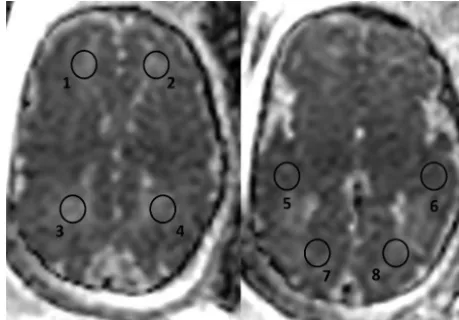
ADC calculation was performed on 8 circular ROIs: 2 on the white matter of both frontal, parietal, temporal, and occipital lobes. A circular ROI was used, placed over the desired anatomic area, ranging from 75 to 98 mm2, similar to that accepted in the recent literature.11
Examples of ROI placements over the various anatomic areas can be seen inFig 1. The ROIs were placed with the following anatomic considerations: Frontal lobe ROIs were placed in the anterior part of the frontal lobes above the lateral ventricles, parietal lobe ROIs were placed in the posterior part of the parietal lobes above the lateral ventricles, temporal lobe ROIs were placed anterior to the temporal horns, and occipital lobe ROIs were placed posterior to the lateral ventricles. The ADC was calculated using IntelliSpace workstation software (Phillips Healthcare, Best, the Netherlands; https://www. usa.philips.com/healthcare/solutions/clinical-informatics/ enterprise-imaging-pacs).
White Matter T2 Hyperintense Signal
WMHS is a subjective interpretation of the signal from the fetal brain white matter on T2 sequences, made by the radiologist. This diagnosis is established when specific areas of the brain white matter appear hyperintense in comparison with other areas or with that expected according to the gestational age (Fig 2).16
Interobserver Validity of ADC Measurements
To validate the consistency of measurements and reliability of results, 2 observers evaluated 10 fetuses. Interobserver variability was assessed by the interclass correlation coefficient (ICC). We considered an ICC value ofⱖ0.8 as excellent agreement.
Neurodevelopmental and Hearing Outcome
Children were assessed by the Vineland Adaptive Behavior Scales, 2nd edition (VABS), which is a structured parent interview assess-ing 4 different domains of behavior: communication, daily livassess-ing skills, socialization, and motor skills. All 4 domains are included in an adaptive composite score.17,18VABS assessment was
con-ducted by a phone interview by 2 medical students trained and supervised by a pediatric neurologist and child development ex-pert experienced in conducting VABS (O.B.-Y.). Validation of the phone interviews were assessed by correlation of the ICC to the VABS scores of 15 children (divided into 7 and 8 children between the students) evaluated by the pediatric neurologist (O.B.-Y.). The ICC was⬎0.83 for the 4 VABS domains and the composite score. Scores of children were considered abnormal if the stan-dard score was⬍70.
Hearing outcome was assessed 2–3 days after delivery by Tran-sient Evoked Otoacoustic Emissions. Neonates positive for CMV were tested routinely before 1 month of age by brain stem audi-tory evoked potential, then at 3, 6, and 12 months by Transient Evoked Otoacoustic Emissions and behavioral assessment.
Statistical Analysis
Categoric variables were expressed as number and percentage. Distribution of continuous variables was assessed using a
[image:2.594.302.532.48.208.2]gram and Q-Q plot. Continuous variables were described using median and interquartile range or mean and SD as appropriate. Categoric variables were compared using the2test, Fisher exact
test, or McNemar test as appropriate. Continuous variables were compared using the Studentttest or Mann-Whit-ney test as appropriate. A 2-tailedP⬍
.05 was considered statistically signifi-cant. Analyses were performed with SPSS (Version 24.0, 2016; IBM, Ar-monk, New York).
RESULTS
Demographic and Clinical Characteristics of the Study Population
The study subjects comprised 43 single-ton pregnant women and 1 single twins pregnant woman who underwent fb-MRI scans. All 44 fetuses included in this study were in their third trimester of pregnancy at the time of the fetal MR imaging, with a median gestational age of 32 weeks (interquartile range, 28.25–35 weeks). The average maternal age was 33 years (interquartile range, 25–32 years). The indications for MR imaging included the following: maternal
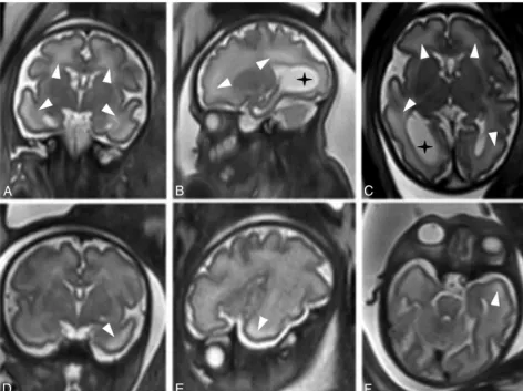
[image:3.594.56.529.48.401.2]cy-FIG 2. T2 MR images (single-shot fast spin-echo T2-weighted sequences in 3 orthogonal planes using a half-Fourier technique, NEX⫽0.53) of 2 fetuses at 33 weeks of gestational age. The CMV-positive fetus has diffuse WMHS (white arrowheads), unilateral ventriculomegaly, and intraventricular adhesions (black asterisks), suggesting ventriculitis (A–C). The CMV-negative fetus has WMHS located in the white matter in the temporal lobes (white arrowhead) (D–F).
Table 1: Demographic and clinical characteristics of fetuses with white matter T2 hyperintense signala
Characteristic
CMV-Positive (n= 22)
CMV-Negative (n= 22)
P Value
Maternal age (yr) 30.5 (26.7–34.2) 31 (28.7–34.2) .74
Gestational age at MRI (wk) 33 (32–34) 33.5 (32–35) .69
Previous pregnancies 2 (2–3) 2 (1–4) .94
Previous labors 1 (1–1.75) 1 (0–2.25) .95
Abnormal outcome in previous labors 6 (27%) 7 (32%) .74
Abnormal maternal medical background 0 6 (27%) .02
Mode of conception 19 (86%) 22 (100%) .35
Spontaneous
IVF 2 (9%) 0 (0%)
Induced pregnancy 1 (5%) 0 (0%)
Sex (male) 12 (54%) 8 (38%) .36
Normal nuchal translucency scan results 21 (95%) 22 (100%) .99 Normal first trimester biochemical test results 21 (94%) 21 (94%) ⬎.99 Normal second trimester biochemical test results 22 (100%) 22 (100%) ⬎.99 Normal early anatomic scan findings 22 (100%) 16 (72%) .02
Normal late anatomic scan findings 21 (94%) 11 (60%) .009
Note:—IVF indicates in vitro fertilization.
aData are presented as median (interquartile range) for continuous variables or number (percentage) for categoric
[image:3.594.55.380.475.662.2]tomegalovirus infection (n⫽31), family history of central ner-vous system pathology (n⫽3), and abnormal findings during fetal sonography (n⫽10). The abnormal sonographic findings included the following: lateral ventricular asymmetry (n⫽4), mega cisterna magna (n⫽2), small head circumference (n⫽2), fisted hands (n⫽1), and hyperechogenic bowel (n⫽1). Twenty-two (50%) of the fetuses were found to be positive for CMV by amniocentesis or by saliva or urinary CMV DNA testing after birth. Thus, to examine the association between ADC values and etiology, we divided the main study group into a CMV-positive group consist-ing of 22 fetuses with confirmed CMV infection and a CMV-negative group consisting of 22 fetuses with WMHS from an unknown etiology.
These 2 distinct patient populations are described separately and compared by their demographic, clinical, imaging, and neu-rodevelopment characteristics.
The median gestational age, pregnancy history, and sex did not differ statistically between the groups. All mothers in the CMV-positive group had normal medical background, com-pared with 6 (27%) mothers in the CMV-negative group who had significant abnormal medical background including thrombophilia, Raynaud disease, and a history of cerebellar infarction (P⫽.02).
In the CMV-positive group, there were significantly less early and late anatomic scan findings (P⫽.02,P⫽
.009, respectively) (Table 1).
Distribution of Hyperintense Signal and Additional MR Imaging Findings
Both groups had similar rates of WMHS in the temporal lobe (P ⬎
.999). WMHS was not depicted in the occipital lobe in both groups. How-ever, the CMV-positive group had sta-tistically higher rates of parietal hyper-intense signal (P⫽.002) and a trend toward higher rates in the frontal lobe (P⫽.067). The CMV-positive group had more extended WMHS involving all 3 lobes (P⫽.004). The CMV-nega-tive fetuses had statistically significantly higher rates (P⫽.046) of minor addi-tional findings on imaging, including subarachnoid cyst (n⫽2), slightly en-larged subarachnoid space (n⫽1), and lateral ventricle asymmetry (n ⫽ 4) (Table 2andFig 2).
Interobserver Validity of ADC Measurements
ADC measurements showed excellent interobserver agreement for all regions, with the ICC ranging between 0.81 and 0.97.
ADC Value Measurements
ADC values for each group were compared with the normal ADC values as published by Hoffmann et al.11Fetuses in the
CMV-positive group were found to have statistically higher ADC values in the temporal lobe bilaterally (P⫽.002 andP⬍.001) and the left parietal lobe (P⫽.033) and a trend in the right parietal lobe (P⫽.057) (Table 3).
ADC values were compared for each region between the 2 study groups. Fetuses in the CMV-positive group were found to have higher ADC values in the left frontal lobe (P⫽.026), the parietal lobe bilaterally (right,P⫽.001; left,P⫽.002), and the temporal lobe bilaterally (right,P⫽.011; left,P⫽.002) (Table 4).
Delivery Data, Hearing, and Neurodevelopmental Assessment
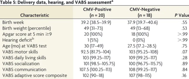
Neurodevelopmental assessment (VABS) and delivery data were analyzed for 20 children in the CMV-positive group and 18 chil-dren in the CMV-negative group (Table 5). Six patients (2 from the CMV-positive group and 4 from the CMV-negative group) refused to participate in the neurodevelopmental assessment. De-livery data and age at developmental assessment were not statisti-cally different between the groups. Comparison of the VABS score for the 4 domains and the adaptive composite score showed only
Table 2: Radiologic MR imaging findingsa
CMV-Positive CMV-Negative PValue
Gestational age at MRI (wk) 33 (32–34) 33.5 (32–35) .6
Radiologic finding
Frontal T2 hyperintense signal 13/22 (59%) 6/22 (27.3%) .067 Parietal T2 hyperintense signal 14/22 (63.6%) 3/22 (13.6%) .002 Temporal T2 hyperintense signal 22/22 (100%) 21/22 (95.5%) ⬎.999 Occipital T2 hyperintense signal 0/22 (0%) 0/22 (0%) ⬎.999 T2 hyperintense signal at 3 lobes (frontal,
parietal, and temporal)
13/22 (59%) 3/22 (13/6%) .004
Additional findings 1/22 (4.5%) 7/22 (31.8%) .046
a
[image:4.594.52.375.198.416.2]Data are presented as median (interquartile range) for continuous variables or number (percentage) for categoric variables.
Table 3: Comparison of ADC values of the study group with the control groupa
Lobe, Side Study Group ADC ADC Control PValue
Frontal
Right CMV⫺ 1793 (191) 1809 (165) .754
Right CMV⫹ 1872 (160) .160
Left CMV⫺ 1741 (141) .115
Left CMV⫹ 1858 (193) .302
Parietal
Right CMV⫺ 1659 (200) 1748 (192) .099
Right CMV⫹ 1840 (140) .057
Left CMV⫺ 1690 (188) .268
Left CMV⫹ 1852 (138) .03
Temporal
Right CMV⫺ 1702 (169) 1692 (164) .843
Right CMV⫹ 1835 (161) .002
Left CMV⫺ 1689 (193) .933
Left CMV⫹ 1846 (122) ⬍.001
Occipital
Right CMV⫺ 1659 (224) 1731 (167) .171
Right CMV⫹ 1723 (129) .830
Left CMV⫺ 1675 (169) .224
Left CMV⫹ 1725 (111) .882
Note:—CMV⫹indicates CMV-positive; CMV⫺, CMV-negative. a
Data are presented as mean (SD). ADC units are 106 mm2
a trend for motor skills (P⫽.07). Four children in the CMV-positive group and 1 child in the CMV-negative group had at least 1 VABS score⬍70; however, this was not statistically significant. Only 1 child had a hearing deficit; this child was CMV-positive with a low VABS score.
DISCUSSION
WMHS is a puzzling finding in fetal MR imaging, and its signifi-cance is not completely understood. The aims of our study were initially to explore the etiologies associated with this finding, then to examine whether the different etiologies are characterized by different ADC values, and, last, to investigate the association of WMHS with neurodevelopmental outcome.
Diverse etiologies were found to be associated with WMHS in this study. The major one (half of the study group), in accordance with fetal MR imaging literature, is congenital CMV infec-tion.13,19,20The precise etiologies of the second half of the group
are not completely clear. Ten fetuses had further imaging find-ings; thus, the WMHS might be a different aspect of the overall brain pathology.
Comparison between the 2 groups demonstrated a difference in the distribution of the WMHS. Both groups had the same prev-alence of WMHS in the temporal lobes, but the CMV-positive group had a higher rate in the parietal lobes and a trend in the frontal lobes. In general, the CMV-positive group had more dif-fuse WMHS.
The median ADC values were higher in the CMV-positive group than in the CMV-negative group in the temporal, parietal,
and left frontal lobes. The ADC values of the CMV-negative group did not differ from those of the control group. Thus, although the incidences of WMHS were similar in the temporal lobes, the median ADC values were different. Because ADC is a quanti-tative measure and not a subjective interpretation, it is possible that some WMHS of the CMV-negative group is actually overinterpretation.
As for the CMV-positive group, the specific involvement of the temporal lobes was previously described in fbMRI. WMHS is part of a spectrum of findings localized to this area, including cysts, dilation of the temporal horns, and reduced temporal lobe volume.4,11,21,22The reason for the specific vulnerability of the
temporal lobe is unclear. Most interesting, in addition to the pres-ence of WMHS in the temporal lobe, most of the fbMRIs in our study depicted a similar signal in the parietal lobe. This finding was previously described in children with congenital CMV.23-25
Thus, the combination of temporal and parietal hyperintense T2 signal is potentially a sign of CMV infection involving the brain. Further involvement of WMHS including the frontal lobes may serve as an additional sign of congenital CMV infection.
The combination of WMHS and high ADC values in the CMV-positive group suggests a possible etiology for this finding. Hyperintense signal was associated at postmortem examination with astrogliosis and extracellular brain edema in the subacute stage of hypoxic-ischemic injury.2Although hypoxic-ischemic
in-jury pathogenesis is different from CMV infection, the simplest explanation of the T2 finding is an increase in extracellular fluid content in the white matter. Kotovich et al26found low ADC
values in fetuses with congenital CMV infection without T2 hy-perintense signal. In a previous study of the same group, Yaniv et al4described postmortem histology consisting of edema and
cel-lular infiltration of plasma cells, lymphocytes, and microglia. The difference in T2 hyperintense signal and ADC values between the 2 latter studies and ours might reflect a different ratio between the edema and cellular component in the brain tissue. When the microgliosis and edema component are more prominent, the ADC values are larger than those of controls; whereas when the component of cellular degeneration and immune cell infil-tration is more prominent, the ADC values are lower than those of controls.2,14,27-29
The association between WMHS and abnormal neurodevel-opmental outcome was not found in this study. In the CMV-positive group, 4 children (20%) had abnormal neurodevelop-mental findings or hearing loss. Based on neurodevelopmental data from pre-vious studies, the expected range of these abnormal findings in children with asymptomatic congenital CMV (normal prenatal imaging findings) is 10%– 15%.30,31Thus, it is difficult to assess
whether the WMHS and the increased ADC values predict worse prognosis.
Combining the results from our study and others2,11-14,26 investigating
[image:5.594.52.285.414.550.2]the association of ADC and brain pa-thologies suggests the following practi-cal advice for clinicians: The combina-tion of WMHS and normal ADC
Table 4: Comparison of ADC values between fetuses with CMV-positive infection and fetuses with isolated white matter hyperintense signala
Lobe, Side CMV-Positive CMV-Negative PValue Frontal
Right 1872 (160) 1793 (192) .147
Left 1858 (193) 1741 (141) .026
Parietal
Right 1840 (140) 1659 (200) .001
Left 1852 (138) 1690 (188) .002
Temporal
Right 1835 (161) 1702 (169) .011
Left 1846 (122) 1689 (193) .002
Occipital
Right 1723 (129) 1659 (224) .259
Left 1725 (111) 1675 (169) .254
a
Data are presented as mean (SD). ADC units are 106 mm2
/s.
Table 5: Delivery data, hearing, and VABS assessmenta
Characteristic
CMV-Positive (n= 20)
CMV-Negative
(n= 18) PValue
Birth week 39.2 (38.5–39.9) 37.9 (39.7–40.6) .55
Birth weight (percentile) 49 (31–73) 49 (13–68) .53
Apgar score at 5 minⱖ9 20 (100%) 18 (100%) ⬎.99
Hearing deficitb 1 (5%) 0 (0%) ⬎.99
Age (mo) at VABS test 30 (17–49) 27.5 (17.2–28.5) .75
VABS motor skills 92.5 (81.75–104) 103 (95.25–108) .07
VABS daily living skills 103 (99.25–117) 109 (99.25–117) .28 VABS socialization 101 (98.5–105.75) 100 (96.75–115.75) .89 VABS communication 107.5 (100.25–113) 108 (99.25–117) .84 VABS adaptive score composite 102 (90–18) 107 (98–115) .16
Child with any VABS score⬍70 4 (20%) 1 (5.5%) .34
a
[image:5.594.54.377.578.714.2]suggests that the source of WMHS is most likely without clinical significance. As for cases of abnormal ADC either below or above the expected value for gestational age, they raise the suspicion of brain pathology such as CMV infection, ventriculomegaly, or ischemia. However, the association of abnormal ADC with neu-rodevelopmental outcome is unclear.
Our study is limited by the size of the study group. A larger number of patients could be used to correlate the distribution of T2 hyperintense signal or ADC values with neurodevelopment. On the other hand, the small number of children in each group strengthens the significance of the difference in T2 hyperintense signal and ADC values between the CMV-positive and -negative groups.
CONCLUSIONS
ADC measurements supported the validity of T2 hyperintense signal in fbMRI only in fetuses with CMV infection. The associa-tion of this finding with neurodevelopmental outcome requires further investigation.
REFERENCES
1. Griffiths PD, Bradburn M, Campbell MJ, et al MERIDIAN collabor-ative group.Use of MRI in the diagnosis of fetal brain abnormalities in utero (MERIDIAN): a multicentre, prospective cohort study.
Lancet2017;389:538 – 46CrossRef Medline
2. Guimiot F, Garel C, Fallet-Bianco C, et al.Contribution of diffusion-weighted imaging in the evaluation of diffuse white matter isch-emic lesions in fetuses: correlations with fetopathologic findings.
AJNR Am J Neuroradiol2008;29:110 –115CrossRef Medline
3. Malinger G, Lev D, Lerman-Sagie T.Imaging of fetal cytomegalovi-rus infection.Fetal Diagn Ther2011;29:117–26CrossRef Medline 4. Yaniv G, Hoffmann C, Weisz B, et al. Region-specific reductions
in brain apparent diffusion coefficient in cytomegalovirus-infected fetuses. Ultrasound Obstet Gynecol 2016;47:600 – 07 CrossRef Medline
5. Fazekas F, Kleinert R, Offenbacher H, et al.Pathologic correlates of incidental MRI white matter signal hyperintensities. Neurology 1993;43:1683– 89CrossRef Medline
6. Schneider JF, Confort-Gouny S, Le Fur Y, et al.Diffusion-weighted imaging in normal fetal brain maturation. Eur Radiol2007;17: 2422–29CrossRef Medline
7. Sartor A, Arthurs O, Alberti C, et al.Apparent diffusion coefficient measurements of the fetal brain during the third trimester of pregnancy: how reliable are they in clinical practice?Prenat Diagn 2014;34:357– 66CrossRef Medline
8. Neil JJ, Shiran SI, McKinstry RC, et al.Normal brain in human newborns: apparent diffusion coefficient and diffusion anisotropy measured by using diffusion tensor MR imaging.Radiology1998; 209:57– 66CrossRef Medline
9. Girard N, Confort-Gouny S, Schneider J, et al.MR imaging of brain maturation.J Neuroradiol2007;34:290 –310CrossRef Medline 10. Toft PB, Leth H, Peitersen B, et al.The apparent diffusion coefficient
of water in gray and white matter of the infant brain.J Comput Assist
Tomograph1996;20:1006 –11CrossRef Medline
11. Hoffmann C, Weisz B, Lipitz S, et al.Regional apparent diffusion coefficient values in 3rd trimester fetal brain.Neuroradiology2014; 56:561– 67CrossRef Medline
12. Erdem G1, Celik O, Hascalik, et al.Diffusion-weighted imaging eval-uation of subtle cerebral microstructural changes in intrauterine fetal hydrocephalus.Magn Reson Imaging2001;25:417–22Medline 13. Yaniv G, Katorza E, Bercovitz R, et al.Region-specific changes in
brain diffusivity in fetal isolated mild ventriculomegaly.Eur Radiol 2016;26:840 – 48CrossRef Medline
14. Baud O, Daire JL, Dalmaz Y, et al.Gestational hypoxia induces white matter damage in neonatal rats: a new model of periventricular leu-komalacia.Brain Pathol2004;14:1–10CrossRef Medline
15. Gat I, Hoffmann C, Shashar D, et al.Fetal brain MRI: novel classifi-cation and contribution to sonography.Ultraschall Med2016;37: 176 – 84CrossRef Medline
16. Lipitz S, Hoffmann C, Feldman B, et al.Value of prenatal ultrasound and magnetic resonance imaging in assessment of congenital pri-mary cytomegalovirus infection.Ultrasound Obstet Gynecol2010;36: 709 –17CrossRef Medline
17. Limperopoulos C, Majnemer A, Steinbach CL, et al.Equivalence re-liability of the Vineland Adaptive Behavior Scale between in-per-son and telephone administration.Phys Occup Ther Pediatr2006;26: 115–27Medline
18. Sparrow SS. Cicchetti D, Balla DA.Vineland Adaptive Behavior Scales. 2nd ed. San Antonio; Pearson: 2005
19. Oosterom N, Nijman J, Gunkel J, et al.Neuro-imaging findings in infants with congenital cytomegalovirus infection: relation to tri-mester of infection.Neonatology2015;107:289 –96CrossRef Medline 20. de Vries LS, Gunardi H, Barth PG, et al.The spectrum of cranial ultrasound and magnetic resonance imaging abnormalities in congenital cytomegalovirus infection.Neuropediatrics2004;35: 113–19CrossRef Medline
21. Cannie MM, Devlieger R, Leyder M, et al.Congenital cytomegalovi-rus infection: contribution and best timing of prenatal MR
imag-ing.Eur Radiol2016;26:3760 – 69CrossRef Medline
22. Doneda C, Parazzini C, Righini A, et al.Early cerebral lesions in cytomegalovirus infection: prenatal MR imaging.Radiology2010; 255:613–21CrossRef Medline
23. van der Knaap MS, Vermeulen G, Barkhof F, et al.Pattern of white matter abnormalities at MR imaging: use of polymerase chain reaction testing of Guthrie cards to link pattern with congenital cytomegalovirus infection. Radiology 2004;230:529 –36 CrossRef Medline
24. Manara R, Balao L, Baracchini C, et al.Brain magnetic resonance findings in symptomatic congenital cytomegalovirus infection.
Pe-diatr Radiol2011;41:962–70CrossRef Medline
25. Hart CK, Wiley S, Choo DI, et al.Developmental disabilities and intracranial abnormalities in children with symptomatic cytomeg-alovirus and cochlear implants.ISRN Otolaryngol2012;2012:502746 CrossRef Medline
26. Kotovich D, Guedalia JS, Hoffmann C, et al.Apparent diffusion co-efficient value changes and clinical correlation in 90 cases of cyto-megalovirus-infected fetuses with unremarkable fetal MRI results.
AJNR Am J Neuroradiol2017;38:1443– 48CrossRef Medline
27. Vannucci RC, Christensen MA, Yager JY.Nature, time-course, and extent of cerebral edema in perinatal hypoxic-ischemic brain
dam-age.Pediatr Neurol1993;9:29 –34CrossRef Medline
28. Lodygensky GA, West T, Moravec MD, et al.Diffusion characteris-tics associated with neuronal injury and glial activation following hypoxia-ischemia in the immature brain.Magn Reson Med2011;66: 839 – 45CrossRef Medline
29. Miller SP, Vigneron DB, Henry RG,et al.Serial quantitative diffu-sion tensor MRI of the premature brain: development in newborns with and without injury. J Magn Reson Imaging2002;16:621– 62 CrossRef Medline
30. Naing ZW, Scott GM, Shand A, et al.Congenital cytomegalovirus infection in pregnancy: a review of prevalence, clinical features, di-agnosis and prevention.Aust N Z J Obstet Gynaecol2016;56:9 –18 CrossRef Medline