PATIENT SAFETY
Functional Contrast-Enhanced CT for Evaluation of
Acute Ischemic Stroke Does Not Increase the
Risk of Contrast-Induced Nephropathy
F.O. Lima M.H. Lev R.A. Levy G.S. Silva M. Ebril E´.C. de Camargo S. Pomerantz A.B. Singhal D.M. Greer H. Ay R. Gilberto Gonza´lez W.J. Koroshetz W.S. Smith K.L. Furie
BACKGROUND AND PURPOSE: Concerns have recently grown regarding the safety of iodinated con-trast agents used for CTA and CTP imaging. We tested whether the incidence of AN, defined by a ⱖ25% increase in the post⫺contrast scan creatinine level, was higher among patients with ischemic stroke who underwent a functional contrast-enhanced CT protocol compared with those who had no iodinated contrast administration.
MATERIALS AND METHODS: The contrast-exposed group consisted of 575 patients with acute isch-emic stroke who underwent CTA (n⫽313), CTA/CTP (n⫽224), or CTA/CTP followed by conventional angiography (n⫽38) within 24 hours of stroke onset and were consecutively enrolled in a prospective cohort study. The nonexposed group consisted of 343 patients with ischemic stroke, consecutively admitted to the same institution, who did not receive iodinated contrast material. Patients were stratified by baseline eGFR. In the primary analysis, the Fisher exact test was used to compare the incidence of AN between the contrast-exposed and the nonexposed patients at 24, 48, and 72 hours and on a cumulative basis. A secondary analysis compared the incidence of AN in patients who underwent conventional angiography following CTA/CTP versus patients who underwent CTA/CTP only.
RESULTS: The incidence of AN was 5% in the exposed and 10% in the nonexposed group (P⫽.003). Patients who underwent conventional angiography after contrast CT were at no greater risk of AN than patients who underwent CTA/CTP alone (26 patients, 5%; and 2 patients, 5%, respectively;P⫽.7). CONCLUSIONS:Administration of a contrast-enhanced CT protocol involving CTA/CTP and conven-tional angiography in selected patients does not appear to increase the incidence of CIN.
ABBREVIATIONS:AN⫽acute nephropathy; CIN⫽contrast-induced nephropathy; Cr⫽creatinine; CTA⫽CT angiography; CTP⫽CT perfusion; eGFR⫽estimated glomerular filtration rate; IQR⫽ interquartile range; kV(p)⫽kilovolt (peak); NIH⫽National Institutes of Health; STOP⫽Screening Technology and Outcomes Project in Stroke
M
ultimodality imaging with MR imaging and CT technol-ogy is increasingly being used to aid in the diagnosis and treatment of acute stroke. The advantages of CT over MR im-aging include its rapid accessibility, lower costs, shorter scan-ning-time intervals, and better patient tolerability. CTA can rapidly and noninvasively identify intra- and extracranial vas-cular stenoses or occlusions.1,2CTP complements theangio-graphic data, defining vascular territories with reduced perfu-sion but potentially salvageable tissue.3CTA and CTP identify vascular and tissue targets for reperfusion strategies.
A limitation of using a CT-based imaging platform has been the concern about the safety profile of the iodinated con-trast agents used for CTA and CTP imaging. CIN is one of the most common causes of hospital-acquired acute renal failure and is associated with increased morbidity and mortality.4,5
To minimize the risk, the American College of Radiology and the European Society of Uroradiology have developed guide-lines for the administration of contrast material in patients with renal failure.6A serum creatinine level determination has become a prerequisite for contrast-enhanced studies in many radiology practices.
Variability in the reported incidence of CIN can be ex-plained by disparate definitions, patient populations, con-trast doses, routes of administration, and timing of patient follow-up.7,8Many of the published studies examined car-diac patients who underwent angiography and could have had other procedural- or perfusion-related causes of renal insufficiency.9,10Very few studies have been performed in
patients with ischemic stroke by using a control group that did not receive contrast material, and none of the studies have stratified patients according to the baseline renal function.11-13
Increases in creatinine levels are not uncommon in
hospi-Received July 3, 2009; accepted after revision September 4.
From the Department of Neurology, (F.O.L., G.S.S., A.B.S., E´.C.d.C., D.M.G., H.A., K.L.F.), Stroke Service, Department of Radiology (M.H.L., S.P., R.G.G.), and Department of Neu-rology, (D.M.G.), Neurocritical Care Service, Massachusetts General Hospital, Boston, Massachusetts; Harvard University (R.A.L.), Cambridge, Massachusetts; Tufts Medical School (M.E.), Boston, Massachusetts; Department of Radiology (H.A.), Martinos Center for Biomedical Imaging, Boston, Massachusetts; National Institute of Neurological Disorders and Stroke (W.J.K.), Bethesda, Maryland; and Department of Neurology (W.S.S.), University of California, San Francisco, San Francisco, California.
Dr. Ay receives grant support from the NIH grant 1-R01-NS059710-01A2. Dr. Singhal receives grant support from the NIH grant R01-NS051412. This work was also supported by the Agency for Healthcare Research and Quality RO1 HS011392, NIH P50 NS051343, the American Heart Association-Bugher Foundation, and the Deane Institute for Integrative Research in Atrial Fibrillation and Stroke. We recognize the generous support of the Esther U. Sharp Fund, the Conway Fellowship Fund, the Lakeside Fund, and the Levitt Fund.
Please address correspondence to Fabricio Oliveira Lima, MD, J. P. Kistler Stroke Research Center, Massachusetts General Hospital, 175 Cambridge St, Suite 300, Boston MA, 02114; e-mail: [email protected]
Indicates open access to non-subscribers at www.ajnr.org
DOI 10.3174/ajnr.A1927
PATIENT
talized patients. In a recent study, Newhouse et al14found that 27% of hospitalized patients with baseline creatinine values between 0.6 and 1.2 mg/dL and 16% of those with baseline creatinine values⬎2.0 mg/dL met the definition of having CIN (a 25% increase in creatinine compared with baseline level) without receiving contrast.
On the basis of these facts, we sought to test whether the incidence of AN was higher among patients with ischemic stroke who underwent CTA, CTP, and conventional angiog-raphy compared with those who had no iodinated-contrast-agent administered.
Materials and Methods
Study Population
The contrast-exposed group consisted of 575 consecutive patients with acute (⬍24 hours from stroke presentation) ischemic stroke enrolled in a prospective cohort study (STOPStroke) at a single aca-demic medical center between March 2003 and June 2005. STOP-Stroke is an observational study to evaluate the utility of emergency CT/CTA/CTP in patients admitted with suspected acute ischemic stroke. The nonexposed group consisted of 343 consecutive patients with ischemic stroke who did not receive iodinated contrast media for tests or procedures such as CTA and intra-arterial thrombolysis, con-secutively presenting to the same institution between September 1999 and June 2000 or between March 2003 and November 2004. Patients already on dialysis treatment were excluded from the study. The study was approved by the institutional review board. Patients provided informed consent for the collection of data.
Neuroimaging Protocol
Nonenhanced CT and CTA acquisitions were performed according to standard departmental protocols with 8- or 16-section multidetector CT scanners (LightSpeed; GE Healthcare, Milwaukee, Wisconsin). Nonenhanced CT was performed in the transverse plane with the patient in a head holder. Representative sample parameters, with minimal variations between scanners shown as ranges, were as fol-lows: 120 – 40 kV(p), 170 mA, 2-second scanning time, and 5-mm section thickness. Imaging with these parameters was immediately followed by biphasic helical scanning, performed at the same head tilt as nonenhanced CT. CTA was performed after a 25-second delay (40 seconds for patients in atrial fibrillation) and administration of a non-ionic contrast agent at an injection rate of 3 mL/s by using a power injector (Medrad Power Injector; Medrad, Indianola, Pennsylvania) via an 18-gauge intravenous catheter. Parameters were 140 kV(p), 220 –250 mA, 0.8- to 1.0-second rotation time, 2.5-mm section thick-ness, 1.25-mm reconstruction intervals, 3.75 mm per rotation table speed, and 0.75:1 pitch. Images were obtained from the C6 vertebral body level through the circle of Willis. Immediately afterward, a sec-ond set of images was obtained from the aortic arch to the skull base. Afterward, source images were reconstructed into standardized max-imum-intensity-projection views of the intracranial and extracranial vasculature.
CTP was initiated 5 seconds after the administration of contrast at 7 mL/s. Four contiguous CT sections were acquired simultaneously every second, during 45– 60 seconds. Scanning sections were 5-mm-thick. Postprocessing of cerebral blood flow and cerebral blood vol-ume maps was done by using commercially available software (CT Perfusion 3; GE Healthcare). CTA/CTP was the standard protocol for patients with stroke considered within the window for reperfusion
therapies (⬍12 hours from stroke-symptom onset). In the 12- to 24-hour window, the decision as to whether to perform CTA or CTA/ CTP was based on clinician preference.
Contrast Agent and Procedures
All patients in the contrast-exposed group received the nonionic io-dinated contrast agent iopamidol (Isovue; Bracco Diagnostics, Princeton, New Jersey) at a dose range from 100 (CTA alone; 313 patients, 54%) to 140 mL (CTA and CTP; 224 patients, 39%). Some patients also underwent transfemoral angiography for intra-arterial thrombolysis after the CTA/CTP (38 patients, 7%), receiving a larger dose of contrast agent (approximately 230 mL total). The clinical institutional protocol recommends hydration and pretreatment with N-acetylcysteine in patients considered at “high risk” for contrast-induced nephropathy (eg, patients with diabetes, baseline creatinine level⬎1.9 mg/dL). Patients in the nonexposed group did not receive iodinated contrast agents.
Patient age, sex, race, and medical history, including diabetes, hy-pertension, congestive heart failure, and coronary artery disease were collected on all patients through interviews, prospective clinical ex-aminations, and review of the medical records by trained staff. Arte-rial hypertension was defined as a positive history or antihypertensive treatment. Diabetes mellitus was defined as a positive history or the presence of oral hypoglycemic medication or insulin treatment. Con-gestive heart failure was defined as a previous positive clinical diag-nosis with episodes of dyspnea requiring emergency treatment or in-patient treatment or appropriate medical treatment. Coronary artery disease was defined by a history of myocardial infarction, typical or atypical angina, electrocardiogram evidence of old myocardial infarc-tion, or a history of a cardiac revascularization procedure. All serum creatinine values available at baseline (admission or pre-CTA) and at 24, 48, and 72 hours after the admission were recorded. The eGFR was calculated with the Modification of Diet in Renal Disease Study equa-tion by using the admission creatinine value.15The term
“contrast-induced nephropathy” cannot be applied to patients who did not receive contrast agents and implies causality. Therefore, the term “AN” was used to define aⱖ25% increase in creatinine from baseline level within 3 days.
Baseline renal dysfunction is an important predictor of a subse-quent increase in creatinine levels.8To control for baseline renal
func-tion, we performed a stratified analysis based on admission eGFR (ⱖ60 mL/min/1.73 m2, 30 –59 mL/min/1.73 m2, and⬍30 mL/min/
1.73 m2), according to guidelines of the National Kidney Foundation
for classification of chronic kidney disease.16
Statistical Analysis
contrast-exposed group (n⫽575). Patients who did not receive io-dinated contrast agent for any tests or procedures were classified un-der the nonexposed group (n⫽343).
We used the Fisher exact test to compare the incidence of AN between the contrast-exposed group and the nonexposed group. A multivariate regression model was used to compare the risk of AN between the exposed and the nonexposed group after adjusting for possible confounders. We also performed a secondary analysis com-paring patients who had conventional angiography following CTA/ CTP (n⫽38) versus patients who underwent CTA and/or CTP alone (n⫽537). In the secondary analysis, we used the Fisher exact test to compare the incidence of AN between patients who underwent con-ventional angiography after CTA/CTP and patients who underwent only CTA and/or CTP. The incidence of AN was compared at each time point (24, 48, and 72 hours) and on a cumulative basis. A 2-sided Pvalue⬍.05 was considered significant. A probability of 4% for developing CIN was assumed on the basis of previous studies with patients with ischemic stroke.9,17,18We had 80% power to detect a
difference of 3% in the incidence of AN between patients who re-ceived iodinated contrast and patients who did not receive iodinated contrast agents (␣⫽0.05;P⬍.05). On the basis of the same assump-tions and a 15% incidence of CIN following angiographic procedures, we were 80% powered to detect a difference of 11% in the incidence of AN between patients who underwent CTA and/or CTP and patients who underwent CTA/CTP followed by conventional angiography.19
Results
The mean age of the population was 68⫾15 years, 48% of the patients were women, and 92% were white. The proportion of patients with conditions that increased the risk of contrast administration, such as diabetes and congestive heart failure, was higher in the noncontrast group (Table 1). Patients in the control group were also older (P⫽.004) and had high baseline levels of creatinine (P⬍.001) and, therefore, lower eGFRs at baseline (P⬍.001).
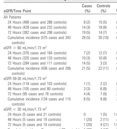
[image:3.594.53.537.59.174.2]The overall incidence of AN was 7% (63 patients). The incidence of AN in contrast-exposed patients was 5% (28 sub-jects), which was lower than the 10% incidence (35 subjects) in the nonexposed group (P⫽.002). Significantly higher inci-dences of AN were noted in the nonexposed group at 24 and 48 hours, but these were not significantly different by 72 hours (Table 2). In the multivariate regression model, after adjusting for age, sex, admission eGFR, hypertension, diabetes, coro-nary artery disease, and congestive heart failure, patients in the contrast-exposed group presented a lower risk for developing
AN than patients in the nonexposed group (odds ratio, 0.42; 95% confidence interval, 0.24 – 0.71).
Thirty-eight patients underwent conventional angiogra-phy following the contrast-enhanced CT. The patients who underwent angiography did not differ significantly from the patients who underwent CTA and/or CTP alone on the basis of age (67⫾17 years versus 65⫾15 years,P⫽.8), sex (53% women versus 47% women,P⫽.5), race (92% white versus 93% white,P⫽.7), hypertension (67% versus 58%,P⫽.4), diabetes (21% versus 16%,P⫽.4), congestive heart failure (10% versus 2%,P⫽.2), coronary artery disease (32% versus 22%,P⫽.2), baseline creatinine level (0.9 mg/dL; IQR, 0.8 – 1.2 versus 1.0 mg/dL; IQR, 0.9 –1.1;P⫽.9) or eGFR (72 mL/ min/1.73 m2; IQR, 63– 84 versus 76 mL/min/1.73 m2; IQR, 60 –90), respectively. The overall incidence of AN in both groups was 5% (P⫽.7). The incidence of AN at 24, 48, and 72 hours did not differ between the angiography and the CTA/ CTP only group.
Table 2: Incidence of AN according to baseline eGFR
eGFR/Time Point
Cases (%)
Controls (%) Pa All Patients
24 Hours (499 cases and 288 controls) 8 (2) 15 (5) .007 48 Hours (428 cases and 232 controls) 14 (3) 18 (8) .01 72 Hours (382 cases and 206 controls) 19 (5) 14 (7) .4 Cumulative incidence (575 cases and 343
controls)
28 (5) 35 (10) .003
eGFR⬎60 mL/min/1.73 m2
24 Hours (376 cases and 164 controls) 7 (2) 12 (7) .004 48 Hours (320 cases and 133 controls) 10 (3) 10 (8) .05 72 Hours (284 cases and 111 controls) 14 (5) 3 (3) .4 Cumulative incidence (436 cases and 206
controls)
21 (5) 22 (11) .01
eGFR 59-30 mL/min/1.73 m2
24 Hours (118 cases and 103 controls) 1 (1) 2 (2) .6 48 Hours (105 cases and 80 controls) 3 (3) 6 (8) .2 72 Hours (95 cases and 76 controls) 4 (4) 7 (9) .2 Cumulative incidence (134 cases and 115
controls)
6 (5) 9 (8) .3
eGFR⬍30 mL/min/1.73 m2
24 Hours (5 cases and 21 controls) 0 1 (5) 1.0 48 Hours (5 cases and 19 controls) 1 (20) 2 (11) .4 72 Hours (5 cases and 19 controls) 1 (20) 4 (21) 1.0 Cumulative incidence (5 cases and 22 controls) 1 (20) 4 (18) .9
a
[image:3.594.299.532.229.485.2]Fisher exact test.
Table 1: Baseline characteristics
Characteristics
Study Population (N⫽918)
Contrast-Exposed (N⫽575)
Nonexposed
(N⫽343) P
Age, mean (yr) 68⫾15 67⫾15 70⫾14 .005a
Sex, % women 48% 48% 48% 1.0b
Race, % white 92% 93% 91% .3b
Hypertension 60% 59% 62% .5b
Diabetes 19% 17% 24% .006b
Congestive heart failure 8% 6% 10% .03b
Coronary artery disease 23% 22% 24% .7b
Admission Cr (mg/dL), median 1.07 (0.8–1.2) 1.0 (0.8–1.1) 1.2 (0.8–1.4) ⬍.001c Admission eGFR (mL/min/1.73 m2), median 73 (56–88) 76 (61–89) 68 (47–87) ⬍.001c
a
Studentttest.
bFisher exact test. c
Discussion
This is the largest reported retrospective cohort study exam-ining the risk of contrast-induced nephropathy in patients with acute stroke. We found that the incidence of AN in the contrast-exposed group did not exceed the incidence of AN in the nonexposed group. In fact, after adjusting for possible confounders, the contrast-exposed group had a lower risk for developing AN than the nonexposed group. The addition of contrast used for conventional angiography after CTA/CTP did not increase the incidence of AN.
Contrast-induced nephropathy is the third most common cause of renal failure, accounting for 11% of cases of hospital-acquired renal insufficiency.4The most commonly accepted definition of CIN is an increase in creatinine ofⱖ25% of the baseline value or an absolute increase ofⱖ0.5 mg/dL in creat-inine above the baseline value within 48 –72 hours of exposure to contrast material.8Serum creatinine level typically peaks 3–5 days after contrast administration and returns to baseline within 1–3 weeks.20Several conditions increase the risk for
CIN. Pre-existing renal disease with an elevated level of serum creatinine level is the primary risk factor for developing of CIN.8 A baseline test of renal function is strongly
recom-mended by the American College of Radiology and European Association of Uroradiologists as a way to assess the risk of CIN.6
Expanded use of CT-based multimodal imaging has been constrained by concerns about potential nephrotoxicity, par-ticularly because time pressures in evaluating patients with acute stroke can necessitate making decisions regarding con-trast administration in the absence of a baseline creatinine value. Smith et al1demonstrated that waiting for the baseline
creatinine level resulted in lengthy delays (average time, 73.3⫾ 51 minutes) in patients with acute stroke. Because acute stroke management protocols seek to minimize the time from symptom onset to thrombolytic delivery, any delay in the process of evaluation is detrimental. This study supports the safety of intravenous contrast agents in patients with acute stroke independent of baseline eGFR when standard prophy-lactic measures are taken (including the use of low-osmolar contrast agents, adequate intravenous hydration, and pre-treatment withN-acetylcysteine in high-risk patients).
A combination of several mechanisms is thought to be re-sponsible for the development of CIN. Renal vasoconstriction and direct tubular injury are thought to be the main factors. The medullary portions of the kidney are particularly vulner-able to reductions in blood flow, given the long length of the vasa recta, the low levels of partial pressure of oxygen encoun-tered, and the high oxygen requirements of the renal tubules responsible for salt reabsorption. Direct tubular injury by con-trast agents may be exacerbated by renal vasoconstriction.21
The incidence of CIN might have been overestimated by ex-trapolating data from cardiology patients who underwent conventional angiography. Local renal hypoxia may be aggra-vated by other complications frequently found in this popula-tion of patients, such as transient reduced cardiac output and perturbations in the pulmonary ventilation-perfusion relationship.
Our data are consistent with the low rates of CIN reported after CTA/CTP in patients with stroke.17,18,22The incidence of
CIN might have been overestimated in previous studies
though. As many as 27% of hospitalized patients with baseline creatinine values between 0.6 and 1.2 mg/dL and 16% of those with baseline creatinine values of⬎2.0 mg/dL would be in-cluded under the definition of CIN (a 25% increase in creati-nine compared with baseline level) without receiving contrast material. Very few studies have used control groups to com-pare the risk of AN in patients receiving and not receiving iodinated contrast.12,13
Pre-existing renal dysfunction is the most important risk factor for the development of CIN. Although an elevated base-line creatinine level is a marker of pre-existing nephropathy, it is not reliable enough to identify patients at risk for CIN.8
Creatinine clearance is the most reliable way to evaluate renal function. Its estimation can be easily performed by using the Modification of Diet in Renal Disease Equation.15The risk of
CIN increases as the eGFR falls, particularly below 60 mL/min/ 1.73 m2.23Our study is the first retrospective cohort study to stratify on the basis of baseline renal function.
There are limitations to our study. Patients undergoing the CTA/CTP were highly selected, as evidenced by the higher rates of diabetes, congestive heart failure, and renal insuffi-ciency in the control population. We believe that this recapit-ulates routine clinical practice, however. We did not collect data on compliance with recommendations for renal protec-tion after contrast administraprotec-tion (including intravenous hy-dration and pretreatment with N-acetylcysteine), though given the acute nature of the studies, hydration was likely the only potential intervention. Because of the low number of cases, the results are not generalizable to patients with severe renal insufficiency (eGFR⬍30).
Conclusions
Although, to our knowledge, it is yet to be established that acute CTA and CTP imaging improve stroke outcomes, they are commonly used in evaluating patients with stroke. This study provides additional evidence that contrast administra-tion does not increase the risk of nephrotoxicity, provided standard prophylactic measures are taken.
Acknowledgments
We sincerely thank the patients who participated and mem-bers of the Massachusetts General Hospital Stroke Service who provided invaluable support.
References
1. Smith WS, Roberts HC, Chuang NA, et al.Safety and feasibility of a CT proto-col for acute stroke: combined CT, CT angiography, and CT perfusion imag-ing in 53 consecutive patients.AJNR Am J Neuroradiol2003;24:688 –90 2. Lev MH, Farkas J, Rodriguez VR, et al.CT angiography in the rapid triage of
patients with hyperacute stroke to intraarterial thrombolysis: accuracy in the detection of large vessel thrombus.J Comput Assist Tomogr2001;25: 520 –28
3. Wintermark M, Reichhart M, Thiran JP, et al.Prognostic accuracy of cerebral blood flow measurement by perfusion computed tomography, at the time of emergency room admission, in acute stroke patients.Ann Neurol2002;51: 417–32
4. Nash K, Hafeez A, Hou S.Hospital-acquired renal insufficiency.Am J Kidney Dis2002;39:930 –36
5. Gruberg L, Mintz GS, Mehran R, et al.The prognostic implications of further renal function deterioration within 48 h of interventional coronary proce-dures in patients with pre-existent chronic renal insufficiency.J Am Coll Car-diol2000;36:1542– 48
a consensus report—Contrast Media Safety Committee, European Society of Urogenital Radiology (ESUR).Eur Radiol1999;9:1602–13
7. McCullough PA, Adam A, Becker CR, et al.Epidemiology and prognostic im-plications of contrast-induced nephropathy.Am J Cardiol2006;98:5K–13K 8. Mehran R, Nikolsky E.Contrast-induced nephropathy: definition,
epidemiol-ogy, and patients at risk.Kidney Int Suppl2006:S11–15
9. Rao QA, Newhouse JH.Risk of nephropathy after intravenous administra-tion of contrast material: a critical literature analysis.Radiology2006; 239:392–97
10. Wyman RM, Safian RD, Portway V, et al.Current complications of diagnostic and therapeutic cardiac catheterization.J Am Coll Cardiol1988;12:1400 – 06 11. Langner S, Stumpe S, Kirsch M, et al.No increased risk for contrast-induced
nephropathy after multiple CT perfusion studies of the brain with a nonionic, dimeric, iso-osmolal contrast medium.AJNR Am J Neuroradiol 2008;29: 1525–29
12. Cramer BC, Parfrey PS, Hutchinson TA, et al.Renal function following infu-sion of radiologic contrast material: a prospective controlled study.Arch In-tern Med1985;145:87– 89
13. Heller CA, Knapp J, Halliday J, et al.Failure to demonstrate contrast nephro-toxicity.Med J Aust1991;155:329 –32
14. Newhouse JH, Kho D, Rao QA, et alFrequency of serum creatinine changes in the absence of iodinated contrast material: implications for studies of con-trast nephrotoxicity.AJR Am J Roentgenol2008;191:376 – 82
15. Levey AS, Bosch JP, Lewis JB, et al.A more accurate method to estimate glo-merular filtration rate from serum creatinine: a new prediction equation—
Modification of Diet in Renal Disease Study Group.Ann Intern Med
1999;130:461–70
16. National Kidney Foundation.K/DOQI clinical practice guidelines for chronic kidney disease: evaluation, classification, and stratification.Am J Kidney Dis
2002;39(2 suppl 1):S1–266
17. Krol AL, Dzialowski I, Roy J, et al.Incidence of radiocontrast nephropathy in patients undergoing acute stroke computed tomography angiography.Stroke
2007;38:2364 – 66
18. Hopyan JJ, Gladstone DJ, Mallia G, et al.Renal safety of CT angiography and perfusion imaging in the emergency evaluation of acute stroke.AJNR Am J Neuroradiol2008;29:1826 –30. Epub 2008 Aug 21
19. McCullough PA, Wolyn R, Rocher LL, et al.Acute renal failure after coronary intervention: incidence, risk factors, and relationship to mortality.Am J Med
1997;103:368 –75
20. McCullough PA, Sandberg KR.Epidemiology of contrast-induced nephropa-thy.Rev Cardiovasc Med2003;4(suppl 5):S3–9
21. Persson PB, Hansell P, Liss P.Pathophysiology of contrast medium-induced nephropathy.Kidney Int2005;68:14 –22
22. Josephson SA, Dillon WP, Smith WS.Incidence of contrast nephropathy from cerebral CT angiography and CT perfusion imaging. Neurology
2005;64:1805– 06