SEQUENTIAL THERAPY VERSUS STANDARD TRIPLE THERAPY IN HELICOBACTER PYLORI ERADICATION
Muzzafar Mohi-Ud-Din, Muzzafer Mohamad Mir, Sajad Sumji, Yawar Yaseen, Majid Khalil
Rather, Mir Intikhab, S. Asif Rafiq and Ursilla
Government Medical College, Srinagar, India
ARTICLE INFO ABSTRACT
Background
shuttered by Barry Marshall and Robbin Warren in 1982 when they identified the organism
Helicobacter pylori. H. pylori
adenocarcinoma and mucosa associated lymphoid tissue lymphoma. The aim of treatment of
infection in any clinical situation is eradication of bacterium from the foregut with armamentarium of antibiotic to choice.
Aim: to study whether
eradication of Helicobacter pylori. Material and methods:
therapy versus standard triple therapy in Helicob
concluded in post graduate department of Medicine, tertiary care institute in 2012. Three hundred patients with documented H. pylori infection studied, were randomized into 3 groups to receive standard or sequent
Results:
triple therapy for 10 days (Group A), second group clarithromycin based sequential therapy days (Group B)and third levofloxacin based sequential therapy for 10 days (Group C). Group A achieved eradication rate of 68% only. While sequential therapy group B and C showed a success of 81 and 86% respectively. In our study, Group B sequential
as compared to standard triple therapy. Conclusion:
sequential treatment for eradicating
The sequential regimen is less expensive and is more effective than conventional therapy for patients with clarithromycin
first line treatment for H. p
Copyright © 2018, Muzzafar Mohi-Ud-Din et al. This unrestricted use, distribution, and reproduction in any medium,
INTRODUCTION
Helicobacter pylori (H. pylori) was discovered by two Australian Nobel Prize winning scientists, Barry
Robin Warren in 1982. It plays a key role in the development of both stomach and intestinal ulcers. It is a spiral, highly mobile, microaerophilic gram negative bacterium with multiple unipolar sheathed flagella (Goodwin, 1987
resides in the deeper mucus gel coating gastric mucosa and between the mucus layer and the gastric epithelium of antrum and proximal segments of the stomach. The genome in pylori encodes-1500 proteins (Fauci, 1989). Multiple virulence factors of Helicobacter pylori that promote colonization and induce tissue injury.
*Corresponding author: Muzzafer Mohamad Mir
DNB Scholar Medical Gastroenterology, Government Medical College, Srinagar, India.
ISSN: 0975-833X
Vol. 10, Issue, 09,
Article History:
Received 29th June, 2018
Received in revised form 20th July, 2018
Accepted 15th August, 2018
Published online 30th September, 2018
Citation:Muzzafar Mohi-Ud-Din, Muzzafer Mohamad Mir, Sajad Sumji, Yawar Yaseen, Majid Khalil Rather, Mir Intikhab, S. Asif Rafiq and UrsillaTaranum
2018. “Sequential therapy versus standard triple therapy in Helicobacter pylori Key Words: Helicobacter pylori, Standard therapy, Sequential therapy, Eradication.
RESEARCH ARTICLE
SEQUENTIAL THERAPY VERSUS STANDARD TRIPLE THERAPY IN HELICOBACTER PYLORI ERADICATION
Din, Muzzafer Mohamad Mir, Sajad Sumji, Yawar Yaseen, Majid Khalil
Intikhab, S. Asif Rafiq and Ursilla Taranum
Government Medical College, Srinagar, India
ABSTRACT
Background: The belief that no organism can survive in the acidic environment of the stomach was shuttered by Barry Marshall and Robbin Warren in 1982 when they identified the organism
Helicobacter pylori. H. pylori is the main cause of gastritis, peptic ulcer disea adenocarcinoma and mucosa associated lymphoid tissue lymphoma. The aim of treatment of
infection in any clinical situation is eradication of bacterium from the foregut with armamentarium of antibiotic to choice.
: to study whether sequential therapy is more effective than standard triple therapy in terms of eradication of Helicobacter pylori.
Material and methods: Our hospital based, prospective, randomized study entitled “Sequential therapy versus standard triple therapy in Helicobacter pylori eradication” was conducted and concluded in post graduate department of Medicine, tertiary care institute in 2012. Three hundred patients with documented H. pylori infection studied, were randomized into 3 groups to receive standard or sequential (clarithromycin or levofloxacin based) anti H. pylori therapy.
Results: Three hundred patients studied were randomized into 3 groups, one group received standard triple therapy for 10 days (Group A), second group clarithromycin based sequential therapy days (Group B)and third levofloxacin based sequential therapy for 10 days (Group C). Group A achieved eradication rate of 68% only. While sequential therapy group B and C showed a success of 81 and 86% respectively. In our study, Group B sequential therapy achieved 13% higher eradication as compared to standard triple therapy.
Conclusion: In conclusion, our large, prospective, hospital based study shows the superiority of sequential treatment for eradicating H. pylori infection compared with convent
The sequential regimen is less expensive and is more effective than conventional therapy for patients with clarithromycin-resistant organisms. Our data suggest that sequential therapy may have a role as a first line treatment for H. pyloriinfection.
This is an open access article distributed under the Creative Commons medium, provided the original work is properly cited.
Helicobacter pylori (H. pylori) was discovered by two Australian Nobel Prize winning scientists, Barry Marshall and Robin Warren in 1982. It plays a key role in the development of both stomach and intestinal ulcers. It is a spiral, highly mobile, microaerophilic gram negative bacterium with Goodwin, 1987). It mostly des in the deeper mucus gel coating gastric mucosa and between the mucus layer and the gastric epithelium of antrum of the stomach. The genome in H. Multiple virulence that promote colonization and
DNB Scholar Medical Gastroenterology, Government Medical College,
Factors promoting Colonization Flagella (Eaton, 1989 Urease (Itoh et al., 1987 Adherence Factors ( Factors Inducing tissue injury
Lipopolysaccharide
Leukocyte recruitment and activating factors (Ernst, 1997)
Vacuolating Cytotoxin (Vac A) Cytotoxin-associated
1998; Yamaoka, 1998
Outer membrane inflammatory protein (Oip A) (Yamaoka, 2000)
Heat shock protein (HSP A, HSP B)
The prevalence of H. pylori
socioeconomic conditions with over 80% of the population
International Journal of Current Research
Vol. 10, Issue, 09, pp.73731-73738, September, 2018
DOI: https://doi.org/10.24941/ijcr.31918.09.2018
Din, Muzzafer Mohamad Mir, Sajad Sumji, Yawar Yaseen, Majid Khalil Rather, Mir Intikhab, S. Asif Rafiq and UrsillaTaranum
Sequential therapy versus standard triple therapy in Helicobacter pylori eradication”, International Journal of Current Research
Available online at http://www.journalcra.com
SEQUENTIAL THERAPY VERSUS STANDARD TRIPLE THERAPY IN HELICOBACTER PYLORI ERADICATION
Din, Muzzafer Mohamad Mir, Sajad Sumji, Yawar Yaseen, Majid Khalil
Taranum
: The belief that no organism can survive in the acidic environment of the stomach was shuttered by Barry Marshall and Robbin Warren in 1982 when they identified the organism is the main cause of gastritis, peptic ulcer disease, gastric adenocarcinoma and mucosa associated lymphoid tissue lymphoma. The aim of treatment of H. Pylori
infection in any clinical situation is eradication of bacterium from the foregut with armamentarium of
sequential therapy is more effective than standard triple therapy in terms of
Our hospital based, prospective, randomized study entitled “Sequential acter pylori eradication” was conducted and concluded in post graduate department of Medicine, tertiary care institute in 2012. Three hundred patients with documented H. pylori infection studied, were randomized into 3 groups to receive
ial (clarithromycin or levofloxacin based) anti H. pylori therapy.
Three hundred patients studied were randomized into 3 groups, one group received standard triple therapy for 10 days (Group A), second group clarithromycin based sequential therapy for 10 days (Group B)and third levofloxacin based sequential therapy for 10 days (Group C). Group A achieved eradication rate of 68% only. While sequential therapy group B and C showed a success of therapy achieved 13% higher eradication
In conclusion, our large, prospective, hospital based study shows the superiority of infection compared with conventional triple therapy. The sequential regimen is less expensive and is more effective than conventional therapy for patients resistant organisms. Our data suggest that sequential therapy may have a role as a
Commons Attribution License, which permits
Factors promoting Colonization 1989) Itoh et al., 1987)
(Boren, 1993) Factors Inducing tissue injury
Lipopolysaccharide (Moran, 1996)
Leukocyte recruitment and activating factors
Vacuolating Cytotoxin (Vac A) (Blaser, 1996) associated antigen (Cag A) (Van Doorn, 1998; Yamaoka, 1998)
Outer membrane inflammatory protein (Oip A)
Heat shock protein (HSP A, HSP B)
The prevalence of H. pylori is strongly correlated with socioeconomic conditions with over 80% of the population in
INTERNATIONAL JOURNAL OF CURRENT RESEARCH
Din, Muzzafer Mohamad Mir, Sajad Sumji, Yawar Yaseen, Majid Khalil Rather, Mir Intikhab, S. Asif Rafiq and UrsillaTaranum,
developing countries and 20-50% in industrialized countries affected (Leal-Herrera, 2003). Infection is acquired by oral ingestion of the bacterium in vomitus, saliva or feces and is mainly transmitted within families in early childhood. There is frequent reinfections following eradication therapy in adults (Soto et al., 2003; Suerbaum, 2002). H. pylori infection has pathogenic role in majority of duodenal and gastric ulcers, and there is strong evidence that it also increases the risk of gastric cancer and gastric mucosa associated lymphoid tissue lymphomas (Leal-Herrera et al., 2003).H. pylori infection that involves the antrum predominantly, while relatively sparing the acid-secreting portion of the stomach, will predispose to duodenal ulceration whereas intense inflammation in the oxyntic mucosa will result in gastric atrophy with a decreased acid output and a predisposition to gastric ulceration and cancer (Graham, 1998). Non-gastrointestinal tract diseases (Leon Tiadis, 1999) possibly associated with H. pylori infection has come up recently although the data supported these associations are weak.
The diseases include Iron deficiency anemia, Coronary artery disease, Cerebrovascular disease, Hypertension, Raynaud’s phenomena, Migraine headaches, Vomiting of pregnancy, Immune thrombocytopenic purpura, Hyperammonemia, Sudden infant death syndrome, Growth retardation, Anorexia of aging, Rosacea and Chronic urticaria. Diagnoses of H. pylori may be divided into that do (Biopsy based tests) and that do not require sampling of gastric mucosa (non-invasive tests). In biopsy based tests at least three samples (e.g. from the lesser curvature angularis, the greater curve pre-pyloric antrum, and the greater curve body) are taken. The standard hematoxylin and eosin (H&E) stain is excellent to determine histological chronic or chronic active inflammation (gastritis) and demonstrates H. pylori if large number of organisms are present. A special stain (e.g. silver stain) is better at detecting the organism if small numbers of bacteria are present. Attributes of both H&E and a special stain are found in the genta and El-Zimaity ‘triple’ stains, which combine the H&E stain, H. pylori selective stains, and alcian blue to detect intestinal metaplasia (Gents, 1994). The alternative is to use two different stains, a combination of an H&E and a Diff-Quik stain is probably the best alternative (El-Zimaity et al.,1998). Rapid urease test is a rapid test for diagnosis of H. pylori. The basis of the test is the ability of H. pylori to secrete the urease enzyme, which catalyzes the conversion of urea to ammonia and bicarbonate. The test is performed at the time of gastroscopy. A biopsy of mucosa is taken from the antrum of the stomach, and is placed into a medium containing urea and an indicator such as phenol red. The urease produced by H. pylori hydrolyzes urea to ammonia, which raises the pH of the medium, and changes the color of the specimen from yellow (negative) to red (positive).There is evidence to suggest that H. pylori moves proximal in the stomach in patients on therapy with proton pump inhibitors, and, as such, samples from the fundus and antrum should be taken in these patients. Non-invasive tests include serological tests, urea breath tests and stool antigen tests. Serological tests (IgG antibodies) are generally not useful to confirm cure after antimicrobial therapy, a fall in antibody titers of 20% or more 6 months after completion of therapy may be sensitive in confirming cure of infection (Cutler, 1996). Urea breath tests using urea labeled with either 13C or 14C that is ingested (Graham, 2001) and are preferred means of evaluating the success of antimicrobial therapy in clinical practice, but patient should be off PPIs for at least 7 days before the test can be done otherwise one third of
patients will give false negative tests (Graham, 2003). H2 receptor antagonists can be continued up to the day before urea breath testing and provide an alternative for the patient who derives continued antisecretory therapy. Another new, non-invasive diagnostic test is a stool antigen test based on detection of H. pylori antigens in stool. Overall, studies using pretreatment H. pylori stool antigen tests have shown that the sensitivity and specificity of the tests are comparable to histology or urea breath tests (Gilbert, 2001). Cure of H. pylori infection is not easy and requires combination of antibiotics often with additional non-antibiotic adjunctive agents; single agents are ineffective. The finding that the elimination of H. pylori infection changes the natural history of peptic ulcer disease (Hentschel et al., 1993)and gastric mucosa associated lymphoid tissue lymphoma (Steinbach, 1999) has led to the development of successful strategies to clear the organisms from persons with these disorders, keeping in view the prevalence of peptic ulcer disease (gastric ulcer and duodenal ulcer) in the United States, with four million individuals affected per year. Lifetime prevalence of peptic ulcer disease in United States is ~12% in men and-10% in women (John Del Valli, 1989). Several combination therapies have been an effective standard of treatment, however resistance rates have been rising and eradication failures have increased to 1 in 5 patients (Vakil, 2006). Reported clarithromycin resistance is 10 to 12% in patients infected with H. pylori and that of metronidazole is 25.1% during the period from 1999 through 2002 (Duck et al., 2004). Several therapies have come like dual therapy (PPI plus amoxicillin, PPI plus clarithromycin, ranitidine bismuth citrate (tritec) plus clarithromycin) is not recommended in view of eradication rates of <80-85%. At present the standard treatment for H. pylori infection that has been endorsed relay on clarithromycin or metronidazole in conjunction with other antibiotics and PPI (European, 2000; Hauden, 1998) i.e. amoxicillin 1g BD + clarithromycin 500mg BD + PPI like Pantoprazole 40mg BD for 10-14 days. But the rate of eradication with such a regimen has decreased (88% in 1996 and 69.4% in 1999) (Kadayific, 1996; Urgun, 1999). Novel first line anti H. pylori therapy in 2011 include sequential therapy, concomitant quadruple therapy, hybrid (dual-concomitant) therapy and bismuth containing quadruple therapy. In bismuth containing Quadruple therapy we use (bismuth + metronidazole / clarithromycin+ tetracycline + PPI) where clarithromycin is substituted for metronidazole (or vice versa) (John Del Velli, ?). So it remains to be determined which therapy is best and cost effective in terms of H. pylori eradication.
Aim: To study whether sequential therapy is more effective
than standard triple therapy in terms of eradication rates of H. pylori.
MATERIALS AND METHODS
indicated that test was positive for H. pylori and subject henceforth was included in study group. Rapid urease test kit was supplied to us by hospital (Allied Marketing Co). A total of 654 patients participated in our study out of which rapid urease test was positive in 463 and 163 subjects dropped out of study, the rest 300 were randomized into three groups (A,B&C) of 100 each, by using method of simple random sampling to avoid selection bias. Group A received standard triple therapy, Group B clarithromycin based sequential therapy and Group C levofloxacin based sequential therapy.
Exclusion criteria
Patients less than 18 years or more than 70 years of age. Pregnant and lactating mother.
Patients on prolonged PPI therapy, anticoagulants, steroids and/or NSAID.
Malignancy of esophagus and stomach, chronic liver disease (CLD) patients.
Comorbid medical conditions, severe or unsuitable cardiovascular, pulmonary or endocrine disease, clinically significant hepatic or renal disease or dysfunction.
Patients or their guardians signed consent before participation in the study. A detailed clinical history, relevant physical and abdominal examinations were carried out. Routine laboratory studies were performed and USG abdomen of subjects was also done.
Treatment regimens used in three groups:
(1)Standard Triple Therapy (Group A): Pantoprazole 40mg
(twice daily) +Clarithromycin 500mg (twice daily) +Amoxicillin 1g (twice daily)
Total duration of treatment (10 Days)
(2) Sequential Therapy
(I) Clarithromycin Based (Group B)
(DAY 1-5):
1.Pantoprazole (40mg twice daily) 2.Amoxicillin (1g twice daily)
(DAY 6-10):
1.Pantoprazole (40mg twice daily) 2.Clarithromycin (500mg twice daily) 3.Tinidazole (500mg twice daily)
(II) Levofloxacin Based (Group C)
(DAY 1-5)
1.Pantoprazole (40mg twice daily) 2.Amoxicillin (1g twice daily)
(DAY 6-10):
1.Pantoprazole (40mg twice daily) 2.Levofloxacin (250mg twice daily) 3.Tinidazole (500mg twice daily)
The aim of our study was eradication of H. pylori infection. All the participants in the study were re-endoscoped four
weeks after completion of drug regimen for H. pylori eradication and test used was RUT.
Statistical Analysis
The difference between the proportions of eradicated infections for the three treatments was calculated by using the method recommended by Newcombe and Altmen.
The level of significance was assessed by using Mannwhitney ‘U’ test and Kruskal Wallis test for Non metric variables. Student’s t test and ANOVA was used for metric Variables. Intergroup variance was checked at 95% CI. MS Excel, Minitab and SPSS software was used for data analysis. The analysis of data enabled us to determine whether sequential treatment regimen is better than standard triple therapy and henceforth can be recommended as initial therapy for H. pylori eradication.
RESULTS
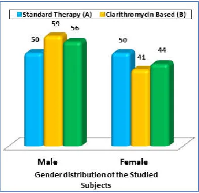
The age of 300 patients in our study ranged from 18 to 70 (mean age 44.3±15 yrs.). Most of the patients in our study belonged to age group 41 to 50 and least between 61 to 70 yrs. Distribution of subjects across age and gender in our three study groups is as given in graph 1 and 2.
Graph 2. Top of columns gives no’s in each group
Most of patients in studied groups presented with dyspepsia, which was seen in 59% in Group A, 66% in Group B and 66% in Group C. Malena was seen in 29%, 29% and 26% subjects in different groups. Hematemesis was seen in 15, 12 and 13% patients in Group A Group B and Group C respectively. The intergroup difference was not statistically significant as depicted in graph 3.
Graph 3. Top of columns gives no’s in each group
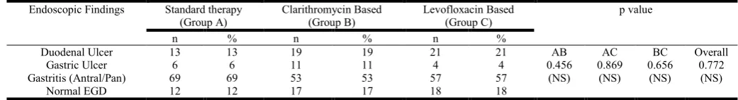
Graph 4. Top of columns gives percentage in each group
Esophagogastroduodenoscopy (EGD) was normal in 94 study subjects. Rest subjects have findings as depicted in table 1 and graph 4. Rapid urease test determined eradication of H. pylori in patients receiving levofloxacin based sequential therapy (Group C) was 86% whereas the subsequent reduction in clarithromycin based sequential therapy (Group B) and standard triple therapy group (Group A)were 81% and 68%. The intergroup difference of A and C was significant (p<0.05), besides A and B was also significant (p<0.05) as depicted in table 2 /graph 5.
Top of columns gives percentage in each group
DISCUSSION
[image:4.595.65.262.53.244.2]The mechanism proposed for the success of the sequential therapy is that bacteria develop efflux channels for clarithromycin, which rapidly transfers the drug out of the bacterial cell, preventing the antibiotic from binding to the ribosome (De Francesco et al., 2006). Because amoxicillin acts on the bacterial cell wall and weakens it, the initial phase of treatment prevents the development of efflux channels by weakening cell wall of bacterium (De Francesco et al., 2006).
Our hospital based, prospective, randomized study entitled “Sequential therapy versus standard triple therapy in Helicobacter pylori eradication” was conducted and concluded in post graduate department of medicine in 2012. Three hundred patients studied were randomized into 3 groups, one group received standard triple therapy for 10 days (Group A), second group clarithromycin based sequential therapy for 10 days (Group B)and third levofloxacin based sequential therapy for 10 days (Group C). Group A achieved eradication rate of 68% only. While sequential therapy group B and C showed a success of 81 and 86% respectively.
To understand the relative efficacy of sequential therapy compared with standard triple therapy, we performed a systematic literature review and meta-analysis of randomized, controlled trials (RCTs) comparing these 2 treatment. Patients taken in our study were between 18 and 70 years of age. The mean age in standard therapy group (Group A) was 43.3+14.7 years, in clarithromycin based study group (Group B) mean age was 45.4+14.4 years and in levofloxacin therapy based sequential therapy group (Group C) mean age was 44.1+16.1. In a Study by De Francesco et al in 2004 mean age of standard therapy was 46 and sequential was 44.2 years. Dyspepsia is defined as pain or discomfort in the central upper abdomen which originates in the upper gastrointestinal tract. Most of patients in studied groups presented with dyspepsia, which was seen in 59% in Group A, 66% in Group B and 66% in Group C. Malena was seen in 29, 29 and 26% subjects in different groups. Hematemesis was seen in 15, 12 and 13% patients in Group A Group B and Group C respectively. Endoscopically most subjects had gastritis (Antral/Pan) across all groups. It was 69% in Group A (standard therapy group), 53% in Group B and 57% in Group C. Duodenal ulcer was seen in 13, 19 and 21% of patients in Group A, Group B and Group C respectively. This was followed by normal EGD i.e. non-ulcer dyspepsia (12, 17, and 18%), least common finding seen endoscopically was gastric ulcer in studied subjects (6, 11 and 4%).
The pathophysiological mechanisms by which the infection may cause dyspepsia are unclear, but may include changes in acid secretion, abnormal motility, or altered visceral perception. Most researchers believe that there is a relation, although an imperfect one, between non-ulcer dyspepsia and infection with H. pylori. In our study we involved even non ulcer dyspepsia patients. Bruley Des Varannes et al in 200145 showed that there are benefits for eradicating H. pylori in patients with non-ulcer dyspepsia, although in majority of patients relief of symptoms is less likely. Prevalence of peptic ulcer was quiet similar to study by MS Khuroo et al in 198946 showed the life time prevalence of peptic ulcer 11.2% with peak incidence in 5th decade of life. R A Moore MA DPhil in 199447 showed peptic ulcers are found in 25% of dyspeptic patients whose blood tests positive for H. pylori, compared with only 3% of similar patients who test negative. Combining data from three separate studies shows that rates of gastric and duodenal erosions, and gastric cancer, are also higher in patients who test positive for H pylori.This indicates that testing blood for H. pylori can be a useful way of determining which patients would benefit from conventional conservative therapy (acid-suppressing medicines) and those who would benefit from curing H. pylori infection.
The overall eradication rate with our standard triple therapy of 10 day duration (Group A) was 68%, with clarithromycin based sequential therapy (Group B) showed a success rate of 81% and levofloxacin based sequential therapy (Group C) 86% respectively. The results of this study show that sequential therapy is superior to triple therapy for the eradication of H. Pylori infection. The study also demonstrates that triple therapy, which is the current standard treatment, has low eradication rate. Our study supported most other studies on H. Pylori eradication that show higher eradication rates with sequential therapy than standard therapy. Choi WH et al48 conducted a study in Asia in 2008 showed eradication by 10 day sequential therapy by 77.9% and 71.6% by standard triple regimen, quiet similar to our findings. Nadim S Jafrri et al in 200849 showed the eradication rates by sequential therapy (clarithromycin based) by 93.4% and by standard triple therapy by 76.9%,which was an European study and showed higher eradication rates as compared to our study, reason may be that in it study 2747 patients were taken as compared to ours were only 300 participated in study and difference in H. pylori between east and west (Rupert et al., 2009). Sanchez-Delgado et al. in 2008 showed eradication rates by sequential therapy (clarithromycin based) by 84.2% results quiet similar to our Table 1. Endoscopic Findings in the Studied Subjects
Endoscopic Findings Standard therapy (Group A)
Clarithromycin Based (Group B)
Levofloxacin Based (Group C)
p value
n % n % n %
Duodenal Ulcer 13 13 19 19 21 21 AB AC BC Overall
Gastric Ulcer 6 6 11 11 4 4 0.456
(NS)
0.869 (NS)
0.656 (NS)
0.772 (NS)
Gastritis (Antral/Pan) 69 69 53 53 57 57
[image:5.595.38.559.81.152.2]Normal EGD 12 12 17 17 18 18
Table 2. Rapid Urease Test
Standard Therapy (Group A)
Sequential Therapy Result
Clarithromycin Based (Group B)
Levofloxacin Based (Group C)
n % n % n % AB AC BC Overall
Rapid Urease Test before treatment +Ve 100 100 100 100 100 100 1.000 (NS) 1.000 (NS) 1.000 (NS) 1.000 (NS)
Rapid Urease Test after treatment +Ve 32 32 19 19 14 14 0.035 (Sig) 0.003 (Sig) 0.342 (NS) 0.006 (Sig)
study in which eradication rate of H. Pylori was 81% respectively by clarithromycin based therapy. Uygun et al. in 2008 also conducted a study in which eradication by sequential therapy was 80.1% and 63% by standard triple regimen again, slightly deviating from our study, causes may be it used tetracycline and eradication was confirmed 6 weeks after therapy as against ours where were endoscoped subjects after 4 weeks. Moayyedi P et al in 2009 identified 12 trials with 3271 patients and concluded that sequential therapy achieved 12% better eradication rates. In our study sequential therapy was 13% superior to standard triple regimen. Romano M et al in 2010 compaired levofloxacin and clarithromycin based sequential therapy with eradication of 80.8% with clarithromycin sequential therapy, 96% with levofloxacin 250 mg sequential therapy and 96.8% with levofloxacin 500 mg sequential therapies. Our study also showed higher efficacy of levofloxacin based regimen as compared to clarithromycin based therapy. Recent several studies have shown that a novel 10 day sequential therapy can achieve success rate of 90-94% (Vaira et al., 2007). Gatta L et al in 2009 reported a systemic review of 13 trials (3271 patients) and showed that sequential therapy achieved 12% higher eradication rate than standard triple therapy. In our study, Group B i.e. clarithromycin based sequential therapy achieved 13% higher eradication as compared to standard triple therapy.
Data of H. pylori eradication rate in Indian patients are available from several clinical trials.
Author Treatment regimen
No. of patients
Time of testing
Eradication rate
Dayal (1997)57 BC/T4/M 57 4 weeks 54%
Ahuja (1998)58 LAS 21 6 & 12 wks 86%
LCS 18 6 & 12 wks 83% LPS 21 6 & 12 wks 71%
Bhasin (1999)59 OC 22 4 wks 68%
OAC 20 4 wks 70%
BC/A/M 22 4 wks 59%
Bhasin (2000060 LAC 2wk 24 6 wks 96%
LAC 2wk 22 6 wks 54%
Pari (2003)61 LAC 35 4 wks 82%
L/BC/T4/M 33 4 wks 72%
L – Lansoperazole; A = Amoxicillin, S = Secnidazole; O = Omeperazole; C = Clarithromycin; BC = Bismuth Citrate; T4 = Tetracycline; M = Metronidazole
Our trial has limitations; the results may not be applicable to other countries and populations. It does not tell us about the percentage of subjects having clarithromycin resistance and our study design does not tell us whether the improved effect with sequential therapy is due to the sequential administration or to the additional antibiotic (tinidazole) that is not contained in the standard regimen. Although sequential therapy is an improvement over current therapies, it does not decrease the duration of therapy. In conclusion, our large, prospective, hospital based study shows the superiority of sequential treatment for eradicating H. pylori infection compared with conventional triple therapy. The sequential regimen is less expensive and is more effective than conventional therapy for patients with clarithromycin-resistant organisms. Side effects with both regimens were similar and consisted mostly of diarrhoea and abdominal discomfort. Our data suggest that sequential therapy may have a role as a first line treatment for
H. pylori infection.
Acknowledgements: We would like to thank the Department
of Medicine, Government Medical College Srinagar.
Funding: No specific funding was sought for this work.
Availability of data and materials: The data on which this
study has been based are freely and publicly available from hospital record department.
Authors' contributions: All authors contributed in every
aspect of study. All authors read and approved the final manuscript.
Competing interests: No competing interest.
Consent for publication: Consent to participate is not
provided as no individual data is provided and it is not possible for patients to be identified from the anonymised data used.
REFERENCES
Ahuja V, Dhor A, Bal C, Sharma MP. 2006. Lansoperazole, secnidazole with clarithromycin, amoxicillin or levofloxacin in the eradication of H. pylori. Null Jan. Bhasin DK, Sharma BC, Sinha SK et al. 2000. Comparison of
7 and 14 days of lansoperazole, clarithromycin and amoxicillin therapy for eradication of H. pylori: report from India. Helicobacter, 5: 84-87.
Bhasin DK, Sharma BC, Sinha SK et al. 2000. Comparison of 7 and 14 days of lansoperazole, clarithromycin and amoxicillin therapy for eradication of H. pylori: report from India. Helicobacter, 5: 84-87.
Bhasin DK, Sharma BC, Sinha SK, Ray P et al. 1992. H. pylori eradication comparison of three treatment regimens in India. Clin Gastroenterol., 28: 348-55.
Bigard MA, Delchier JC, Riachi C et al. 1998. One week triple therapy using omeperazole, amoxicillin and clarithromycin for the eradication of H. Pylori in patients with non-ulcer dyspepsia. Influence of dosage of omeperazole and clarithromycin. Aliment Pharmacol Ther., 12: 383-388. Blaser MJ. 1996. Role of Vac A and the Cag A locus of
Helicobacter pylori in human disease. Aliment Pharmacol Ther., 10(suppl 1): 73.
Boren T, Falk P, Roth KA, et al. 1993. Attachment of
Helicobacter pylori to human gastric epithelium mediated by blood group antigens. Science, 262: 1892.
Bruley Des varannes S, Flejou JF, Colin R, Zaim M, Meunier A, Bidaut Mazel C. 2001. There are some benefits for eradicating Helicobacter pylori in patients with non-ulcer dyspepsia. Aliment pharmacol ther., 15(8): 1177-85. Choi WH, Park DI, Oh SJ, Baek YH, Hong CH, Hong EJ,
Song MJ, Park SK, Park JH, Klin HJ, Cho YK, Sohn CI. 2008. Effectiveness of 10 day-sequential therapy for
Helicobacter pylori eradication in Korea. Korean J Gastroenterol, 51(5): 280-4.
Current concept in the management of Helicobacter pylori
infection – Maastricht III. Consensus Report. Gut 2006; 101: 634.
Cutler AL, Prasad AM. 1996. Long term follow-up of H. pylori Serology after successful eradication. Am J Gastroenterol., 91: 85.
Dayal VM, Kumar P, Kemal S, Shehli SK. 1997. Triple drug therapy of H. Pylori infection in DU disease. Indian J Gastroenterol., 16: 46-48.
De Francesco V, Margiotta M, Zullo A, Hassan C, troiani L, Burattini O et al. 2006. clarithmomycin-resistant genotypes and eradication of H. pylori. Ann intern Med., 144: 94-100. De Francesco V, Zullo A, Margiotta M, Marangi S, Burattini
O, Berloco P, et al. 2004. Sequential treatment for
therapy failure. Aliment Pharmacol Ther., 19: 407-14. [PMID:14871280].
Duck WM, Sobel J, Pruckler JM, et al. 2004. antimicrobial resistance, incidence and risk factors among Helicobacter pylori infected persons in the United States. Emerg Infect Dig., 10(6): 1088-1094.
Eaton KA, Morgan DR, Krakawka S. 1989. Camphylobacter pylori Virulence factors in gnotobiotic piglets. Infect Immun., 57: 1119.
El-Zimaity HM, Segura AM, Gents RM, et al. 1998. Histological assessment of H. pylori status after therapy: comparison of Giemsa, Diff Quick, and Gents stains. Mod Pathol., 11: 288.
Ernst PB, Crowe SE, Reyes VE. 1997. How does Helicobacter pylori cause mucosal damage? The inflammatory response.
Gastroenterology, 113: S35.
Essa AS, Kramer JR, Graham DY, Treiber G. 2009. Meta-analysis: four-drug, three-antibiotic, non-bismuth-containing “concomitant therapy” versus triple therapy for Helicobacter pylori eradication. Helicobacter 14: 109-118. European Helicobacter pylori study group (EUPSG). Current
concepts in the management of Helicobacter pylori
infection- the maastrichit 2000. concesus repart. Aliment Pharmal Ther 2002; 16: 1676-80.
Fauci, Braunwald, Kasper, Hauser, Longo, Jameson. H Pylori
the bacterium. Page 1858 17TH Edition.
Gatta L, Vakil N, Leandro C et al. 2009. Sequential therapy or triple therapy for Helicobacter pylori infection: systematic review and meta-analysis of randomized controlled trial in adults and children. Am J Gastroenterol., 104: 3069-3074. Gents RM, Robason GO, Graham DY. 1994. simultaneous
visualization of H. pylori and gastric morphology: A new stain. Hum Pathol., 25: 221.
Gilbert JP, Pajaras JM. 2001. diagnosis of H. pylori infection by stool antigen determination: A systematic review. Am J Gastroenterol., 96: 2829
Goodwin CS, Mc Cullouch RK, Armstrong JA, Wee SH. 1987. Unusual cellular fatty acid and distinctive ultra-structure in a new spiral bacterium (Camphylo bacter pyloridis) from the human gastric mucosa. J. Med Microbial., 19: 257-67.
Graham DY, Opeken AR, Hammad F, et al. 2003. Studies regarding the mechanism of false negative ures breath tests with protein pump inhibitors. Am J Gastroenterol., 98: 1005.
Graham DY, Qureshi WA. 2001. Markers of infection. In: Mobley HLT, Mendz GL, Hazell SL, ed. H. pylori
physiology and genetics, Washington, DC: ASM press, 499 Graham DY, Yamaoka Y. 1998. H. pylori and cagA: Relationships with gastric cancer, duodenal ulcer, and reflux esophagitis and its complications. Helicobacter., 3: 145.
Gumurdulu Y, Serin E, Ozer B et al. 2004. Low eradication rate of Helicobacter pylori with triple 7-14 days and quadruple therapy in Turkey. World J Gastroenterol., 10: 668-671.
Hauden CW, Hunt RH. 1998. Guidelines for the management of H. pylori infection. Adhoc committee on practice parametery of the American College of Gastroenterology.
Am J Gastroenterology., 93: 2330-8.
Hentschel E, Brnd Statter G, Dragoscics B, Hirschl AM, Nemec H, Schutze K et al. 1993. Effect of ranitidine and amoxicillin plus metronidazoli on the eradication of
Helicobacter pylori and the recurrence of duodenal ulcer. N Engl J Med., 328: 308-12.
Itoh T, Yanagawa Y, Shingaki M, et al. 1987. Isolation of
Camphylobacter pylori from human gastric mucosa and characterization of the isolates- Microbal Immunal., 31: 603.
Jafri NS, Honung CA, Houden CW. 2008. Meta-analysis Sequential therapy appears superior to standard therapy for
H.pylori infections in patients naïve to treatment. Ann Intern Med., 148: 923-31.
John Del Valli. Peptic ulcer disease. By Kasper, Braunweld, Lavci, Truser, Lango, Jameson, 1989. Herrison’s principles of internal medicine edition 17th page 1855. Eaton KA, Morgan DR, Krakawka S. Camphylobacter pylori
Virulance factors in gnotobiotic piglets. Infect Immun., 57: 1119.
John Del Velli, Peptic ulcer disease and related disorders. By Kasper, Braunwald, Lavi, Hauser, Lango, Jameson. Harrison’s principle of internal medicine, 16th edition page 1754.
Kadayific A, Simsek H, Tatar G. 1996. The efficiency of omeprazole and dual antibiotic treatment schemes for H. pylori eradication. Turk J Gastroenterol., 7: 228-32. Laine L, Fennerty MB, Osato M,Sugg J, Suchower L, Probst P
et al. 2000. Esomeprazole based H. pylori eradication therapy and the effect of antibiotic resistance: results of three multicenter, double blind trial. Am J Gastroenterol.,
95: 3393-8.
Leal-Herrera Y, Torrey J, Monath TP et al. 2003. High rates of recurrence and of transient reinfections of Helicobacter pylori in a population with high prevalence of infection.
Am J Gastroenterol., 98: 2395.
Leon Tiadis GI, Sharma VK, Howden CW. 1999. Non-gastrointestinal tract associations of helicobacter pylori
infection. Arch Intern Med., 159: 925.
Moayyedi P, Malfertheiner P. Editorial, 2009. Sequential therapy for eradication of Helicobacter pylori: a new guiding light or a false dawn. The American Journal of Gastroenterology Dec, 104: 3081-3083.
Moore, R. A., MA DPhil, 1994. Helicobacter Pylori and Peptic Ulcer A systematic review of effectiveness and an overview of the economic benefits of implementing what is known to be effective treatment strategy. Pain Research The Churchill Headington Oxford December.
Moran AP. 1996. The role of lipopolysacchaides in
Helicobacter pylori pathogenesis. Aliment pharmacol thes,
10(suppl): 39.
MS Khuroo, R Mahajan, SA Zargar, G Javid, S Munshi, 1989. Prevalence of peptic ulcer in India: an endoscopic and epidemiological study in urban Kashmir.Deptt of Gastroenterology SKIMS Sgr. Gut., 30, 930-934.
O’Morain C, Borody T, Farley A, De Boer WA, Dallaire C, Schuman R, Piotrowski J, Fallone CA, Tytgat G, Mégraud F, Spénard J. 2003. Efficacy and safety of single-triple capsules of bismuth biskalcitrate, metronidazole and tetracycline, given with omeprazole, for the eradication of
Helicobacter pylori: an international multicentre study.
Aliment Pharmacol Ther., 17: 415-420.
Robin Warren and Barry Marshall won noble prize for Stomach Ulcer discovery. http://www.xys.org/cgi-bin/ mainpage.pl
Romano M, Cuoma A, Gvavino AG, Miranda A, Lovene MR, Tiso A, Sica M, Rocco A, Salerno R, Marmo R, Fedrico A, Nardone G. 2010. Empirical levofloxacin-containing versus clarithromycin-containing sequential therapy for
Rupert W. Leong, MBBS, MD, FRACP. 2009. Differences in Peptic Ulcer Between the East and the West. Gastroenterol Clin N Am., 38: 363-379.
Sanchez-delgade J, Calvet X, Bujande L, Gilbert JP, Castro M. 2008. Ten-day sequential treatment for Helicobacter pylori
eradication in clinical practice. Am J Gastroenterol.,
103(9):2220-3.
Satoshi Mamori, Akihiro Higashida, Fumiaki Kawara, Katsuhiro Ohnishi, Akihiko Takeda, Eri Senda, Cho Ashida, Hajime Yamada, 2010. Age-dependent eradication of Helicobacter pylori in Japanese patients. World J Gastroenterol., 7; 16(33): 4176-4179
Shimbo I, Yamaguchi T, Qdaka T et al. 2005. Effect of clostridium butyricum on fecal flora in Helicobacter pylori
eradication therapy. World J Gastroenterol., 11: 7520-7524.
Soto G, Bautista CT, Roth DE, et al. 2003. Helicobacter pylori
reinfection is common in peruvian adults after antibiotic eradication therapy. J infect Dis., 188: 1263.
Steinbach G, Ford R, Glober G, Sampli D, Hagemeister LB, Lynch PM et al. 1999. Antibiotic treatment of gastric lymphoms of mucosa-associated lymphoid tissue. An uncontrolled trail. Ann Intern Med., 131: 88-95.
Suerbaum S, Michetti P. 2002. Helicobacter pylori infection.
N Engl J Med., 347: 1175-86.
Takahisa Faruta and David Ibrahim, 2010. Pharmacologic aspects of eradication therapy for Helicobacter pylori
infection. Gastroenterol Clin N Am., 39: 465-480.
Urgun A, Ates Y, Erdil A et al. 1999. efficiency of omeprazole plus two antimicrobials for the eradication of H. pylori in a Turkish population. Clin ther., 21: 1539-48.
Uygun A, Kadayifli, Yesilora Z, Safali M, Llgan S, Karaeren N. 2008. Comparison of sequential and standard triple-drug regimen for Helicobacter pylori eradication: a 14-day, open-label, randomized, prospective, parallel-arm study in adult patients with nonulcer dyspepsia. Clin ther., 30(3): 528-34.
Vaira D, Zullo D, Vakil N et al. 2007. Sequential therapy versus standard triple therapy for Helicobacter pylori
eradication: a randomized trial. Ann Intern Med., 146: 556-563.
Vakil N. 2006. Helicobacter pylori treatment; a practical approach. Am J Gastroenterol., 101: 497-9.
Van Doorn LJ, Ligueiredo C, Sanna R et al. 1998. Clinical relevance of the Cag A, Vac A and Ice A status of
Helicobacter pylori. Gastroenterology., 115: 58
Yamaoka Y, Kwan DH, Graham DY. 2000. AM(R) 34,000 proinflammatory after membrane protein (OIPA) of
Helicobacter pylori Ivoc Na ti A cad Sui USA, 97: 7533. Yamaoka Y, Kodama T, Kita M, et al. 1998. Relationship of
vacA genotypes of Helicobacter pylori to cagA status, cytotoxin production, and clinical outcome. Helicobacter, 3:241
Zullo A, De Francesco V, Hassan C, Morini s, Vaira D. 2007. The sequential therapy segmin for H. pylori eradication: a pooled-date analysis. Gut., 56(10): 1353- 1357.
Zullo A, Hassan C, Morini S et al. 2004. Sequential therapy for
H. pylori an “aberrant” therapy ready for general use. Dig liver dis., 36: 377-379