6.3
Other parasitic infections
6.3.A Systemic protozoal infections
6.3.A.a African trypanosomiasis
BOX 6.3.A.A.1 Minimum standards
Hydration, nutritional support, and treatment of intercur-rent infections.
Confi rm the diagnosis, including lumbar puncture for clinical staging.
Pentamidine, suramin, melarsoprol, efl ornithine and nifurtimox.
Prednisolone.
Public health measures and vector control.
Introduction
Gambian trypanosomiasis, caused by Trypanosoma brucei gambiense, is a slowly progressive disease of West and Central Africa. Rhodesian trypanosomiasis, caused by
T. b. rhodesiense, is a subacute infection found in East and Southern Africa. Trypanosomiasis of wild and domestic animals is often caused by other ‘subspecies’ of T. brucei
which are indistinguishable morphologically from those that cause human infection.
Transmission
By the bite of infected tsetse fl ies (Glossina).
Riverine tsetse (Glossina palpalis group) are responsi-ble for transmission of T. b. gambiense, chiefl y from a human reservoir. Infection may be endemic or epidemic. Savannah tsetse fl ies (Glossina morsitans group) are mainly responsible for sporadic transmission of T. b. rhodesiense from animals to humans.
Congenital transmission is also well recognised.
Clinical features
A painful bite lesion (the trypanosomal chancre) may form at the site of the infected bite and last for up to 3 weeks. Among indigenous people in endemic areas, this is more commonly seen in T. b. rhodesiense (19%) than in T. b. gambiense infections. However, a chancre may be seen in 25–40% of early presentations of T. b. gambiense among expatriates. Clinical staging is essential for planning treat-ment, and depends on evidence of CNS involvement based on lumbar puncture fi ndings.
Haemolymphatic stage 1
Symptoms of fever and malaise that last for about a week are associated with waves of parasitaemia.
Lymph nodes (especially those at the back of the neck in Gambian disease) become enlarged.
There may be short- lived oedematous swellings of the face or limbs, and sometimes a patchy circular erythe-matous rash or skin itching.
Early symptoms are often milder in Gambian disease, and this stage may last for months to years.
In Rhodesian disease, patients are usually more ill with tachycardia, high fever, hepatosplenomegaly, myocar-ditis, anaemia and sometimes jaundice.
Meningo- encephalitic stage 2
Severe headache and altered behaviour are often seen. Patients may become apathetic, depressed or frankly psychotic.
Sleep is disturbed, so that patients are often awake during the night and sleep by day; eventually deep coma results.
Ataxia and cerebellar signs are frequent.
Delayed response to pain after deep pressure, the appearance of primitive refl exes and altered tendon refl exes may be seen.
Death often results from intercurrent infection.
Diagnosis
In T. b. rhodesiense infections, trypanosomes can usu-ally be observed in thick blood fi lms. These are also useful for T. b. gambiense infections, but may be nega-tive during periods of low parasitaemia.
More sensitive methods of examining the blood include microhaematocrit centrifugation, use of the quantita-tive buffy coat (QBC) technique, and the mini- anion exchange column method.
When there are enlarged lymph nodes, particularly posterior cervical nodes in T. b. gambiense infections (Winterbottom’s sign), microscopy of a node aspirate may demonstrate trypanosomes.
parasites. No serological screening tests are cur-rently available for T. b. rhodesiense.
Treatment depends on evaluation of the stage of infec-tion, so lumbar puncture is essential. Criteria for stage 2 disease in a previously untreated patient include either the presence of trypanosomes in the CSF, or a raised CSF lymphocyte count (> 5 cells/mm3) in the absence
of another cause. CSF protein levels are usually raised. CSF IgM (if available) may be useful as an early marker of CNS invasion.
Treatment
Drug resistance is becoming more widespread. Check local resistance patterns and treatment recommendations. The drugs used for treatment are toxic. They should only be started after a parasitological diagnosis has been con-fi rmed and, particularly in stage 2 T. b. gambiense disease, after the patient’s general condition has been improved by attention to hydration, nutrition and intercurrent infections.
T. b. gambiense stage 1
Give pentamidine isethionate 4 mg/kg IM daily for 7–10 days.
Children should be given a meal or a sweet drink 1 hour prior to treatment (to reduce the risk of hypoglycaemia), and must lie down for an hour after an injection and have careful checks of pulse and blood pressure (there is a
risk ofsevere hypotension).
Side effects: hypoglycaemia (may occur up to 7 days after treatment), arrhythmias, bone- marrow suppres-sion, electrolyte disturbances (low K+, Ca2+, Mg2+).
Monitoring is recommended if possible.
T. b. gambiense stage 2
Recommended treatment is nifurtimox–efl ornithine combination treatment (NECT).
Give nifurtimox 5 mg/kg orally three times daily for 10 days plus efl ornithine 200 mg/kg every 12 hours by IV infusion (over 2 hours) for 7 days.
Second choice, if nifurtimox is not available and the patient is under 12 years of age, is to give efl ornithine 150 mg/kg every 6 hours by IV infusion (over 2 hours) for 14 days. If the patient is over 12 years of age, give efl ornithine 100 mg/kg every 6 hours by IV infusion (over 2 hours) for 14 days.
There is a risk of infection and phlebitis at the IV site. Care is needed with regard to sterile procedures and securing the IV line. Change the IV site every 2 days. Side effects include CNS abnormalities (due to nifur-timox), convulsions, and bone- marrow suppression (due to efl ornithine).
Relapse after NECT or efl ornithine: Give melarsoprol, 2.2 mg/kg/day slowly IV for 10 days. Encephalopathy occurs in up to 15% of patients treated with melar-soprol, and is associated with a 50% case- fatality
rate. Co- administration of prednisolone reduces the risk of encephalopathy to less than 5%. Prior to the fi rst dose of melarsoprol, start prednisolone orally 1 mg/kg (maximum 40 mg/day) daily for 10 days, then taper and stop over 3 days.
Side effects include encephalopathy, peripheral neu-ropathy, skin reactions including Stevens–Johnson syndrome, and phlebitis. Note that melarsoprol IV is very painful, particularly if extravasation occurs, and may cause tissue necrosis.
T. b. rhodesiense stage 1
Suramin: initial test dose of 4–5 mg/kg slowly IV over 5 minutes on day 1, then 20 mg/kg slowly IV on days 3, 10, 17, 24 and 31. Maximum single dose 1 g/injection. The initial test dose is to reduce the risk of idiosyncratic anaphylactic reactions to suramin. Have IM adrenaline available (see Section 5.1.B).
Test the urine for albumin before each dose, and modify the regime if more than a trace of protein is seen. This regime may also be used for stage 1 T. b. gam-bienseif pentamidine is unavailable.
Side effects include hypersensitivity, nephrotoxic-ity (monitor urine albumin levels before each dose, and modify the regime if more than a trace of protein is seen) and peripheral neuropathy.
T. b. rhodesiense stage 2
Melarsoprol: 3.6 mg/kg slowly IV for 3 or 4 days repeated three or four cycles with an interval of 7–10 days between treatment series.
Prednisolone: 1 mg/kg (maximum 40 mg/day) orally daily throughout the course of melarsoprol, then gradu-ally taper and stop. Note that the recommendation for use in T. b. rhodesiense stage 2 is largely based on evidence for use in T. b. gambiense stage 2.
Side effects: see previous notes.
Follow- up
Notify all cases so that effective surveillance and public health action is taken.
All patients should have follow- up lumbar puncture for 2 years (T. b. gambiense, lumbar puncture 6- monthly;
T. b. rhodesiense, 3- monthly for 1 year and then 6- monthly).
If initially stage 1 but at follow-up:
(i) CSF 6–19 white blood cells/mm3: repeat lumbar
puncture in 1–2 months.
(ii) CSF 20 white blood cells/mm3: treat as stage 2.
If initially stage 2, CSF white cell count trend at follow- up is more important than the actual value.
6.3.A.b American trypanosomiasis (Chagas disease)
BOX 6.3.A.B.1 Minimum standards Bed nets.
Vector control. Benznidazole. Nifurtimox.
Introduction
American trypanosomiasis is potentially life- threatening and is caused by the protozoan parasite, Trypanosoma cruzi.
An estimated 10 million people are infected worldwide, mostly in Latin America, where it is endemic. In 2008 it killed more than 10 000 people. It is increasingly being detected in the USA, Canada, many European and some Western Pacifi c countries.
In Latin America, T. cruzi is mainly transmitted by the infected faeces of blood- sucking triatomine bugs. These bugs typically live in the cracks of poorly constructed homes in rural or suburban areas. They become active at night when they feed on human blood by biting an exposed area of skin such as the face, where the bug defecates close to the bite. The parasites enter the body when the person instinctively smears the bug faeces into the bite, the eyes, the mouth, or any break in the skin.
T. cruzi can also be transmitted in the following ways: via food contaminated with the parasite through, for example, contact with triatomine bug faeces
by blood transfusions from infected donors
by transmission from an infected mother to her newborn during pregnancy or childbirth.
Clinical management
Signs and symptomsThe disease presents in two phases. The initial acute phase lasts for about 2 months after infection. During the acute phase, a high number of parasites circulate in the blood. In most cases, symptoms are absent or mild, but can include fever, headache, enlarged lymph glands, pallor, muscle pain, diffi culty in breathing, swelling and abdominal or chest pain. In less than 50% of people bitten by a triatomine bug, the characteristic fi rst visible signs can be a skin lesion or a purplish swelling of the lids of one eye.
During the chronic phase, the parasites congregate in the heart and digestive tract. Up to 30% of patients suffer from cardiac disorders, and up to 10% suffer from diges-tive (typically enlargement of the oesophagus or colon), neurological or mixed pathology. The infection can lead to sudden death or heart failure caused by progressive destruction of the heart muscle.
Treatment
Benznidazole and nifurtimox are both almost 100% effective in curing the disease if given soon after infection. However, the effi cacy of both diminishes the longer a person has been
infected. Treatment is also indicated for those in whom the infection has been reactivated (e.g. due to immunosuppres-sion), for infants with congenital infection, and for patients during the early chronic phase. The potential benefi ts of medication in preventing or delaying the disease should be weighed against the long duration of treatment (up to 2 months) and possible adverse reactions (occurring in up to 40% of treated patients).
Benznidazole and nifurtimox should not be taken by pregnant women or by people with kidney or liver failure. Nifurtimox is also contraindicated in people with a history of neurological or psychiatric disorders. In addition, specifi c treatment for cardiac or digestive manifestations may be required.
Benznidazole 100 mg tablets
Acute or early chronic phase:
Full term newborn infant give 5 mg/kg daily in 3 divided doses increasing after 3 days to 10 mg/kg daily if no leu-kopenia or thrombocytopenia occurs. Treat for 60 days. Infant or child, 40 kg body weight give 7.5 mg/kg daily in 2–3 divided doses for 60 days.
Child > 40 kg give 5 mg/kg daily in 2–3 divided doses for 60 days.
Chronic phase:
Infant or child 5 mg/kg daily in 2–3 divided doses for 60 days.
Nifurtimox Tablets 30, 120 and 250 mg
Acute or early chronic phase:given after meals
Neonate, infant or child < 40 kg give15–20 mg/kg daily in 3 divided doses for 60 days
Child > 40 kg 12.5–15 mg/kg daily in 3 divided doses for 60 days
Chronic phase:
Infant or child 8–10 mg/kg daily in 3 divided doses for 60 days.
Vector control and prevention
There is no vaccine for Chagas disease. Vector control is the most effective method of preventing this disease in Latin America. Blood screening is necessary to prevent infection through transfusion.
The WHO recommends:
insecticide spraying of houses and surrounding areas house improvements to prevent vector infestation personal preventive measures such as bed nets good hygiene practices in food preparation, transporta-tion, storage and consumption
screening of blood donors
6.3.A.c Leishmaniasis
BOX 6.3.A.C Minimum standards Public health measures and vector control.
Leishmaniasis: visceral
Bone- marrow, splenic and lymph- node aspirate. Pentavalent antimonials.
Amphotericin B. Paromomycin.
Leishmaniasis: cutaneous and mucocutaneous
Pentavalent antimonials.
Topical 15% aminosidine plus 12% methyl benzethonium.
Ketoconazole.
Introduction
Leishmaniasis is caused by Leishmania, a protozoon whose reservoir is in animals, including rodents and dogs, and in some areas (e.g. India) in humans. The vector is the female sandfl y.
There are three main clinical types of disease: cutaneous (CL)
mucocutaneous (MCL)
visceral leishmaniasis (VL) or kala- azar.
Parasite and life cycle
About 21 of the 30 or more species of Leishmania infect humans. They are morphologically similar and can only be differentiated by isoenzyme analysis which identifi es the zymodeme in the cultured parasite.
In animals and humans, Leishmania lives in mac-rophages in the reticulo- endothelial system in the form of amastigotes (Leishman–Donovan bodies). When taken up by the biting sandfl y it transforms into a promastigote, which has a fl agellum.
There are two main genera of sandfly responsi-ble for transmission, Phlebotomus in the Old World and Lutzomyia in the New World (Central and South America). Sandfl ies breed in organic material in dark moist sites, such as cracks in masonry, termite hills, or leaves on the forest fl oor. The female obtains her blood meal at night by feeding on animals, and also on humans if they are living or working in the vicinity.
Epidemiology
The Old World comprises Africa, Asia and Europe (col-lectively known as Afro- Eurasia), plus the surrounding islands. It is used in the context of, and contrast with, the ‘New World’ (i.e. the Americas and sometimes Oceania). Old world CL and VL are found in the Mediterranean basin, the Middle East, the Sudan, Ethiopia, Kenya, Afghanistan, the Indian subcontinent, and southern regions of the former Soviet Union, and China. Where HIV infection and VL coex-ist, there are major problems in the treatment of VL. Drug resistance in VL is a serious concern in India and the Sudan. Bihar State has 90% of VL in India and 45% of world cases.
In the New World, CL and MCL are the main forms of infection. VL occurs mainly in North- East Brazil.
Currently, leishmaniasis occurs in four continents and is considered to be endemic in 88 countries, 72 of which are resource- limited:
90% of all visceral leishmaniasis cases occur in Bangladesh, Brazil, India, Nepal and Sudan
90% of mucocutaneous leishmaniasis cases occur in Bolivia, Brazil and Peru
90% of cutaneous leishmaniasis cases occur in Afghanistan, Brazil, Iran, Peru, Saudi Arabia and Syria.
Leishmaniasis is a disease of poverty associated with malnutrition, displacement, poor housing and migration of non- immune people to endemic areas. It is linked with deforestation and urbanisation.
Immunology
A strong cell- mediated immune (CMI) response is required for control of and recovery from disease. Polyclonal stimulation of B cells results in high levels of IgG.
Subclinical infection is common. CL usually heals spon-taneously, but untreated MCL will progress, and VL will result in death. Development of VL indicates that the host’s CMI is unable to control the infection, and if untreated, progressive immunosuppression will develop. Death is usually due to a secondary infection (e.g. res-piratory tract or gut infection).
Cutaneous and mucocutaneous
leishmaniasis
Cutaneous leishmaniasis
The species responsible are L. tropica, L. major, L. aethi-opica in the Old World, and L. mexicana and L. amazonensis
in the New World. Single or multiple nodules develop on exposed areas, especially the face or extremities, and usu-ally ulcerate. Most heal spontaneously within months to a year or so, leaving scars.
Mucutaneous leishmaniasis
The species responsible is L. braziliensis. A nodule devel-ops initially, as in CL, but at about the time of healing, metastatic lesions occur on mucosal surfaces, such as the nasal mucosa and oropharynx. If these are left untreated, progressive destruction of local tissue occurs.
Diagnosis
Slit skin smear or aspiration should be undertaken from the raised margin of the lesion (not the base of the ulcer). Material is spread on a slide, dried, fi xed in methanol and stained with Giemsa or Leishman.
If a biopsy is undertaken (e.g. in MCL), impression smears should be done before fi xing.
If available, the specimen should be cultured.
Management
multiple, large and disfi guring lesions and all MCL. Clean the lesion, and give antibiotics if necessary.
Standard treatment for CL and MCL is with pentavalent antimonials (Sb): sodium stibogluconate (Pentostam, 100 mg Sb/mL) or meglumine antimoniate (Glucantime, 85 mg Sb/mL). It is essential to remember that the doses of these two drugs are different, because they contain dif-ferent concentrations of antimony (Sb). Give 20 mg Sb/kg/ day IV or IM in a single dose for 20–28 days depending on the species of Leishmania (e.g. MCL requires 28 days or more). The IV infusion is stopped if coughing or substernal pain occurs. Urinary excretion of Sb is rapid (its half- life is 2 hours), although slow accumulation occurs.
For L. major, weekly or twice weekly intra- lesional injections of Sb (which are painful) may be administered (1 mL/lesion at four sites per ulcer) to adolescents or adults, using a 1- mL syringe and a fi ne (24- gauge) needle, for 4–8 weeks. A topical ointment containing 15% aminosidine and 12% methylbenzethonium chloride applied twice daily for 10–20 days may be tried. Effi cacy is variable, but it may be combined with intra- lesional Sb injections.
Oral fl uconazole, 3 mg/kg once daily (maximum 100 mg) for up to 6 weeks, may be effective, but there is a danger of liver dysfunction.
In areas where there is antimonial resistance, pentami-dine (IM 2 mg/kg every second day for seven injections), amphotericin B and oral miltefosine may be required (see management of VL below).
All three of these drugs have potentially serious side effects.
Visceral leishmaniasis (kala azar)
Epidemics occur in situations of famine, complex emer-gencies and mass movements of populations. It has a high fatality rate if untreated. It is estimated that there are 360 000 new cases every year globally, of which more than 60% occur in Northern India (Bihar).
The species responsible are L. donovani and L. infantum
in the Old World, and L. chagasi in the New World. The major presenting features include the triad of prolonged fever, anaemia and moderate to marked splenomegaly. In the early stages the child is often only mildly unwell and may have a reasonable appetite. In a minority of cases, the onset may be acute, with a high temperature, toxaemia and mild splenomegaly. Pancytopenia is the main laboratory fi nding.
Diagnosis
In children, the diagnosis is usually confi rmed by dem-onstrating amastigotes on bone- marrow aspirate. Splenic aspirates have a higher sensitivity, and this procedure is safe in skilled hands so long as the platelet count is above 40 × 109/litre and coagulation is normal.
Repeat bone- marrow or splenic aspiration to monitor progress if required.
If there is lymphadenopathy, diagnosis may be attempted by fi ne- needle aspiration.
Serological antibody tests such as ELISA have a high sensitivity, and are particularly helpful if a parasitological diagnosis cannot be obtained. If a microscopic diagnosis cannot be made, the poly-merase chain reaction (PCR) should be undertaken. The value of the PCR is being evaluated.
Differential diagnosis
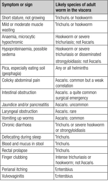
Differential diagnosis of marked hepatosplenomegaly, anaemia and pancytopenia includes hyper- reactive splenomegaly (tropical splenomegaly) syndrome and schistosomiasis, as well as myeloid leukaemia and myelofi brosis.
In acute- onset disease, malaria, disseminated tuber-culosis, typhoid, brucellosis, African trypanosomiasis, relapsing fever and leukaemia should be considered.
[image:5.595.289.499.214.393.2] [image:5.595.289.497.416.517.2]HIV infection greatly increases the risk of visceral leishmaniasis, and thus co- infection is common.
TABLE 6.3.A.C.1 Clinical features of visceral leishmaniasis Incubation period: 2–4 months (weeks to two years) Fever: intermittent at fi rst
Anaemia: bone- marrow depression, hypersplenism Splenomegaly: progressive enlargement
Hepatomegaly Weight loss
Epistaxis: haemorrhage from other sites may occur in advanced disease
Diarrhoea: invasion of gut by amastigotes, secondary infection Cough
Oedema: hypoalbuminaemia
Hair and skin signs of malnutrition in chronic forms Lymphadenopathy: in some African countries
TABLE 6.3.A.C.2 Clinical pathology of visceral leishmaniasis Haemoglobin: low; normochromic, normocytic fi lm White blood cells: low, 2–3 × 10⁹/litre Eosinophils low Platelets: low, < 100 × 109/litre
Reticulocytes: low Serum albumin: low Serum globulin: elevated
Liver transaminases and serum bilirubin: normal
Management of visceral leishmaniasis
Consider HIV co- infection and secondary disorders such as malaria, respiratory and gut infections, and tuberculosis. Blood transfusion for anaemia is seldom required, as the child has usually adapted to the low haemoglobin level. Give haematinics and vitamin supplements during nutritional rehabilitation and convalescence.Liposomal amphotericin B used to be expensive, but following a campaign the WHO has brought about a 90% reduction in price, and consequently this is now the treatment of choice.
The alternative treatment is with antimoniates (Sb), for which again the WHO has obtained a substantial reduction in cost. Meglumine antimoniate and sodium stibogluco-nate are available. The duration of Sb treatment is usually 4 weeks, but prolonged treatment (up to 6 weeks) may be necessary in resistant cases (see Table 6.3.A.c.3).
disorders should be undertaken. Serious toxicity may require dimercaprol to chelate and remove the antimony.
In areas where there is resistance to Sb, such as Bihar state in India, and the Sudan, alternative drugs are required as follows: amphotericin B by slow infusion; paromomycin (aminoglycoside, identical to aminosidine); or
[image:6.595.99.527.156.514.2]oral miltefosine. Combinations of drugs (e.g. paromomycin and Sb) may be more effective. In patients with HIV/VL co- infection, management is diffi cult because of frequent relapse when treatment is stopped. HAART combined with maintenance anti- leishmanial therapy is important.
TABLE 6.3.A.C.3 Drugs used in the treatment of visceral leishmaniasis
Drug Doses Contraindications and
cautions Side effects
Liposomal amphotericin B 4 mg/kg IV over
30–60 minutes once daily by IV infusion on days 1, 2, 3, 5 and 10
An initial test dose of 100 micrograms/kg (maximum 1 mg) is infused over
15 minutes. Observe for 1 hour to ensure that anaphylaxis does not occur, then proceed
May produce hypotension, fever, vomiting, headache, and muscle and joint pain. Less commonly, chest pain, hypoxia, severe abdominal pain, fl ushing, urticaria, and fl ank or leg pain
Sodium stibogluconate (100 mg Sb/mL) or meglumine antimoniate (81 mg Sb/mL)
20 mg/kg (minimum 200 mg) IV infusion or deep IM injection over over 5–10 minutes once daily for 28 days
Prolonged treatment for 6 weeks in resistant cases
Pre- existing severe cardiac, liver, renal, pancreatic or haematological abnormalities Not to be given during pregnancy
Filter solution through 5- micron fi lter immediately before infusion
Vomiting, abdominal pain, myalgia and arthralgia, headache, metallic taste. Rarely sudden death with prolonged QT interval; therefore monitor ECG and stop infusion if QT exceeds 0.5 seconds
Conventional amphotericin B Slow IV infusion, 1 mg/kg every second day for 15 days, or daily for 20 days. (daily dose must never exceed 1.5 mg/kg)
An initial test dose of 100 micrograms/kg (maximum 1 mg) is infused over
15 minutes. Observe for 1 hour to ensure that anaphylaxis does not occur, then proceed
May produce hypotension, fever, vomiting, headache, and muscle and joint pain
Paromomycin Daily IV or IM injections
16–20 mg/kg/day for 21 days Do not give at the same time as gentamicin or other aminoglycosides.
Avoid if there is renal impairment
Vomiting, diarrhoea, abdominal pain, fever and ototoxicity
Miltefosine 2.5 mg/kg/day orally for 28 days
Not in pregnancy Nausea and vomiting
A limited stock of the above drugs is kept by the WHO in Geneva for rapid response to an epidemic.
A study looking at the effectiveness of a single- dose treatment (by IV infusion) using liposomal amphotericin B is currently in progress in India.
Follow- up and prognosis
Symptomatic improvement usually occurs within a few days, and a haematological response occurs within 2 weeks. Splenomegaly slowly regresses, but may take a year or more to resolve. Prolonged follow- up (at least 1 year) is necessary to detect relapse. Relapse is treated with a repeat prolonged course of antimonials (up to 8 weeks). Unresponsiveness will require alternative drugs such as liposomal amphotericin B (if available), aminosidine, stand-ard amphotericin B or pentamidine. Trials of miltefosine are in progress.
Prevention and control
Prevention is similar to that of malaria, and includes insect repellents and the use of fi ne- mesh bed nets impregnated with permethrin. Control includes spraying of sandfl y resting sites and human dwellings, destruction of animal reservoirs and treatment of cases.
Further reading
Murray HW, Berman JD, Davies CR et al. (2005) Advances in leishmaniasis. Lancet, 366, 1561–77.
6.3.A.d Malaria
BOX 6.3.A.D.1 Minimum standards for an effective malaria control programme 1 Prevention:
Impregnated bed nets (ITNs), preferably long- lasting insecticidal nets (LLINs).
Where appropriate, intermittent preventive treatment in infants (IPTi) and seasonal malaria chemoprevention (SMC) for older children. Other methods of vector control, such as indoor residual spraying (IRS), personal protection (e.g. mosquito coils, impregnated clothing, repellents, etc.).
2 A well- informed population to improve early care seeking and adherence to treatment and preventive regimes.
3 Good case management:
Early accurate diagnosis: all patients should have a biological test before treatment:
— Quality- assured thick blood fi lm and/or rapid diagnostic test (RDT).
— Haemoglobin measurement to detect and treat malaria anaemia (e.g. using a haemocue machine).
— In severe disease, facilities to measure blood glucose levels and provide safe blood transfusion.
Effective treatment:
— Treatment for simple malaria.
» Artemisinin combination therapy (ACT) (following the national protocol for recom-mended ACT).
— Treatment for severe disease.
» IV or IM artesunate (IM artemether, rectal artesunate, or IV or IM quinine if artesunate is not available).
— Pre- referral treatment.
» Rectal artesunate (if artesunate is not avail-able, rectal quinine) or if injections are safe IV or IM artesunate or IM artemether, or IM quinine.
4 Accessible, acceptable and affordable care: Consider training community health workers in remote areas to diagnose and treat malaria. Make treatment free for pregnant girls and children under fi ve.
Set up a good referral system from community and fi rst- level health facilities to facilities with means to treat severe cases, including transport and facilities to access resources to pay for treatment if needed.
Introduction
Malaria is an extremely important public health burden in Africa, disproportionately affecting the youngest and most vulnerable. Children under 5 years and pregnant women, especially in the fi rst pregnancy, suffer from severe forms of the disease. In Asia, the disease is more common in men and older children.
Nearly 80% of the world’s malaria burden is in Africa.
Malaria is estimated to cause at least 650 000 deaths each year, mostly among African children.
Unlike anywhere else in the world, children aged 6–24 months in Africa are most at risk of the worst forms of malaria. Every 30 seconds an African child dies of malaria.
There are fi ve Plasmodium species known to be infective to humans, namely Plasmodium falciparum, P. vivax, P. ovale,
P. malariae and P. knowlesi.
P. falciparum causes severe disease and is the most prevalent form in sub- Saharan Africa (most sub- Saharan Africans are protected against P. vivax due to lacking a pro-tein in their red blood cells (the Duffy antigen)). P. falciparum
differs from the other species in that infected erythrocytes adhere to capillary epithelium, thus disappearing from the circulation and evading destruction by the spleen.
P. vivax and P. ovale can cause recurrent malaria attacks due to the formation of a dormant form existing as hypno-zoites in the liver, which are periodically released into the blood. Drugs to eliminate the hypnozoites from the liver are limited (primaquine).
P. malariae can cause long- term problems, including kidney failure, and P. knowlesi is a newly emerging form which has caused severe disease in Asia (Papua New Guinea and Thailand).
Life cycle
The infected Anopheles female mosquito injects sporo-zoites into the bloodstream of an individual. Sporosporo-zoites circulate for less than 30 minutes before being phagocy-tosed or entering liver parenchymal cells. The blood and liver phase prior to re- entry into the circulation is called the pre- erythrocytic phase, and it varies in length according to the species. At the end of this phase, merozoites invade the red blood cells and begin the erythrocytic phase. Parasites rapidly multiply within the red blood cells, which fi nally burst, releasing more merozoites into the bloodstream to invade further red blood cells.
Periodic bouts of fever are associated with the release of the merozoites. After some time, sexual forms of the parasites (gametocytes) are formed which are then ingested by a female mosquito to complete the cycle in humans. In the mosquito stomach, the gametocytes merge and eventually form sporozoites which migrate to the salivary
BOX 6.3.A.D.2 Minimum standards for hospital treatment of severe malaria
A triage system.
RDTs and microscopy for initial diagnosis, plus labora-tory facilities to determine levels of parasitaemia. Antimalarial drugs for IV, IM and oral treatment. Oxygen.
Antibiotics and anticonvulsants. Safe blood transfusion services. Nasogastric feeding.
glands, where they are injected into the bloodstream by the mosquito as it takes a blood meal to support its own reproductive effort.
In two species (P. vivax and P. ovale) some hepatic- stage parasites remain within the liver cells with the formation of the dormant phase, called hypnozoites. For various reasons (perhaps including waning immunity), at a later date the dormant phases activate and reseed blood. This leads to manifestations of malaria not from a new infection but from the latent exo- erythrocytic phase.
P. falciparum differs from the other species in that infected erythrocytes adhere to capillary epithelium, thus disappearing from the circulation and evading destruction by the spleen.
P. falciparum is the most likely species to cause life- threatening disease, and is a major cause of mortality in children.
Plasmodium falciparum
Clinical featuresTypical symptoms include high- grade fever alternating with cold spells, rigors, chills and sweating. There are usually associated myalgias and arthralgias.
However, features in children under 5 years of age may be non- specifi c, with fever, vomiting, diarrhoea and abdominal pain being the main symptoms.
In older immune individuals the only symptoms may be fever with headache and joint pains.
All fevers in children from a malaria- endemic area are therefore due to malaria until proven otherwise.
Diagnosis
Microscopy
Blood smear for malaria remains the gold standard: a thick fi lm for diagnosis, and a thin fi lm to confi rm the type of malarial parasite. Typically species- specifi c ring forms inside red blood cells are seen, but there may also be gametocytes.
The level of parasitaemia is usually scored as 1–5+. If the malarial smear is 3+ or more, there is a high level para-sitaemia. In areas where parasitic density is measured the smear is reported as parasites/mm3.
Malaria microscopy in district hospitals can be of very poor quality. A quality assurance programme should be in place that includes the following:
— a properly trained and regularly updated microscopist — adequate time to look at slides, particularly for low-
level parasitaemia
— the correct stains and good- quality slides
— a binocular microscope that is properly serviced and
maintained
— a system of internal and preferably external cross-
checking of a sample of slides, especially the low parasitaemias and negative slides.
— If possible examination requires a reliable electricity
supply or good lighting near a window in the day time. Many modern microscopes have an inbuilt LED light.
Rapid diagnostic tests
Antigen- capture test kits use a rapid simple dipstick test from a fi nger- prick blood sample to give a result in 10–20 minutes. RDTs should be used in circumstances where microscope facilities and/or diagnostic expertise are limited.
There are two main forms of rapid test.
Histidine- rich protein 2 (HRP2) tests
These only detect P. falciparum. HRP2 tests have a sensitivity of 97–100% (i.e. there are very few false- negative results). These tests can lack specifi city (which may be as low as 59% in some studies), so there can be a high frequency of false- positive results, especially in a high trans-mission zone where malaria infection is frequent (children can have as many as six attacks a year). HRP2 remains in the bloodstream for at least 2 weeks after all viable para-sites have been killed, and often for considerably longer (6–8 weeks), so patients returning with fever within 4 weeks after treatment cannot be diagnosed using an HRP2- based RDT. However, a presumptive diagnosis that fever equals malaria has an even lower specifi city.
HRP2 tests are very heat stable, but are sensitive to humidity. They have a shelf- life of 2 years, and their use can be taught to healthcare workers, even at village level, in a few hours. They are especially suitable for use in sub- Saharan Africa, where other species of malaria are rare.
Parasite lactate dehydrogenase (pLDH) tests
The parasite lactate dehydrogenase (pLDH) antigen is produced by all four Plasmodium species. The pLDH- based tests detect the antigen using a panel of monoclonal antibodies. They can have high sensitivity for P. falciparum, and are more specifi c than HRP2. They return to negative in 3–14 days (the majority do so within 7 days).
Some pLDH tests are able to differentiate between
P. falciparum and other Plasmodium species, and between viable and non- viable parasites, thereby enabling their use for monitoring therapy and for detecting new infections within 2 weeks of successful treatment.
The tests currently on the market are available in two forms. The fi rst has a pan- pLDH antibody that can detect any species of malaria. When positive, it produces a single test line. The second produces two test lines, a pan- specifi c line and a line that detects P. falciparum. In theory, there are monoclonal antibodies that can individually detect all of the different species, but these have not yet been validated.
pLDH tests are not as heat stable as HRP2 tests. Although pLDH has a high sensitivity for P. falciparum, its sensitivity for P. vivax appears to be less satisfactory if the patient has a low parasitaemia. pLDH tests are more expensive than HRP2 tests, and are not therefore recom-mended in sub- Saharan Africa, where 97% of infections are due to P. falciparum.
Advantages of RDTs over microscopy
The result is available within 15–20 minutes and one person can set up a new test every 1 or 2 minutes. In contrast, there are more steps involved in microscopy (i.e. slide preparation, drying, staining, and drying stained slides), and a negative slide requires 6 minutes of reading time (a microscopy report can be delayed up to an hour from collecting the blood).
Training takes 2 hours with minimally educated workers. Many more tests can be done in one clinic or outreach session.
with last Malarial Treatment (CMT) is based, and at project level after transportation, to ensure that tests remain in good condition (lot testing). Monitoring of the conditions to which the tests are subjected during transportation may account for problems with their function at project level.
Field teams need to monitor the performance of healthcare staff regularly to ensure that tests are per-formed properly.
Other diagnostic tests that should be available in malaria programmes
Haemacue to determine haemoglobin levels.
Tests to deliver safe transfusion: two instant HIV tests, syphilis, hepatitis B and hepatitis C screen.
Tests for G6PD defi ciency if primaquine is to be used for radical treatment to eliminate hypnozoites and/or gametocytes of P. falciparum.
Polymerase chain reaction (PCR) tests. These can be used to detect very low levels of parasitaemia. Work is progressing to develop a bedside PCR detection machine. PCRs are very important in elimination sce-narios to detect very low parasitaemias, and in drug effi cacy studies.
Case defi nitions of malaria
Suspected malaria: a patient with a fever or history of fever in the last 48 hours who lives in or has come from a malaria- endemic area.
Uncomplicated (simple malaria): a patient with a fever or history of fever in the last 48 hours who has a positive biological test and no symptoms of severe disease.
Complicated malaria: a patient with the signs and symptoms of simple malaria who is unable to take oral drugs.
Non- severe malaria may be associated with a vari-ety of other symptoms, including cough, vomiting, diarrhoea, abdominal pain, myalgia, headache, sweat-ing and rigors.
Severe malaria
A patient with one or more of the following signs or symp-toms, with biologically confi rmed P. falciparum infection (and occasionally P. vivax) and parasitaemia:
prostration (inability to sit, or to drink or breastfeed) impaired consciousness (cerebral malaria) respiratory distress
multiple convulsions circulatory collapse
severe anaemia (haemoglobin concentration < 5 grams/ dL or haematocrit of < 15%) may be the presenting symptom, especially in children and pregnant women, and can rapidly lead to death.
Other conditions that may be associated with
severe malaria
Hyperparasitaemia may be associated with severe malaria, but is not pathognomonic of severe disease in itself. It has been associated with a higher risk of mortality and needs to be rigorously treated, preferably in the fi rst instance with parenteral medications. If there are no other signs of sever-ity, the patient may not need hospital admission.
Hypoglycaemia often causes unconsciousness or death if not detected and treated rigorously. It is especially
dangerous in children, malnourished patients and pregnant women, and is exacerbated by quinine treatment.
Pulmonary oedema is a grave and often fatal complica-tion of malaria. It can occur spontaneously (particularly during pregnancy), but it is often a result of fl uid overload during treatment.
Metabolic (lactic) acidosis: see section on severe malaria below.
Abnormal bleeding is associated with thrombocyto-paenia, and leads to bleeding of gums and epistaxis, and sometimes more severe internal bleeding.
Jaundice is more common in adults than in children. Mild jaundice only refl ects haemolysis, whereas very high bilirubin levels suggest hepatic dysfunction.
Haemoglobinuria is common, but its more extreme form, blackwater fever, is rare. It is associated with quinine therapy.
Oliguria/anuria can be a sign of renal dysfunction, but make sure that the patient is adequately rehydrated before commencing therapy for renal failure. Fluid balance charts should be instituted and monitored closely for all patients with severe malaria.
Uncomplicated/simple malaria
There is a fever and a positive blood smear. There is no evidence of altered consciousness, hypoglycaemia, severe anaemia, jaundice or respiratory diffi culties.
Management
Management of children who have always lived in an endemic area
There is no need to admit the child to hospital (unless they are under 4 months of age or less than 5 kg in weight, or pregnant).
A diagnostic test should be done before treatment (microscopy if available and quality assured, or an RDT). This will confi rm malaria and also ensure that patients who do not have malaria receive appropriate treatment.
Note that malaria is frequently accompanied by other serious infections, such as pneumonia. Signs of bacterial or viral infections should be looked for and treated appropriately even if the malaria diagnostic test is positive.
Give fi rst- line antimalarial treatment (ACTs) as recom-mended in local national guidelines.
Ensure that tablets or syrup are swallowed and not vomited.
Give the fi rst dose under direct observation and advise the carer on how to administer the drug to young chil-dren by dissolving tablets in breast milk or syrup and giving this slowly with a syringe.
If the child vomits within the fi rst 30 minutes, repeat the full dose. If they vomit after 1 hour give a half dose. Advise the carer to return if further doses are vomited. Remember to advise the carer to give the dose with food if artemether/lumefantrine is used, to improve absorption of the lipophilic lumefantrine.
and folic acid, but if sulfadoxine- pyrimethamine has been used for malaria treatment, do not give folic acid for 2 weeks).
Management of children visiting or returning from an endemic area for the fi rst time
Hospital admission for management of P. falciparum is always advisable.
Treat with an ACT
The WHO recommends the use of fi xed- dose combina-tions (FDCs) if available, or pre- packaged drugs if FDCs are not available. The WHO discourages the use of mono-therapies, to reduce the risk of resistance developing.
In particular, the use of artesunate monotherapy, which is commonly available on the private market, is strongly discouraged.
ACTs recommended by the WHO
Artesunate/amodiaquine (AS/AQ FDC).
Artesunate + mefl oquine (AS+MQ or AS/MQ FDC). Artesunate + sulphadoxine/pyrimethamine (AS + SP). Artemether + lumefantrine (AM/LM FDC).
DHA/piperaquine (Duo- Cotecxin, Eurartesim) FDC. Artesunate/pyronaridine (Pyramax) FDC.
Non- ACTS
Malarone (atovaquone/proguanil) FDC: this is very expensive and usually only used where there is arte-misinin resistance, or for prophylaxis in western travellers. Quinine tablets in IV, IM and rectal forms: for true treat-ment failures.
Chloroquine: only for non- P. falciparum malaria. Primaquine and its derivatives (tafenoquine): for radical treatment of P. vivax (and P. falciparum in elimination areas).
Paediatric formulations
AS/AQ infant dose is dispersible, and suitable for chil-dren who weigh 4.5–8 kg.
Paediatric Coartem® (AM/LM) is dispersible and
avail-able as cherry- fl avoured tavail-ablets for children who weigh 5–25 kg.
Artequin (Mepha) FDC AS/MQ is available as mango- fl avoured pellets/granules that can be swallowed directly without water. It is not WHO prequalifi ed.
AS/MG FDC produced in Brazil for Drugs for Neglected Diseases (DNDi).
Drugs frequently available but not WHO prequalifi ed
ASMQ Artequin (also in paediatric granules). Artemisinin/piperaquine (Artequick). Artemisinin and naphthoquine.
Drugs in development
Artemisone (partner drug not yet decided). Synthetic AS called OZ (Sanofi Aventis). Semi- synthetic artemisinin (One World Health).
Advice for carers
Discuss preventive efforts with carers (e.g. bed net at night, ideally impregnated with insecticide). Give LLIN if possible.
Tell the mother to return after 2 days if fever persists, and earlier if the child deteriorates.
If the child is repeatedly vomiting and the area is remote and admission to hospital diffi cult, give rectal artesunate until the vomiting settles. Then give a full 3- day course of ACT.
Management of severe malaria
Severe malaria is a complex multi- system disease that constitutes a medical emergency.
Mortality approaches 100% without treatment, and death often occurs within the first few hours. Prompt initiation of antimalarial treatment in peripheral healthcare facilities and comprehensive management in hospital are necessary to prevent deaths.
Neurological sequelae of cerebral malaria affect about 10% of African children who survive cerebral malaria. These sequelae are severe and permanent in up to 19 000 children annually, and include spastic paresis and epilepsy.
Care should be provided within 15 minutes of arrival at a healthcare facility. Triage systems should be in place in health centres and hospitals to pick up severely ill patients, referral should be rapid, and emergency facilities must be instituted in hospitals, with a high standard of medical and nursing care available 24 hours a day.
Any seriously ill or unconscious patient in a malaria- endemic area must be tested for malaria by RDT (remember that parasites may not be present in the peripheral blood of a patient with cerebral malaria). Malaria should be assumed in any child with severe anaemia, convulsions, hyperpyrexia and/or hypoglycaemia either in hospital or in a peripheral healthcare facility.
Even if a diagnostic test is not available, the patient should be given an antimalarial drug (IV, IM or rectally, depending on the skill of the staff in the facility) before transfer to the hospital. This can be repeated if transfer is impossible or is delayed for more than 12 hours. A note of what has been given should be sent with the patient as soon as transfer can be arranged.
If any doubt exists, it is safer to treat than not to treat before transfer.
Immediate measures (in hospital)
Vital signs: temperature, pulse, blood pressure, and respiratory rate and depth.
State of hydration.
Estimate or ideally measure body weight. Estimate of weight by age in well- nourished children:
— For an infant up to 1 year of age, birth weight doubles
by 5 months and triples by 1 year.
— For children over 1 year, use the following formula:
weight (kg) = 2 (age in years + 4).
Be careful in HIV- endemic areas where body weights are often very different from those derived by this formula. Weigh the child if at all possible.
Level of consciousness (AVPU or Glasgow or Adelaide coma scales) (see Section 5.16.A).
The depth of coma may be assessed rapidly in children using the coma scale for children or by observing the response to standard vocal or painful stimuli (rub your knuckles on the child’s sternum; if there is no response, apply fi rm pressure on the thumbnail bed with a hori-zontal pencil).
disease. Do not wait for a malaria smear result before initiating treatment, as it can take up to an hour. If the RDT is positive, commence treatment immediately.
Perform lumbar puncture if the patient is unconscious to eliminate meningitis if there are no contraindica-tions. Contraindications include papilloedema or suspicion of raised intracranial pressure (irregu-lar breathing and pupil(irregu-lary responses, posturing), bleeding problems or respiratory diffi culty such that fl exing the back would compromise respiration.In such a situation, give IV antibiotics to treat menin-gitis as well as malaria.
Measurement of glucose (fi nger prick), haemoglobin and haematocrit (packed cell volume, PCV).
Group and cross- match blood and search for a suit-able donor.
Parenteral IV or IM treatment
In Africa and many other regions, sodium artesunate or quinine are the drugs of choice for severe malaria. In South- East Asia and the Amazon Basin, quinine is no longer always effective and should be accompanied by doxycycline in adults or clindamycin in children. Large trials in mainly Asian Adults (SEAQUMAT study) and in African Children (AQUAMAT study) have proved that parenteral artesunate reduces mortality by over 30% and should be used in preference to quinine.
Initially give treatment intravenously, if possible; oth-erwise use the IM route. Change to oral therapy as soon as possible.
Especially in the malaria- endemic areas of Africa, the following initial antimalarial medicines are recommended. Artesunate has been shown to reduce mortality compared with quinine, but it is important to use whichever drug is available locally.
artesunate IV or IM
artemether IM (its absorption may be erratic in children in shock).
quinine (IV infusion or divided IM injection)
First- line antimalarial drugs
Sodium artesunate IV or IM
Give 2.4 mg/kg IV (by slow injection) or IM on admission (time 0), followed by 2.4 mg/kg IV or IM at 12 hours and again at 24 hours, and then once daily for a minimum of 3 days until the child can take oral treatment with an ACT.
OR second choice Artemether IM
Give 3.2 mg/kg IM as loading dose, then 1.6 mg/kg IM once daily (every 24 hours) for a minimum of three days until oral treatment can be taken. Use a 1 mL tuberculin syringe to give the small injection volume (note: absorption may be erratic and therefore only use if quinine and artesunate are not available) and if shocked do not use this drug as absorption is too unreliable.
Intravenous IV quinine (quinine dihydrochloride)
This is the second choice, to be used if sodium artesunate is not available. Give 20 mg/kg quinine dihydrochloride (maximum 1.4 grams) in 5% glucose at a concentration of 1 mg of quinine to 1 mL of 5% glucose over 2–4 hours (never more rapidly than over 2 hours). If possible use an in- line infu sion chamber (100–150 mL) to ensure that the
loading dose does not go in too quickly. Alternatively, ensure that the IV giving bag contains only the amount needed for each dose. There is a major risk of cardiac side effects if it is infused too quickly.
Subsequently give 10 mg/kg in 10 mL/kg fl uid (5% glu-cose) IV every 12 hours for 24 hours, or longer if the child remains unconscious. These latter doses must be given over at least 2 hours.
Never give quinine as an IV bolus. The infusion rate must not exceed a total of 5 mg quinine salt/kg/hour.
If safe control over the rate of infusion of IV quinine is not possible (e.g. there are insuffi cient or only untrained nursing staff available), give a loading dose intramuscularly (with initial doses of 10 mg/kg quinine salt IM at 0 and 4 hours and then 12- hourly).
For IM injections, dilute the quinine solution to allow better absorption and less pain.
As soon as the child is able to take medication orally, switch to quinine tablets 10 mg/kg every 8 hours for a total of 7 days, or the locally available fi rst- line ACT treatment for malaria.
Side effects:
Common: cinchonism (tinnitus, hearing loss, nausea and vomiting, uneasiness, restlessness, dizziness, blurring of vision).
Uncommon: hypoglycaemia, although this is a common complication of severe malaria.
Serious cardiovascular problems (QT prolongation on the ECG) and neurological toxicity are rare.
If overdosed by mistake with quinine tablets, give activated charcoal orally or by nasogastric tube as a suspension in water (1 gram/kg).
Chloroquine IV
This drug should never be used to treat severe falciparum malaria but only cases of non- resistant vivax or ovale malaria. Give 5 mg base/kg every 6 hours for a total of 25 mg base/kg (fi ve doses) as an infusion in 5% glucose (give over 2 to 4 hours).
Antimalarial treatment after IV or IM regimes have ended
Following parenteral administration, usually for a minimum of 24 hours or until the child can take oral drugs, the treat-ment of severe malaria must be completed by giving a full course of one of the artemisinin- based combination thera-pies (ACT) described below. In some parts of the world, oral quinine combined with clindamycin to complete 7 days of treatment is used
The following ACTs are recommended: artemether plus lumefantrine
artesunate plus amodiaquine
artesunate plus sulfadoxine- pyrimethamine dihydroartemisinin plus piperaquine artemether plus clindamycin artesunate plus mefl oquine.
The choice of ACT in a particular country or region will be based on the level of resistance of the partner medicine in the combination.
areas without multi- drug resistance (mainly Africa), any of the ACTs, including those containing amodiaquine, may still be effective. Every country has a national malaria policy in which the fi rst- line therapy is described and should be used.
If possible avoid using mefl oquine if the patient has presented with an impaired conscious level.
Treatment for HIV- infected patients with
P. falciparum
malaria
Patients with HIV infection who develop malaria should receive prompt effective antimalarial treatment regimens as recommended above.
Treatment with ACT involving sulfadoxine- pyrimethamine should not be given to HIV- infected patients who are receiving co- trimoxazole (trimetho-prim plus sulfamethoxazole) prophylaxis.
Treatment of HIV- infected patients who are on zidovudine or efavirenz should, if possible, avoid amodiaquine- containing ACT regimens.
Treatment of
P. falciparum
malaria in
malnourished patients
Although there are many reasons why antimalarial phar-macokinetics may differ between malnourished patients and those who are well nourished, there is insuffi cient evidence to change current mg/kg body weight dosing recommendations.
Always check local guidelines on drug sensitivities.
With all antimalarial drugs, change to an oral therapy when the child can tolerate it.
Additional treatment where needed
Insert a nasogastric tube to minimise the risk of aspira-tion pneumonia if the patient’s level of consciousness is low. This can also be used to give food to prevent hypoglycaemia if the child is unconscious for a long period and is unable to eat. Alternatively, sucrose (sugar) can be placed under the tongue.
Insert an IV cannula and restore the circulating volume.
— Fluids should be given with caution and the
need for them assessed on an individual basis after ascertaining the nutritional status and degree of dehydration present.
— In general, children with metabolic acidosis who have not previously received parenteral fl uids are dehydrated and should be managed accordingly. Give oxygen if SpO2 is < 94% (to keep SpO2 in the range
94–100%) or if there is respiratory distress and no pulse oximeter available.
Treat severe anaemia with a safe blood transfusion if the child is showing signs of decompensation.
Give anticonvulsants (diazepam is preferred) if the patient is convulsing (see below) to prevent long- term neu-rological damage (see Section 5.16.E). Convulsions associated with cerebral malaria should be distinguished from febrile convulsions common in children under 4 years of age. The child usually recovers rapidly, within a few minutes, from a febrile convulsion. Convulsions in malaria are common before or after the onset of coma. They are signifi cantly associated with morbidity and sequelae. They may present in a very subtle way. Important signs include intermittent nystagmus, saliva-tion, minor twitching of a single digit or a corner of the mouth, and an irregular breathing pattern.
Prophylactic anticonvulsants have been recommended in the past, but recent evidence suggests that phenobarbital may be harmful in this situation.
Paracetamol, 15 mg/kg of body weight 4- hourly, may also be given orally or rectally as an antipyretic. Use tepid sponging and fanning to try to keep the rectal temperature below 39°C. Relatives are usually happy to do this when instructed.
High- dose IV or IM antibiotics should be given routinely to an unconscious or shocked patient.
Avoid using harmful ancillary drugs.
The patient will need intensive nursing care at least until they regain consciousness. They may urgently need glucose or a blood transfusion if hypoglycaemia or haemolysis is severe.
Management of associated causes of
mortality in severe malaria
Some children with P. falciparum malaria go on to develop altered consciousness, severe anaemia, acidosis, or any combination of these. Where transmission of P. falciparum
is endemic, malaria is the commonest cause of coma in children, especially in those aged 1–5 years.
Cerebral malaria (coma, confusion and convulsions)
Coma develops rapidly, often within 1 or 2 days of onset of fever, and sometimes within hours. Convulsions are usual and may be repeated. Clinical features suggest a metabolic encephalopathy, with raised intracranial pres-sure. Opisthotonos, decorticate or decerebrate posturing, hypotonia and conjugate eye movements are common. Oculovestibular refl exes and pupillary responses are usu-ally intact. Papilloedema is found in a small minority of cases. A unique retinopathy with patchy retinal whitening and pallor of vessels is found. In fatal cases, brain swelling is commonly present at autopsy, but cerebral herniation is not usually found even in patients who have undergone lumbar puncture.
Hypoglycaemia, acidosis, hyperpyrexia and convul-sions (sometimes undetectable without EEG) are common accompaniments of cerebral malaria, and require appropri-ate management (see below).
No physical signs are diagnostic of coma due to malaria, and incidental parasitaemia is common in endemic areas, so other causes of coma, especially hypoglycaemia and meningitis, must always be carefully sought, and if neces-sary treated on the basis of presumptive diagnosis.
Even with optimal treatment, the case fatality rate is 15–30%, and about 10% of survivors have residual neuro-logical sequelae (hemiparesis, spasticity, cerebellar ataxia) that may partially or completely resolve over time.
Investigations
Management
Coma
Ensure that the airway is open at all times and that the patient is breathing adequately. Give oxygen by face mask with a reservoir or nasal cannulae (to keep SpO2 in the
range 94–98% if a pulse oximeter is available). If the child stops breathing, give assisted ventilation with a bag-mask of suitable size (500 mL or 1600 mL).
Ensure that a bag-mask is available at all times. Nurse the patient in the recovery position to avoid aspiration of secretions or vomit.
Exclude other treatable causes of coma (e.g. hypogly-caemia, bacterial meningitis).
Treat convulsions (see Section 5.16.A on coma and Section 5.16.E on convulsions).
Treat hypoglycaemia.
Convulsions
Convulsions are common before and after the onset of coma.
Ensure that the airway is open, and give oxygen by face mask with a reservoir or nasal cannulae.
If the child stops breathing, give assisted ventilation with a bag-mask of suitable size (500 mL or 1600 mL). Examine all children with convulsions for hyperpyrexia and hypoglycaemia. Treat hypoglycaemia with IV or oral glucose if identifi ed on blood testing, but also treat as for hypoglycaemia if blood glucose levels cannot be measured and the child is drowsy, unconscious or fi tting (see below).
Give anticonvulsant treatment with rectal diazepam or paraldehyde or IM paraldehyde.
If the patient has a fever of 39°C ( 102.2°F), give paracetamol rectally (if available).
Treat seizures lasting for more than 5 minutes with drugs.
Ensure that a bag-mask is available at all times in case of apnoea following the use of diazepam. Apnoea is usually short- lived and improves quickly with ventilation via bag and mask.
Note that seizure activity needs to be looked for care-fully, as it may appear as just a twitching of the thumb or mouth.
Give IV diazepam:
— Children: 300 microgram/kg of body weight as an IV
infusion over 2 minutes or 400–500 microgram/kg of body weight intra- rectally. This dose can be repeated after 10–15 minutes if still fi tting.
— Pregnant girls: 10 mg rectally or by slow IV injection.
This dose can be repeated after 10–15 minutes if still fi tting.
— Do not exceed 10 mg per dose.
Alternatively, paraldehyde 0.1 mL/kg of body weight may be given by deep IM injection or 0.8 mL/kg of body weight (maximum 20 mL) intra- rectally using a sterile glass syringe (a disposable plastic syringe may be used provided that the injection is given immediately after the paraldehyde is drawn up, and the syringe is never reused).
Hypoglycaemia
Hypoglycaemia is common and is due to poor intake, increased metabolic needs of the patient and parasites and impaired hepatic gluconeogenesis. It is easily overlooked
because clinical signs may mimic those of cerebral malaria.
Check for hypoglycaemia in all patients who are uncon-scious, in shock or deteriorating. Also regularly (every hour in the fi rst instance) check pregnant girls, children under 5 years, and the malnourished, and all patients receiving quinine.
Hypoglycaemia is defined as blood glucose levels < 2.5 mmol/litre (< 45 mg/dl).
Prevent hypoglycaemia with a maintenance quantity of 5% glucose in 0.9% Ringer- lactate or Hartmann’s solu-tion (50 mL of 50% glucose in a 500- mL bag). If the child develops hypoglycaemia despite this, give maintenance as 10% glucose in 0.9% Ringer- lactate or Hartmann’s solution (100 mL of 50% glucose in a 500- mL bag). Do not exceed maintenance fl uid requirements for the child’s weight (see
Section 9 Appendix). If the child develops signs of fl uid overload, stop the infusion; repeat the 10% glucose boluses (5 mL/kg) if there is hypoglycaemia identifi ed by making regular checks of blood glucose levels.
If IV access is not possible and the child is hypoglycae-mic, place an intra- osseous needle (see Section 8.4.B).
Treat hypoglycaemia or suspected hypoglycaemia with an IV glucose infusion or bolus:
Children: 1 mL/kg of 50% dextrose, diluted with four times the volume of infusion fl uid (usually Ringer- lactate or Hartmann’s solution) infused over 5 minutes or 5 mL/ kg of 10% glucose as a bolus.
Pregnant girls: 50 mL of 50% dextrose diluted with an equal volume of infusion fl uid (usually Ringer- lactate or Hartmann’s solution) over 15 minutes (irritating to veins).
Re- test 15 minutes after completion of the infusion, and repeat the infusion if blood glucose remains low. Repeat until blood glucose recovers, then infuse with 5–10% glucose in Ringer- lactate or Hartmann’s solution (accord-ing to hypoglycaemia risk) to prevent recurrence. Ensure regular feeding when oral intake can be sustained. Fluids used to treat hypoglycaemia must be included in daily fl uid requirements.
If blood glucose levels cannot be measured and hypogly-caemia is a possibility, always give IV glucose as described above.
If the child is still unable to swallow after 48 hours, start nasogastric feeds. If a gag refl ex is present and the child is able to swallow, feed them as soon as this is possible. For young children breastfeed every 3 hours if possible, or give milk feeds of 15 mL/kg 3- hourly if the child can swallow. If they are not able to feed without risk of aspiration, give milk, especially breast milk, by nasogastric tube or sugar sublin-gually (see Section 5.8.B). Continue to monitor the blood glucose levels, and treat accordingly (as described above) if these are found to be < 2.5 mmol/litre or < 45 mg/dL.
Hypoglycaemia is a major cause of death in severe malaria patients, especially in young children and pregnant girls. Remember that quinine will potentiate hypoglycaemia. Young children should receive regu-lar feeding, including by nasogastric tube, if they are unable to take oral foods.
Severe anaemia
and, rarely, pulmonary oedema (fast breathing, fi ne basal crackles on auscultation) may be present (see above).
Severe haemolytic anaemia is defi ned as < 5 grams of haemoglobin/dL or haematocrit < 15%.
Severe anaemia may be the presenting feature in malaria. Patients with severe anaemia, especially pregnant girls, should be tested for malaria.
Give a safe blood transfusion as soon as possible to: all children or pregnant girls with a haematocrit of 12% or Hb of 4 g/dL
less severely anaemic children (haematocrit > 12–15%; Hb 4–5 g/dL) with any of the following:
— clinically detectable dehydration (as well as
rehydrat-ing orally if possible)
— shock
— impaired consciousness — deep and laboured breathing — heart failure
— very high levels of parasitaemia (> 10% of red blood
cells parasitised).
Give packed cells (10–20 mL/kg body weight for chil-dren and 500 mL for pregnant girls), if available, over three to four hours in preference to whole blood. Allow red blood cells to settle at the bottom of the bag, and stop the infusion when the cells have been used.
If not available, give fresh whole blood (20 mL/kg body weight) over 3–4 hours.
A diuretic is not usually indicated (unless pulmonary oedema or fl uid overload is developing), because many of these children have a low blood volume (hypovolaemia).
Check the respiratory rate and pulse rate every 15 min-utes. If one of them rises, transfuse more slowly. If there is any evidence of fl uid overload due to the blood transfu-sion, give IV furosemide (1–2 mg/kg body weight) up to a maximum total of 20 mg for children, and give 40 mg IV for pregnant girls.
After the transfusion, if the haemoglobin level remains low, repeat the transfusion.
In severely malnourished children, fl uid overload is a common and serious complication. Give whole blood (10 mL/kg body weight rather than 20 mL/kg) once, and only repeat the transfusion if there are no signs of overload.
Perform microscopy following transfusion, and repeat or extend antimalarial treatment if parasitaemia is increasing.
Respiratory distress due to acidosis
This presents with deep laboured breathing while the chest is clear on auscultation, sometimes accompanied by lower chest wall indrawing. It is caused by systemic metabolic acidosis (frequently lactic acidosis) and may develop in a fully conscious child, but more often in children with cerebral malaria or severe anaemia. Always exclude other causes, such as pneumonia or pulmonary oedema.
Metabolic acidosis in severe malaria has been attrib-uted to the combined effects of several factors that reduce oxygen delivery to tissues:
Increased production of lactic acid by parasites (through direct stimulation by cytokines).
Decreased clearance by the liver.
Marked reductions in the deformability of uninfected red blood cells may compromise blood fl ow through tissues. Dehydration and hypovolaemia can exacerbate micro-vascular obstruction by reducing perfusion pressure.
Destruction of red blood cells and anaemia further compromise oxygen delivery.
Mean venous blood lactate concentrations have been found to be almost twice as high in fatal cases as in survivors, and to correlate with levels of tumour necrosis factor and interleukin 1- alpha. The lactate concentra-tions fell rapidly in survivors but fell only slightly, or rose, in fatal cases. Sustained hyperlactataemia has been found to be the best overall prognostic indicator of outcome.
Treatment
Give oxygen to all patients (even if they are not hypoxae-mic), and if a pulse oximeter is available keep SpO2 in the
range 94–100%.
Correct reversible causes of acidosis, especially dehy-dration and severe anaemia.
If Hb is 5 g/dL, give 10 mL/kg of 0.9% Ringer-lactate or Hartmann’s solution IV as a bolus and then reassess. If haemoglobin level is < 5 grams/dL, give whole blood (10 mL/kg) over 30 minutes, and a further 10 mL/kg over 1–2 hours without diuretics. Check the respiratory rate and pulse rate every 15 minutes. If either of these shows any rise, transfuse more slowly to avoid precipitating pulmonary oedema (see Section 1.7).
Monitor ECG for cardiac arrhythmias if possible. The use of sodium bicarbonate is controversial.
Respiratory distress due to pulmonary oedema
This is different to that due to acidosis, and there is usually more chest recession, hypoxaemia (cyanosis, SpO2 < 94%),
basal lung crepitations, enlarging liver, gallop rhythm, and raised jugular venous pressure. It may be due to fl uid over-load, often in the presence of severe anaemia. The most effective treatment is to tilt the bed of the patient head up so that the venous blood fl ow to the heart is reduced. If the bed cannot be tilted, sit the patient up, give furosemide 1 mg/kg for children and 40 mg IV for pregnant girls, and proceed with a careful transfusion of packed blood cells. Repeat furosemide as needed.
Respiratory distress due to pulmonary aspiration or pneumonia
Prevent aspiration pneumonia if possible, because it can be fatal. Place the comatose patient in the recovery position and ensure that the airway is open. If it is safe to intubate and maintain this, do so in order to protect the airway if the patient is unconscious (U on the APVU scale, or Glasgow Coma Scale score of < 9).
Give oxygen if the SaO2 is < 94% or, if pulse oximetry
is not available, if there is cyanosis, severe lower chest wall indrawing or a respiratory rate of 70 breaths/ minute. Keep SpO2 94–100%. Give IM or IV antibiotics
as described for pneumonia (see Section 5.3.A), and add in metronidazole 7.5 mg/kg 8 hourly (maximum individual dose 500 mg) until the patient can take these orally, for a total of 7 days.
Shock