GROSS AND MICROSCOPIC CHANGES ON RAT TESTES DUE TO
THE EFFECT OF CISPLATIN
Muna Kadel*1, Chandra Bhusan Jha2 and Himal Sangraula3
1
Department of Anatomy, KIST Medical College Lalitpur,
2
Department of Anatomy,
3
Department of Clinical Pharmacology, B.P. Koirala Institute of Health Sciences, Dharan.
ABSTRACT
Cisplatin is an efficient platinum-derived anticancer drug which acts in
nonspecific phases of the cell cycle. Because of increasing number of
long term survival of cancer patient treated with cisplatin, the long
term chemotherapy induced side-effects of cardiovascular system and
reproductive system are of great concern. The objective of the study is
to observe dose dependent histomorphological changes in rat testes
due to cisplatin and to find out the effect is either reversible or not.
Methods: Experimental study was carried out in 45 healthy adult male
albino rats weighing 150-220 gm. They were divided into 3 groups,
one control and two experimental groups (n=15 per group). One experimental group was
exposed to 3 rounds of 1 mg/kg body weight of cisplatin whereas other group received 3
rounds of 2.5-mg/kg body weight of cisplatin. Control group received the equal volume of
normal saline instead of cisplatin (1 injection daily for 5 days with a recovery phase of 16
days between the cycles). On 63rd day, the rats were anaesthetized and sacrificed to take out testes for histological study. Results: A dose-dependent reduction in weight, diameter and
volume of testes was observed. Significant reduction of germ cells and sertoli cells were
observed in both experimental groups (p<0.01). Besides that, high doses revealed severe
atrophy and loss of normal architecture of seminiferous tubules. Conclusion: Cisplatin
produces dose dependent effect in rat testes. Effect is reversible in low dose.
KEYWORDS: Germ cells, Leydig cells, seminiferous tubules, Sertoli Cells.
Volume 6, Issue 17, 1041-1047. Research Article ISSN 2277–7105
*Corresponding Author
Dr. Muna Kadel
Department of Anatomy,
KIST Medical College
Lalitpur, Nepal.
Article Received on 24 Oct. 2017,
Revised on 15 Nov. 2017, Accepted on 06 Dec. 2017,
INTRODUCTION
Cisplatin is platinum coordinated anti cancer drug that comes under the class of alkylating
agent. It is widely used to treat testicular cancer, ovarian cancer, cancers of bladder,
esophagus, lungs and colon.[1,2] Cytotoxic effect of cisplatin is due its interaction with DNA.[3] It forms DNA-platinum complex causing inhibition of cellular processes like replication, transcription, translation and DNA repair.[4] Cisplatin chemotherapy is mainly used in children and adolescents.[5] Most germ cell malignancies, even after metastasis, are efficiently eliminated by a combination of cisplatin, surgery and radiotherapy, leading to an
overall cure rate of about 95%.[6] Because of increased survival rate of cancer patients treated with cisplatin, the side effects of cispaltin on cardiovascular and reproductive system are of
great clinical concern.[7]
Testes consist of numerous seminiferous tubules. Each seminiferous tubule contains
spermatogonia, primary spermatocyte, secondary spermatocyte, and spermatozoa.[8] Seminiferous tubules are the sites for spermatogenesis. During spermatogenesis as the germ
cells are rapidly dividing, the cells are highly sensitive to the detrimental effects of various
physical and chemical agents.[9] The inhibition of nucleic acid synthesis is apparently responsible for anti –tumor action on the dividing germ cells as well as regression of lining
epithelium of seminiferous tubule.[10] Techniques such as spermatozoa cryopreservation and intracytoplasmic sperm injection (ICSI) has been developed to preserve the fertility in men
undergoing traditional anticancer therapies.11 Fertility is an important issue in cancer patients. So, the study aimed to observe the effect of cisplatin on rat testes.
MATERIALS AND METHODS
Forty-five healthy Wistar Albino rats weighing 150-220gm were obtained from the animal
house of BPKIHS, Dharan. They were housed in well ventilated room at controlled ambient
temperature (25±50C) with 12 hours alternating light-dark cycle and fed pellet diet and Bengal gram.
Rats were equally divided into 3 groups, one control and two experimental groups (n=15 per
group). One experimental group was exposed to 3 rounds of 1 mg/kg body weight of cisplatin
intraperitonelly whereas other group received 3 rounds of 2.5-mg/kg body weight of cisplatin.
Control group received the equal volume of normal saline. Rats were administered 1 injection
On 63rd day, all the rats were anaethesized with chloroform. The fully anesthetized rats were sacrificed to take out testes for histological study. Testes tissue were fixed and processed for
slide preparation. Haematoxyline and Eosine staining was done and studied under light
microscope.
Experimental protocol for this study was approved by protocol Evaluation Committee of B.
P. Koirala Institute of Health Sciences, Dharan. All the experimental works were carried out
as per ethical guidelines of Nepal Health Research Council (NHRC).
SPSS version 19 was used for data entry and analysis. NPar Tests and Wilcoxon Signed
Ranks Test were used to see the level of significance of differences. P<0.05 was considered
as statistically significant.
RESULTS
Quantiative measurement
Weight of testes: Weight of both testes in high dose experimental group was found to be
significantly reduced as compared to control group. Weight of testes of different groups is
expressed in table I.
Diameter of testes: Diameter of both testes was significantly reduced in high dose
experimental group as compared to control group, which is as illustrated in table II.
Diameter of seminiferous tubules and interstitial space: Diameter of seminiferous tubules
was found significantly decreased in both the experimental groups as illustrated in table III.
The increment of interstitial space was also highly significant in both test groups in
comparison to control group.
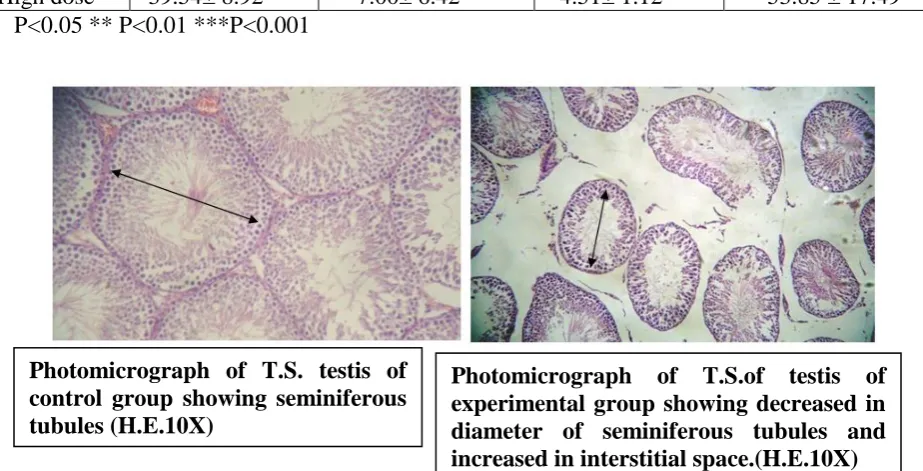
Qualitative changes: In control group numerous semeniferous tubules were found to be
closely placed and their shape was round or oval with different stages of spermatogenesis.
In low dose test group, some of the seminiferous tubules showed spermatogenic arrest and
some cells showed cytoplasmic vacuolation but there was preservation of normal
architecture. Whereas in high dose group most of the tubules showed necrosis with
sloughing of the lining epithelium. Some multinucleated giant cells were also visible inside
the tubules. Lumen of the tubules was filled with eosinophilic necrotic material instead of
Table. I: Weight of testes of control and test group.
Group Right testes (gm) Left testis (gm)
Control 1.31±.11 1.24 ± .12
Low Dose 1.10±.21 * 1.09±.35
High dose 0.453±.10*** 0.41 ±.10***
Table. II: Diameter of testes of control and test group.
Group Right testes (mm) Left testes (mm)
Control 1.87 ± 0.11 1.74 ± 0.17
Low Dose 1.85 ± 0.12 1.76± 0.12
High Dose 1.45± 0.15*** 1.40 ± 0.14**
P<0.05 ** P<0.01 ***P<0.001
Table-III: Interstitial space and diameter of seminiferous tubules of control and test
group.
Group Mean diameter of seminiferous
tubules(μm)
Interstitial space (μm)
Control 329.10 ± 23.81 35.68 ± 10.87
Low dose 256.58 ± 16.46 *** 76.93 ± 13.58***
High dose 171.23 ± 19.02*** 91.93 ± 9.22***
P<0.05 ** P<0.01 ***P<0.001
Table. IV: Number of different types of germ cells (primary spermatocytes and
spermatids) and non-germ cells (sertoli cells and leydig cells) per seminiferous tubules
Types No of primary
spermatocytes
No of spermatids No of sertoli cell No of leydig cells
Control 96.60± 4.62 202.18± 43.64 27.54± 2.93 46.01 ± 7.40
Low dose 78.03± 7.11 144.53± .74** 20.87± .0*** 29.85 ± 5.46***
High dose 39.54± 8.92*** 7.00± 6.42*** 4.51± 1.12*** 53.85 ± 17.49 P<0.05 ** P<0.01 ***P<0.001
Photomicrograph of T.S.of testis of experimental group showing decreased in diameter of seminiferous tubules and increased in interstitial space.(H.E.10X) Photomicrograph of T.S. testis of
[image:4.595.65.527.532.767.2]DISCUSSION
The present study had been designed with the rationale to observe the effect of cisplatin on
the testes of rat by using healthy rat as an experimental model. The study tried to show the
effect of cisplatin mimicking the human clinical regimen.
There was significant reduction in the experimental rat’s testicular weight with the dose of
2.5mg/kg body weight of cisplatin. At the same time, the reduction in testicular weight was
not significant in the rat administered with 1mg/kg body weight of cisplatin. This finding is
similar to the findings of Pragati Sawhney et al (2005).[12] Diameter of seminiferous tubules may get affected by many cytotoxic drugs among which cispaltin is the commonest one .[10] In this study significant reduction of diameter of seminiferous tubules was also observed.
This may be due to shrinkage of tubules caused by cisplatin. Decrease in diameter of tubules
and increase in the interstitial space were also found to be dose dependent. The diameter of
tubules and the interstitial spaces are thus found to be inversely proportional. Significant
reduction in the number of primary spermatocytes and round spermatids per tubules with
increasing dose of cisplatin[13] was also found in this study but there is no significant loss of germ cells in low dose. Acute loss of germ cells can result in temporary infertility but the
testes has ability to repopulate itself with mature cells.[12]So the gonadal effect of cisplatin in low dose is reversible. The cause of death and maturation arrest of the germ cells can be due
to penetration of the adluminal compartment of semineferous tubules after crossing the blood
testes barrier by low molecular weight substance like cisplatin.[14]
Irreversible damage in the tubules with substantial loss of spermatogonia was found in this
Photomicrograph of testis (high dose test
group) showing small, irregular
seminiferous tubules with increased
interstitial space (H&E stain 10X)
Presence of few spermatogonia may result in permanent infertility.[16] So it can be stated that prolonged exposure to systemically less toxic dose (2.5 mg/kg) of cisplatin results in
sustained injury to seminiferous epithelium and consequently infertility.
CONCLUSION
The present study shows that there is cisplatin dose-dependent reduction in weight and
diameter of the testes of rat. Number of primary spermatocyte is unaltered in low dose group.
On the other hand, high dose of cisplatin resulted in atrophy and loss of normal architecture
of seminiferous tubules with maturation arrest and cytoplasmic vacuolization of germ cells
and presence of multinucleated giant cells. These findings suggested that cisplatin produces
dose dependent effect to the male gonads of rat.
ACKNOWLEDGMENT
I am immensely grateful towards Prof. Dr. Kishore Singh Basnet, Ms Shanta Hada, Lecturer
Department of Anatomy, and KIST Medical College for their support and valuable
suggestions. I am thankful to Dr. Surya Niraula, Department of Community Medicine, BP
Koirala Institute of Health Sciences, Dharan for his suggestions and help in statistical
analysis.
REFERENCES
1. Joel G. Hardman, Lee E. Limbird Perry Goodman and Gillman. Cytotoxic Drugs (10th ed):. The Pharmacological Basis of Therapeutics, 1996; 1432-1433.
2. Lebwohl D, Canetta R. Clinical development of platinum complexes in cancer therapy: an
historical perspective and an update. Eur J Cancer, 1998; 34(10): 1522-34.
3. Parker, R. J. Eastman, A.Bostick –Bruton, F & Reed, E. Acquired cisplatin resistance in
human ovarian cancer cells is associated with enhanced repair of cisplatin-DNA lesions &
reduced drug accumulation. J. Clin. Invest, 1991; 87: 772-777.
4. Recognition and repair of DNA-cisplatin adducts. Woźniak K(1), Błasiak J. Acta
Biochim Pol, 2002; 49(3): 583-96.
5. Kobrinsky NL, Sposto R, Shah NR, Anderson JR, Delaat C Morse M, Warkentin P,
Gilchrist GS, Cohen MD, Shina D, Meadows AT. Outcomes of treatment of children and
adolescents with recurrent non-Hodgkin’s lymphoma and Hodgkin’s disease with
dexamethasone, etoposide, cisplatin, cytarabine and I-asparagine, maintenance
6. Einhorn L.H. Treatment of testicular cancer: a new and improved model. J Clin Oncol
1990; 8: 1777-1781.
7. D. Strumber S. Brügge, M. W. Korn, S. Koeppen, J. Ranft, G. Scheiber, C. Reiners.
Evaluation of long-term toxicity in patients after cisplatin-based chemotherapy for
non-seminomatous testicular cancer.Annals of Oncology, 2002; 13: 229-236
8. Sharpe RM: Regulation of spermatogenesis. In: The physiology of Reproduction (EDs E.
Knobil & JD Neill), Raven Press, New York, 1994; 1363-1434.
9. Principles and methods of toxicology, 3rd edition, A Wallace Hayes pg 939-940
10.Huang HF, Pogach LM, Nathan E, Giglio W. Acute and chronic effects of cisplatinum
upon testicular function in the rat. J Androl, 1990; 11: 436-445.
11.Tracy E. Naysmith, Deborah A. Blake Vernon J. Harvey and Neil P. Johnson. Do men
undergoing sterilizing cancer treatments have a fertile future? Human Reoroduction,
1998; 13(11): 3250-55
12.Pragati Sawhne, C. John Giammon, marvin Meistrich and John H. Richbur. Cisplatin
induced long term failure of spermastogenesis. Andrology, 2005; 26: 136-45.
13.Prillaman HM, Turner TT. Rescue of testicular function after acute experimental torsion.
J Urol, 1999; 157: 340-5.
14.Okumura K, Lee IP, Dixon RL. Permeability of selected drugs and chemical across the
blood testis barrier of the rat. J Pharmacol Exp Ther. 1975; 194: 89-95.
15.Reddy KP, Madhu P, Reddy PS. Protective effect of resveratrolagainst cisplatin-induced
testicular and epididymal toxicity in rats. Food chem. Toxicol. 2016; 91: 65-72.
16. Peterson PM, Giwercman A, Skakkebaek NE, Roth M. Gonadal function in men with