www.wjpr.net Vol 8, Issue 13, 2019. 1641
FORMULATION AND
IN VIVO
EVALUATION OF RISPERIDONE
SPHERICAL AGGLOMERATES PREPARED FOR EARLIER
ABSORPTION OF RISPERIDONE
Moataz Farid*1,2 and Mohamed Jaffer3
1
Department of Pharmaceutics, Batterjee Medical College, Jeddah, Saudi Arabia.
2
Department of Pharmaceutics, National Organization for Drug Control and Research, Cairo,
Egypt.
3
Department of Pharmacology, Batterjee Medical College, Jeddah, Saudi Arabia.
ABSTRACT
Risperidone (RES) is a second generation antipsychotic drug with
potent serotonin-5HT2 and potent dopamine-D2 antagonistic
properties. RES belongs to class II of the Biopharmaceutical
Classification System (BCS) meaning that it has low water solubility
and high permeability and the dissolution rate is the limiting factor for
its absorption. The main aim of this study was to improve the
solubility, the dissolution rate and hence the bioavailability of
risperidone using spherical agglomeration technique with methanol,
water and chloroform as good solvent, poor solvent and bridging
liquid, respectively. The quasi emulsion solvent diffusion technique
was used as a method for spherical agglomeration. Polyethylene
Glycol 4000 and inutec SP1 were used as hydrophilic polymer in agglomeration process. The
agglomerated crystals were evaluated by water solubility, drug content, in-vitro release
studies, Powder X- Ray diffraction (PXRD), Differential Scanning Calorimetry (DSC),
Fourier Transform Infra Red Spectroscopy (FT-IR) and Particle size analysis. The water
solubility of all RES agglomerated crystals was improved, it was found to be in the range of
15.6 ± 0.27 to 188.2 ± 0.2 µg/ml when compared with the solubility of raw risperidone 2.16 ±
0.12 µg /ml. The dissolution rate of risperidone agglomerates within 60 minutes increased to
about 3 - 6 folds in simulated gastric fluid. DSC and FT-IR outcome showed no chemical
alteration in the recrystallized drug. DSC and PXRD studies showed that crystallinity of RES
was retained in all of the prepared agglomerated crystals. The in vivo activity of F2 (showing
Volume 8, Issue 13, 1641-1655. Research Article ISSN 2277– 7105
Article Received on 22 Oct. 2019,
Revised on 11 Nov. 2019, Accepted on 01 Dec. 2019,
DOI: 10.20959/wjpr201913-16539
*Corresponding Author
Moataz Farid
Department of
Pharmaceutics, Batterjee
Medical College, Jeddah,
highest dissolution rate in simulated gastric fluid) was assessed in mice. The selected formulae
showed significantly higher activity in comparison to the pure drug and control.
KEYWORDS: Risperidone, Spherical agglomeration, Solubility, Dissolution rate.
INTRODUCTION
It is well-known that the active ingredient in a solid dosage form must go into solution before
it is available for absorption and it is estimated that between 40% and 70% of all new
chemical entities identified in drug discovery programs are insufficiently soluble in aqueous
media[2] and the oral delivery of such drugs is frequently associated with low bioavailability, high intra- and inter-subject variability and a lack of dose proportionality.[3] Often, poor drug dissolution and solubility rather than limited permeation through the epithelia of the
gastrointestinal tract are responsible for low bioavailability of orally taken drugs and as these
compounds have low solubility, limited bioavailability is observed.[4-5] Therefore, the solubility of a drug is an important factor in determining the rate and extent of its
absorption.[6]
At the present time, most of importance is given to enhance the dissolution rate of the poorly
soluble drugs, so, it increases the bioavailability of drugs[7-8] as it is recognized that dissolution of a drug from a solid dosage form is frequently the rate-limiting step in
gastrointestinal absorption. Thus, when considering a drug of low solubility, the relationship
between dissolution rate and absorption is particularly distinct. Consequently, numerous
attempts have been made to attain more rapid and more complete absorption. When
dissolution is the controlling step in the overall process, absorption is said to be dissolution
rate-limited. Since the dissolution process precedes the absorption process, any factor
influencing the rate of dissolution must influence also the rate of absorption. Consequently,
dissolution rate may affect the onset, intensity and duration of biological response.[9-10] Various techniques, such as milling or grinding of particles, solid dispersion[11-13], making liquisolid formulations, micronization[14-17] and .spherical crystallization have commonly been used to improve the dissolution and bioavailability of poorly water-soluble drugs.[18]
Risperidone (RES) is an atypical antipsychotic agent indicated for the treatment of
schizophrenia with reduced side effects especially the extra-pyramidal symptoms.
Risperidone has very low solubility in water and high permeability so that it belongs to class
limiting step for its absorption[19] and therefore, RES shows dissolution rate inadequate absorption that gives rise to difficulties in pharmaceutical formulations for oral delivery,
which may lead to variable bioavailability. This factor motivated the development of drug
delivery technologies to overcome the barrier of solubilization and it explains the rationale
for the selection of Risperidone as the model drug for this research. Very few attempts to
enhance the aqueous solubility and dissolution of RES have been reported in literature using
complexation techniques with cyclodextrin and amberlite resins.[20-23] The present work aims to increase the solubility, the dissolution rate and hence the bioavailability of RES using
quasi emulsion solvent diffusion technique as a method for spherical agglomeration with
methanol, water and chloroform as good solvent, poor solvent and bridging liquid,
respectively. Polyethylene Glycol 4000 and inutec SP1 were used as hydrophilic polymers in
the agglomeration process as to our knowledge no work was done before for the enhancement
of solubility of RES by quasi emulsion solvent diffusion technique. By performing this
approach an attempt has been made to develop dosage forms with improved oral absorption,
and increased therapeutic efficiency.
1. MATERIALS AND METHODS
Materials
Risperidone was provided as a gift sample from Delta Pharma. Pharmaceutical Co., Egypt.
Chloroform, Fisher Scientific UK Ltd., United Kingdom, Methanol, chloroform and
hydrochloric acid, Sigma-Aldrich Chemie GmbH, Germany. Polyethylene glycol 4000 (PEG
4000), Fluka GmbH, United States. Inutec SP1 (inulin lauryl carbamate) kindly supplied by
Beneo BBC, Belgium. Diazepam was generously provided as a gift sample from Amoun
Pharmaceutical Co, Egypt.
2. Preparation of RES spherical agglomerates
RES spherical agglomerates were prepared using quasi emulsion solvent diffusion technique.
RES powder was dissolved in methanol (good solvent) so as to form the saturated solution of
the drug. The solution was poured drop wise into distilled water only (poor solvent) or
distilled water containing PEG4000 or Inutec SP1 at three different concentrations (0.0125, 0.
025 and 0.05% w/v) with a stirring rate 600 rpm[24] using propeller type agitator at room temperature. When fine crystals of RES begun to precipitate (about 2 to 3 min), 5 mL of
chloroform was added and the stirring was continued for 30 min then the prepared
were washed with distilled water and left for drying in air for 24 h and then stored in desicator
[image:4.595.74.526.167.296.2]over silica gel. The composition of the different prepared spherical agglomerates is shown in
Table I.
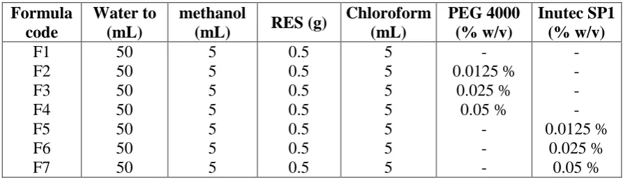
Table I: Composition of different prepared Res spherical agglomerates.
Formula code
Water to (mL)
methanol
(mL) RES (g)
Chloroform (mL) PEG 4000 (% w/v) Inutec SP1 (% w/v) F1 F2 F3 F4 F5 F6 F7 50 50 50 50 50 50 50 5 5 5 5 5 5 5 0.5 0.5 0.5 0.5 0.5 0.5 0.5 5 5 5 5 5 5 5 - 0.0125 % 0.025 % 0.05 % - - - - - - - 0.0125 % 0.025 % 0.05 %
3. Evaluation of prepared RES spherical agglomerates
3.1.Determination of the yield of spherically agglomerated crystals[25]
The yield of spherical agglomerates was calculated by comparing the whole weight of the
agglomerates formed against the collective weight of the polymer and drug.
Percentage yield = (Weight of agglomerates obtained/Total weight of drug and polymer) ×
100.
3.2. Drug Content[26]
The assay of the weighed amount of RES spherical agglomerates was carried out to
determine the drug content. The weighed samples (about 10 mg) were dissolved in 10 mL of
0.1 N HCl and stirred by vortex mixer. The solution was subjected to serial dilution, filtered
using Whatman filter paper. The content was estimated spectrophotometrically (Shimadzu,
model UV-2450, Japan) at 237 nm.
3.3. Solubility study[27]
Half gram of RES and its different spherical crystals were introduced separately into a series
of vials in 10 mL of distilled water. Vials were shaken in a thermostatically controlled shaker
for 48 h at 25 ± 0.5 °C at 100 rpm. The time required for equilibrium was established by
repetitive sampling. After equilibrium, the samples were centrifuged then filtered through
0.45 μm membrane filter. The drug saturation solubility in the systems under investigation
was determined spectrophotometrically after appropriate dilution of the filtrate using distilled
3.4 Particle size analysis
The particle size of RES powder and its prepared spherical agglomerates was determined by
laser scattering particle size analyzer (Horiba, model LA-920, Japan).
3.5. In vitro dissolution study[28]
In vitro dissolution tests for pure RES and its prepared spherical agglomerates equivalent to
10 mg of RES were carried out with the USP dissolution method type II using USP
dissolution testing apparatus (Pharma Test, PTW, Germany) at 37 ± 0.5 °C and rotated at 100
rpm using 900 mL of simulated gastric fluid (SGF) as dissolution medium (n=3). Samples
were withdrawn at specified time intervals, and replaced with an equivalent volume of fresh
dissolution medium to keep the volume constant. Collected samples were filtered and assayed
spectrophotometrically at λmax 237 nm using SGF as blank. The cumulative amount of drug
released was calculated and plotted versus time. The study was done in triplicate.
3. Solid state characterization[29-30]
3.1. Microscopical observation
RES powder as well as its selected agglomerated crystals was examined under the optical
microscope (model Bio-pal2-TP, Japan) and photomicrographs at appropriate magnification
were taken. Scanning electron microscopy (model JSM 840A, Jeol, Japan) was used to access
the surface morphology of the agglomerates; the crystals were splutter covered with gold
before scanning.
3.2. Differential scanning calorimetric (DSC) studies
DSC analysis was performed using differential scanning calorimeter (Shimadzu -DSC-50,
Japan). Samples of RES and its selected agglomerates were heated under nitrogen atmosphere
as carrier gas on aluminum pan at a flow rate 25 mL/min and a heating rate of 10 °C/ min over
temperature range of 20-400 °C.
3.3. Fourier Transform Infrared Spectroscopic Studies
Fourier transform infrared (FT-IR) spectroscopy was employed to further characterize the
possible interactions between the drug and the carrier in the solid state on a FT-IR
spectrophotometer (Jasco-FTIR-1700 spectrophotometer, Japan) by the conventional KBr
3.4. Powder X-ray diffraction (PXRD)
PXRD spectra of RES and the selected spherical agglomerates were traced by employing an
X-ray Diffractometer (Philips, England) for pure drug, and the selected spherical agglomerates Ni
filtered Cu K (α) radiation, a voltage of 40 kV, a current of 20 mA and receiving slit of 0.2
inch. The samples were analyzed over 2Ø range from 2◦ C -50◦C at the rate of 2º C per min at 0.02◦ at 2Ø step size.
4. In vivo Evaluation
Albino mice of 7–17 weeks of age, weighing 20 to 32 g were used. Mice were housed in
groups of two to four with free access to food and water. A 12-hr light–dark cycle was
maintained (light on from 6.00 a.m. until 6.00 p.m.) at a temperature of 22°C and a relative
humidity of 60%. All experiments were conducted in accordance with the U.S. guide for the
care and use of laboratory animals and approved by local authorities (NIH publication No.
86-23, revised 1985 and the current version of the German Law on the Protection of
Animals).
Effect of RES on Diazepam-induced Sleeping Time in Mice
The animals were divided into 3 groups (n=6). Diazepam (25 mg/ kg, i.p.) was administered
to the first (control) group. To the second and third group RES or F2 (2.5 mg / kg, p.o.) was
administered 6.0 h prior to the diazepam injection. The time interval between the loss and
regaining of righting reflex (falls asleep) was measured as sleeping time, the duration of
action of diazepam per se and in combination with RES or F2 was noted.[31]
RESULTS AND DISCUSSION
1. Evaluation of the prepared RES spherical agglomerates
The percentage yield of different spherical agglomerated crystals was in the range of 92.5 ±
1.14 to 98.6 ± 2.16. The percentage of drug content of the prepared spherical agglomerated
crystals ranged from 91.2 5 ± 1.85 to 98.45 ± 0.51. These results were very close to the
theoretical values for all of the prepared spherical agglomerated crystals. These values were
taken to calculate the amounts needed for achieving the dissolution studies under sink
conditions. Data are presented in Table II.
1.1. Solubility study
The solubility of all RES agglomerated crystals was improved, it was found to be in the
2.16 ± 0.12 µg/mL (Table II). Agglomerated RES containing polymers showed highest
solubility than those without polymers. This expected result was due to wetting effect of the
hydrophilic polymers.[32] An increase in polymer concentration was accompanied by a decrease in RES solubility. According to phase solubility method of Higuchi and Connors this
type of solubility can be classified as AN subtype (negatively deviating isotherms) because at
higher polymer concentrations a negative deviation from linearity exists, meaning that they
are less effective at higher concentrations.[33] The highest improvement in solubility was observed with polyethylene glycol 4000 at 0.0125% w/v concentration level.
1.2. Particle size determination
The pure drug exhibited a small particle size, 174 ± 6.7 µm, whereas the size of the prepared
agglomerates ranged from 312 ± 5.1 to 412 ± 8.1 µm; this indicates agglomeration of crystals
(Table II).
Table II: Percentage yield, Drug content, Solubility and Particle Size of different
spherical agglomerated crystals.
Formula Percentage yield
± SD
Drug content ± SD
Solubility (µg/mL ± SD)
Particle size (µm ± SD) RES F1 F2 F3 F4 F5 F6 F7 --- 93.4 ± 2.11 98.6 ± 2.16 96.1 ± 3.15 92.5 ± 1.14 97.88 ± 2.17
96.9 ± 2.21 95.25 ± 1.31
--- 96.73 ± 1.5 97.45 ± 1.14
95.94 ± 1.15 98.45 ± 0.51 95.55 ± 1.34 97.24 ± 1.25 91.2 5 ± 1.85
2.16± 0.12 15.6 ± 0.27 188.2 ± 0.2 172.5 ± 0.28 162.4 ± 0.49 176.3 ± 0.15 155.5 ± 0.37 75.4 ± 0.72
174 ± 6.7 372 ± 9.4 348 ± 4.6 412 ± 8.1 351 ± 14.6
340 ± 7.1 312 ± 5.1 366 ± 9.4
1.3. Dissolution study
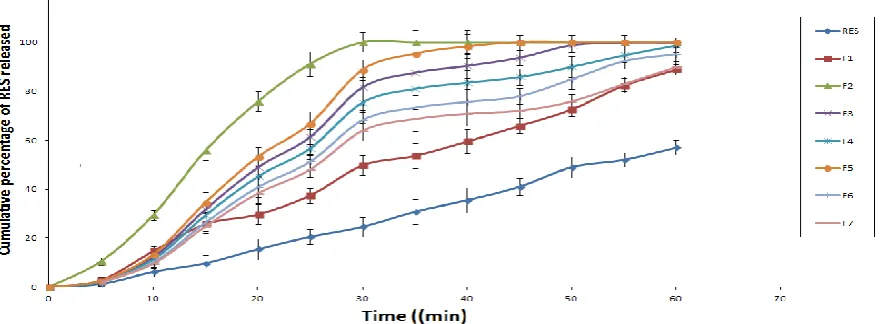
Dissolution profiles of RES and different prepared agglomerates are illustrated in Figure 1
Recrystallization of RES into spherical agglomerates successfully improved dissolution rate
comparing to RES powder in SGF. The enhancement in the rate of dissolution in comparison
to the pure drug was a result of improved porosity resulting from diffusion of solvents during
solidification process.[34] Data of dissolution study were consistent with that of solubility study where RES agglomerates containing polymers showed higher dissolution rate than
those without polymers (F1). Also, highest dissolution rate enhancement was associated with
agglomerates containing polymers at low concentration levels. In SGF: RES spherical
agglomerates showed 3.45 - 6.16 times improvement in the dissolution compared to RES
while the pure drug gave only 57.4% release after 60 min. the results indicated that, there was
a dramatic improvement in the rate of dissolution in comparison with pure RES. However,
[image:8.595.77.518.178.340.2]the increase in dissolution rate was in order of F2 > F5> F3> F4> F6> F7 > F1 as shown in
Figure 1.
Figure 1: Dissolution profiles of RES from its agglomerates in SGF.
2. Solid state characterization of RES spherical agglomerates
Light microscopy photographs of RES powder and its prepared agglomerates, Figure 2
showed that the agglomerated crystals are nearly spherical in shape while the crystals of pure
RES were irregular shaped. SEM (Figure 3) showed that particles of unprocessed RES were
smaller in size than processed ones. Unprocessed RES particles were flat shaped while the
processed ones were spherical agglomerates. It was apparent that degree of surface roughness
of spherical agglomerates crystallized in the presence of polymers was higher compared to
agglomerates with no polymer due to adhesion of polymers to the surface of the agglomerates
[image:8.595.76.520.583.688.2]which is supported by DSC and XRD analysis.
Figure 2: Microscopic photographs of RES, F2 and F5.
Figure 3: SEM images of RES, F2 and F5.
2.1. Differential scanning calorimetery studies
DSC thermograms of RES, and selected agglomerates are illustrated in figure 4. A sharp
endothermic peak appeared in pure RES thermogram with the following peak parameters:
onset at 166.69°C, peak at 175.20°C, with an area of 150.06m J and Delta H value of 50.020
which is indicative of its high crystalline nature.[35-37] A broad single endothermic peak was observed at 155°C and 195°C (for PEG4000 and Inutec SP1 respectively) which indicated the
nature of the polymers. The peaks of the prepared agglomerated crystals using PEG4000 and
Inutec SP1 were 178.65°C and 180.2°C respectively. The non-appreciable change in RES
endotherms in all of the tested samples indicates that no interaction has occurred between
RES and polymers during crystallization of particles.
Figure 4: DSC patterns of RES, polymers and selected agglomerates prepared using
different polymers at 0.0125% w/v concentration.
2.2. Fourier Transform infrared spectroscopy
The FT-IR spectra of pure RES, F2 and F5 are shown in figure 5. The following
[image:9.595.96.493.454.619.2]stretching); 820 cm-1 (C-Cl stretching). The FT-IR spectrum of all selected agglomerates shows the characteristic bands of RES. Also, there was no great difference in the fingerprint
region (almost unique for every organic compound) between FT-IR spectrum of the drug and
that of its selected spherical agglomerated crystals.[38] These findings indicated that there was no remarkable interaction occurred between drug and different polymers used during
[image:10.595.127.472.218.369.2]crystallization process confirming DSC study data.
Figure 5: FT-IR spectra of pure RES, F2 and F5.
2.3. Powder X-ray diffraction studies
X-ray diffractograms of RES powder, and the selected crystal agglomerates are represented
in Figure 6. The diffraction pattern of RES showed that the drug has high crystallinity
because of the presence of numerous distinct peaks. The most characteristic peaks in
ascending order of intensity are at 2θ diffraction angles of 6, 9.5, 11, 12.5, and 16.5°. The
results of PXRD of the spherical agglomerates showed presence of several distinct peaks
characteristic for RES which revealed that the agglomerates are in a crystalline form.
Although the agglomerates showed crystalline pattern in PXRD diffractograms, but the
intensity of the peaks have been reduced in comparison to the pure RES suggesting the
conversion of crystalline RES to partially disordered molecules (decreased crystallinity or
Figure 6: PXRD spectra of pure RES, F2 and F5.
3. In vivo Evaluation
Both RES and F2 enhanced the diazepam-induced sleeping time in mice. The average
sleeping time due to diazepam (25 mg / kg, i.p.) per se was found to be 82.6 ± 3.5 min., while
the average sleeping time due to diazepam and RES was increased to 92.2± 5.5 min and the
average sleeping time due to diazepam and F2 was increased significantly to 160.5 ± 6.5
min. The data were subjected to one way analysis of variance (ANOVA) followed by post hoc
analysis (LSD). The results demonstrated that RES powder and F2 significantly (p < 0.05)
enhanced the diazepam-induced sleeping time in mice as compared to the control. F2
significantly increased the diazepam-induced sleeping time in mice in comparison to the RES
powder (p < 0.05), indicating that there is a significant improvement in the bioavailability of
RES demonstrated by the significant enhancement of RES activity.[41–44]
CONCLUSIONS
The spherically agglomerates crystals of RES were successfully prepared by quasi emulsion
solvent diffusion technique and the incorporation of hydrophilic polymers as polyethylene
glycol 4000 and Inutec SP1 to such systems resulted in an improvement in the quality of the
produced spherical agglomerates. Our result revealed that the F2 formula significantly
improved RES activity (manipulated by a significant increase in the diazepam-induced
sleeping time in mice) in comparison to the RES powder. From these results we can conclude
that this technique may be applied fruitfully to enhance the bioavailability of RES powder
ACKNOWLEDGEMENTS
Our deepest thanks and appreciation to Dr. Rola Bayram, Assistant prof. of analytical
chemistry for her continuous help during our research.
REFERENCES
1. Daniel Markl, J. Axel Zeitler. A Review of Disintegration Mechanisms and Measurement
Techniques, Pharm Res, 2017; 34: 890–917.
2. Shweta Gupta, Rajesh Kesarla, and Abdelwahab Omri. Formulation Strategies to Improve
the Bioavailability of Poorly Absorbed Drugs with Special Emphasis on Self-Emulsifying
Systems ISRN Pharm., 2013; 2013: 1-16.
3. Gursoy R.N. and Benita S. Self emulsifying drug delivery systems (SEDDS) for
improved oral delivery of lipophilic drugs. Biomed Pharmacother, 2004; 58(3): 173-182.
4. Krishnaiah Y.S. Pharmaceutical technologies for enhancing oralbioavailability of poorly
soluble drugs. J Bioequiv Bioavail, 2010; 2: 28-36.
5. Kesisoglou F., Panmai S., Wu Y. Nanosizing-oral formulation development and
biopharmaceutical evaluation. Adv Drug Deliv Rev, 2007; 59(7): 631-644.
6. Orienti I., Bigucci F., Luppi B., Cerchiara T., Zuccari G., Giunchedi P. and Zecchi V.
Polyviny lalcohol substituted with triethyleneglycolmonoethylether as a new material for
preparation of solid dispersion of hydrophobic drugs. Eur J Pharm Biopharm, 2002;
54(2): 229-233.
7. Ben J.Boyda, Christel, A.S. Bergströmb, Zahari Vinarovc, Martin Kuentzd, Joachim
Brouwerse, Patrick Augustijnse, Martin Brandlf, Andreas Bernkop-Schnürchg, Neha
Shresthah, Véronique Préath, Anette Müllertzi, Annette Bauer-Brandlf, Vincent Janninj.,
Successful oral delivery of poorly water-soluble drugs both depends on the intraluminal
behavior of drugs and of appropriate advanced drug delivery systems, European Journal
of Pharmaceutical Sciences, 2019; 137: 1049672.
8. Chaudhary A., Upendra N., Neha G., Sharma V. K. and Khosa R., Enhancement of
solubilization and bioavailability of poorly soluble drugs by physical and chemical
modifications: A recent review. JAPER, 2012; 2(1): 32-67.
9. Tereza Školáková, Michaela Slámová, Andrea Školáková, Alena Kadeřábková, Jan
Patera, and Petr Zámostný, Investigation of Dissolution Mechanism and Release Kinetics
of Poorly Water-Soluble Tadalafil from Amorphous Solid Dispersions Prepared by
Various Methods Pharmaceutics, 2019; 11(8): 383.
Modification: A unique solutions to Solubility problem Journal of Drug Delivery &
Therapeutics, 2019; 9(2): 542-546.
11. Choi JS, Lee SE, Jang WS, Byeon JC, Park JS. Solid dispersion of dutasteride using the
solvent evaporation method: Approaches to improve dissolution rate and oral
bioavailability in rats. Mater Sci Eng C Mater Biol Appl., 2018; 1(90): 387-396.
12. Phuong Tran, Yong-Chul Pyo, Dong-Hyun Kim, Sang-Eun Lee, Jin-Ki Kim and
Jeong-Sook Park. Overview of the Manufacturing Methods of Solid Dispersion Technology for
Improving the Solubility of Poorly Water-Soluble Drugs and Application to Anticancer
Drugs Pharmaceutics, 2019; 11: 132-158.
13. Vaibhav R. Toche, Pratik R. Ugale, Amol S. Deshmukh. Solid Dispersion: A Technique
To Enhance Solubility Of Poorly Soluble Drugs Asian Journal of Research in Biological
and Pharmaceutical Sciences, 2018; 6(2): 85-93.
14. Amit T., Pravin K. and Dinesh S. An improvement of micromeritic properties and
dissolution behaviors of carvedilol spherical agglomerates crystallized in presence of
inutec SP1. Turk J Pharm Sci, 2012; 9(1): 101-112.
15. Gadekar S., Kavade V. and Kale V. Development of directly compressible metformin
hydrochloride tablets. Int J Pharm Sci Rev Res, 2011; 9(1): 37-41.
16. Borut K., Franc V. and Odon P. Spherical crystallization of drugs. Acta Pharm, 2012; 62:
1-14.
17. Mahenty S., Sruti J., Niranjan P. and Bhanoji M.E. Particle design of drugs by spherical
crystallization techniques. IJPSN, 2010; 3(2): 912-918.
18. Chetan B. Chure, and Swati Rawat. Overview of Particle Engineering: Spherical
Crystallization Techniques. Research Journal of Pharmaceutical, Biological and Chemical
Sciences, 2018; 9(2): 1172-1180.
19. M Campbell P I Young D N Bateman, J M Smith and S H L Thomas. The use of atypical
antipsychotics in the management of schizophrenia. Br J Clin Pharmacol, 1999; 47(1):
13–22.
20. Shukla D., Chakraborty S., Singh S., Mishra B. Fabrication and evaluation of taste
masked resinate of risperidone and its orally disintegrating tablets. Chem. Pharm. Bull.,
2009; 57: 337-345.
21. Shukla D., Chakraborty S., Singh S., Mishra B. Preparation and in-vitro characterization
of risperidone-cyclodextrin inclusion complexes as a potential injectable product. DARU,
2009; 7(4): 226-235.
taste-masking resin: Novel application of near infra-red and chemical imaging to evaluate
complexes. Pharm. Dev. Tech., 2009; 14: 409–421.
23. Mansoor A.K., Rakhi B.S., Mobin A.T., Vilayat A.S. Complexation between risperidone
and amberlite resin by various methods of preparation and binding study. Drug Dev. Ind.
Pharm., 2009; 1: 1–10.
24. Tapas A.R., Kawtikwar P.S., Sakarkar D.M. - Enhanced dis- solution rate of felodipine
using spherical agglomeration with Inutec SP1 by quasi emulsion solvent diffusion
method.- Res. Pharm. Sci., 2009; 4(2): 77-84.
25. MuthukumarN, Harry Thomas RodriguezA, Evaluation of spherical agglomerated
crystals of lomefloxacin by IR and optical microscopy. Journal of Research in biology,
2014; 4(5): 1405 – 1416.
26. Shital P., Rakesh M., SV Shirlkar. Spherical agglomeration a novel approach for
solubility and dissolution enhancement of simvastatine. Asian Journal of Pharmaceutical
And Clinical Research, 2016; 9(6): 65-72.
27. Ghurghure S M., Chandakavathe B N. Design and Development of Spherical
Agglomerates of Drug for the Enhancement of Solubility. Indian Journal of Noval Drug
Delivery, 2016; 8(2): 93-99.
28. Biswal S., Sahoo J., Murthy P.N., Giradkar R.P., Avari J.G. Enhancement of
dsissoltuion rate of glicliazide using solid dispersions with polyethylen glycol 6000.
AAPS Pharm Scitech., 2008; 9: 563-570.
29. Marwah M. Hareeja and Eman B.H.Al-Khedairy, Formulation and In- Vitro
Evaluation of Spherical Crystal Agglomerates of Ebastine by Quasi Emulsion
Solvent Diffusion Method Iraqi J Pharm Sci, 2018; 27(2): 77-92.
30. Motaz Farid, Doaa Ahmed El-Setouhy, Mohamed Ahmed El-Nabarawi, and Tahany
El-Bayomi1 Particle engineering/different film approaches for earlier absorption of
meloxicam. Drug Deliv, 2016; 23(7): 2309–2317.
31. Pal D, Nandi M. CNS activities of Celesia coromandeliane Vahl. in mice. Acta Pol
Pharm, 2005; 62: 355–361.
32. Fadke J, Desai J, Thakkar H. formulation development of spherical crystal
agglomerates of itraconazole for preparation of directly compressible tablets with
enhanced bioavailability. AAPS Pharm Sci Tech., 2015; 16(6): 1434-1444.
33. Moataz Farid, Alyah Al Atwi, Ebtihal Al Qahtani, Lama Allam and Lilian Saher.
Improvement of Bioavailability of A Drug Belonging To Biopharmaceutical
2018; 7(4): 1872-1883.
34. Kadajji VG, Betageri GV. Water soluble polymers for pharmaceutical applications.
Polymers, 2011; 3(4): 1972-2009.
35. Leuner C., Dressman J. Improving drug solubility for oral delivery using solid
dispersions. Eur. J. Pharm. Biopharm., 2000; 50: 47–60.
36. Craig D.Q.M. The mechanism of drug release from solid dispersions in water soluble
polymers. Int. J. Pharm., 2002; 231: 131–144.
37. Dhirendra K.L., Udupa N., Atin K. Solid dispersions: A review. Pak. J. Pharm. Sci.,
2009; 22: 234-246.
38. Fülöp Ibolya, Árpád Gyéresi, Piroska Szabó-Révész, Zoltán Aigner, Solid
dispersions of flufenamic acid with PEG 4000 and PEG 6000, Farmacia, 2011; 59(1):
60-69.
39. Usha A.N., Mutalik S., Reddy M.S., Ranjith A.K., Kushtagi P., Udupa N. Preparation
and, in vitro, preclinical and clinical studies of aceclofenac spherical agglomerates. -
Eur. J. Pharm. Biopharm., 2008; 70(2): 674-683.
40. M. Farid, D.A. El-Setouhy, M.A. El-Nabarawi, T. El-Bayomi, Recrystallized
agglomerated meloxicam: evaluation of anti-nociceptive effect. J. Drug Del. Sci.
Tech., 2014; 24(6): 645-652.
41. Samah S, Mohammad F, Alsayed Z, Radwan El. H, Mohammad J. S. Evaluation of oral
hypoglycemic potency of Medicago polymorpha and Zygophyllum simplex: A Drug –
Drug interaction study. Journal of Pharmacognosy and Phytochemistry, 2017; 6(6):
648-651.
42. Shabana S, Fouad M, Zaki A, El-Haggar R, Sadiq M.J, Osman NMS and Sindi IA:
Evaluation of antihypertensive activity of natural mixture and investigation of herb-herb
interaction. Int J Pharmacognosy., 2018; 5(8): 455-60.
43. Jaffar S. M, Padmanabha R.Y, Kalava B, Narayana G. A Study on Antidepressant
Activity of Eugenol Excluded Clove Extract. Research Journal of Pharmaceutical,
Biological and Chemical Sciences., 2012; 3(2): 632 – 638.
44. Jaffar S. M, Vigneshwaran. E, Shareen T, Srinath. B, Padmanabha R. Y, Chandrasekhar
K.B. A study on antiepileptic activity of eugenol excluded aqueous extract of eugenia