PHYTO ACTIVE COMPOUNDS FROM HERBAL PLANT EXTRACTS:
ITS EXTRACTION, ISOLATION AND CHARACTERIZATION
Akshada Amit Koparde*1, Dr. C. S. Magdum2 and Dr. R. C. Doijad3
1
Asst Professor, Department of Pharmaceutical Chemistry, KIMSDU‟S Krishna Institute of
Pharmacy, NH4, Krishna Hospital Campus, Malkapur, Karad 415110.
2
Principal, Rajarambapu College of Pharmacy, Near Padayatri Smarak, Kasegaon 415404.
3
Dean, KIMSDU‟S Krishna Institute of Pharmacy, NH4, Krishna Hospital Campus,
Malkapur, Karad 415110.
ABSTRACT
Herbal extracts its pure compounds provide opportunities for new drug
discovery. Herbal Plants are widely used in the pharmaceutical
industry for herbal drug development due to their structural diversity
and presence of various pharmacological activities. The biological
active compounds that are present in plants referred as phytochemicals.
These phytochemicals derived from different parts of plants and
thereby used as sources of lead molecules. The secondary metabolic
compounds present in the plants describes the phytochemistry.
Naturally occurring chemical compounds are present in plants and
contains structurally diverse bioactive molecules. This review article
highlights on the analytical methodologies which include the
extraction, isolation and characterization of active ingredients from
herbal plants. Different techniques of extraction are explained as
extraction is the most important first step towards analysis of active constituents. This paper
also highlights the isolation of active molecules by chromatographic techniques like TLC,
column etc. The most important step towards analysis of bioactive compounds present in the
plant extracts is characterization which includes phytochemical screening assays, HPLC(High
Performance Liquid Chromatography), HPTLC (High Performance Thin Layer
Chromatography), FTIR Fourier Transform Infra-Red spectroscopy (FTIR), NMR (Nuclear
Magnetic Resonance), GCMS (Gas Chromatography and Mass Spectrometry) through which
Volume 6, Issue 8, 1186-1205. Research Article ISSN 2277–7105
*Corresponding Author Akshada Amit Koparde Asst Professor, Department
of Pharmaceutical
Chemistry, KIMSDU‟S
Krishna Institute of
Pharmacy, NH4, Krishna
Hospital Campus, Malkapur,
Karad 415110., Article Received on 01 June 2017,
Revised on 21 June 2017, Accepted on 12 July 2017
lead molecule structure identification can be done .Thus key challenges in research related to
herbal drug development is discussed in this paper.
KEYWORDS: Herbal extracts, Phytochemicals, Extraction, Chromatographic techniques,
Herbal drug development.
INTRODUCTION
Plants are natural reservoir of medicinal agents. These are almost free from the side effects.[1]
Natural products, such as plants extract, open a new horizon for the discovery of new
therapeutic agents. It also provide unlimited opportunities for new drug discoveries because
of the unmatched availability of chemical diversity.[2] In most developing countries
traditional medicine and medicinal plants are used for the maintenance of good health and
about 80% of the world‟s population relies on herbal medicines as better curative measures
without or with minimum side effects.[3] Primary health care treatment, most of which
involve the use of herbal preparations which include plant extracts containing active
constituents.[4,5] Thus Plants contain a wide range of chemical compounds that can be used to
treat chronic as well as infectious diseases.[6] Due to the development of adverse effects and
microbial resistance to the chemically synthesized drugs, men turned to
ethnopharmacognosy. They found literally thousands of phytochemicals from plants as safe
and broadly effective alternatives with less adverse effect. According to the World Health
Organization, a medicinal plant is any plant which, in one or more of its parts, contains
substances that can be used for therapeutic purposes, or which are precursors for
chemo-pharmaceutical semi synthesis. Parts of such plants including leaves, roots, rhizomes, stems,
barks, flowers, fruits, grains or seeds, employed in the control or treatment of a disease
condition and therefore contains chemical components that are medically active. These plant
chemical compounds or bioactive components are often referred to as phytochemicals
(„phyto‟- from Greek - phyto meaning „plant‟) or phytoconstituent. They protect plants
against microbial infections or infestations by pests.[7,8] These constituents have been in use
by the ancient therapist for the use of various human aliments also. The science of application
of these indigenous or local medicinal remedies including plants for treatment of diseases is
currently called ethno pharmacology. Many beneficial biological activity such as anticancer,
antimicrobial, antioxidant, antidiarrheal, analgesic, antiinflammatory and wound healing
activity were reported. In many cases the people claim the good benefit of certain natural or
show evidence based approach towards medicine from herbal source. The evidences can only
be obtained by performing research and thus when we have pure form of phytochemicals ,the
researcher can prove it through characterization and pharmacological activity. According to
the World Health Organization (WHO), nearly 20,000 medicinal plants exist in 91 countries
including 12 mega biodiversity countries. The process to obtain the biologically active
compound from herbal plant are extraction, isolation, characterization of bioactive
compound, pharmacological screening, toxicological evaluation and clinical evaluation. This
review focus on different available techniques for the extraction of these phytochemicals ,its
isolation and characterization for structure identification ,number of hydrogen atom, carbon
atom present in the molecule and its molecular weight through phytochemical screening
assay, chromatographic techniques, such as NMR, HPLC, HPTLC, GCMS, and FTIR. etc.
Strategies in the search of new natural compounds.[9]
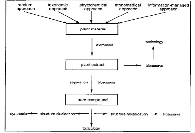
To study medicinal plants, it is first of all necessary to know which plant to select and what
type of biological activity to look for. The selection criteria of plants, which potentially
contain new biological agents, is based on five principle approaches: the random, the
taxonomic, the phytochemical, the ethnomedical and the information-managed approach. In
the random approach all available species are collected, irrespective of prior knowledge and
experience. In the taxonomic approach, plants of a specific genus or family are deemed to be
of interest, and sought from diverse locations. The phytochemical (chemo-taxonomic)
[image:3.595.132.451.339.556.2]anticipated to produce related compounds are collected. Taxonomic and the phytochemical
approach are closely related and can not be clearly divided from each other. In the
ethnomedical approach, credence is given to information on the medicinal use of the plant.
Based on this information, the plant is collected and evaluated.
Practical Aspects of Herbal Drug Discovery[10,11]
The following scheme represents a summary of the stages involved in the development of
pure drug from a plant source.
Collection and identification of the plant and deposition of voucher sample in local and
major herbarium.
Literature survey on the plant species selected for studies.
Extraction with solvent and preparation of non-polar and polar extracts for initial
biological testing.
Evaluation of plant extract against a panel of biological test methods, as exemplified by
receptor biding, enzyme inhibition, and /or cytotoxicity assays.
Activity guided fractionation on the extract showing activity, by monitoring each
chromatographic fraction with bioassay chosen from the panel available to the
investigation.
Structure elucidation of pure active isolates using spectroscopic techniques and chemical
methods, if necessary.
Test each active compound (whether of novel or known chemical structure) in all in vitro
and in vivo biological test methods available, in order to determine potency and
selectivity of the drug.
Perform molecular modeling studies and prepare derivatives of the active compound of
interest.
Sometime requires to carry out large scale reisolation of interesting active compounds for
toxicological, pharmacological and for mutation studies. Clinical trials.
Extraction Methods for Studying Phytochemicals
Extraction is the first important step in the analysis of medicinal plants, because for further
separation and characterization it is necessary to extract the desired chemical components
from the plant materials. The extraction of plant constituents is essential to isolate
the analysis of medicinal plants. The basic steps included are pre-washing, drying of plant
materials or freeze drying, grinding to obtain a homogenous sample. Extraction from the
plant is important since different solvents are utilized at varying conditions such as time and
temperature of extraction. It must be assured that the potential active constituents are not lost
or distorted during the process of extraction. If the plant was selected on the basis of
traditional uses,[12] then it is needed to prepare the extract as described by the traditional
healer in order to mimic as closely as possible the traditional „herbal‟ drug. Different type of
solvent systems are available to extract the active compound. The specific solvent system to
be used depends on the nature of the bioactive compound which is being targeted. The
extraction of hydrophilic compounds uses polar solvents such as methanol, ethanol or
ethyl-acetate. For extraction of more lipophilic compounds, dichloromethane or a mixture of
dichloromethane/methanol in ratio of 1:1 are used. In some instances, extraction with hexane
is used to remove chlorophyll.[13] As the target compounds may be non-polar to polar and
thermally labile, the suitability of the methods of extraction must be considered. Various
methods, such as sonification, heating under reflux, soxhlet extraction and others are
commonly used[14-16] for the plant samples extraction. In addition, plant extracts are also
prepared by maceration or percolation of fresh green plants or dried powdered plant material
in water and/or organic solvent systems. Further fractionation of extracted compounds done
on the basis of their acidity, polarity or molecular size. The extraction methods mostly used
has been discussed below:
Hot Continuous Extraction (Soxhlet)
In this method, the finely ground crude drug is placed in a porous bag or “thimble” made of
strong filter paper, which is placed in chamber E of the Soxhlet apparatus. The extracting
solvent in flask A is heated, and its vapors condense in condenser D. The condensed
extractant drips into the thimble containing the crude drug, and extracts it by contact. When
the level of liquid in chamber E rises to the top of siphon tube C, the liquid contents of
chamber E siphon into flask A. This process is continuous and is carried out until a drop of
solvent from the siphon tube does not leave residue when evaporated. The advantage of this
method, compared to previously described methods, is that large amounts of drug can be
extracted with a much smaller quantity of solvent. This effects tremendous economy in terms
of time, energy and consequently financial inputs. At small scale, it is employed as a batch
process only, but it becomes much more economical and viable when converted into a
Microwave-assisted extraction (MAE)
It simply termed as microwave extraction, that combines microwave and traditional solvent
extraction. It has application in extraction of high-value compounds from natural sources
including phytonutrients, nutraceutical and functional food ingredients and pharmaceutical
actives from biomass. Heating the solvents and plant tissue using microwave increases the
kinetic of extraction, is called microwave-assisted extraction.[18] The target for heating in
dried plant material is the minute microscopic traces of moisture that occurs in plant cells.
The heating up of this moisture inside the plant cell due to microwave effect, results in
evaporation and generates tremendous pressure on the cell wall. The cell wall is pushed from
inside due to the pressure and the cell wall ruptures. Thus the exudation of active constituents
from the ruptured cells occurs, hence increasing the yield of phytoconstituents.[19,20] It offers
following advantages:[21]
1. Increased purity of crude extracts, improved products, improved stability of marker
compounds, possibility to use less toxic solvents.
2. Increased recovery and purity of marker compounds, reduced processing costs, very fast
extraction rates, reduced energy and solvent usage.
The other modern extraction techniques include solid-phase micro-extraction,
supercritical-fluid extraction, pressurized-liquid extraction, microwave-assisted extraction, solid-phase
extraction, and surfactant-mediated techniques, which possess certain advantages. These are
the reduction in organic solvent consumption and in sample degradation, elimination of
improvement in extraction efficiency, selectivity, and/ kinetics of extraction. The ease of
automation for these techniques also favors their usage for the extraction of plants
materials.[22]
Isolation, Identification and Characterisation of Phytochemicals
Combination of various types of bioactive compounds or phytochemicals with different
polarities are usually present in different plant extracts. Separation, identification and
characterization of bioactive compounds is a big challenging job in herbal drug development
process. A number of different separation techniques are used for identification and
characterization of these bioactive compounds to obtain pure compounds such as TLC,
column chromatography, flash chromatography, HPTLC and HPLC. The pure compounds
are then used for the determination of structure and pharmacological activity. Various
techniques such as phyto-chemical screening assay, Fourier-transform infrared spectroscopy
(FTIR), NMR(Nuclear Magnetic Resonance), GCMS (Gas Chromatography and Mass
Spectrometry) are specifically used for the identification of the bioactive compounds through
which lead molecule structure identification can become easy.[23]
Phytochemical screening assay[24-29]
Phytochemical screening assay is a simple, quick, and inexpensive procedure that tells about
various types of phytochemicals in a mixture and an important tool in bioactive compound
analyses. Phytochemical examinations are carried out for all the extracts as per the standard
methods.
1. Tests for Carbohydrates
Preparation of test solution: The test solution was prepared by dissolving the test extract with
water. Then it was hydrolyzed with 1 volume of 2N HCl and subjected to following chemical
tests.
Molisch's test (General test): To 2-3 ml aqueous extract, added few drops of -naphthol
solution in alcohol, shaken and added concentrated H2SO4 from sides of the test tube and
then observed for violet ring at the junction of two liquids.
Fehling's test: 1 ml Fehling's A and 1ml Fehling's B solutions were mixed and boiled for
one minute. Added equal volume of test solution. Heated in boiling water bath for 5-10
Benedict's test: Equal volume of Benedict's reagent and test solution in test tube were
mixed. Heated in boiling water bath for 5 min. Solution may appear green, yellow or red
depending on amount of reducing sugar present in test solution.
Barfoed's test: Equal volume of Barfoed's reagent and test solution were added. Heated
for 1-2 min, in boiling water bath and cooled. Observed for red precipitate.
2. Tests for Proteins
Biuret test (general test): To 3 ml test solution (T.S.) add 4% NaOH and few drops of 1%
CuSO4 solution observed for violet or pink colour.
Million's test (for proteins): Mixed 3 ml T.S. with 5 ml million's reagent, white precipitate. Precipitate warmed turns brick red or precipitate dissolves giving red colour.
Xanthoprotein test (For protein containing tyrosine or tryptophan): Mixed 3 ml T.S. with
1 ml concentrated H2SO4 observed for white precipitate.
3. Tests for Amino acids
Ninhydrin test (general test): 3 ml T.S. and 3 drops 5% Ninhydrin solution were heated in
boiling water bath for 10 min. observed for purple or bluish colour.
Test for tyrosine: Heated 3 ml T.S. and 3 drops Million's reagent. solution observed for
dark red colour.
Test for tryptophan: To 3 ml T.S. added few drops glycoxalic acid and concentrated
H2SO4 observed for reddish violet ring at junction of the two layers.
Test for cysteine: To 5 ml. T.S. add few drops of 40% sodium hydroxide and 10% lead
acetate solution. Boil. Black ppt. of lead sulphate is formed.
4. Tests for Steroid and Triterpenoid
Salkowaski reaction: Mixed 2 ml of extract, 2 ml chloroform and 2 ml concentrated
H2SO4, Shake well, whether chloroform layer appeared red and acid layer showed
greenish yellow fluorescence was observed.
Liebermann-burchard reaction: Mixed 2ml extract with chloroform add 1-2 ml acetic
anhydride and 2 drops concentration H2SO4 from the side of test tube observed for first
red, then blue and finally green colour was observed.
Liebermann‟s reaction: Mixed 3 ml extract with 3 ml acetic anhydride. Heated and
Tests for Glycosides
Preparation of test solution: The test solution was prepared by dissolving extract in the
alcohol or hydro-alcoholic solution.
A) Tests for cardiac glycosides
Baljet's test: A test solution observed for yellow to orange colour with sodium picrate. Legal's test (For cardenoloids): To aqueous or alcoholic test solution, added 1 ml pyridine
and 1 ml sodium nitroprusside observed for pink to red colour.
Test for deoxysugars (Keller killiani test): To 2 ml extract, add glacial acetic acid, one
drop of 5% FeCl3 and concentrated H2SO4 observed for reddish brown colour at junction
of the two liquid and upper layers bluish green.
Liebermann‟s test (for bufadenolids): Mixed 3 ml extract with 3 ml acetic anhydride.
Heat and cooled. Added few drops concentrated H2SO4 observed for blue colour.
B) Tests for saponin glycosides
Foam test: The extract was shaken vigorously with water. Persistent foam was observed. Haemolytic test: Added test solution to one drop of blood placed on glass slide.
Haemolytic zone appears.
C) Tests for anthraquinone glycosides
Borntrager‟s test: To 3 ml. extract, add dil.H2SO4. Boil and filter. To cold filtrate, add
equal volume benzene or chloroform. Shake well. Separate the organic solvent. Add
ammonia. Ammoniacal layer turns pink or red.
Modified borntrager‟s test: To 5 ml. extract, add 5 ml. 5% FeCl3 and 5 ml. dil. HCl. Heat
for 5 min. in boiling water bath. Cool and add benzene, shake well and separate organic
layer. Add equal volume dil. ammonia in organic layer. Ammoniacal layer shows pinkish
red colour.
Tests for Flavonoids
The flavonoids are all structurally derived from the parent substance called flavone. The
flavonoids occur in the free form as well as bound to sugars as glycosides. For this reason,
when analyzing flavonoids it is usually better to examine the flavonoids in hydrolyzed plant
Preparation of test solution
i. To a small amount of extract added equal volume of 2M HCl and heated in a test tube for
30 to 40 min. at 100C.
ii. The cooled extract was filtered, and extracted with ethyl acetate.
iii. The ethyl acetate extract was concentrated to dryness, and used to test for flavonoids.
Shinoda test: To extract, add 5 ml 95% ethanol, few drops concentrated HCl and 0.5 g magnesium turnings. Pink colour was observed. To small quantity of residue, acetate
solution was added, observed for yellow coloured precipitate. Addition of sodium
hydroxide to the residue showed yellow colouration, which was decolourised after
addition of dilute hydrochloric acid.
Ferric chloride test: Test solution with few drops of ferric chloride solution shows intense
green colour.
Alkaline reagent test: Test solution was treated with sodium hydroxide solution shows
intense yellow colour which becomes colourless on addition of few drops of dilute
hydrochloric acid.
Lead acetate solution test: Test solution with few drops of lead acetate solution (10%)
gives yellow precipitates.
Tests for Alkaloids
Mayer‟s test: Test solution treated with mayer‟s reagent (Potassium mercuric iodide)
cream coloured precipitate was not obtained.
Wagner‟s reagent: The test solution treated with wagner‟s reagent (Iodine in potassium iodide) brown precipitate was not obtained.
Hager‟s test: The test solution treated with hager‟s reagent (Saturated picric acid solution)
gives yellow precipitate.
Dragendorff‟s test: The test solution treated with dragendorff‟s reagent (Pottasium
bismuth iodide) reddish brown precipitate was not obtained.
Tests for Tannins and Phenolic compounds
To 2-3 ml of extract, add few drops of following reagents:
5% FeCl3 solution:deep blue-black colour.
Lead acetate solution: white precipitate.
Bromine water: decoloration of bromine water.
Acetic acid solution: red colour solution
Dilute iodine solution: transient red colour.
Dilute HNO3: reddish to yellow colour.
Dilute KMnO4: Pink colour disappears.
Chromatography techniques
Chromatography is a technique where the molecules are separated based on their shape, size
and charge.[30] In any extract, there are hundreds of unknown components and many of them
are in very low amount. During chromatography analyte in solvent and move through solid
phase that acts as a sieving material. As molecule proceeds further through molecular sieve it
gets separated. Moreover, there usually exists variability within the same herbal materials.
Hence it is very important to obtain reliable chromatographic fingerprints that represent
pharmacologically active and chemically characteristic components of the herbal medicine.
Thin layer chromatography are the chromatographic techniques which readily provides
qualitative information and through which it become possible to obtain quantitative data.
Thin layer chromatography (TLC)
Stahlgiven the first practical application of thin layer chromatography.[31] Advantage of TLC
is its versatility, speedy and sensitivity. TLC is an adsorption chromatography[32] where
samples are separated based on the interaction between a thin layers of adsorbent attached on
the plate. The technique mostly used for the separation of low molecular weight compounds.
Different adsorbent like silica gel, aluminium, cellulose powder, starch etc can be used to
separate various compounds like amino acids, alkaloids, phenols, steroids, vitamins etc.
It is being employed extensively for the following reasons:
1) It enables rapid analysis of herbal extracts with minimum sample clean-up requirement.
2) It provides qualitative and semi quantitative information of the separated compounds
Table no 1: TLC mobile phase for important classes of phytoconstitutents.[45]
Plant constituents Stationary
Phase Mobile phase Detection
Carbohydrates Silica gel Ethyl acetate: toluene(1:1) 10% ethanolic
sulphuric acid Alkaloids/
phenanthrenes Silica gel
Toluene: Ethyl acetate:
diethylamine (7:2:1)
Dragendroff Reagent
Flavonoids Silica gel Ethyl acetate:Formic acid:glacial
acetic acid:water (10:1.1:1.1:2.6)
UV 254nm or 366nm
Tannins Silica gel Ethyl acetate:Formic acid:glacial
acetic acid:water (7.5:0.3:0.2:2)
Vanillin Sulfuric acid reagent
Saponin glycoside Silica gel Chloroform:cglacial acetic acid:
Methanol: water (6.4:3.2:1.2:0.8)
Vanillin Sulfuric acid reagent Specific Mobile phases
Betasitosterol Silica gel Benzene: Ethylacetate (9:1) Vanillin Sulfuric
acid reagent
Rutin Silica gel Ethyl acetate:Formic acid:glacial
acetic acid: water (10:1.1:1.1:2.6)
UV 254nm or 366nm
Curcumin Silica gel Chloroform:methanol(9.8:0.2) Visible light
Gingerol Silica gel Toluene: ethylacetate (9.3:0.7) Vanillin Sulfuric
acid reagent
Stigmasterol Silica gel Petroleum ether:ethyl acetate (7:3) Vanillin Sulfuric
acid reagent
High performance thin layer chromatography (HPTLC)
HPTLC is a more powerful separation tool for quantitative analysis and it uses the technique
in a more optimized way. High performance thin layer chromatography (HPTLC) is a planar
chromatography where separation of sample components is achieved on high performance
layers with detection and data acquisition. These high performance layers are pre-coated
plates coated with a sorbent of particle size 5-7 microns and a layer thickness of 150-200
microns. The reduction in thickness of layer and particle size results in increasing the plate
efficiency as well as nature of separation. HPTLC gives chromatogram i.e. separated samples
after chromatography can be inspected by the eyes only in case of HPTLC. The procedure
used is as follow:[33] A silica gel 60 F254 pre-coated plate (20 x 10 cm) are used with any
developed solvent system. Different extracts are to be spotted on pre-coated HPTLC plates.
Spots of different concentration in (l micro lit) was applied on HPTLC plates to study the
exact separation of spots. Saturation time will be 20 minutes and room temperature 25o C +
2o C. TLC Plates was developed upto 8 cm. After air drying, a plate was heated at 110o C for
2-3 minutes. In TLC fingerprinting, the data that can be recorded using a high performance
TLC (HPTLC) scanner includes the chromatogram, retardation factor (Rf) values, the colour
resolved bands. All of these, together with the profiles on derivatization with different
reagents, represent the TLC fingerprint profile of the sample. The information so generated
has a potential application in the identification of an authentic drug, in excluding the
adulterants and in maintaining the quality and consistency of the drug.
Column chromatography (CC)
Column chromatography involves ion exchange, molecular sieves, and adsorption
phenomenon. The flushing in conventional chromatography greatly dilutes the material, and
the fractions usually require another step for concentration. A newer method called
displacement chromatography elute with some compounds that has great affinity for the
adsorbent. Fractions of elute materials can be more concentrated than the original solution
applied to column. The column was prepared using silica for column chromatography. The
fraction was dissolved in smallest possible volume of solvent and it was mixed with 2 gms of
silica for column chromatography. The mixture was dried to obtain free flowing powder and
it was added to column. Then the column was eluted with solvent of various proportions. The
eluent was collected in properly cleaned test tube.[34]
High performance liquid chromatography (HPLC)[35]
High performance liquid chromatography (HPLC) is a versatile, robust, and widely used
technique for the isolation of natural products. Currently, this technique is gaining popularity
among various analytical techniques as the main choice for fingerprinting study for the
quality control of herbal plants. The biologically active entity is often present only as minor
component in the extract and the resolving power of HPLC is ideally suited to the rapid
processing of such multicomponent samples on both an analytical and preparative scale.
HPLC instruments now are modular in design and comprise a solvent delivery pump, a
sample introduction device such as an auto-sampler or manual injection valve, an analytical
column, a guard column, detector and a recorder or a printer. Chemical separations can be
accomplished using HPLC by utilizing the fact that certain compounds have different
migration rates given a particular column and mobile phase. The extent or degree of
separation is mostly determined by the choice of stationary phase and mobile phase.
Generally the identification and separation of phytochemicals can be accomplished using
isocratic system (using single unchanging mobile phase system). Gradient elution in which
the proportion of organic solvent to water is altered with time may be desirable if more than
under the conditions employed. Identification of compounds by HPLC is a crucial part of any
HPLC assay. In order to identify any compound by HPLC, a detector must first be selected.
Once the detector is selected and is set to optimal detection settings, a separation assay must
be developed. The parameters of this assay should be such that a clean peak of the known
sample is observed from the chromatograph. The identifying peak should have a reasonable
retention time and should be well separated from extraneous peaks at the detection levels
which the assay will be performed. UV detectors are popular among all the detectors because
they offer high sensitivity and also because majority of naturally occurring compounds
encountered have some UV absorbance at low wavelengths (190-210 nm). The high
sensitivity of UV detection is bonus if a compound of interest is only present in small
amounts within the sample. Besides UV, other detection methods are also being employed to
detect phytochemicals among which is the diode array detector (DAD) coupled with mass
spectrometer (MS). Liquid chromatography coupled with mass spectrometry (LC/MS) is also
a powerful technique for the analysis of complex botanical extracts. It offers accurate
determination of molecular weight of proteins, peptides. Isotopes pattern can also be detected
by this technique. Recent advances includes electro spray, thermo spray, and ion spray
ionization techniques which offer unique advantages of high detection sensitivity and
specificity.[36,37] It provides abundant information for structural elucidation of the compounds
when tandem mass spectrometry (MSn) is applied. Therefore, the combination of HPLC and
MS facilitates rapid and accurate identification of chemical compounds in medicinal herbs,
especially when a pure standard is unavailable. The processing of a crude source material to
provide a sample suitable for HPLC analysis as well as the choice of solvent for sample
reconstitution can have a significant bearing on the overall success of natural product
isolation. The source material, e.g., dried powdered plant, will initially need to be treated in
such a way as to ensure that the compound of interest is efficiently liberated into solution. In
the case of dried plant material, an organic solvent (e.g., methanol, chloroform) may be used
as the initial extractant and following a period of maceration, solid material is then removed
by decanting off the extract by filteration. The filtrate is then concentrated and injected into
HPLC for separation. The usage of guard columns is necessary in the analysis of crude
extract. Many natural product materials contain significant level of strongly binding
components, such as chlorophyll and other endogenous materials that may in the long term
compromise the performance of analytical columns. Therefore, the guard columns will
that cannot be vaporized or that decompose under high temperature, and it provides a good
complement to gas chromatography for detection of compounds.[38]
METHODS OF DETECTION
Fourier-transform infrared spectroscopy (FTIR)
FTIR has proven to be a valuable tool for the characterization and identification of
compounds or functional groups (chemical bonds) present in an unknown mixture of plants
extract.[39,40] It helps for identification and structure determination of the molecule. In
addition, FTIR spectra of pure compounds are usually so unique that they are like a molecular
"fingerprint". For most common plant compound, the spectrum of an unknown compound
can be identified by comparison to a library of known compounds. Samples for FTIR can be
prepared in a number of ways. For liquid samples, the easiest is to place one drop of sample
between two plates of sodium chloride. The drop forms a thin film between the plates. Solid
samples can be milled with potassium bromide (KBr) to and then compressed into a thin
pellet which can be analyzed.[41] The region in IR spectrum above 1200cm-1 shows spectral
bands or peaks due to the vibrations of individual bonds or functional groups under
examination. The region below 1200 cm-1 indicates bands due to the vibrations of the whole
molecule and because of its complexity is known as the „Fingerprint region‟. Intensities of
the various bands are recorded subjectively on a simple scale as being either strong (S),
medium (M) or weak (W).[42] And as per new techniques developed, the advanced
instruments of company bruker, jasco has made easier by application of one drop or pinch of
sample on the instruments and this softerware will give the results and samples can be reused.
Mass spectrometry (MS)
Mass spectrometry is a powerful analytical technique for the identification of unknown
compounds, quantification of known compounds and to elucidate the structure and chemical
properties of molecules. Through MS spectrum the molecular weight of sample can be
determined. The value of the technique is that it requires only microgram amounts of
material, that it can provide an accurate molecular weight and that it may yield a complex
fragmentation pattern which is often characteristic of that particular compound.[42] This
technique works successfully for the structural elucidation of organic compounds, for peptide
or oligonucleotide sequencing and for monitoring the existence of previously characterizes
compounds in complex mixtures with a high specificity by defining both the molecular
equipment can be directly coupled with rapid scan mass spectrometer (GCMS) of various
types. High resolution analysis can be performed due to coupling of equipments.
Liquid Chromatography- Mass Spectroscopy (LC-MS) offers accurate determination of
molecular weight of proteins, peptides. Isotopes pattern can also be detected by this
technique. Recent advances includes electro spray, thermo spray, and ion spray ionization
techniques which offer unique advantages of high detection sensitivity and specificity.[43,37]
Nuclear Magnetic Resonance Spectroscopy (NMR)
Nuclear Magnetic Resonance Spectroscopy gives physical, chemical and biological
properties of matter. C13 NMR is used to identify the types of carbon are present in the
compound. H1- NMR is used to find out types of hydrogen are present in the compound and
to find out how the hydrogen atoms are connected. Proton NMR spectroscopy is basically
provides a method for determining the structure of an organic compound by measuring the
magnetic moments of its hydrogen atoms. In most compounds, hydrogen atoms are attached
to different groups (as -CH2-, -CH-, -CHO, -NH2, -CHOH-, etc.) and the proton NMR
spectrum provides a record of the number of hydrogen atoms in these different situations.
However, it cannot give any direct information on the nature of the carbon skeleton of the
molecule; this can only be obtained by carbon 13 NMR spectroscopy. 13C-NMR
spectroscopy is complementary to proton NMR and the combination of the two techniques
provides a very powerful means of structural elucidation for new terpenoids, alkaloids or
flavonoids. It is useful in the analysis of glycosides, in indicating the linkage between sugar
moieties and their configurations. Both proton and 13C-NMR measurements have been
successfully applied to structural and other analyses of proteins and other macromolecules.42
Liquid Chromatography- Nuclear Magnetic Resonance (LC-NMR) is a combination of
chromatographic separation technique with NMR spectroscopy. It is one of the most
powerful and time saving method for the separation and structural elucidation of unknown
compound and mixtures, especially for the structure elucidation of light and oxygen sensitive
substances.[44]
CONCLUSION
With growing interest towards herbal drugs development with minimum side effects there are
better opportunities to explore the medicinal and other biological properties of previously
inaccessible natural products. To establish its usefulness, it is mandatory to focus on
on extraction, its isolation and characterization of phytochemicals which is gift of the nature
in rational and scientific way. There is an unmet need for utilization of the natural products
for the benefit of human kind and development of new lead for drug discovery. Once the
phytochemical is obtained this can be used for the further exploration through QSAR studies,
molecular modeling, animal studies followed by clinical trial.
REFERENCES
1. Fennell, C.W.; Lindsey, K.L., McGaw, L.J., Sparg, S.G., Stafford, G.I., Elgorashi, E.E.,
Grace, O.M. & van Staden, J. Assessing African medicinal plants for efficacy and safety:
Pharmacological screening and toxicology. Journal of Ethnopharmacoly, 2004; 94:
205-17.
2. Cosa, P., Vlietinck, A.J., Berghe, D.V., Maes, L. Anti-infective potential of natural
products: How to develop a stronger in vitro „proof-of-concept‟. J. Ethnopharmacol,
2006; 106: 290–302.
3. UNESCO. Culture and Health, Orientation Texts – World Decade for Cultural
Development 1988 – 1997, Document CLT/DEC/PRO –Paris; France, 1996; 129.
4. Kamboj, V.P. Herbal medicine. Current Science, 2000; 78(1): 35-39.
5. Yadav, N.P. & Dixit, V.K. Recent approaches in herbal drug standardization.
International Journal of Intergrative Biology, 2008; 2(3): 195-203.
6. Duraipandiyan V, Ayyanar M, Ignacimuthu S. Antimicrobial activity of some
ethnomedicinal plants used by Paliyar tribe from Tamil Nadu, India. BMC
Complementary Altern. Med, 2006; 6: 35-41.
7. Nweze, E.L.; Okafor, J.L. and Njoku O. Antimicrobial Activityies of Methanolic extracts
of Trume guineesis (Scchumn and Thorn) and Morinda lucinda used in Nigerian Herbal
Medicinal practice. Journal of Biological Research and Biotechnology, 2004; 2(1): 34-46.
8. Doughari, J.H.; Human, I.S, Bennade, S. and Ndakidemi, P.A. Phytochemicals as
chemotherapeutic agents and antioxidants: Possible solution to the control of antibiotic
resistant verocytotoxin producing bacteria. Journal of Medicinal Plants Research, 2009;
3(11): 839-48.
9. Mukherjee P, Quality control of herbal drugs – An approach to evaluation of botanicals,
5th Edition, 2005; 86.
10.Bhanu PS, Sagar, Zafar R. Herbal drugs. The Indian Pharmacist, 2003; 2(12): 13-6.
11.Samanta MK, Mukherjee PK, Prasad MK, Suresh B. The Eastern Pharmacist, 2000;
12.Fabricant, D.S. and Farnsworth, N.R. The value of plants used in traditional medicine for
drug discovery. Environ. Health Perspect, 2001; 109: 69–75.
13.Cosa, P., Vlietinck, A.J., Berghe, D.V., Maes, L. Anti-infective potential of natural
products: How to develop a stronger in vitro „proof-of-concept‟. J. Ethnopharmacol,
2006; 106: 290–302.
14.United States Pharmacopeia and National Formulary, USP 25, NF 19, United States
Pharmacopeial Convention Inc., Rockville, 2002.
15.Pharmacopoeia of the People‟s Republic of China. English ed., The Pharmacopeia
Commission of PRC, Beijing, 2000.
16.The Japanese Pharmacopeia, Fourteenth ed., JP XIII, The Society of Japanese
Pharmacopeia, Japan, 2001.
17.Handa SS, Khanuja SPS, Longo G, Rakesh DD. Extraction Technologies for Medicinal
and Aromatic Plants. International centre for science and high technology, Trieste, 2008;
21- 5.
18.Delazar A, Nahar L, Hamedeyazdan S, Sarker SD. Microwave-assisted extraction in
natural products isolation. Methods Mol Biol., 2012; 864: 89-115.
19.Gordy WWV, Smith RF Trambarulo. Microwave Spectroscopy. New York; Wiley, 1953.
20.Goldman R. Ultrasonic Technology. Van Nostrand Reinhold, New York, 1962.
21.Patil, P.S. & Shettigar, R. An advancement of analytical techniques in herbal research J.
Adv. Sci. Res., 2010; 1(1): 08-14.
22.Huie, C.W. A review of modern sample-preparation techniques for the extraction and
analysis of medicinal plants. Anal. Bioanal. Chem., 2002; 373: 23-30.
23.Sasidharan S, Chen Y, Saravanan D, Sundram KM, Yoga Latha L. Extraction, isolation
and characterization of bioactive compounds from plants‟ extracts. Afr J Tradit
Complement Altern Med., 2011; 8(1): 1-10.
24.Finar IL. Organic Chemistry. England, ELBS, 6th Edn., 1975; 2: 518.
25.Khandelwal KR. Practical Pharmacognosy. Techniques and experiments. Nirali
prakashan Pune; 18th Edn., 2002; 149-153.
26.Kokate CK, Purohit AP. Gokhale SB. Pharmacognosy. Nirali prakashan Pune, 1999; 549.
27.Trease, Evans. Text Book of Pharmacognosy. England, ELBS, 13th Edn,1994; 289:
342- 388.
28.Tyler VE, Brady LR, Robbers JE. Pharmacognosy. K.M. Varghese company, 8th Edn.,
29.Chatwal G. Organic Chemistry of Natural Products. Himalaya publishing house Delhi,
1956; 119.
30.Heftmann F. Chromatography: Fundamentals and Application of Chromatographic and
Electrophoretic Techniques. 5th edn., Elsevier, Amsterdam, The Netherlands, 1992.
31.Stahl E. Thin Layer Chromatography. Springer-verlag, Berlin, 1965.
32.Hahn-Deinstrop E. Applied Thin Layer Chromatography: Best practice and avoidance of
Mistakes. Wiley-VCH, Weinheim, Germany, 2000.
33.Raina A, Kumar A. Pareek S. HPTLC Analysis of hepatoprotective diterpenoid
andrographoloide from Andrographis Paniculata Nees. Indian Journal of Pharmaceutical
Sciences, 2007; 69(3): 473-75.
34.orchem.colorado.edu.
35.S. Sasidharan1, Y. Chen1, D. Saravanan, K.M. Sundram, L. Yoga Latha. Extraction,
Isolation and Characterization of Bioactive Compounds From Plants Extracts. Afr J
Tradit Complement Altern Med, 2011; 8(1): 1-10.
36. Oleszek, W. & Marston A. Saponins in food and medicinal plants. Kluwer academic
publishers. Ney York, 2000; 1-95.
37.Philipson, J.D. Phytochemistry and pharamacognosy. Phytochemistry, 2007; 68:
2960- 972.
38.Katz ED. High Performance Liquid Chromatography: Principle and Methods in
Biotechnology (Separation science Series), New Jersey; USA. John wiley & sons, 1995.
39.Eberhardt, T.L., Li, X., Shupe, T.F. and Hse, C.Y. Chinese Tallow Tree (Sapium
Sebiferum) utilization: Characterization of extractives and cell-wall chemistry. Wood
Fiber Sci., 2007; 39: 319-24.
40.Hazra, K. M, Roy R. N, Sen S. K. and Laska, S. Isolation of antibacterial pentahydroxy
flavones from the seeds of Mimusops elengi Linn. Afr. J. Biotechnol, 2007; 6(12):
1446-49.
41.Anonymous. Characteristic Infrared absorption frequencies, 2009.
http://www.chem.csustan/edu/Tutorials/INFRARED.HTM.
42.Harborne JB. Phytochemical Methods: A Guide to Modern Techniques of Plant Analysis,
3rd edition., New York; London. Thomson science, 1998; 21-29.
43.Oleszek, W. & Marston A. Saponins in food and medicinal plants. Kluwer academic
44.Daffre, S., Bulet, P., Spisni, A., Ehret-sabatier, L., Rodrigues, EG. &Travassos, L.R.
Bioactive natural peptides. In: Atta-ur-Rahman (Ed.) Studies in Natural Products
Chemistry, Elsevier, 2008; 35: 597-691.
45.Dr.S.S. Khadabadi, Dr.S.L.Deore, B.A.Bhaviskar. Experimental Phytopharmacognosy-A