Base-specific spin-labeling of RNA for structure
determination
Nelly Piton
1, Yuguang Mu
3, Gerhard Stock
2, Thomas F. Prisner
2,
Olav Schiemann
2and Joachim W. Engels
1,*
1
Institute of Organic Chemistry and Chemical Biology, J. W. Goethe-University, Max-von-Laue Strasse 7, 60438 Frankfurt am Main, Germany, 2Institute of Physical and Theoretical Chemistry, J. W. Goethe-University, Max-von-Laue Strasse 7, 60438 Frankfurt am Main, Germany and 3Center of Biological Magnetic Resonance, Nanyang Technological University, 60 Nanyang Drive, Singapore 637551, Singapore
Received January 30, 2007; Revised and Accepted March 6, 2007
ABSTRACT
To facilitate the measurement of intramolecular distances in solvated RNA systems, a combination of spin-labeling, electron paramagnetic resonance (EPR), and molecular dynamics (MD) simulation is presented. The fairly rigid spin label 2,2,5,5-tetra-methyl-pyrrolin-1-yloxyl-3-acetylene (TPA) was base and site specifically introduced into RNA through a Sonogashira palladium catalyzed cross-coupling on column. For this purpose 5-iodo-uridine, 5-iodo-cytidine and 2-iodo-adenosine phos-phoramidites were synthesized and incorporated into RNA-sequences. Application of the recently developed ACEÕ chemistry presented the main
advantage to limit the reduction of the nitroxide to an amine during the oligonucleotide automated synthesis and thus to increase substantially the reliability of the synthesis and the yield of labeled oligonucleotides. 4-Pulse Electron Double Resonance (PELDOR) was then successfully used to measure the intramolecular spin–spin distances in six doubly labeled RNA-duplexes. Comparison of these results with our previous work on DNA showed that A- and B-Form can be differentiated. Using an all-atom force field with explicit solvent, MD simulations gave results in good agreement with the measured distances and indicated that the RNA A-Form was conserved despite a local destabilization effect of the nitroxide label. The applicability of the method to more complex biological systems is discussed.
INTRODUCTION
RNA and its structural diversity has gained major attention in the last 10 years, in particular through the finding of RNAi as a natural antiviral mechanism of cells (1,2) and of riboswitches, a class of RNAs in bacteria, specialized in translational regulation (3). Furthermore, the folding of RNA and its conformational changes, induced by interactions with proteins, metal ions or small molecules, are essential for its biological function. This, in conjunction with a growing number of X-ray structures, makes RNA an ever increasingly interesting target for drug interactions and design.
In order to rationally approach RNA as a 3D target, simple, fast and accurate methods to gain structural and dynamical information are necessary. Electron Paramagnetic Resonance (EPR) has already proved its efficiency in characterizing the structural environment of paramagnetic centers (4–7), as well as the global arrange-ment of domains in proteins and protein complexes (8–13). Yet, EPR-based studies on the local structure of, e.g. metal ion binding sites (14,15) or of tertiary structure elements in RNA (16) and RNA/protein complexes (17,18) are rare. One reason for the lack of EPR studies related to tertiary RNA structures is that they require site directed and efficient labeling of RNA domains with nitroxides and subsequent measurements of the distance between these nitroxides. It is only recently that strategies were developed to spin label the phosphate backbone (17,19), the sugar moiety (16,20) or the uridine base (21–23) of RNA. Furthermore, pulsed EPR sequences like pulsed electron double resonance (PELDOR) (24–26) or double quantum coherence EPR (DQC-EPR) (27) had to be introduced, which are capable to reliably and
*To whom correspondence should be addressed.Tel:þ49-69-798-29150; Fax:þ49-69-798-29148; Email: [email protected] Correspondence may also be addressed to Olav Schiemann. Tel:þ49-69-798-29786; Fax:þ49-69-798-29404; Email: [email protected]
ß2007 The Author(s)
This is an Open Access article distributed under the terms of the Creative Commons Attribution Non-Commercial License (http://creativecommons.org/licenses/ by-nc/2.0/uk/) which permits unrestricted non-commercial use, distribution, and reproduction in any medium, provided the original work is properly cited.
at St Andrews University Library on December 5, 2013
http://nar.oxfordjournals.org/
precisely measure spin–spin distance of up to 8 nm (28) and overcome thereby the distance limit of 2 nm for continuous wave EPR techniques (29). First applications of PELDOR (30,31) and DQC (32) to duplex RNAs have been reported.
Despite these advances, each of the RNA spin labeling strategies mentioned above has its disadvantages and limitations. The major disadvantage of spin labeling RNA bases is the restriction to uridine. Either a 5-iodouridine (21) or a 4-thiouridine (22,23) is incorpo-rated into the RNA during the automated phos-phoramidite synthesis and then coupled with the acetylenic nitroxide derivative 2,2,5,5-tetramethyl-pyrrolin-1-yloxyl-3-acetylene (TPA) or a methanethio-sulfonate nitroxide (MTSSL), respectively. Advantageous of the TPA labeling is the chemically stable and geometrically fairly rigid acetylenic linker, whereas the disulfide bridge formed by MTSSL is chemically unstable and leads to the loss of the N3 imino proton, inducing structural distortions. The spin labeling of sugar moieties is also restricted with respect to label sites, at the moment to the 20 site of pyrimidines
(20). The largest flexibility with respect to the choice of label site is given by spin labeling a specific phosphate group using phosphorothioates in combination with a iodomethylnitroxide (19). However, in this case the 20site
of the nucleotide 50to the label side has to be protected or
replaced with a 20 deoxyribose to avoid strand cleavage
and the mixture of RP and SP diastereomers makes a
translation of the measured distance into RNA structure more difficult. With respect to PELDOR, it should be mentioned that a parameter free and reliable extraction of a distance from the time trace requires the observation of a dipolar modulation. This can be achieved if most of the sample is labeled (480%) and the distance distribution is small. Thus, highly efficient labeling strategies with rigid labels, a broad flexibility with respect to label sites and small structural perturbations are needed.
Here, we report an extension of RNA base specific labeling to cytosine and the purine adenine, in addition to a considerable increase of the yield of TPA labeled RNAs using ACetoxyEthyl orthoester (ACEÕ) chemistry. PELDOR measurements for each duplex-RNA yielded a dipolar modulation, from which the distance between the spin labels distances could be extracted. Furthermore, molecular dynamics (MD) simulations gave results in good agreement with the measured distances and indi-cated that the TPA label induces only a small and local structural distortion.
RESULTS AND DISCUSSION
As we already had learned from our DNA work (33), the introduction of acetylenic spin labels postsynthetically on 5-iodo-20-deoxyuridine during the oligonucleotide
solid-phase synthesis presents several advantages in comparison to the derivatization in solution: the required amount of spin label is smaller, the yields are better and a simple washing step removes all excess reagents. Furthermore, the method can be extended to various
labels, e.g. pyrene for fluorescence studies (34) or Fluorescence Resonance Energy Transfer (FRET) mea-surements (35). In order to successfully apply this method to RNA, we synthesized a series of iodinated RNA-building blocks (Figure 1).
In addition to the pyrimidine bases U and C iodinated at the 5 position, we also modified a purine base A at the 2 position, which brings more flexibility in the choice of the labeled nucleotide and therefore of the spin-label position in the strand. This flexibility is particularly important for future EPR studies or NMR based ‘Paramagnetic Relaxation Enhancement’ measurements on biological systems. We took also into consideration the orientation of the nitroxide spin label in the RNA, which can be directed for duplexes either into the major (for U, C and potentially for A) or into the minor groove [for A and G (36)]. Therefore, RNA–protein interactions, for example, could be studied without any interference of the label with the structure of the complex.
Initially, we decided to use the current standard phosphoramidite chemistry with the acid-labile 4,40-dimethoxytrityl group (DMT) and the fluoride-labile
tert-butyldimethylsilyl (TBDMS) group for protection of the 50-OH and 20-OH, respectively (37,38).
5-Iodouridine-phosphoramidite1is commercially available but can also be easily obtained in three steps from 5-iodouridine using standard methods. The key step for the synthesis of 5-iodocytidine was the iodination of the partially protected cytidine with iodic acid and iodine (39,40) (for the scheme and numbering of the compounds see supporting information Scheme 1). Standard depro-tection/protection steps led to the phosphoramidite. For the minor groove modification, the nucleoside 2-iodoadenosine was synthesized in four steps from guanosine according to procedures described in the literature (41–43). In particular, the iodination was performed with iodine, copper iodide and methylene iodide via a radical mechanism. The phosphoramidite was obtained without difficulties after protection of the exocyclic amino group with formamidine (21).
The phosphoramidites 1 and 4 were coupled success-fully during synthesis of RNA 12 mers with the same efficiency as the phosphoramidites of the natural nucleo-bases. Key reaction for the derivatization on solid-phase was the Sonogashira palladium(II)-catalyzed cross-cou-pling reaction of the above iodo compounds with TPA 6(Figure 2).
Direct transfer of the procedure reported for DNA failed for RNA, in part due to its lower reactivity: three successive cross-couplings were necessary to achieve nearly quantitative yields in the case of 5-iodo-uridine in RNA (44), instead of two for 5-iodo-desoxyuridine in DNA. Hereby we envisaged also a serious side reaction for some of the RNA-building blocks, in particular for A. Detailed analysis showed it to be the reduction of the nitroxide to the corresponding amine. For example, a molecular mass of 16 g mol1 less than the calculated one (minus oxygen) was often observed in MALDI-MS, as well as a minor peak corresponding to a mass of30 g mol1 (cleavage of NO). Purification by anion-exchange HPLC did not allow a complete separation of these
at St Andrews University Library on December 5, 2013
http://nar.oxfordjournals.org/
side-products. When checking the RNA-synthesis cycle routinely performed for the oligonucleotides synthesis using TBDMS as a 20-protecting group, we observed
the same problem with the oxidation step iodine in pyridine/water as already suggested by Gannettet al. (45).
The spin label could be first oxidized to the nitrone by the halogen and then react with water as proton donor to yield the hydroxylamine. Under the acidic conditions for the cleavage of the DMT group with dichloroacetic acid (DCA), this hydroxylamine could be protonated,
N
N=CHNMe2
O N
O
OACE O
BzHO
I
P OCH3 N
HN
O N
O
OTBDMS O
DMTO
I
P O
N CN
HN O
O N
O
OACE O
BzHO
I
P OCH3 N
N
N N
N N=CHNMe2
O
OTBDMS O
DMTO
P
O CN
N I
N
N N
N N=CHNMe2
O
OACE O
BzHO
P OCH3 N
I BzH = Si
O
O O Si
Si
ACE =
O
O
O
O
O O
1 2 3
4 5
[image:3.612.105.492.72.376.2]O
Figure 1. Phosphoramidites prepared for incorporation of iodinated bases into protected RNA oligomers.
O
O R1O
PdIICl2(PPh3)2, CuI
CH2Cl2/Et3N
N O
N O
OR2
TBDMS-chemistry: R1 = DMT
R2 = TBDMS
ACE-chemistry : R1 = BzH Si
O O
O
Me3Si
SiMe3
O
O
OAc
OAc R2 = ACE
N
N N N
N=CHNMe2
I
O
O R1O
OR2 N
N N N
N=CHNMe2
6
Figure 2. Sonogashira cross-coupling during the solid-phase synthesis (example of 2-iodoadenosine).
at St Andrews University Library on December 5, 2013
http://nar.oxfordjournals.org/
[image:3.612.122.482.419.633.2]the leaving water could give itself the necessary hydride for the formation of the amine (46).
As we recently have had excellent results with the newly developed ACE chemistry (47–49), we changed our protocol to this procedure. This chemistry does not use iodine for the oxidation but water-freet-butyl hydroper-oxide instead. In addition, the 50-deprotection in each
step is accomplished by fluoride, which is a much milder reagent than the acid DCA (46). The acid is responsible for further degradation of the oxidized nitroxide label. Thus the combination of the mild oxidation agentt-butyl hydroperoxide with the neutral fluoride is advantageous for the survival of the nitroxide spin label during RNA synthesis. The ACE protected 5-iodouridine-phosphoramidite 2 was purchased from Dharmacon, Chicago, IL, USA, whereas 2-iodoadenosine-phosphoramidite 5 and 5-iodocytidine-phosphoramidite
3 were synthesized in five steps from the protected nucleoside as previously described (50). The synthesis of
5is shown in Scheme 1.
The 50 and 30 hydroxyl groups were simultaneously
protected with the Markiewicz silyl group (51) in a 72% yield for 2-iodoadenosine and 71% for 5-iodocytidine. Selective introduction of the ACE-group at the 20-OH
was then performed with tris(2-acetoxyethyl)orthoformate under acidic catalysis (pyridinium-para-toluenesulfonate, ppTs). Once the reaction started, 4-tert -butyldimethyl-siloxy-3-penten-2-one was added to increase the speed of the reaction by shifting the equilibrium towards the product. The completion of the reaction is achieved within 36–48 h after addition of the ketone, which led to very good yields of about 90% for both nucleobases. After quantitative deprotection of the Markiewicz group with a freshly prepared
tetramethylethylenediamine (TEMED), HF solution in acetonitrile, the 50-hydroxyl group was protected at 08C
with benzhydryloxy-bis(trimethylsilyloxy)chlorosilane under basic conditions (67% for 2-iodoadenosine and 79% for 5-iodocytidine). A pure phosphoramidite was formed by reaction of the 30-hydroxyl group with
methyl-N,N,N0,N-tetraisopropylphosphor-diamidite, activated
with tetrazole.
Phosphoramidites3and5were incorporated into RNA sequences (Table 1) on a 0.2mol scale, and labeled with the nitroxide TPA by the same method and the same amount of reagents as in case of the TBDMS-chemistry (1mol synthesis). In this case, only two successive Sonogashira cross-couplings with the spin label were necessary to obtain quantitative yields. Analysis of the identity and purity of the synthesized oligonucleotides were performed with MALDI-Tof mass spectrometry, analytical HPLC and enzymatic digestion (see supporting information).
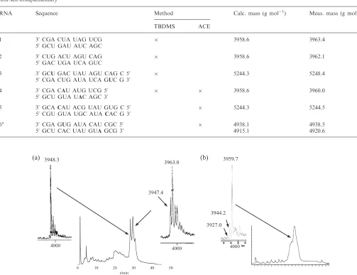
To stress the advantage of using the ACE chemistry, we compared the results obtained by both methods for RNA 4, modified with a spin-labeled adenosine. Using the TBDMS chemistry, a significant amount of reduced oligonucleotide was observed, as shown on the HPLC-chromatogram in Figure 3, and a second purifica-tion was necessary to obtain a pure enough sample for the EPR measurement, but which therefore lowered the yield. The ACE chemistry enabled us to achieve much better yields, as the proportion of reduced RNA was significantly decreased already after the first HPLC run yielding comparable MS-spectra (Figure 3). According to experiments in solution, this side reaction is due to the treatment of the oligonucleotides with disodium-2-carbamoyl-2-cyanoethylene-1,1-dithiolate-trihydrate to O OH O O Si Si O O O O O Si Si O O O O O O O O O O O O O O O O
CH2Cl2, ppTs, RT, 87% O O Si O OACE OH HO TEMED,HF
CH3CN, 90%
O OACE OH BzHO O O Si Si
CH2Cl2, DIPA, 0°C, 67%
P OMe
N N
CH2Cl2, tetrazol in MeCN, 75%
O OACE O BzHO P MeO N N N N N=CHNMe2 I N N N N I N N N N N=CHNMe2 I N N N N N=CHNMe2 I N N N N N=CHNMe2 I O OH OH HO N (a) (b) N N N NH2 I
10 12 13
14 15 5 = BzHCl a, b N=CHNMe2 N Si Cl O
Scheme 1. Synthetic pathway to 2-iodoadenosine phosphoramidite (ACEÕ chemistry); (a) HC(OMe)
2NMe2, DMF, 508C, 69% (compound 11);
(b) TIPS-Cl2, pyridine, 08C, 72%.
at St Andrews University Library on December 5, 2013
http://nar.oxfordjournals.org/
deprotect the phosphate groups at the end of the oligonucleotide synthesis. For all the labeled RNAs, reproducible results could be obtained with the ACE chemistry. Yields were 7–10 OD for a 12-mer (35–50%) to be compared with 4–10 OD with the TBDMS chemistry on a five times larger scale (4–10%).
To determine if the spin label disturbs the RNA structure, UV-melting and Circular Dichroism (CD) studies were performed. The UV-melting curves showed a destabilization of the duplexes between 1.5 and 5.18C, slightly higher than for spin labeled DNA-duplexes (33), which probably results from the geometry of the A-helix, with a very deep but narrow major groove and a very wide but shallow minor groove. CD spectroscopy confirmed that the A-helix is conserved with a similar ellipticity for both modified and unmodified RNAs. Both data together indicate that TPA does not significantly perturb the A-form RNA structure (for detailed data see supporting information).
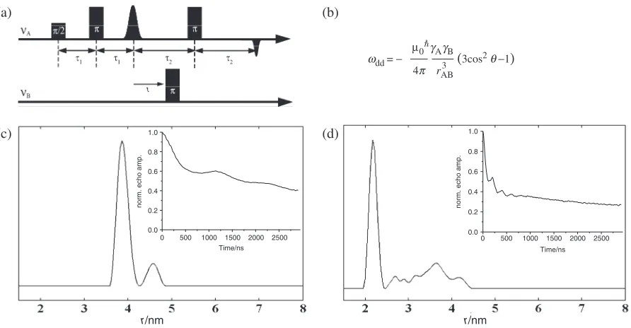
We then subjected RNA-duplexes 1–6 to 4-pulse-ELDOR measurements (Figure 4a). The P4-pulse-ELDOR pulse sequence recovers the magnetic dipole coupling !dd
between two electron spins A and B from which the spin–spin distancerABcan be calculated according to the
equation given in Figure 4b. The principle of the pulse sequence is the following: The detection sequence is applied at a microwave frequencyAwhich is in resonance
with the A spin and creates an refocused echo. The amplitude of this echo is monitored as function of the time position t of an inversion pulse applied at a microwave frequencyB, which is in resonance with the B
spin. This stimulated flip of the B spin induces a sudden change in the Larmor frequency of the spin A by!dd, so
[image:5.612.44.547.87.474.2]that the A spins precess with this altered frequency in the transversal plane, leading to a non-perfect refocusing of the A spins. By variation of the time position t of the inversion pulse, the dephasing angle can be changed which induces a periodic modulation of the A-spin echo intensity
Table 1. Spin-labeled RNA and their corresponding masses; the chemistry used for the preparation is indicated by a cross, a: RNA6 is non-self-complementary
RNA Sequence Method Calc. mass (g mol1
) Meas. mass (g mol1
)
TBDMS ACE
1 30 CGA CUA UAG UCG 3958.6 3963.4
50 GCU GAU AUC AGC
2 30 CUG ACU AGU CAG 3958.6 3962.1
50 GAC UGA UCA GUC
3 30 GCUGAC UAU AGU CAG C 50 5244.3 5248.4
50 CGA CUG AUA UCA GUC G 30
4 30 CGA CAU AUG UCG 50 3958.6 3960.0
50 GCU GUA UAC AGC 30
5 30 GCACAU ACG UAU GUG C 50 5244.3 5244.5
50 CGU GUA UGC AUACAC G 30
6a 30 CGA GUG AUA CAU CGC 50 4938.1 4938.5
50 GCU CAC UAU GUAGCG 30 4915.1 4920.6
0 10 20 30 40 50
3963.0
3947.4
3959.7
3944.2
4000 4000
3948.3
4000
3927.0
(a) (b)
[image:5.612.43.557.97.473.2]t/min
Figure 3.(a) HPLC-chromatogram of RNA4synthesized with the TBDMS chemistry and the corresponding MS spectra of the separated fractions as indicated with an arrow. The reduced oligonucleotide strand shows a mass of 3947–3948 g mol1and the spin labeled strand of 3963 g mol1.
(b) HPLC-chromatogram and MS spectrum of RNA4, synthesized with the ACEÕ chemistry without second HPLC purification. The calculated
mass of the spin labeled RNA4is 3958.6 g mol1.
at St Andrews University Library on December 5, 2013
http://nar.oxfordjournals.org/
by the dipolar coupling !dd. The observation of this
dipolar modulation is crucial for a reliable and parameter free distance calculation.
Figure 4 shows as an example the measured PELDOR time traces and the corresponding distances obtained by Tikhonov regularization (52–54) for RNA3 and RNA4. Both RNAs show clearly visible oscillations in the time traces and one dominant peak in the Tikhonov regular-izations at 38.7 and 21.9 A˚ for RNA3and4, respectively. The peaks of weak intensity at larger distances are most likely due to a slight orientation selection, which is compensated by the Tikhonov regularization by adding more distances. An additional explanation may be coaxially end-to-end stacking of the helices (see support-ing information) (30).
Also all other RNAs exhibit visible oscillations (supporting information) and the extracted mean dis-tances are listed in Table 2. The observation of the dipolar modulation for each RNA proves that the labeling efficiency is high and that the label is sufficiently rigid to yield small distance distributions. Compared to earlier PELDOR studies on RNA (30,31), the dipolar oscillations are deeper for the spin-label TPA used here, which might be attributed to its increased rigidity. Accordingly, the modulation depth is comparable with TPA-labeled duplex DNAs (33). A recent PELDOR study on duplex DNAs labeled at the phosphate backbone also shows oscillations of comparable depth (55).
To be able to translate the measured N–O/N–O distances into RNA structures, we performed 50 ns all-atom MD simulations of all doubly labeled RNAs in explicit water solvent. The simulations yielded single-peaked distance distributions (measured between the oxygen atoms), whose mean and width are reported
in Table 2. Mean distances from the PELDOR measure-ments compare well with the ones from the MD simulations started from generic A-form RNA duplexes. A linear fit through all data points yields r(MD)¼0.93r(PELDOR)þ1.0 A˚ with a standard deviation of 1.9 A˚ and a correlation coefficient of 0.976 (Figure 5). This correlation indicates that the RNA and DNA duplexes retain their conformations in frozen aqueous buffer solution if 20% ethylene glycol is added. Furthermore, the PELDOR experiments allow us to distinguish between A- and B-form helices, as long as the peak width is smaller than the distance difference between both conformations. Thus, RNA/DNA 1 and3
can clearly be assigned to A- and B-conformations, whereas the distance difference is below this limit for RNA/DNA 2.
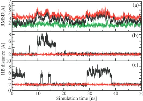
Apart from the distances, MD simulations may provide detailed information on the structure and conformational dynamics of these RNA systems (56–62). Here, we are interested in to what extent the label disturbs the RNA structure and dynamics. As a representative example, Figure 6a shows the time evolution of the root mean squared deviations (RMSD) of the MD trajectory obtained for RNA 1. The black line displays the RMSD from the standard A-form, while the red line reflects the RMSD from the standard B-form of RNA 1. During the 50 ns simulation, the RMSD from the A-form (2.7 A˚ in average) is always smaller than the RMSD from the B-form (3.7 A˚ in average). Note that there are no transitions between the A and B-form. The time evolu-tions of the RMSDs pertaining to the two RNA forms are correlated, indicating that the structure simultaneously moves away from both the A and the B-form that is, it approaches a non-standard structure of RNA. Besides
0 500 1000 1500 2000 2500 0.0
0.2 0.4 0.6 0.8 1.0
norm. echo amp.
Time/ns 0 500 1000 1500 2000 2500
0.0 0.2 0.4 0.6 0.8 1.0
norm. echo amp.
Time/ns
(3cos2q−1)
(c) (a)
(d) (b)
wdd = − m0
បg AgB
4p rAB3
t
[image:6.612.90.536.69.301.2]r/nm r/nm
Figure 4. (a) 4-Pulse ELDOR sequence. (b) Relation between the dipolar coupling!ddand the spin–spin distancerAB.0is the vacuum permeability, are the magnetogyric ratios of the spins A and B,h is the Planck’s constant divided by 2andABis the angle betweenrABand the external
magnetic field. (c) Plot of the Tikhonov regularization and of the corresponding PELDOR time trace (inset) for RNA3and (d) for RNA4.
at St Andrews University Library on December 5, 2013
http://nar.oxfordjournals.org/
the overall comparison of the structures with standard A-form or B-form by RMSD values, the classification of a dinucleotide step as A-form or B-form based on the positioning of phosphorus atoms with respect to the middle frame was performed using the program X3DNA (63). All dinucleotide steps maintain A-form quite well (80%), except for step 6 (i.e. between base pairs U6:A19 and A7:U18), which is only to 20% in A-form (Figure 6).
To study to what extent the spin labels affect the overall structure of RNA1, the green line in Figure 6a shows the RMSD from the A-form obtained for unlabeled RNA1. As may be expected, the latter is smaller (1.6 A˚ in average) than in the case of the spin-labeled RNA 1 (2.7 A˚ in average). The RMSD from the B-form of unlabeled RNA
1(data not shown) looks quite similar with an average of 3.9 A˚. Moreover, the time evolution of the RMSD of unlabeled RNA 1 misses the characteristic modulations seen in the RMSD of labeled RNA1, e.g. at timest¼10
[image:7.612.42.557.84.252.2]and 30 ns. A closer analysis reveals that the latter are caused by occasional openings of hydrogen bonds between canonical Watson–Crick pairs, in particular in the vicinity of the labeled bases uracil 8 and uracil 20 of RNA 1. To illustrate this effect for the two base pairs between the two labeled bases, Figure 6b shows the distance between the H3 atom of uracil 6 and the N1 atom of adenine 19 and Figure 6c shows the distance between the N1 atom of adenine 7 and the H3 atom of uracil 18 (for the numbering of the bases see Table 2). When a hydrogen bond is formed the distance remains around 2.0 A˚, while the distance may increase up to 7–8 A˚, when the hydrogen bond is broken. It is interesting to note that the spin labels hardly perturb the two labeled bases uracil
Table 2. RNA sequences and the corresponding N–O/N–O distances from PELDOR and Molecular Dynamics (MD) simulations
RNA Sequence R(PELDOR) [A˚] r(MD) [A˚]
1 30CGA CU20
A19UAG UCG 19.31.2 [DNA 23.30.6] 18.0 (2.4) [DNA 21.4 (1.6)] 50GCU GAU6AU8C AGC
2 30CUG ACU AGU CAG 33.73.9 [DNA 34.71.4] 30.5 (2.4) [DNA 33.0 (2.7)] 50GAC UGA UCA GUC
3 30GCUGACUAUAGUCAGC 38.71.3 [DNA 44.85.0] 36.2 (3.1) [DNA 43.3 (2.5)] 50CGACUGAUAUCAGUCG
4 30CGACAUAUGUCG 21.90.8 24.7 (0.8)
50GCUGUAUACAGC
5 30GCACAUACGUAUGUGC 33.62.6 34.3 (1.8)
50CGUGUAUGCAUACACG
6 30GCAGUGAUACAUCGC 26.91.3 24.6 (2.4)
50GCUCACUAUGUAGCG
The PELDOR distances are given along with the experimental error determined from the full width of the peak at half height. The MD distances are given including the width of the distance distribution in brackets. For RNA1–3, the mean distances for the sequence analog B-form DNA (33) is given in square brackets. The superscript numbers in the sequence of RNA1are used in the discussion of the MD part to identify the bases.
15 20 25 30 35 40 45 50
15 20 25 30 35 40 45 50
r
(MD)/Å
r(PELDOR)/Å
[image:7.612.59.271.302.472.2]Figure 5. Correlation of the PELDOR and MD distances for RNAs 1–6(squares) and DNAs 1–3(open circles).
Figure 6. Molecular dynamics simulation results obtained for RNA 1. (a) Time evolution of the RMSD from the standard A-form (black line) and B-form (red line) for labeled RNA1. For comparison, the RMSD from the A-form obtained for unlabeled RNA1is also shown (green line). (b) and (c): Hydrogen bonding as monitored by (b) the distance between the H3 atom of uracil 6 and the N1 atom of adenine 19 and (c) the distance between the N1 atom of adenine 7 and the H3 atom of uracil 18. While the hydrogen bonds of labeled RNA 1 (black lines) open occasionally, the hydrogen bonds of unlabeled RNA1(red lines) remain stable.
at St Andrews University Library on December 5, 2013
http://nar.oxfordjournals.org/
[image:7.612.315.548.303.468.2]8 and uracil 20, but mainly affect the structure of dinucleotide step 6 including the base pairs U6:A19 and A7:U18. For comparison, Figure 6 also shows (red lines) the corresponding results obtained for unlabeled RNA1, which shows no indication of hydrogen bond opening. The opening of the Watson–Crick hydrogen bonds may be a reason for the observed lower melting temperatures found for the spin-labeled RNAs.
In conclusion, the spin-label TPA was introduced into RNA by Sonogashira cross-coupling on column during oligonucleotide solid phase synthesis utilizing the bases A, U and C.
Application of the recently developed ACE chemistry presented the main advantage to limit the reduction of the nitroxide TPA into the corresponding amine during the oligonucleotide synthesis and thereby to increase substantially the reliability of the synthesis and the yield of labeled oligonucleotides. Thus, we are able now to introduce the spin label either into the major or minor groove of duplex RNA and to adenine, uridine and cytosine. The combination of site specific labeling with the advantage of the rigid label renders this method advanta-geous for distance measurements.
PELDOR experiments enabled us to measure the intramolecular distances in the six doubly labeled RNA-duplexes with significant modulations depth of up to 40% and allowed us to distinguish A-form RNA from B-form DNA duplexes. MD simulations on the same oligonucleotides gave results in good agreement with the measured distances and showed that the destabilization effect of the label is only local. Thus the combination of this spin-label strategy with PELDOR and MD opens a way to study structures in complex RNA folds and how they change upon binding of metals, small organic ligands or proteins.
EXPERIMENTAL PART
Chemical synthesis
The reactions were monitored by thin-layer chromato-graphy (TLC) analysis on silica gel aluminum plates (silica gel 60 F254, 0.2 mm, Merck, Columbus, OH, USA).
Column chromatography was performed on silica gel (40–63m, 230–400 mesh, Merck). Technical solvents were used after distillation for chromatography; absolute solvents, dried over molecular sieve, were purchased from FLUKA. 1H, 13C and 31P NMR spectra were recorded with a Bruker AMX250/DPX 250 at 250 MHz or AMX400 at 400 MHz as indicated. Electron Spray Ionization (ESI) masses were collected on a VG Platform II (Fisons Instruments, San Carlos, CA, USA). Elemental analyses were performed on CHN-O-Rapid from Foss-Heraeus, Hanau, Germany.
5-Iodo-uridine-phosphoramidites 1 and 2. Commercially available from Glen Research, Sterling, VA, USA and Dharmacon, respectively.
2-Iodo-adenosine-phosphoramidites 4 and 5. All reactions were carried out under a protective argon atmosphere.
20,30,50-Tri-O-acetyl-guanosine 7. A mixture of 35 g (0.12 mol) guanosine, 70 ml (0.74 mol, 6.2 eq) acetic anhydride in 140 ml of dried dimethylformamide/pyridine 5/2 was heated at 758C for 4 h. The resulting solution was cooled to 48C, at which the product crystallized overnight. After filtration, washing with isopropanol and drying overnight, 46 g (91%) 20,30,50-tri-O-acetyl-guanosine were
obtained.
Rf (CH2Cl2/MeOH: 9/1): 0.64; 1H NMR (250 MHz,
DMSO-d6):d[ppm] 10.80 (brs, 1H, NH), 7.95 (s, 1H, H8),
6.57 (s, 2H, NH2), 6.01 (d, J¼6 Hz, 1H, H10), 5.81 (dt,
J¼6.0 Hz, 1H, H20), 5.52 (dd,J¼5.8 Hz, 1H, H30), 4.42–
4.24 (m, 3H, H40and H50), 2.12 (s, 3H, OAc), 2.06 (s, 3H,
OAc), 2.05 (s, 3H, OAc); 13C NMR (62.9 MHz, DMSO-d6):d[ppm] 170.1–169.4–169.2 (3C¼O), 156.7 (C6), 153.7
(C2), 151.1 (C4), 136.7 (C8), 116.8 (C5), 84.4 (C10), 79.5
(C40), 72.0 (C20), 70.3 (C30), 63.1 (C50), 20.5–20.3–20.1
(3CH3); ESI-MS (þ): calc. 409.1, found 410.0 [MH]þ.
20,30,50-Tri-O-acetyl-10-deoxy-10
-(2-amino-6-chloropurine)-b-D-ribofuranose 8. To a solution of pre-dried 20,30,50
-tri-O-acetyl-guanosine (30 g, 73 mmol) in 150 ml abs acetoni-trile were added at room temperature, in this order, 24.3 g (0.14 mol, 2 eq) tetraethylammoniumchloride, pre-dried overnight at 808C over P2O5, 9.3 ml N,N-dimethylaniline
(73 mmol, 1 eq) and 41 ml freshly distilled phosphoryl chloride (0.43 mol, 6 eq). After stirring for 10 min under reflux (the oil bath was preheated to 1008C), volatile materials were evaporated immediately in vacuo. The resulting oily yellow foam was dissolved in 300 ml chloroform and 200 ml cold water and stirred for 15 min under ice-cooling. The layers were separated and the aqueous phase was extracted with 3100 ml chloroform. The combined organic phases were washed with 650 ml of cold water and 6100 ml of a 5% NaHCO3aqueous
solution. After drying over Na2SO4, 150 ml isopropanol
was added to the organic phase, which was then slowly evaporated to 100 ml. The product crystallized at 48C, was filtered, washed with isopropanol and dried overnight in vacuo. The crude product could directly be used for the next step without any further purification. Yield: 19 g (61%). Rf (CH2Cl2/MeOH: 95/5): 0.52; 1H NMR
(250 MHz, CDCl3): d[ppm] 7.84 (s, 1H, H8), 5.95 (d,
J¼4.8 Hz, 1H, H10), 5.89 (dt, J¼5.1 Hz, 1H, H20), 5.67
(dd,J¼4.9 Hz, 1H, H30), 4.41–4.25 (m, 3H, H40and H50),
2.07 (s, 3H, OAc), 2.03 (s, 3H, OAc), 2.01 (s, 3H, OAc);
13
C NMR (62.9 MHz, CDCl3):d[ppm] 170.5–169.7–169.4
(3C¼O), 159.2 (C6), 153.1 (C2), 151.8 (C4), 140.7 (C8), 126.7 (C5), 86.6 (C10), 79.9 (C40), 72.7 (C20), 70.5 (C30),
64.0 (C50), 20.7–20.5–20.4 (3CH
3); ESI-MS (þ): calc.
427.1, found 428.0 [MH]þ.
20,30,50-Tri-O-acetyl-10-deoxy-10-(6-chloro-2-iodopurine)-b
-D-ribofuranose 9. Iodine (11.3 g, 44 mmol), diiodo-methane (36 ml, 10 eq), copper iodide (9.3 g, 49 mmol) and isopentyl nitrite (17.8 ml, 0.13 mol) were added to a solution of 19.0 g of 20,30,50-tri-O-acetyl-10-deoxy-10
-(2-amino-6-chloropurine)-b-D-ribofuranose (44 mmol) in 200 ml abs THF. The suspension was stirred at reflux for 45 min. After cooling at RT, the mixture was filtered off and the solution evaporated. Followed a purification
at St Andrews University Library on December 5, 2013
http://nar.oxfordjournals.org/
through a flash column chromatography (CH2Cl2/MeOH:
99/1). Yield: 19.4 g (82%).Rf(CH2Cl2/MeOH: 98/2): 0.28; 1
H NMR (250 MHz, CDCl3): d[ppm] 8.16 (s, 1H, H8),
6.25 (m, 1H, H10), 5.73 (m, 1H, H20), 5.53 (m, 1H, H30),
4.39 (m, 1H, H40), 4.34 (m, 1H, H50), 2.11 (s, 3H, OAc),
2.08 (s, 3H, OAc), 2.04 (s, 3H, OAc); 13C NMR (62.9 MHz, CDCl3): d[ppm] 170.4–169.9–169.6 (3C¼O),
152.0 (C6), 150.9 (C4), 143.2 (C2), 132.2 (C8), 117.0 (C5), 84.7 (C10), 80.8 (C40), 73.3 (C20), 70.6 (C30), 62.9 (C50),
20.8–20.6–20.4 (3CH3); ESI-MS (þ): calc. 538.0, found
538.9 [MH]þ.
2-Iodo-adenosine 10. To 750 ml of abs ethanol saturated with ammonia at 08C was added 20,30,50-tri-O-acetyl-10
-deoxy-10-(6-chloro-2-iodopurine)-b-D-ribofuranose
(7.16 g, 13.3 mmol). The solution was stirred 1 h at 08C and 24 h at RT. The solvent was then removed under reduced pressure and the residue treated with a NaOCH3
solution (25 mM) for 1 h at RT to remove the last acetyl group. After neutralization with DOWEX 50W-8, filtra-tion of the resin and evaporafiltra-tion of the solvent, the crude product was purified by crystallization in water. Yield: 4.45 g (85%). Rf (CH2Cl2/MeOH: 9/1): 0.24; 1H NMR
(400 MHz, DMSO-d6): d[ppm] 8.29 (s, 1H, H8), 7.73
(brs, 2H, NH2), 5.80 (d, 1H, H10, J¼6.2 Hz), 5.46
(d, J¼6.2 Hz, 1H, 20-OH), 5.20 (d, J¼3.7 Hz, 1H,
30-OH), 5.04 (t, J¼6.3 Hz, 1H, 50-OH), 4.52
(dd,J¼6.2 Hz and 11.2 Hz, 1H, H20), 4.11 (m, 1H, H30),
3.93 (m, 1H, H40), 3.67–3.51 (m, 2H, H50); 13
C NMR (100.6 MHz, DMSO-d6): d[ppm] 155.9 (C6), 149.7 (C4),
136.4 (C8), 121.3 (C2), 120.8 (C5), 87.2 (C10), 85.8 (C40),
73.6 (C20), 70.7 (C30), 61.4 (C50); ESI-MS (): calc. 393.1,
found 391.9 [M-H].
N,N-Dimethyl-N0-formamidine-2-iodo-adenosine 11. A
mixture of 2-iodo-adenosine (4.44 g, 11.3 mmol), 7.5 ml N,N-dimethylformamide-dimethylacetal (56.5 mmol, 5 eq) in 90 ml abs dimethylformamide (DMF) was heated at 508C for 1 h. After evaporation of the solvent, the obtained oil was purified through a flash chromatography (CH2Cl2/MeOH: 95/5). Yield 3.5 g (69%). Rf (CH2Cl2/
MeOH: 9/1): 0.36; 1H NMR (250 MHz, DMSO-d6):
d[ppm] 8.83 (s, 1H, CH), 8.42 (s, 1H, H8), 5.87–5.50– 5.23 (3 s, 3H, 3OH), 5.03 (d, 1H, H10, J¼5.25 Hz), 4.57
(m, 1H, H20), 4.47–4.09 (m, 1H, H30), 3.96 (m, 1H, H40),
3.72–3.35 (m, 2H, H50), 3.25 (s, 3H, CH
3), 3.24 (s, 3H,
CH3); 13
C NMR (62.9 MHz, DMSO-d6): d[ppm] 159.3
(CH), 158.4 (C6), 151.9 (C4), 141.0 (C2), 125.6 (C8), 120.3 (C5), 87.2 (C10), 86.8 (C40), 73.5 (C20), 70.4 (C30), 61.4
(C50), 34.6 (2CH
3); ESI-MS (þ): calc. 448.0, found 448.8
[MH]þ
; Anal. calc. for C13H17IN6O4: C 34.84%, H 3.82%,
N 18.75%; found: C 34.60%, H 4.10%, N 18.50%.
30,50-Tetraisopropyldisiloxane-N,N-dimethyl-N0 -formami-dine-2-iodo-adenosine 12. N,N0-dimethyl-N
-formamidine-2-iodo-adenosine (600 mg, 1.33 mmol) was dissolved in 5 ml abs pyridine and cooled at 08C. 1,3-Dichloro-1,1,3,3-tetraisopropyldisiloxane (0.46 ml, 1.1 eq) was added dropwise for 1 h. After further stirring for 30 min at 08C, the reaction was quenched with 0.2 ml water, the solution concentrated in vacuo and coevaporated with toluene.
The residue was dissolved in 30 ml CH2Cl2 and washed
with 5% aqueous NaHCO3. After separation of the two
layers, the aqueous phase was extracted once with 15 ml CH2Cl2. The combined organic phases were washed with
20 ml saturated aqueous NaCl. The separated aqueous phase was extracted with 15 ml CH2Cl2. The organic
phases were combined, dried with Na2SO4, filtered and
evaporated. A flash chromatography (EtOAc/n-hex: 8/2 to 10/0) afforded 660 mg product (72%).Rf(CH2Cl2/MeOH:
98/2): 0.33;1H NMR (250 MHz, DMSO-d6):d[ppm] 8.80
(s, 1H, CH), 8.25 (s, 1H, H8), 5.86 (1 d, 1H, OH), 5.62 (d, 1H, H10), 4.66–4.56 (m, 2H, H20 and H30), 4.01–3.97
(m, 3H, H40and H50), 3.23 (s, 3H, CH
3), 3.14 (s, 3H, CH3),
1.15–0.90 (m, 28H, Si-iPr);13C NMR (62.9 MHz, DMSO-d6):d[ppm] 159.4 (CH), 158.2 (C6), 152.7 (C4), 141.1 (C2),
125.8 (C8), 120.3 (C5), 89.4 (C10), 80.8 (C40), 73.3 (C20),
70.8 (C30), 61.1 (C50), 40.9 and 34.8 (2CH
3), 17.3–16.9
(CH(CH3)2), 13.9–12.4 (CH(CH3)2); ESI-MS (þ): calc.
690.19, found 691.2 [MH]þ; Anal. calc. for C25H43IN6O5Si2: C 43.47%, H 6.27%, N 12.17%;
found: C 43.44%, H 6.33%, N 12.03%.
20-O-bis-(Acetoxyethyloxy)methylester-30,50
-tetraisopropyl-disiloxane-N,N-dimethyl-N0-formamidine-2-iodo-adenosine
13. A solution of 12 (970 mg, 1.4 mmol), tris(2-acetox-yethyl)orthoformate (1.04 g, 2.3 eq), 3 ml abs dichloro-methane and pyridinium p-toluenesulfonate (70 mg, 0.2 eq) was stirred at RT. As soon as the reaction has progressed well (6 h after TLC analysis), 4-tert -butyldi-methylsiloxy-3-penten-2-one (0.6 ml, 1.8 eq) was added, the solution stirred for 40 h at RT and finally neutralized with 105l TEMED (0.5 eq). Followed a direct flash chromatography (EtOAc/n-hex/MeOH/TEMED: 50/50/ 1/0.5) to yield a yellow foam (1.11 g, 87%). Rf (CH2Cl2/
MeOH: 95/5): 0.42;1H NMR (250 MHz, CDCl3):d[ppm]
8.80 (s, 1H, CH), 8.00 (s, 1H, H8), 6.04 (m, 1H, H10), 5.65
(s, 1H, CH(ACE)), 4.61–4.55 (m, 1H, H20), 4.43–4.41 (m,
1H, H30), 4.30–4.08 (m, 6H, CH
2(ACE), H40and H50),
3.99–3.80 (m, 5H, H50and CH
2(ACE)), 3.18 (s, 3H, CH3),
3.15 (s, 3H, CH3), 1.99 and 1.93 (2 s, 6H, OAc), 1.92–1.05
(m, 28H, Si-iPr); 13C NMR (62.9 MHz, CDCl3): d[ppm]
170.9 (C¼O), 159.7 (CH), 158.5 (C6), 151.1 (C4), 139.4 (C2), 126.7 (C8), 120.0 (C5), 112.3 (CH(ACE)), 88.6 (C10),
81.4 (C40), 76.5 (C20), 69.1 (C30), 63.6, 63.3, 63.2, 61.0
(CH2(ACE)), 59.9 (C50), 41.6 and 36.5 (2CH3
formamidine), 20.9 (CH3(ACE)), 17.6–16.9 (CH(CH3)2),
13.3–12.7 (CH(CH3)2); MALDI-MS (þ): calc. 908.3,
found 909.3 [MH]þ
and 931.2 [MNa]þ .
20-O-bis-(Acetoxyethyloxy)methylester-N,N-dimethyl-N0 -formamidine-2-iodo-adenosine 14. TEMED/HF was freshly and separately prepared: 0.89 ml TEMED (5.9 mmol) was dissolved in 2 ml acetonitrile at 08C and 0.15 ml hydrofluoric acid (48% in water, 4.1 mmol) was added slowly in 2 min. This mixture was stirred for 5 min at 08C and added dropwise in 5 min at RT to a solution of 1.08 g 20-O-ACE-30,50-O-TIPS-N,N-dimethyl-N0
-formami-dine-2-iodo-adenosine 13 in 2 ml acetonitrile. After 2 h stirring, the solution was concentrated in vacuo, the oily residue was dissolved in 5 ml CH2Cl2, 1 mln-hexane and
0.1 ml TEMED and purified through a flash
at St Andrews University Library on December 5, 2013
http://nar.oxfordjournals.org/
chromatography (95/5/0.5: EtOAc/MeOH/TEMED). Yield: 711 mg (90%).Rf(EtOAc/MeOH: 95/5): 0.13; 1H
NMR (250 MHz, CDCl3): d[ppm] 8.81 (s, 1H, CH), 7.78
(s, 1H, H8), 5.84 (d, J¼7.8 Hz, 1H, H10), 5.17 (s, 1H,
CH(ACE)), 4.96 (m, 1H, H20), 4.45 (m, 1H, H30), 4.29
(m, 1H, H40), 4.06–3.92 (m, 5H, CH
2(ACE) and H50),
3.72–3.45 (m, 5H, H50and CH
2(ACE)), 3.21 (s, 3H, CH3),
3.20 (s, 3H, CH3 formamidine), 1.99 and 1.93 (2 s, 6H,
OAc); 13C NMR (62.9 MHz, CDCl3): d[ppm] 170.8 and
170.8 (C¼O), 160.2 (CH), 158.7 (C6), 150.9 (C4), 141.6 (C2), 127.6 (C8), 119.1 (C5), 112.7 (CH(ACE)), 89.1 (C10),
87.7 (C40), 76.6 (C20), 71.9 (C30), 63.4 (CH
2(ACE)), 63.3
(C50), 62.8 and 62.6 (CH
2(ACE)), 41.7 and 35.6 (CH3
formamidine), 20.9 (CH3(ACE)); ESI-MS (): calc. 666.1,
found: 700.9 [MCl].
20-O-bis-(Acetoxyethyloxy)methylester-50
-O-bis(trimethyl-silyloxy)benzhydryloxysilyl-N,N-dimethyl-N0 -formamidine-2-iodo-adenosine 15. Compound 14 (660 mg, 0.99 mmol) and diisopropylamine (169l, 1 eq) were dissolved in 5 ml abs dichloromethane and cooled to 08C. Simultaneously, 210l diisopropylamine were added dropwise in 1 min to a solution of benzhydryloxy-bis(trimethylsilyloxy) chlorsilane (BzHCl, 630 mg, 1.5 eq) in 2 ml abs CH2Cl2.
After 5 min stirring at RT, this last solution was added dropwise and in portions to the previous one: first 0.5 eq BzHCl in 15 min, then twice 0.2 eq in 10 min and finally portions of 0.1 eq until the completion of the reaction (TLC analysis). The mixture was washed with 10 ml 8% aqueous NaHCO3 and 10 ml brine. The layers were
separated and the organic phase dried with Na2SO4and
evaporated. The crude product was purified through a flash chromatography (EtOAc/n-hex/acetone/Et3N:
35/45/20/0.5), which afforded 700 mg (67%) of 15. Rf
(CH2Cl2/MeOH: 95/5): 0.36; 1H NMR (250 MHz,
CDCl3): d[ppm] 8.92 (s, 1H, CH), 8.04 (s, 1H, H8),
7.31–7.12 (m, 10H, phenyl), 6.09 (d, 1H, H10,J¼4.5 Hz),
5.88 (s, 1H, CH-Ph), 5.33 (s, 1H, CH(ACE)), 4.58 (m, 1H, H20), 4.21–4.00 (m, 6H, H30, H40 and CH
2(ACE)),
3.88–3.82 (m, 1H, H50), 3.76–3.64 (m, 5H, H50 and
CH2(ACE)), 3.21 (s, 3H, CH3formamidine), 3.15 (s, 3H,
CH3 formamidine), 1.97 (s, 6H, OAc), 0.02 to 0.01
(2 s, 18H, SiMe3);13C NMR (62.9 MHz, CDCl3):d[ppm]
169.3 (C¼O), 158.4 (CH), 157.7 (C6), 150.2 (C4), 142.5 (CAr), 138.3 (C2), 126.7 (C8), 126.7, 124.9, 124.9, 124.7
(Ph-H), 118.5 (C5), 111.3 (CH(ACE)), 85.5 (C10), 83.0
(C40), 76.7 (CH-Ph), 75.4 (C20), 68.8 (C30), 61.6
(CH2(ACE)), 61.6 (C50), 61.4 and 60.8 (CH2(ACE)),
40.01 and 33.8 (CH3 formamidine), 19.4 and 19.3
(CH3(ACE)), 0.00 (SiMe3); ESI-MS (þ): calc. 1054.3,
found: 1055.3 [MH]þ; Anal. calc. for C41H59IN6O13Si3:
C 46.67%, H 5.64%, N 7.97%; found: C 46.63%, H 5.88%, N 7.32%.
20-O-bis-(Acetoxyethyloxy)methylester-30
-O-(N,N-diisopropylamino)methoxyphosphinyl-50 -O- bis-(trimethylsilyloxy)benzhydryloxysilyl-N,N-dimethyl-N0-formamidine-2-iodo-adenosine 5. About 216l
Methyl-N,N,N0,N0-tetraisopropylphosphordiamidite (0.75 mol)
and 666l of a 0.45 M tetrazole solution in acetonitrile (0.5 eq) were solved in 2 ml abs dichloromethane.
After 5 min at RT, this solution was added dropwise to a solution of 20-O-ACE-50-O-BzH-N,N
-dimethyl-N0-formamidine-2-iodo-adenosine (632 mg, 0.60 mmol) in
2 ml abs dichloromethane, cooled at 08C. After 5 min at 08C and 11 h at RT, the reaction mixture was quenched with 175l ethanol (5 eq) and evaporated. A purification with a flash chromatography (n-hex/acetone/Et3N: 70/30/
0.5) yielded 546 mg of the phosphoramidite (75%). Rf (n-hex/acetone: 70/30): 0.20 and 0.11; 1H NMR
(250 MHz, CDCl3): d[ppm] 9.00 (s, 2H, CH), 8.11 and
8.08 (2 s, 2H, H8), 7.25–7.37 (2 m, 20, Phenyl), 6.20 (s, 2H, CH–Ph), 5.95–5.96 (m, 2H, 2H10), 5.40 and 5.48 (2 s, 2H,
CH(ACE)), 4.72 (m, 2H, H20), 4.50 (m, 2H, 2H30),
4.12–4.18 (m, 10H, 2H40 and 2CH
2(ACE)), 3.61–3.81
(m, 14H, 2H50, 2H500, 2CH
2(ACE) and 4CH(iPr)),
3.34–3.43 (m, 6H, OCH3), 3.26 (s, 6H, CH3formamidine),
3.18 (s, 6H, CH3 formamidine), 2.00–2.06 (4 s, 12H,
CH3(ACE)), 1.16–1.22 (m, 24H, iPr), 0.04–0.11 (s, 36H,
SiMe3);31P NMR (162 MHz, CDCl3):d[ppm] 150.82 and
150.71 (1/1); ESI-MS (þ): calc. 1215.4, found 1215.8.
50-O-(4,40-Dimethoxytriphenylmethyl)-N,N-dimethyl-N0 -formamidine-2-iodo adenosine 16. About 1.50 g N,N -dimethyl-N0-formamidine-2-iodo-adenosine (3.34 mmol)
were dissolved in 35 ml abs DMF. A solution of 4,40-dimethoxytriphenylmethylchloride (1.39 g, 1.2 eq) in
7 ml abs pyridine was added dropwise in three portions. After 3 h at RT, the reaction was quenched with 5 ml methanol, the solution evaporatedin vacuoand coevapo-rated with toluene. The obtained foam was taken up in dichloromethane and washed with a saturated NaHCO3
aqueous solution. The two layers were separated and the organic phase extracted with CH2Cl2. The combined
organic phases were dried with Na2SO4, filtered and
evaporated. A flash chromatography yielded 1.69 g of the purified product (67%). Rf (CH2Cl2/MeOH: 95/5): 0.39; 1
H NMR (250 MHz, CDCl3): d[ppm] 8.87 (s, 1H, CH),
7.87 (s, 1H, H8), 7.26–7.07 (m, 9H, DMT), 6.70–6.66 (m, 4H, DMT), 5.87 (d, J¼6.0 Hz, 1H, H10), 4.71
(dd, J¼5.8 Hz, 1H, H20), 4.34–4.31 (m, 2H, H30 and
H40), 3.70 and 3.69 (2 s, 6H, OCH
3), 3.39–3.22 (m, 2H,
H50), 3.19 (s, 3H, CH
3), 3.18 (s, 3H, CH3); 13
C NMR (62.9 MHz, CDCl3):d[ppm] 159.0 (C6), 158.5 (CH), 151.2
(C4), 144.5 (C2), 140.0 (C8), 135.6 (DMT), 130.0–125.6 (DMT), 119.6 (C5), 113.1 (DMT), 91.8 (C10), 85.3 (C40),
74.7 (C20), 72.4 (C30), 63.8 (C50), 55.2 (OMe), 35.5 and
41.1 (2CH3); ESI-MS (þ): calc. 750.2, found 751.2 [MH] þ
; Anal. calc. for C34H35IN6O6: C 54.41%, H 4.70%,
N 11.20%; found: C 54.00%, H 4.84%, N 10.92%.
50-O-(4,40-Dimethoxytriphenylmethyl)-20
-O-(tert-butyldimethylsilyl)-N,N-dimethyl-N0 -formami-dine-2-iodo-adenosine 17. To a solution of 1.82 g of 50-O-(4,40-dimethoxytriphenylmethyl)-N,N-dimethyl-N0
-formamidine-2-iodo-adenosine in 40 ml of a mixture tetrahydrofurane (THF)/pyridine: 1/1 were added under argon, silver nitrate (536 mg, 1.3 eq) and t-butyltrimethylsilylchloride (1 M in THF, 3.4 ml, 1.4 eq). The suspension was stirred 7 h at RT in the dark. After filtration over CeliteÕ, washing with CH
2Cl2, the
clear solution was evaporated. The residue was dissolved
at St Andrews University Library on December 5, 2013
http://nar.oxfordjournals.org/
in CH2Cl2, the organic phase washed with a saturated
NaHCO3aqueous solution. The aqueous phase was then
extracted twice with dichloromethane. The combined organic phases were finally dried with Na2SO4, filtered,
concentrated in vacuo and coevaporated with toluene. A flash chromatography (EtOAc/n-hex: 7/3 then 100/0) enabled to separate the two isomers. The 20-O-isomer was
obtained in a 61% yield, the unwanted 30-O-isomer in a
10% yield. Rf (CH2Cl2/MeOH: 95/5): 0.39; 1H NMR
(250 MHz, CDCl3):d[ppm] 8.90 (s, 1H, CH), 8.01 (s, 1H,
H8), 7.47–7.16 (m, 9H, DMT), 6.86–6.80 (m, 4H, DMT), 6.03 (d, J¼5.0 Hz, 1H, H10), 4.85 (dd, J¼4.9 Hz, 1H,
H20), 4.32–4.22 (m, 2H, H30and H40), 3.79 (s, 6H, OCH 3),
3.55–3.41 (m, 2H, H50), 3.27 (s, 3H, CH
3), 3.23 (s, 3H,
CH3), 0.88 (s, 9H,tBu), 0.05 and0.05 (2 s, 6H, SiCH3); 13
C NMR (62.9 MHz, CDCl3): d[ppm] 159.1 (C6), 158.4
(CH), 151.8 (C4), 144.5 (C2), 136.7 (C8), 135.6–135.5 (DMT), 130.0–126.3 (DMT), 119.9 (C5), 113.1–113.2 (DMT), 88.2 (C10), 84.1 (C40), 75.9 (C20), 71.5 (C30),
63.4 (C50), 55.1 (OMe), 35.4 and 41.4 (2CH
3), 25.6 and
25.5 (SitBu), 5.3 and 4.9 (SiMe); ESI-MS (þ): calc. 864.8, found 865.4 [MH]þ; Anal. calc. for C40H49IN6O6Si:
C 55.55%, H 5.71%, N 9.72%; found: C 55.46%, H 5.92%, N 9.49%.
30-O-(2-Cyanethoxydiisopropylphosphine)-50-O-(4,40
-dimethoxytriphenylmethyl)-20-O-(tert butyldimethylsilyl)-N,N-dimethyl-N0-formamidine-2-iodo-adenosine 4. A
solu-tion of 705 mg17(0.83 mmol) and 420l diisopropylethy-lamine (3 eq) in 20 ml abs dichloromethane was cooled to 08C. About 280l 2-cyanoethyldiisopropylchlorophos-phoramidite (1.5 eq) were then added under argon. The mixture was stirred for 5 min at 08C then 2 h at RT and finally diluted with 20 ml CH2Cl2. The organic phase
was washed twice with a saturated NaHCO3 solution,
dried with Na2SO4, filtered and concentrated in vacuo.
The crude product was purified through a flash chroma-tography (EtOAc/n-hex: 8/2). Yield: 745 mg (86%). Rf (EtOAc/n-hex: 80/20): 0.43 and 0.38; 1H NMR
(250 MHz, CDCl3): d[ppm] 9.03 (s, 2H, CH), 8.16 and
8.14 (2 s, 2H, H8), 7.61–7.37 (m, 18H, DMT), 6.98–6.93 (m, 8H, DMT), 6.14 (m, 2H, H10), 5.09 (m, 2H, H20),
4.49–4.46 (m, 4H, H30and H40), 3.93 (s, 6H, OCH 3), 3.92
(s, 6H, OCH3), 3.76–3.72 (m, 8H, OCH2and H50), 3.40 (s,
3H, CH3), 3.36 (s, 3H, CH3), 2.82, 2.48 (m, 4H, CH2CN),
1.31–1.21 (m, 24H,iPr), 1.18 and 1.02 (2 s, 18H, SitBu), 0.16–0.04 (m, 12H, SiMe2);31P NMR (162 MHz, CDCl3):
d[ppm] 151.13 and 149.78; ESI-MS(þ): calc. 1064.4, found: 1065.1 [MH]þ.
5-Iodo-cytidine-phosphoramidite 3.
20,30,50-Tri-O-acetyl-cytidine, hydrochloride 18.
Acetylchloride (17.5 ml, 0.24 mol) was added to a solution of 10 g cytidine (40 mmol) in 65 ml acetic acid. After stirring overnight at RT, the solution was concentrated and the obtained white powder recrystallized in ethanol. Yield: 13 g (78%). Rf (CH2Cl2/MeOH: 9/1): 0.64; 1
H NMR (250 MHz, DMSO-d6): d[ppm] 10.19 (brs, 1H,
NH), 9.00 (brs, 1H, NH), 8.04 (d, J¼7.8 Hz, 1H, H6), 6.30 (d,J¼5.8 Hz 1H, H5), 5.92 (d,J¼4.3Hz, 1H, H10),
5.50 (dd,J¼4.5 and 5.8 Hz, 1H, H20), 5.34 (m, 1H, H30),
4.29 (m, 3H, H40-H50), 2.07 (s, 9H, OAc); 13
C NMR (62.9 MHz, DMSO-d6):d[ppm] 170.0 and 169.3 (3C¼O),
159.3 (C4), 146.9 (C2), 145.0 (C6), 94.8 (C5), 89.1 (C10),
79.3 (C40), 72.4 (C20), 69.3 (C30), 62.7 (C50),
20.5–20.3–20.2 (3CH3); ESI-MS (þ): calc. 405.1, found
370.1 [MCl].
20,30,50-Tri-O-acetyl-5-iodo-cytidine (19). Iodine (4.11 g, 14.4 mmol) was added to a suspension of 20,30,50-tri-O
-acetyl-cytidine, hydrochloride (10.0 g, 24 mmol) in 90 ml CH3COOH/CCl4: 1/1. After heating to 408C, iodic acid
(5.2 g, 0.9 eq) was added and the mixture stirred at 408C for 48 h. The suspension was concentrated in vacuo and the residue taken up in 200 ml dichloromethane. The organic phase was washed with 100 ml 5% aqueous NaHCO3, dried with Na2SO4 and evaporated. A flash
chromatography (CH2Cl2/MeOH: 98/2 to 90/10) yielded
10.09 g of 19 (69%). Rf (CH2Cl2/MeOH: 95/5): 0.45; 1
H NMR (250 MHz, DMSO-d6):d[ppm] 8.09 (s, 1H, H6),
8.05 (s, 1H, NH), 6.83 (s, 1H, NH), 5.82 (d, J¼4.3 Hz, 1H, H10), 5.45 (dd, J¼4.5 and 6.3 Hz, 1H, H20), 5.38
(m, 1H, H30), 4.34 (m, 3H, H40), 4.22 (m, 2H, H50), 2.07
(s, 9H, OAc); 13C NMR (62.9 MHz, DMSO-d6): d[ppm]
170.0 and 169.3 (3C¼O), 164.9 (C4), 163.9 (C2), 148.6 (C6), 89.8 (C10), 78.9 (C40), 72.4 (C20), 69.7 (C30), 63.0
(C50), 57.7 (C5), 20.6 and 20.3 (3CH
3); ESI-MS (þ): calc.
495.0, found 495.9 [MH]þ .
5-Iodo-cytidine 20. About 7.61 g 20,30,50
-tri-O-acetyl-5-iodo-cytidine (15.4 mmol) were dissolved in 20 ml of a sodium methanolate solution (0.1 M in methanol) and stirred for 1 h at RT. The mixture was neutralized with DOWEX 50W-X8 (Hþ
), filtered and evaporated in vacuo. Pure 5-iodo-cytidine could be obtained without any further purification in a 86% yield (4.84 g). Rf (CH2Cl2/MeOH: 8/2): 0.36; 1H NMR
(250 MHz, DMSO-d6): d[ppm] 8.43 (s, 1H, H6), 7.81 (s,
2H, NH2), 5.74 (m, 1H, H10), 3.96 (m, 2H, H20and H30),
3.85 (m, 1H, H40), 3.69 (m, 2H, H50); 13
C NMR (62.9 MHz, DMSO-d6): d[ppm] 163.7 (C4), 154.1 (C2),
147.6 (C6), 89.4 (C10), 84.0 (C40), 74.3 (C20), 68.7 (C30),
59.8 (C50), 56.7 (C5); ESI-MS (): calc. 369.0, found
368.1 [M-H].
N,N-dimethyl-N0-formamidine-5-iodo-cytidine
21. 5-Iodo-cytidine 20 (4.07 g, 11.5 mmol) was dissolved under argon in 30 ml abs dimethylformamide. N,N-dimethylformamide-dimethylacetal (8.1 ml, 5 eq) was added and the solution heated at 508C. After completion of the reaction (2 h), the mixture was concentrated and purified through a flash chromatogra-phy (CH2Cl2/MeOH: 9/1). Yield: 4.44 g (95%).Rf
(CH2Cl2/MeOH: 9/1): 0.33; 1H NMR (250 MHz,
DMSO-d6): d[ppm] 8.61 (s, 1H, CH), 8.58 (s, 1H, H6),
5.73 (d, 1H, H10), 5.45–5.26–5.01 (3 brs, 3H, 3OH), 3.98
(m, 2H, H20and H30), 3.89 (m, 1H, H40), 3.67 (m, 2H,
H50), 3.21 (s, 3H, CH
3), 3.14 (s, 3H, CH3); 13C NMR
(62.9 MHz, DMSO-d6): d[ppm] 163.9 (C4), 158.3 (CH),
154.4 (C2), 147.7 (C6), 89.8 (C10), 84.0 (C40), 74.4 (C20),
at St Andrews University Library on December 5, 2013
http://nar.oxfordjournals.org/
68.7 (C30), 68.7 (C50), 59.6 (C5), 34.9 (CH
3); ESI-MS (þ):
calc. 424.0, found 425.0 [MH]þ.
30,50-O-Tetraisopropyldisiloxane-N,N-dimethyl-N0
-forma-midine-5-iodo-cytidine 22. N,N-dimethyl-N0
-formamidine-5-iodo-cytidine (1.0 g, 2.4 mmol) was dissolved in 10 ml abs pyridine and cooled at 08C. 1,3-Dichloro-1,1,3,3-tetraisopropyldisiloxane (810l, 1.1 eq) was added drop-wise in 10 min. After further stirring for 30 min at RT, the reaction was quenched with 0.2 ml water, the solution concentrated in vacuo and coevaporated with toluene. The residue was dissolved in 30 ml CH2Cl2 and washed
with 30 ml 5% aqueous NaHCO3. After separation of
the two layers, the aqueous phase was extracted once with 15 ml CH2Cl2. The combined organic phases were washed
with 15 ml saturated aqueous NaCl. The separated aqueous phase was extracted with 10 ml CH2Cl2.
The organic phases were combined, dried with Na2SO4,
filtered and evaporated. A flash chromatography (CH2Cl2/MeOH: 97/3) followed to yield 1.11 g product
(71%). Rf (CH2Cl2/MeOH: 95/5): 0.30; 1H NMR
(250 MHz, CDCl3): d[ppm] 8.76 (s, 1H, CH), 8.14
(s, 1H, H6), 5.74 (m, 1H, H10), 4.42–4.36 (m, 1H, H20),
4.29–4.23 (m, 2H, H30 and H40), 4.20–3.98 (m, 2H, H50),
3.24 (s, 3H, CH3 formamidine), 3.21 (s, 3H, CH3
formamidine), 1.12–1.02 (m, 28H, iPr); 13C NMR (62.9 MHz, CDCl3): d[ppm] 169.0 (C4), 160.0 (CH),
156.3 (C2), 146.8 (C6), 92.2 (C10), 82.1 (C40), 75.2 (C20),
69.14 (C30), 69.09 (C50), 60.4 (C5), 41.4 and 35.5 (CH 3
formamidine), 17.7–16.9 (CH(CH3)2), 13.4–12.6
(CH(CH3)2); ESI-MS (þ): calc. 666.2, found 667.1
[MH]þ; Anal. calc. for C24H43IN4O6Si2: C 43.24%,
H 6.50%, N 8.40%; found: C 43.41%, H 6.70%, N 8.26%.
20-O-bis-(Acetoxyethoxy)methylester-30,50
-O-tetraisopro-pyldisiloxane-N,N-dimethyl-N0
-formamidine-5-iodo-cyti-dine 23. A solution of 22 (967 mg, 1.45 mmol), tris(2-acetoxyethyl)orthoformate (1.08 g, 2.3 eq), 4 ml abs dichloromethane and pyridinium p-toluenesulfonate (73 mg, 0.2 eq) was stirred at RT. As soon as the reaction has progressed well (overnight after TLC analysis), 4-tert-butyldimethylsiloxy-3-penten-2-one (0.6 ml, 1.8 eq) was added, the solution stirred for 48 h at RT and finally neutralized with 105l TEMED (0.5 eq). Followed a direct flash chromatography (EtOAc/n -hex/MeOH/TEMED: 50/49/1/0.5 to yield 23 (1.12 g, 87%). Rf (CH2Cl2/MeOH: 95/5): 0.30; 1H NMR
(250 MHz, CDCl3): d[ppm] 8.67 (s, 1H, CH), 8.16 (s,
1H, H6), 5.81 (m, 1H, H10), 5.74 (s, 1H, CH(ACE)),
4.20–4.12 (m, 8H, CH2(ACE)), 4.04–3.77 (m, 5H, H20/
H30/H40/H50), 3.16 (s, 3H, CH
3formamidine), 3.15 (s, 3H,
CH3 formamidine), 1.99 (s, 6H, CH3(ACE)), 1.04–0.91
(m, 28H, iPr); 13C NMR (62.9 MHz, CDCl3): d[ppm]
171.0 and 171.0 (C¼O), 169.1 (C4), 158.9 (CH), 155.2 (C2), 146.1 (C6), 112.1 (CH ACE), 90.2 (C10), 81.8 (C40),
76.6 (C20), 69.1 (C30), 67.7 (C50), 63.8–63.4–62.2–60.9
(CH2ACE), 59.2 (C5), 41.4 and 35.5 (CH3formamidine),
21.0 and 20.9 (CH3(ACE)), 17.8–16.9 (CH(CH3)2),
13.5–12.7 (CH(CH3)2); ESI-MS (þ): calc. 884.3, found
885.2 [MH]þ; Anal. calc. for C33H57IN4O12Si2: C 44.79%,
H 6.49%, N 6.33%; found: C 44.96%, H 6.54%, N 6.28%.
20-O-bis-(Acetoxyethyloxy)methylester-N,N-dimethyl-N0
-formamidine-5-iodo-cytidine 24. TEMED, HF was freshly and separately prepared: 0.85 ml tetramethylethylendia-mine (5.7 mmol) was dissolved in 2 ml acetonitrile at 08C and 0.14 ml hydrofluoric acid (48% in water, 4.1 mmol, 3.5 eq) was added slowly in 2 min. This mixture was stirred for 5 min at 08C and added dropwise in 5 min at RT to a solution of 1.0 g 20-O-ACE-30,50-O-TIPS-N,N
-dimethyl-N0-formamidine-5-iodo-cytidine in 4 ml acetonitrile. After
2 h stirring, the solution was concentrated in vacuo, the oily residue was dissolved in 10 ml CH2Cl2, 2 ml hexane
and 0.2 ml TEMED and purified through a flash chromatography (93/7/0.5: EtOAc/MeOH/TEMED). Yield: 726 mg (quantitative); Rf (CH2Cl2/MeOH: 95/5):
0.16; 1H NMR (250 MHz, CDCl3): d[ppm] 8.65 (s, 1H,
CH), 8.10 (s, 1H, H6), 5.56 (d, 1H, H10), 5.46 (s, 1H,
CH(ACE)), 4.74 (m, 1H, H20), 4.31 (m, 1H, H30),
4.17–4.11 (m, 4H, CH2(ACE)), 4.10 (m, 1H, H40),
3.97–3.91 (m, 1H, H50), 3.78–3.70 (m, 5H, H50 and
CH2(ACE)), 3.16 (2 s, 6H, CH3 formamidine), 2.00
(s, 6H, CH3(ACE)); 13C NMR (62.9 MHz, CDCl3):
d[ppm] 170.9 (C¼O), 169.1 (C4), 159.8 (CH), 156.0 (C2), 149.4 (C6), 113.0 (CH ACE), 93.3 (C10), 85.7 (C40), 76.2
(C20), 69.5 (C30), 63.3–63.1–63.0–62.8 (CH
2(ACE)), 62.7
(C50), 41.7 and 35.6 (CH
3 formamidine), 21.0 and 20.9
(CH3(ACE)); ESI-MS (þ): calc. 642.10, found 643.0
[MH]þ
; Anal. calc. for C21H31IN4O11: C 39.26%, H
4.86%, N 8.72%; found: C 39.48%, H 5.09%, N 8.52%.
20-O-bis-(Acetoxyethyloxy)methylester-50
-O-bis-(trimethyl-silyloxy)benzhydryloxysilyl-N,N-dimethyl-N0 -formamidine-5-iodo-cytidine 25. 24 (635 mg, 0.97 mmol) and diisopro-pylamine (136l, 1 eq) were dissolved in 5 ml abs dichloromethane and cooled to 08C. Simultaneously, 204l diisopropylamine were added dropwise in 1 min to a solution of benzhydryloxy-bis(trimethylsilyloxy)-chlorsilane (BzHCl, 617 mg, 1.5 eq) in 2 ml abs CH2Cl2.
After 5 min stirring at RT, this last solution was added dropwise and in portions to the previous one: first 0.5 eq BzHCl in 15 min, then twice 0.2 eq in 10 min and finally portions of 0.1 eq until the completion of the reaction (TLC analysis). The mixture was washed with 20 ml 8% aqueous NaHCO3 and 20 ml brine. The layers were
separated and the organic phase dried with Na2SO4 and
evaporated. The crude product was purified through a flash chromatography (EtOAc/n-hex/acetone/Et3N: 50/
30/20/0.5 then 60/20/20/0.5), which yielded 797 mg (79%) of 21. Rf (CH2Cl2/MeOH: 95/5): 0.28; 1H NMR
(250 MHz, CDCl3): d[ppm] 8.65 (s, 1H, CH); 8.10 (s,
1H, H6); 7.05–7.33 (m, 10H, Phenyl); 5.92 (s, 1H, CH– Ph); 5.77 (m, 1H, H10); 5.58 (s, 1H, CH(ACE)); 4.18–4.10
(m, 3H, H20, H30, H40 and H50); 4.06–4.03 (m, 1H, H50);
3.70–3.86 (m, 8H, CH2(ACE)); 3.13 (s, 3H, CH3
formamidine); 3.07 (s, 3H, CH3 formamidine); 1.95 (2 s,
6H, CH3(ACE)); 0.03 (s, 18H, SiMe3); 13C NMR
(62.9 MHz, CDCl3): d[ppm] 169.2 (C¼O), 167.3 (C4),
at St Andrews University Library on December 5, 2013
http://nar.oxfordjournals.org/
157.3 (CH), 153.7 (C2), 145.0 (C6), 142.5 and 142.4 (CAr),
126.5, 126.5, 125.5, 125.4, 124.9 and 124.7 (CHAr), 111.6
(CH (ACE)), 88.0 (C10), 82.2 (C40), 76.9 (CH
Ar), 75.2
(C20), 67.6 (C30), 66.1 (C50), 61.5, 61.5, 61.4 and 61.4
(CH2(ACE)), 59.7 (C5), 39.7 and 33.8 (CH3formamidine),
19.2 (CH3(ACE)), 0.0 (SiMe3); ESI-MS (þ): calc. 1030.2,
found 1031.2 [MH]þ
and 1053.3 [MNa]þ
; Anal. calc. for C41H61IN6O14Si3: C 46.59%, H 5.77%, N 5.43%; found C
46.34%, H 5.94%, N 5.43%.
20-O-bis-(Acetoxyethyloxy)methylester-30 -O-(N,N-diiso-propylamino)methoxyphosphinyl-50
-O-bis-(trimethylsilylox-y)benzhydryloxysilyl-N,N-dimethyl-N0
-formamidine-5-iodo-cytidine 3. About 0.8 ml of a 0.45 M tetrazole solution in acetonitrile was added to a solution of methyl-N,N,N0,N0-tetraisopropylphosphordiamidite
(260l, 1.25 eq) in 2 ml abs dichloromethane and stirred 5 min at RT. This activated phosphorylating agent was added dropwise to a solution of 20-O-ACE-50-O
-BzH-N,N-dimethyl-N0-formamidine-5-iodo-cytidine (741 mg,
0.72 mmol) in 2 ml abs dichloromethane, previously cooled to 08C. After 5 min at 08C and 10 h at RT, the reaction was quenched with 330l ethanol (5 eq) and the solution evaporated. A flash chromatography (n-hexane/ acetone/Et3N: 55/45/0.5) led to a pure phosphoramidite.
Yield 608 mg (76%). Rf (n-hexane/acetone: 55/45): 0.31; 1
H NMR (250 MHz, CDCl3): d[ppm] 8.74 (s, 2H, CH),
8.20 and 8.19 (2 s, 2H, H6), 7.25–7.40 (2 m, 20, phenyl), 6.07 (s, 2H, CH-Ph), 6.00–6.02 (m, 2H, 2H10), 5.75 and
5.69 (2 s, 2H, CH(ACE)), 4.31–4.32 (m, 2H, H20), 4.16–
4.27 (m, 12H, 2H30, 2H40and 2CH
2(ACE)), 3.80–3.94 (m,
12H, 2H50 and 2CH
2(ACE)), 3.57–3.61 (m, 2H, iPr),
3.31–3.38 (m, 6H, OCH3), 3.26 (s, 6H, CH3formamidine),
3.18 (s, 6H, CH3 formamidine), 2.06–2.07 (4 s, 12H,
CH3(ACE)), 1.15–1.20 (m, 24H, iPr), 0.04–0.12 (s, 36H,
SiMe3);31P NMR (162 MHz, CDCl3):d[ppm] 150.81 and
150.60; ESI-MS (þ): calc. 1191.3, found 1192.4 [MH]þ.
Oligonucleotide synthesis
The oligonucleotides were synthesized on a 1mol scale on a EXPEDITE synthesizer from Perseptive Biosystems, Foster City, CA, USA, with phosphoramidites purchased from Biospring, Frankfurt am Main, Germany (TBDMS chemistry) or on a 0.2mol scale on a rebuilt ABI 392 synthesizer (Applied Biosystems, Foster City, CA, USA) with phosphoramidites purchased from Dharmacon (ACE chemistry). Every RNA synthesis was stopped after incorporation of the iodinated phosphoramidite without deprotecting the 50-hydroxyl group (DMTon). The
column was removed from the synthesizer and maintained under argon atmosphere. In the mean time 9.5 mg copper(I) iodide were dissolved in dried and deoxygenated CH2Cl2/Et3N (1.75/0.75 ml). About 150l of this solution
were added under argon to a mixture of PdII(PPh3)2Cl2
(2.1 mg) and TPA (2 mg). The orange solution was given into the column and moved in it back and forth using two syringes. After a reaction time of 2.5 h the column was washed with 10 ml abs CH2Cl2, dried for 10 min under
vacuum and flushed with argon. The Sonogashira cross-coupling was performed twice or three times depending on
the labeled base and on the chosen chemistry for the oligonucleotide synthesis. Note that the amounts of reagents were not reduced for the 0.2mol synthesis. Then the column was reinstalled on the synthesizer to end the synthesis of the oligonucleotide.
When using the TBDMS chemistry, the oligonucleo-tides were cleaved from the controlled pore glass (CPG) and the amino groups deprotected with a mixture of ammonia (32%)/MeOH (3/1) over 24 h. The TBDMS groups were cleaved with triethylamine, HF over 24 h. After precipitation in abs. ethanol (208C over night), the RNA strands were purified via anion-exchange chromato-graphy (Dionex NucleoPacTM PA 100 column, 2509 mm, flow 5 ml min1
) on a JASCO-HPLC. When using the ACE chemistry, the methyl group on the phosphate was first cleaved on-column with a 0.4 M solution of disodium-2-carbamoyl-2-cyanoethylene-1,1-dithiolate-trihydrate (S2Na2) in DMF/H2O: 98/2 in
30 min. Then, the oligonucleotides were cleaved from the solid support and deprotected with methylamine (40% in water): 10 min at 558C for the unmodified RNA-strands and 12 h at RT for the spin labeled ones. Followed a purification through anion-exchange HPLC.
The oligonucleotides were desalted with PD-10 Sephadex columns from Amersham Biosciences, Piscataway, NJ, USA and finally characterized with a MALDI-Tof VOYAGER DE-PRO mass spectrometer from Applied Biosystems. In case of the ACE chemistry the final deprotection of the 20-ACE groups was
performed under sterile conditions with a TEMED-acetic acid buffer pH 3.8, 30 min at 608C for all the RNA strands.
Analysis
Calf intestine alkaline phosphatase (Sigma-Aldrich, St. Louis, MO, USA) and Penicillium citrinum nuclease P1 (Roche, Nutley, NJ, USA) were used for the enzymatic digestion of ODNs (see (33) for the procedure and supplementary data for HPLC diagram). UV-melting curves (Tm) of the duplexes, dissolved in phosphate
buffer (10 mM Na2HPO4, 10 mM NaH2PO4, 140 mM
NaCl, 2.5M duplex, pH 7), were recorded on a Cary UV-vis spectrophotometer equipped with a Peltier ther-mostat from Varian. The UV absorption was measured at a wavelength of 260 nm, while the temperature was increased with a heating rate of 0.58C min1. CD spectra were measured at a temperature of 20 C between 350 and 200 nm on a JASCO J-710 spectropolarimeter with a Peltier thermostat. The duplexes were dissolved in the same buffer and at the same concentration as for the Tm
measurements.
PELDOR
All EPR samples had a volume of 100l and contained 0.1 mM duplex in phosphate buffer (140 mM NaCl, 10 mM Na2HPO4, 10 mM NaH2PO4, 20% ethylene
glycol, pH 7). The solutions were transferred into sterile standard quartz EPR tubes and shock frozen in liquid nitrogen. The 4-Pulse ELDOR experiments were per-formed on an ELEXSYS E580 pulsed X-band EPR
at St Andrews University Library on December 5, 2013
http://nar.oxfordjournals.org/