University of Warwick institutional repository: http://go.warwick.ac.uk/wrap
This paper is made available online in accordance with
publisher policies. Please scroll down to view the document
itself. Please refer to the repository record for this item and our
policy information available from the repository home page for
further information.
To see the final version of this paper please visit the publisher’s website.
Access to the published version may require a subscription.
Author(s): Christophe Six, Jean-Claude Thomas, Laurence Garczarek,
Martin Ostrowski, Alexis Dufresne, Nicolas Blot, David J Scanlan and
Frédéric Partensky
Article Title: Diversity and evolution of phycobilisomes in marine
Synechococcus spp.: a comparative genomics study
Year of publication: 2007
spp.: a comparative genomics study
Christophe Six
¤*†
, Jean-Claude Thomas
¤‡
, Laurence Garczarek
*
,
Martin Ostrowski
§
, Alexis Dufresne
*
, Nicolas Blot
*
, David J Scanlan
§
and
Frédéric Partensky
¤*
Addresses: *UMR 7144 Université Paris VI and CNRS, Station Biologique, Groupe Plancton Océanique, F-29682 Roscoff cedex, France. †Mount Allison University, Photosynthetic Molecular Ecophysiology Group, Biology Department, E4L 1G7 Sackville, New Brunswick, Canada. ‡UMR 8186 CNRS and Ecole Normale Supérieure, Biologie Moléculaire des Organismes Photosynthétiques, F-75230 Paris, France. §Department of Biological Sciences, University of Warwick, Coventry CV4 7AL, UK.
¤ These authors contributed equally to this work.
Correspondence: Frédéric Partensky. Email: [email protected]
© 2007 Six et al.; licensee BioMed Central Ltd.
This is an open access article distributed under the terms of the Creative Commons Attribution License (http://creativecommons.org/licenses/by/2.0), which permits unrestricted use, distribution, and reproduction in any medium, provided the original work is properly cited.
Phycobilisome diversity and evolution
<p>By comparing Synechococcus genomes, candidate genes required for the production of phycobiliproteins, which are part of the light-harvesting antenna complexes called phycobilisomes, were identified. Phylogenetic analyses suggest that the phycobilisome core evolved together with the core genome, whereas rods evolved independently. </p>
Abstract
Background: Marine Synechococcus owe their specific vivid color (ranging from blue-green to
orange) to their large extrinsic antenna complexes called phycobilisomes, comprising a central allophycocyanin core and rods of variable phycobiliprotein composition. Three major pigment types can be defined depending on the major phycobiliprotein found in the rods (phycocyanin, phycoerythrin I or phycoerythrin II). Among strains containing both phycoerythrins I and II, four subtypes can be distinguished based on the ratio of the two chromophores bound to these phycobiliproteins. Genomes of eleven marine Synechococcus strains recently became available with one to four strains per pigment type or subtype, allowing an unprecedented comparative genomics study of genes involved in phycobilisome metabolism.
Results: By carefully comparing the Synechococcus genomes, we have retrieved candidate genes
potentially required for the synthesis of phycobiliproteins in each pigment type. This includes linker polypeptides, phycobilin lyases and a number of novel genes of uncharacterized function. Interestingly, strains belonging to a given pigment type have similar phycobilisome gene complements and organization, independent of the core genome phylogeny (as assessed using concatenated ribosomal proteins). While phylogenetic trees based on concatenated allophycocyanin protein sequences are congruent with the latter, those based on phycocyanin and phycoerythrin notably differ and match the Synechococcus pigment types.
Conclusion: We conclude that the phycobilisome core has likely evolved together with the core
genome, while rods must have evolved independently, possibly by lateral transfer of phycobilisome rod genes or gene clusters between Synechococcus strains, either via viruses or by natural transformation, allowing rapid adaptation to a variety of light niches.
Published: 5 December 2007
Genome Biology 2007, 8:R259 (doi:10.1186/gb-2007-8-12-r259)
Received: 23 July 2007 Revised: 22 October 2007 Accepted: 5 December 2007 The electronic version of this article is the complete one and can be
Background
Since their discovery almost 30 years ago [1,2], marine repre-sentatives of the Synechococcus genus have been found in the upper illuminated layer of most marine ecosystems, from coastal to offshore waters as well as from low to high latitudes [3-5]. Besides being ubiquitous, Synechococcus are often abundant, with cell densities ranging from a few hundred to over one million cells per milliliter of seawater [6-10].
Synechococcus cells owe their vivid colors mainly to their photosynthetic antenna, called phycobilisomes (PBSs). These water-soluble macromolecular complexes comprise rods sur-rounding a central core and are made of phycobiliproteins, which covalently bind chromophores (phycobilins) by thioether bonds to cysteinyl residues (for reviews, see [11-15]). All phycobiliproteins in cyanobacteria consist of two dis-tinct subunits, α and β, organized either as trimeric (αβ)3 or, in most cases, as hexameric discs (αβ)6. The PBS core of marine Synechococcus is made of allophycocyanin (AP), which binds only the blue-colored chromophore phycocyano-bilin (PCB; Amax = 620 nm). In some strains, phycocyanin (PC) may constitute the whole rod, as it does in many fresh-water cyanobacteria (for example, Synechococcus elongatus PCC 7942, Synechocystis sp. PCC 6803). In that case, it binds only PCB and is of the C-PC type [15]. However, in most phy-coerythrin (PE)-containing marine Synechococcus character-ized so far, PC makes up the basal disc at the core-proximal end of the rods. It binds both PCB and the red-colored chromophore phycoerythrobilin (PEB; Amax = 550 nm) at a molar ratio of 1:2 and thus belongs to the R-PCII type [16]. In strain WH7805, however, the base of the rods is thought to consist of a so-called R-PCIII, an optically unusual PC that binds PCB and PEB at a molar ratio of 2:1 [15,17].
In most PE-containing Synechococcus strains isolated to date, the distal part of the PBS rods is composed of two types of PE (PEI and PEII). PEII always binds both PEB and the orange colored phycourobilin (PUB; Amax = 495 nm), whereas PEI binds either only PEB or both PEB and PUB [18,19]. However, Everroad and Wood [20] have recently suggested that some marine Synechococcus strains may contain rods with a single type of PE that binds only PEB chromophores. In addition, the higher order structure of PBSs is stabilized by linker polypeptides that contribute to the building of a pro-tein environment around the phycobilins [14,21]. These link-ers have very variable sizes (8-120 kDa) but most are in the 27-35 kDa range. In the rods, only those associated with PEII are chromophorylated with PUB [19,21].
Although the Synechococcus genus itself is polyphyletic, marine Synechococcus characterized thus far form a well-defined branch within the cyanobacteria radiation, together with the Prochlorococcus and Cyanobium genera [22-25]. This grouping, called 'Cluster 5' by Herdman and coworkers [26], is a combination of the former Marine Clusters A and B previously defined by Waterbury and Rippka [27]. Cluster 5
thus gathers coastal, euryhaline Synechococcus strains as well as strictly marine strains (that is, with elevated growth requirements for Na+, Mg+ and Ca++). Subclusters 5.1 and 5.2
have also been tentatively defined by Herdman and cowork-ers [26] in order to separate the strictly marine PE-containing strains (5.1) from a group of euryhaline strains lacking PE (5.2), including WH5701 and WH8007. However, Fuller and coworkers [23] have shown that one clade within the subclus-ter 5.1 (clade VIII) gathers euryhaline strains lacking PE and Chen and coworkers [25] have isolated several new members of subcluster 5.2 into culture that do contain PE. Further-more, the latter authors suggested that WH5701 and WH8007 might actually belong to two distinct clusters.
Among the strains containing two PE types, there is a clear consistency between phylogenies based on different molecu-lar markers, including rpoC1 [28], ntcA [29], the 16S rRNA gene [23] and the 16S-23S rDNA internal transcribed spacer [24]. However, none of these phylogenies is congruent with the whole cell ratio of PUB to PEB. This chromophore ratio is known to vary according to the light niche, with open ocean strains predominantly displaying a high PUB:PEB whereas mesotrophic or coastal strains generally have lower ratios or no PUB [6,7,30-32]. Some strains even display a variable PUB:PEB depending on the ambient light quality, that is, they are able to chromatically adapt [33]. These so-called type IV chromatic adapters are not confined to a particular phyloge-netic clade within Cluster 5 [34]. This raises the question of the molecular basis of differences in whole cell PUB:PEB between Synechococcus strains. More generally, one might wonder whether PBS components have undertaken a differ-ent evolutionary trajectory compared to the core genome.
In order to address these questions, we studied 11 Synechoc-occus strains, belonging to various phylogenetic clades according to Fuller et al. [23] and representing the whole variety of PBS pigmentations known so far within Cluster 5. We compared the PBS gene complements of these strains, an approach that revealed a number of novel PBS genes, includ-ing putative lyases and linker polypeptides. By combininclud-ing these genomic data with biochemical and optical properties of isolated phycobiliprotein complexes, we identified several marine Synechococcus pigment types and we propose puta-tive, structural models for their corresponding PBSs. We also examined the phylogeny of each phycobiliprotein type, yield-ing new insights into the evolution of PBS complexes within the marine Synechococcus group.
Results
Synechococcus pigment types
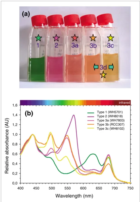
Type 3 can be further subdivided into four subtypes (3a through 3d) based on the ratio of the two chromophores (PEB and PUB) bound to PEs, a ratio that can be low, medium, high or variable. Figure 1a illustrates these different pigment types or subtypes and their corresponding colors. The 11 fully sequenced marine Synechococcus strains cover the whole range of PBS pigmentation known so far in this group [6,23,33]. Pigment type 1 is represented by the blue-green, PE-lacking strains WH5701 and RS9917. These strains absorb light optimally in the wavelength range 600-660 nm, that is, red and orange light (Figure 1b). The genome of the fuchsia pink WH7805 strain (pigment type 2) contains a sin-gle set of PE genes encoding a PEI-like complex, as detailed below. The whole cell absorption maximum of this form of PE devoid of PUB (Amax = 570 nm, corresponding to yellow-green light) is red-shifted relative to other PEs (Figure 1b).
All strains displaying pigment type 3 possess both PEB and PUB chromophores. Subtypes 3a through 3c differ from one another in their whole cell ratio of PUB to PEB (hereafter PUB:PEB), as assessed by their fluorescence excitation maxima (F495 nm : F550 nm) with emission at 580 nm (Table 1). Note that the use of this fluorescence excitation ratio is pref-erable to using the corresponding absorption ratio (A495 nm: A550 nm) to characterize these different subtypes in vivo, since the carotenoids zeaxanthin and β-carotene have a notable contribution to the wavelength range of the PUB absorption peak (Figure 1b). The PUB:PEB can be either low (approxi-mately 0.4) in type 3a strains such as WH7803, medium (approximately 0.8) in type 3b strains such as RCC307 or high (>1.7) in type 3c strains such as in WH8102 and CC9605 (Table 1). Depending on this ratio, PBSs of these strains pref-erentially harvest either green light (550 nm) or blue-green light (495 nm) (Figure 1b). Finally, pigment type 3d includes strains with a variable PUB:PEB (0.7-1.7), depending on whether these cells are grown under white/green or blue light [33,34]. These type IV chromatic adapters include the strains CC9311, RS9916, BL107 and CC9902 as well as a number of other strains that have not yet been sequenced (including WH8020, M16.17, M11.1, RCC61 (a.k.a. Minos 11) and RS9911; Table 1 and data not shown). To this suite of pigment types can be added a 'moderately high' PUB:PEB subtype (PUB:PEB approximately 1.2), represented by strain WH8103 and which is indistinguishable by eye from, and included within, type 3c (Figure 1a). Though as yet unse-quenced, the genome of WH8103 has been screened, in part, by suppression subtractive hybridization [35].
Optical properties of phycobiliproteins
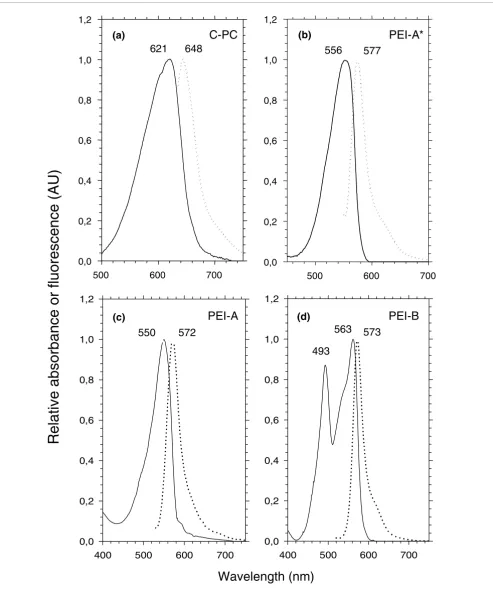
The color and specific absorption properties of whole Syne-chococcus cells (Figure 1) are mainly determined by the major phycobiliprotein form occurring in the PBS rods. Isolated PC and/or PE complexes have been previously characterized in a few marine Synechococcus strains, including WH7803, WH7805, WH8102, WH8103 and the chromatic adapters WH8020 (under white light only), M11.1 and M16.17 [13,16-19,34,36], as summarized in Table 1. In order to explore fur-ther the diversity and possible combinations of these phyco-biliproteins in the different Synechococcus pigment types, we have used sucrose density gradients and isoelectric focusing to isolate PC, PEI and/or PEII from a number of other strains and then have determined their optical properties (Figures 2 and 3 and Table 1).
[image:4.612.53.296.86.436.2]The PC present in WH5701 and RS9917, which formed a sky blue band on isoelectric focusing gels (not shown), had absorption (Amax = 621 nm) and fluorescence (Fmax = 648 nm) properties typical of C-PC (Figure 2a), that is, known to bind only PCB chromophores [15]. We also found C-PC in the PE-containing, PUB-lacking strain WH8018, whereas WH7805 (which, like WH8018, displays pigment type 2) is known to possess R-PCIII [17]. R-PCIII has a molar PCB:PEB of 2:1, like the R-PCI of red algae, but a different spectrum, with an The diversity of pigment types among marine Synechococcus spp
Figure 1
The diversity of pigment types among marine Synechococcus spp. (a) Photograph of representative cultured strains of the major pigment types (1-3) and subtypes (3a-c) of marine Synechococcus grown under low white light and (b) corresponding absorption properties of whole cells. Pigment subtype 3d corresponds to type IV chromatic adapters, which are able to modify their PBS pigmentation from subtype 3b when grown under white or green light to subtype 3c when grown under blue light. The different colors of stars in panel A are a code for the different pigment types.
3c
3b
3d
Wavelength (nm)
400 450 500 550 600 650 700 750
Relative absorbance (AU)
0,0 0,2 0,4 0,6 0,8 1,0 1,2 1,4 1,6
Col 8 vs Col 9 Col 8 vs Col 11 Col 8 vs Col 13 Col 8 vs Col 15 Col 8 vs Col 17
(a)
(b)
Type 1 (WH5701)Type 2 (WH8018) Type 3a (WH7803) Type 3b (RCC307) Type 3c (WH8102)
infrared
Amax at 555 nm and a shoulder at 590 nm [17]. Our isolation protocol did not allow us to obtain a pure PC fraction from any of the PEII-containing strains, because the PC band was always contaminated by variable amounts of PEII. It is known, however, that Synechococcus sp. WH7803, like WH8020 and WH8103, possesses a R-PCII type PC with a molar PEB:PCB of 2:1; it has absorption peaks at 533, 554 and 615 nm and maximal fluorescence emission at 646 nm [16].
Several types of PEI can be distinguished based on their dif-ferent optical properties. The major phycobiliprotein found in WH7805 and WH8018, a PEI-like phycobiliprotein, exhib-ited an Amax at 556 nm and an Fmax at 577 nm (Figure 2b). We
have called it PEI-A* to distinguish it from the PEI-A found in Synechococcus strains displaying the 3a and 3b pigment types. PEI-A has blue-shifted optical properties (Amax = 550 nm; Fmax = 572 nm; Figure 2c) compared to PEI-A*, though both forms bind only PEB chromophores. PEI-B, which has a molar PUB:PEB of 2:3 [18], has been found in all strains exhibiting pigment type 3c examined thus far, as well as in some chromatic adapters, including M11.1 and M16.17 [34]. It has maximal absorption at 493 and 563 nm and fluorescence at 573 nm (Figure 2d).
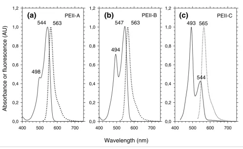
[image:5.612.56.553.119.540.2]Similarly, one can distinguish three optical types of PEII dif-fering by their PUB:PEB. All have two absorption maxima (or
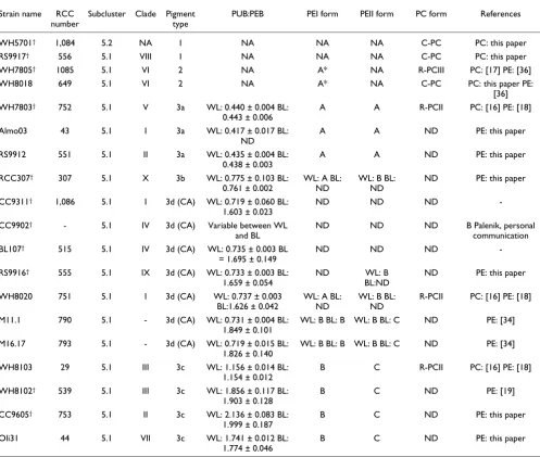
Table 1
Strain numbers, phylogenetic position and PBS characteristics of all marine Synechococcus spp. mentioned in this paper
Strain name RCC number
Subcluster Clade Pigment type
PUB:PEB PEI form PEII form PC form References
WH5701† 1,084 5.2 NA 1 NA NA NA C-PC PC: this paper
RS9917† 556 5.1 VIII 1 NA NA NA C-PC PC: this paper
WH7805† 1085 5.1 VI 2 NA A* NA R-PCIII PC: [17] PE: [36]
WH8018 649 5.1 VI 2 NA A* NA C-PC PC: this paper PE:
[36]
WH7803† 752 5.1 V 3a WL: 0.440 ± 0.004 BL: 0.443 ± 0.006
A A R-PCII PC: [16] PE: [18]
Almo03 43 5.1 I 3a WL: 0.417 ± 0.017 BL:
ND
A A ND PE: this paper
RS9912 551 5.1 II 3a WL: 0.435 ± 0.004 BL:
0.438 ± 0.003
A A ND PE: this paper
RCC307† 307 5.1 X 3b WL: 0.775 ± 0.103 BL: 0.761 ± 0.002
WL: A BL: ND
WL: B BL: ND
ND PE: this paper
CC9311† 1,086 5.1 I 3d (CA) WL: 0.719 ± 0.060 BL: 1.603 ± 0.023
ND ND ND
-CC9902† - 5.1 IV 3d (CA) Variable between WL and BL
ND ND ND B Palenik, personal
communication BL107† 515 5.1 IV 3d (CA) WL: 0.735 ± 0.003 BL
= 1.695 ± 0.149
ND ND ND
-RS9916† 555 5.1 IX 3d (CA) WL: 0.733 ± 0.003 BL: 1.659 ± 0.054
ND WL: B
BL:ND
ND PE: this paper
WH8020 751 5.1 I 3d (CA) WL: 0.737 ± 0.003 BL:1.626 ± 0.042
WL: A BL: ND
WL: B BL: ND
R-PCII PC: [16] PE: [18]
M11.1 790 5.1 - 3d (CA) WL: 0.731 ± 0.004 BL: 1.849 ± 0.101
WL: B BL: B WL: B BL: C ND PE: [34]
M16.17 793 5.1 - 3d (CA) WL: 0.719 ± 0.015 BL: 1.826 ± 0.140
WL: B BL: B WL: B BL: C ND PE: [34]
WH8103 29 5.1 III 3c WL: 1.156 ± 0.014 BL:
1.154 ± 0.012
B C R-PCII PC: [16] PE: [18]
WH8102† 539 5.1 III 3c WL: 1.856 ± 0.117 BL: 1.903 ± 0.128
B C ND PE: [19]
CC9605† 753 5.1 II 3c WL: 2.136 ± 0.083 BL: 1.999 ± 0.187
B C ND PE: this paper
Oli31 44 5.1 VII 3c WL: 1.741 ± 0.012 BL:
1.774 ± 0.046
B C ND PE: this paper
Absorption (continuous line) and fluorescence (dotted line) properties of isolated PBP complexes
Figure 2
Absorption (continuous line) and fluorescence (dotted line) properties of isolated PBP complexes. (a) C-PC (as in Synechococcus spp. RS9917, WH5701 and WH8018); (b) PEI-A* (as in Synechococcus spp. WH8018 and WH7805); (c) PEI-A (as in Synechococcus spp. WH7803, Almo03 and RS9912); (d) PEI-B (as in Synechococcus spp. WH8102, CC9605 and Oli31).
Wavelength (nm)
Relative absorbance or fluorescence (AU)
400 500 600 700 0,0
0,2 0,4 0,6 0,8 1,0 1,2
400 500 600 700 0,0
0,2 0,4 0,6 0,8 1,0 1,2
Oli31
Almo03
PEI-B
573
563
493
572
550
PEI-A
(c)
(d)
500 600 700 0,0
0,2 0,4 0,6 0,8 1,0 1,2
500 600 700 0,0
0,2 0,4 0,6 0,8 1,0 1,2
8
1
0
8
H
W
7
1
9
9
S
R
577
556
648
621
PEI-A*
at least shoulders) around 495 nm and 550 nm, due to the two chromophores they bind, and a maximal fluorescence emis-sion around 565 nm. PEII-A (Figure 3a) is found only in Syn-echococcus pigment type 3a, including WH7803 [18], Almo03 and RS9912 (this study). Its molar PUB:PEB is most likely 1:5, although the cysteinyl site to which the sole PUB chromophore is bound (either α-75 or β-50/61) has not yet been ascertained. PEII-B (Figure 3b) is found in RCC307 (Table 1) and in all white light-grown chromatic adapters that have been screened thus far, including WH8020 [18], M11.1, M16.17 [34] and RS9916 (this study). Its molar PUB:PEB is 2:4. PEII-C (Figure 3c) is found in Synechococcus pigment type 3c, including WH8103 [18], WH8102 [19], Oli31 and CC9605 (this study) as well as in the blue light-grown chro-matic adapters [34]. The molar PUB:PEB of this PEII has been shown to be 4:2 [18].
Comparative analysis of the phycobilisome gene regions
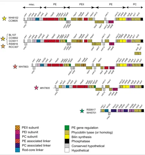
After careful annotation, we compared PBS gene complement (Additional data file 1) and organization in the 11 different genomes. One remarkable trait of marine Synechococcus is that most of the PBS genes are gathered into a few gene
clus-ters [19,37]. As in several other cyanobacteria, a first small cluster groups together four AP core genes, in the order apcE-A-B-C, while two other core genes, apcD and apcF (encoding the minor α-B and β-18 AP subunits, respectively) have no PBS gene in their close vicinity. Most of the PBS rod genes are located in a much larger cluster, the size of which increases with the complexity of the rod structure from approximately 9-10 Kbp in pigment type 1 up to 27-28.5 Kbp in chromatic adapters (Figure 4). Interestingly, the gene organization in this region is very similar for strains belonging to a given pig-ment type. It is also similar between the chromatic adapters and the medium PUB:PEB strain RCC307.
[image:7.612.56.554.89.395.2]In most genomes, the 5'-end of the PBS rod gene region starts with a short gene of unknown function (unk1). In RCC307, however, the unk1 ortholog is found elsewhere in the genome. The 3'-end of the region consists of a well conserved gene pre-dicted to encode a low molecular weight phosphotyrosine phosphatase. In the blue-green, PE-lacking strains, the rest of the region is mainly composed of two identical cpcB-A oper-ons encoding the C-PC α- and β-subunits and of genes encod-ing three rod linkers, one rod-core linker and two types of phycobilin lyases (CpcT and CpcE/F; see below). Both Absorption (continuous line) and fluorescence (dotted line) properties of the isolated PEII complexes
Figure 3
Absorption (continuous line) and fluorescence (dotted line) properties of the isolated PEII complexes. (a) PEII-A (as in Synechococcus sp. WH7803); (b) PEII-B (as in Synechococcus sp. RCC307); (c) PEII-C (as in Synechococcus spp. CC9605 and WH8102). Type IV chromatic adapters have a PEII-B under white or green light and a PEII-C under blue light [34].
400 500 600 700
0,0 0,2 0,4 0,6 0,8 1,0 1,2
CC9605
400 500 600 700
0,0 0,2 0,4 0,6 0,8 1,0 1,2
RS9912
400 500 600 700
0,0 0,2 0,4 0,6 0,8 1,0 1,2
RS9916
493
498
494
544
565
547
544
563
563
Wavelength (nm)
Absorbance or fluorescence (AU)
PEII-C
PEII-A
PEII-B
Comparison of PBS rod gene regions of the different pigment types of marine Synechococcus
Figure 4
Comparison of PBS rod gene regions of the different pigment types of marine Synechococcus. Rectangles above and below the lines have a length proportional to the size of ORFs and correspond to the forward and the reverse strand, respectively. In several genomes, the sense of the rod region was inversed so that the regions all appear in the same direction. For the group formed by the chromatic adapters and RCC307, a few variations can be found with regard to the region shown here, which corresponds to BL107. First, the lyase-encoding gene(s) located near the 3'-end can either be a rpcE-F operon or rpcG, a pecEF-like fusion gene (see text). Second, the gene organization at the 5'-end can vary: unk1 is located elsewhere in the genome of RCC307 and the gene following unk2 is either the lyase gene cpcT in RS9916 and RCC307, unk3 in BL107 and CC9902, or none of these in CC9311. Colored stars indicate the pigment type of each strain (see Figure 1 for color code).
cpeZ cpeY cpeAcpeB
cpeY
cpeZ cpeAcpeB mpeBmpeAmpeC mpe
U
pebApebB rpcBrpcA mpeV
cpcGII cpeC mpeD
aplA cpeE cpeScpeTcpeR mpeY
rpcE rpcF
unk7unk8unk9 unk12
rpcT unk13
unk4 unk5
unk7unk8unk9
unk11
unk12
cpeU
PE associated linker
PEI subunit
PE gene regulation
PC subunit
Rod-core linker
Bilin synthesis
Phosphatase
Conserved hypothetical
Hypothetical
cpcGII cpeC mpeD
aplA cpeE cpeScpeTcpeR mpeY mpeBmpeA mpeC mpeU pebApebB rpcBrpcA
Phycobilin lyase (or homolog)
cpcGII cpeC mpeDaplA cpeE cpeScpeTcpeR rpcF
cpeZ
cpeY cpeAcpeB mpeBmpeA
mpeE
mpeV pebApebBrpcBrpcA rpcE
WH8102 CC9605
WH7803 BL107
CC9902 CC9311 RS9916 RCC307
cpcGII cpeC mpeD cpeE cpeScpeTcpeR rpcF
cpeZ cpeY cpeAcpeB
mpeV pebApebBrpcBrpcArpcE
WH7805
unk13
mpeY
unk1cpcT cpcGII
unk2
cpcCIcpcD cpcBIcpcAI cpcCII cpcBIIcpcAIIcpcE
RS9917 WH5701
cpcF
PC associated linker
PEII subunit
unk3 unk5 unk11 cpeU rpcT
rpcT unk13
unk4 unk5
unk8+7unk9
unk11
unk12
cpeU
unk13
unk4 unk5 unk11
unk12
cpeU cpcT
unk2 unk1
unk2 unk1
unk4
unk2 unk1 unk3
unk2 unk1
PEII PE
or
unk10 unk6
unk6
unk6
unk6
rpcG rpcG
RS9917 and WH5701 have an additional cpcB gene copy out-side the PBS rod gene region but, surprisingly, no additional cpcA.
A part of the PC gene cluster found in the blue-green strains (cpcCI-D-B-A-CII) is replaced in the fuchsia pink strain WH7805 by a set of 19 genes, likely involved in the synthesis and regulation of a PEI-like complex (Figure 2). The pebA and pebB genes, located at the 3'-end of this insertion, are known to be involved in the synthesis of PEB chromophores [38].
This PE region can also be found in all PEII-containing strains, but it is interrupted by an additional sub-region containing 5 to 9 genes, between the PE regulator cpeR [39] and the putative lyase gene cpeY in WH7803 (or cpeZ in the other strains). This inserted sub-region includes genes encod-ing the PEII α- and β-subunits, two phycobilin lyases, one linker polypeptide and two or three uncharacterized proteins.
In addition, all PEII-containing strains have, upstream of cpcGII, an ortholog of aplA. Its product, AplA, which shows homology to the AP α-subunit (ApcA), was recently described in Fremyella diplosiphon as belonging to a new class of cyanobacterial photosensors of unknown function [40].
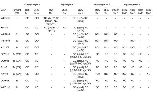
In the following sections, we have analyzed more specifically the phyletic profile (that is, the different patterns of occur-rence of orthologs in the set of Synechococcus genomes) and characteristics of three gene categories: genes encoding linker polypeptides (Table 2), genes encoding putative phycobilin lyases (Table 3) and genes of unknown function specifically located in the PBS rod gene region and, therefore, potentially involved in PBS metabolism or regulation (Table 4).
Phycobilisome linker polypeptides
[image:9.612.53.557.341.643.2]The core-membrane linker LCM, encoded by apcE, possesses three predicted repeat (or linker-like) domains in all marine
Table 2
Presence or absence of genes encoding linker polypeptides in the different marine Synechococcus genomes
Allophycocyanin Phycocyanin Phycoerythrin I Phycoerythrin II
Strain Pigment type
apcC
(LC)
apcE
(LCM)
cpcC
(LR)
cpcD
(LR)
cpcG
(LRC)
cpeC
(LR)
cpeE
(LR)
mpeD*
(LR)†
mpeC
(LR)†
mpeE
(LR)†
mpeF
(LR)†
mpeG
(LR)†
WH5701 1 CC CC‡ RC (cpcCI‡) RC (cpcCII‡) NC
(cpcCIII‡§)
RC GC (cpcGI)‡ RC (cpcGII)
- - -
-RS9917 1 CC CC RC (cpcCI) RC
(cpcCII)
RC GC (cpcGI) RC (cpcGII)
- - -
-WH7805 2 CC CC‡ - - GC (cpcGI)‡ RC
(cpcGII)
RC‡ RC‡ RC‡ - - -
-WH7803 3a CC CC‡ - - GC (cpcGI)‡ RC
(cpcGII)
RC‡ RC‡ RC‡ - RC‡ -
-RCC307 3b CC CC‡ - - GC (cpcGI)‡ RC
(cpcGII)
RC‡ RC‡ RC‡ RC‡ NC‡ - NC
CC9311 3d (CA) CC CC - - GC (cpcGI) RC
(cpcGII) NC (cpcGIII)
RC RC RC RC RC NC
-CC9902 3d (CA) CC CC - - GC (cpcGI) RC
(cpcGII) NC (cpcGIII)
RC RC RC RC NC NC
-BL107 3d (CA) CC CC - - GC (cpcGI) RC
(cpcGII) NC (cpcGIII)
RC RC RC RC NC NC
-RS9916 3d (CA) CC CC‡ - - GC (cpcGI)‡ RC
(cpcGII)
RC‡¶ RC‡ RC‡ RC‡ RC‡ - NC
CC9605 3c CC CC - - GC (cpcGI) RC
(cpcGII) NC (cpcGIII)
RC RC RC RC NC -
-WH8102 3c CC CC - - GC (cpcGI) RC
(cpcGII)
RC RC RC RC NC -
Synechococcus except strains CC9311 and RS9916, in which LCM has four such domains. RCC307 has the shortest LCM sequence (953 amino acids) compared to the other strains due to shorter Arm2 and Arm3 regions (see [15,41] for details on LCM domains). Besides the PC-associated linker genes found in the rod gene region of both blue-green strains (Fig-ure 4), WH5701 has a third cpcC homolog (cpcCIII) located elsewhere in the genome that potentially encodes a chimeric protein since it has an extended carboxyl terminus showing strong similarity to CpcD. None of the PE-containing strains possesses cpcC and cpcD homologs. In all marine
Synechoc-occus genomes, the rod-core linker gene cpcGII is found in the PBS rod region whereas cpcGI is found outside this clus-ter. A third cpcG gene copy, which we refer to as cpcGIII, is present elsewhere in the genomes of BL107, CC9902, CC9311 and CC9605.
[image:10.612.52.562.118.309.2]The total number of putative PE-associated linker genes var-ies from zero in the blue-green strains to six in the group constituted by the chromatic adapters and RCC307 (Table 2 and Figure 4). The location of the mpeE linker gene appears more variable than the other PEII genes, as it can be found
Table 3
Presence or absence of genes encoding putative phycobilin lyases in the different Synechococcus genomes
Phycocyanin Phycoerythrin I and/or II
Strain Pigment type
cpcEF
operon
rpcEF
operon
rpcG* cpcS† cpcT‡ rpcT§ cpeS cpeT cpeU¶ cpeY cpeZ mpeV mpeU mpeY¥ mpeZ¥
WH5701 1 RC - - GC# RC - - - - - - - - -
-RS9917 1 RC - - GC RC - - -
-WH7805 2 - RC - GC RC - RC RC RC RC RC RC - -
-WH7803 3a - RC - GC - RC RC RC RC RC RC RC - RC
-RCC307 3b - RC - GC RC - RC RC RC RC RC RC RC RC NC
CC9902 3d (CA) - RC - GC - RC RC RC RC RC RC RC RC RC NC
CC9311 3d (CA) - RC - GC - RC RC RC RC RC RC RC RC RC NC
BL107 3d (CA) - - RC GC - RC RC RC RC RC RC RC RC RC NC
RS9916 3d (CA) - - RC GC RC RC RC RC RC RC RC RC RC RC NC
CC9605 3c - - RC GC - RC RC RC RC RC RC - RC RC
-WH8102 3c - - RC GC - RC RC RC RC RC RC - RC RC
-GC, gene located within a cluster comprising the cpcGI and cpcS genes; NC, gene unlinked to other PBS genes; RC, gene located within the PBS rod gene cluster. Novel gene names proposed in this study are underlined. *pecE/F-like fusion gene [19]. †Ortholog of Nostoc sp. PCC 7120 'cpeS1' gene [51,52] that we propose to rename cpcS (see text). ‡Ortholog of a Synechococcus sp. PCC 7002 cpcT gene [50]. §Novel cpcT paralog found downstream of rpcA. ¶Novel cpcS paralog found upstream of pebA. ¥These two novel, closely related genes are both paralogs of cpeY. #Gene split into two different reading frames in this strain. CA, type IV chromatic adapter.
Table 4
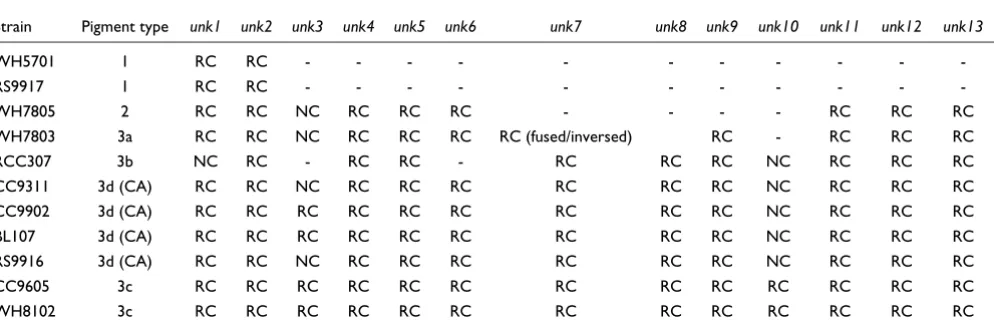
Presence or absence of genes encoding conserved hypothetical genes located in the phycobilisome rod gene region
Strain Pigment type unk1 unk2 unk3 unk4 unk5 unk6 unk7 unk8 unk9 unk10 unk11 unk12 unk13
WH5701 1 RC RC - - -
-RS9917 1 RC RC - - -
-WH7805 2 RC RC NC RC RC RC - - - - RC RC RC
WH7803 3a RC RC NC RC RC RC RC (fused/inversed) RC - RC RC RC
RCC307 3b NC RC - RC RC - RC RC RC NC RC RC RC
CC9311 3d (CA) RC RC NC RC RC RC RC RC RC NC RC RC RC
CC9902 3d (CA) RC RC RC RC RC RC RC RC RC NC RC RC RC
BL107 3d (CA) RC RC RC RC RC RC RC RC RC NC RC RC RC
RS9916 3d (CA) RC RC NC RC RC RC RC RC RC NC RC RC RC
CC9605 3c RC RC RC RC RC RC RC RC RC RC RC RC RC
WH8102 3c RC RC RC RC RC RC RC RC RC RC RC RC RC
[image:10.612.58.555.547.707.2]either in the PBS rod gene region (for example, upstream of cpcGII in CC9311 or downstream of cpcGII in RS9916) or a few genes upstream of this region (in RCC307, BL107 and CC9902) or even in a totally different location of the genome (in CC9605).
Surprisingly, the PEII-lacking strain WH7805 possesses a homolog of mpeD, a gene known to encode a chimeric protein made of a PEII-associated linker (amino terminus) and a PEI-associated CpeD-like linker (carboxyl terminus) [19]. How-ever, closer examination of the amino-terminal part of this protein in WH7805 reveals a relatively low similarity with other MpeD sequences and a notable deletion of the region corresponding to amino acids 43-59 in Synechococcus sp. WH8102 [19] that is conserved in all other MpeD sequences (Additional data file 2). This region includes two cysteinyl residues involved in linking a PUB chromophore via a Δ2,3 double bond, a type of chromophorylation typical of PEII-associated linker polypeptides [21]. Synechococcus sp. WH7803 also lacks the mpeC gene, which encodes the distal PEII-associated linker polypeptide in other strains [19,21]. Finally, both chromatic adapters and RCC307 have, outside
the PBS core region, an additional gene potentially encoding a PEII-associated linker (Table 3). In phylogenetic trees made with all PEII linkers (Additional data file 3), these sequences are both related to the amino terminus of MpeD but are split between two distinct gene clusters, one gathering BL107, CC9311 and CC9902, which we propose to name MpeF, and the other gathering RS9916 and RCC307, which we propose to name MpeG.
In order to compare further the linker composition of marine Synechococcus strains and determine whether they are all present in the PBSs, we performed a lithium dodecyl sulphate (LiDS)-PAGE analysis of intact PBSs. The Coomassie stained gel shown in Figure 5 displays the PBS proteins of two to three strains per pigment type. For WH7803 and RCC307, a Tris-tricine running buffer provided a better separation of the linker polypeptides than Tris-glycine (Figure 5, right). For strains WH5701, WH7805, WH7803, RCC307 and RS9916, all linker polypeptide bands (except ApcC and CpcD, which are not detectable under these electrophoresis conditions) were cut out from the gel and then identified by mass spec-trometry (Table 2). In all five strains, the upper band proved
Coomassie blue stained LiDS polyacrylamide gradient (10-20%) gel of PBS linkers run using a Tris-glycine buffer system (left)
Figure 5
Coomassie blue stained LiDS polyacrylamide gradient (10-20%) gel of PBS linkers run using a Tris-glycine buffer system (left). A Tris-tricine buffer (right) gave higher band resolution for RCC307 and WH7803. Green dots indicate linker polypeptides fluorescing green under UV light due to the presence of a PUB chromophore. Colored stars indicate the pigment type of each strain (see Figure 1 for color code). FNR: ferredoxin:NADP+ oxidoreductase.
Lcm
Lcm’
MpeD
other
linkers
PBP a
a a
a
and b
b
b
b
subunits
FNR
14
20
30
45
66
97
14
20
30*
45
66
97
TRIS-glycine
TRIS-tricine
RCC307
RS9917
W
H
7805
W
H
8018
W
H
7803
A
lmo03
BL107
W
H
5701
RS9916
O
L
i31
CC9605
W
H
8102
RCC307
W
H
7803
to be the core-membrane linker LCM, often accompanied by its degradation product LCM', making a band of lower appar-ent molecular weight. As expected, RS9916, which has an extended apcE gene sequence, possesses the LCM band of low-est electrophoretic mobility. Although the rod-core linker CpcGI was systematically present in all four strains, no CpcGII was detected by mass spectrometry, suggesting either that the cpcGII gene is expressed at a much lower level than cpcGII or that CpcGII is not present in the PBS fraction of these strains. It is worth noting though that we previously observed CpcGII (co-migrating with CpcGI) in a PBS fraction from Synechococcus sp. WH8102 [19]. Interestingly, we iden-tified all three predicted PC rod linkers in WH5701, including the CpcCD-like protein, which is not found in the RS9917 genome. Furthermore, all PEII linkers predicted in WH7803, RCC307 and RS9916 were detected by mass spectrometry, except the products of the mpeF gene of RS9916 and of the mpeG gene of RCC307 (Table 1). This suggests that either these two potential linker genes are not expressed in our standard culture conditions or their products are undetectable on Coomassie-stained LiDS-PAGE gels due to some inherent biochemical properties.
Lyases, lyase-isomerases and related genes
Four types of phycobilin lyases, enzymes involved in the chromophorylation of phycobiliproteins, have been charac-terized so far. One of these, the heterodimeric CpcE/F com-plex, reversibly ligates a PCB molecule to Cys-84 of the α -subunit of C-PC [42,43]. Two genes with strong homology to the characterized cpcE and cpcF genes of Synechococcus spp. PCC 7942 [44] and PCC 7002 [45] are found near the 3'-end of the rod gene region in 7 out of the 11 marine Synechococcus genomes. We have called these cpcE-F in the two C-PC-con-taining strains (RS9917 and WH5701) and rpcE-F in WH7803, CC9311 and CC9902, in agreement with the nomenclature proposed by Wilbanks and Glazer [37]. Indeed, Synechococcus sp. WH7803 (as well as WH8020 and WH8103) possesses a R-PCII type PC that has a PEB at α-84 [16]. Though we have called these genes rpcE/F in strains WH7805 and RCC307 as well (Additional data file 1), it is worth noting that in phylogenetic trees made with concate-nated CpeE-F or RpcE-F protein sequences using Gloeo-bacter violaceus as an outgroup, these two strains cluster with RS9917 and WH5701, with only moderate bootstrap support (Additional data file 4). Both CpeE/F and RpcE/F lyases from marine Synechococcus possess all sites described by Zhao and coworkers [46] to be important for the activity of CpeE/F in Fischerella sp. PCC 7603 (a.k.a. Mastidocladus laminosus), so they cannot be differentiated on this basis. In the four other Synechococcus genomes, including the high PUB:PEB strains WH8102 and CC9605 and the chromatic adapters BL107 and RS9916, these two lyase genes are replaced by a single fusion gene that we propose to call rpcG (Table 3). The amino- and carboxy-terminal parts of the rpcG gene product show strong homology to the PecE and PecF of Fischerella sp., respectively, the two subunits of a PCB
lyase-isomerase, which binds a PCB to Cys84 of the phycoerythro-cyanin α-subunit and concomitantly isomerizes it into phyco-violobilin [47,48]. A conserved motif 'NHCQGN' shown to be crucial for the isomerase activity of Fischerella PecF is present in the carboxyl terminus of the four marine Syne-chococcus RpcG sequences (for example, positions 361-366 of SYNW2005 in WH8102). This suggests that RpcG is also a phycobilin lyase-isomerase, although several other sites defined as potentially important for the activity of the PecE/F enzyme in Fischerella sp. [49] are not conserved in those sequences.
An ortholog of cpcT, shown in Synechococcus sp. PCC 7002 to encode a lyase catalyzing the binding of PCB at Cys153 of the C-PC β-subunit [50], is found in WH5701, RS9917, WH7805, RCC307 and RS9916 (Table 3). This gene belongs to a family of three paralogs, including cpeT, first described in the PE gene cluster of F. diplosiphon [39] and located at a similar position in all PE-containing marine Synechococcus (Figure 4). An uncharacterized gene located near the 5'-end of the PBS rod gene cluster of all PE-containing strains except RCC307 also belongs to this family. We propose to name this gene rpcT, since it is present in the PC-specific gene region of WH7803, which possesses R-PCII. Thus, most marine Syne-chococcus strains possess either cpcT or rpcT. Surprisingly, the RS9916 strain possesses both genes, confirming their par-alogous nature (Additional data file 5).
PE-containing Synechococcus possess several genes in the PEI or PEII gene sub-regions that encode proteins showing homology to other types of lyases, likely involved in binding phycobilins to one or both PEs. These lyases include CpeY and CpeZ, which in F. diplosiphon were presumed to be subunits of a heterodimeric lyase, binding PEB to PE α- or β-subunits [53], but the precise site specificity of this enzyme is hitherto unknown. The mpeU and mpeV genes, which were first observed in Synechococcus sp. WH8020 by Wilbanks and Glazer [37], likely encode two additional lyases. These paral-ogous genes are both present in the chromatic adapters and in RCC307, whereas WH7803 and WH7805 have only mpeV and the high PUB:PEB strains only mpeU (Table 3). Finally, we found two novel, paralogous lyase genes, again closely related to one another and more distantly related to cpeY. We propose to name these genes mpeY and mpeZ. Contrary to CpeY and CpeZ, the products of these putative lyase genes likely do not form heterodimers, given their distinct phyletic profiles (Table 3). Indeed, mpeY is found in the PEII-specific sub-region of all PEII-containing strains (Figure 4) whereas mpeZ is found only in the genomes of the chromatic adapters and of RCC307, outside the PBS gene clusters.
Conserved hypothetical genes located in the phycobilisome gene region
Table 4 reports the phyletic profile of 13 conserved hypothet-ical genes associated with the PBS rod region of all (or a majority of) strains. Many of them are seemingly specific to marine Synechococcus while some are found in other cyano-bacterial genera, including Prochlorococcus and/or Gloeo-bacter. It is worth noting though that there are still very few genomes of phycoerythrin-containing cyanobacteria in cur-rent databases and it is likely that homologs will be found in those as they become available. In this study, we have given these genes the provisional names unk1-13, until a more com-plete characterization is performed.
As already mentioned, the unk1 gene is located upstream of the PBS rod region in all strains except RCC307, in which unk1 is located elsewhere in the genome. Another unknown gene (unk2) immediately follows unk1 in most PE-containing strains (in RCC307, it is the first gene of the PBS rod gene region). The unk2 gene is found three genes downstream of unk1 in the two blue-green strains. The predicted Unk2 pro-tein sequence generally shows a fairly large variability among the different Synechococcus strains, although the BL107 and CC9902 sequences are very closely related (91% identity at the amino acid level). Both Unk1 and Unk2 are short proteins with no recognizable motifs. The unk3 gene is associated with the PBS rod region in only four out of the eleven genomes and encodes a protein with six putative transmembrane helices. It is therefore probably not directly related to PBS structure. The unk4 gene is present upstream of aplA in all PE-contain-ing strains and directly upstream of cpcGII in WH7805, which lacks aplA. The unk5 gene, generally located down-stream of cpcGII, has the same phyletic profile as unk4 (Table
4) and its product possesses pentapeptide repeat motifs. Though very short (57-61 amino acids), the Unk6 protein is very well conserved among the PE-containing Synechococ-cus. A cluster of three consecutive short and conserved hypo-thetical genes (unk7-9) is found only in PEII-containing strains. Localization of these genes in a PEII-specialized sub-region strongly suggests that they are involved in some still unknown function specifically related to PEII. The predicted proteins Unk7 and Unk8 both possess a motif of unknown function (PF07862) also found in the product of a gene located in the nif cluster of several cyanobacteria as well as in the nitrogen-fixing proteobacterium Azotobacter vinelandii [54]. Surprisingly, in WH7803, unk7 and unk8 are fused and reversed with regard to unk9. This suggests that these genes encode two subunits of the same heterodimeric complex. In the high PUB:PEB strains WH8102 and CC9605, the PEII-specialized region ends with unk10, which is strongly con-served between these strains (90% identity at the amino acid level). Homologs of unk10 are also found in the genomes of the chromatic adapters and in RCC307 but outside the PBS rod gene region and have only about 49% identity with sequences of the high PUB:PEB strains. Located in the PEI-specific region, the translated unk11 gene is very variable in length and sequence (especially the 3'-end) among marine Synechococcus strains. In contrast, the neighboring gene unk12 displays low sequence variability between strains. Finally, the unk13 gene, though strongly conserved, was not correctly modeled in WH8102, in which a wrong open read-ing frame (ORF; SYNW2018) was predicted in a different reading frame. The unk12 gene was previously known and was called orf140 in WH8020 by Wilbanks and Glazer [37], who sequenced the 3'-end of the PBS rod gene region from mpeB to the phosphatase. By remodeling this region, we con-firmed that the unk11 and unk13 genes are also present in this strain and were incorrectly assigned by these authors. Though partial, the organization and gene content of this region in WH8020 [37] is clearly similar to that of chromatic adapters (Figure 4), and this is confirmed by the ability of this strain to chromatically adapt (Table 1; see also [33]).
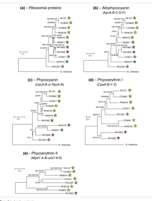
Phylogeny of phycobilisome genes
to a relatively low number of informative sites. Whenever possible, we used the primitive, PE-containing, freshwater cyanobacterium Gloeobacter violaceus as an outgroup to root our trees, in order to better understand evolution of PBSs within the marine Synechococcus group.
The phylogenetic trees obtained with concatenated proteins encoding the AP components (ApcA-B-C-D-F; Figure 6b) share many characteristics with those based on ribosomal proteins (Figure 6a). In both cases, RCC307 and WH5701 are isolated on two long branches well apart from all other strains. Furthermore, WH7803 and WH7805 on the one hand, and CC9902 and BL107 on the other, appear closely related to one another. The only variable positions are those of the closely related strains RS9916 and RS9917, which clus-ter on the same branch as WH7803, WH7805 and CC9311 in the ribosomal tree, and at the base of the branch bearing BL107, CC9902, CC9605 and WH8102 in the AP tree, but with relatively low bootstrap support in the second case.
The phylogenetic trees of concatenated PC α- and β-subunits (CpcGII was not included because mass spectrometry analy-ses suggested it may not be part of the PBS; Table 2) show a number of differences relative to the AP tree, including the fact that the two blue-green strains group together (with high bootstrap support) apart from all others (Figure 6c). This is consistent with the fact that they both have C-PC (binding only PCB), whereas all other strains have a PC form binding both PCB and PEB. The relative positions of WH7805 and RCC307 varied between phylogenetic methods. WH7805 is known to contain R-PCIII [17] and this is probably the case for RCC307 as well, based on their similar PC lyase gene con-tent, including cpcS, cpcT and rpcE-F (Table 3). All strains containing R-PCII (or possibly another, unidentified PC form, for those strains possessing rpcG; Table 3) formed a well-supported cluster with both ML and NJ methods, though the relative positions of CC9311 and RS9916 were variable within this cluster.
The phylogeny obtained for the concatenated PEI proteins CpeA-B-Y-Z - addition of Unk12 did not significantly alter the tree topologies, but gave lower bootstrap support (data not shown) - fits well with the pigment types, as defined in Table 1. Indeed, the two high PUB:PEB strains group together, well apart from the other PE-containing strains. RCC307 is found at the base of a cluster formed by chromatic adapters (Figure 6d), consistent with the fact that all these strains share a sim-ilar PBS gene complement and organization. Likewise, strains WH7803 and WH7805 group together, consistent with the similar organization of their PEI-like region, with cpeZ being located downstream of mpeV instead of upstream of cpeY as in all other PE-containing strains (Figure 4).
Phylogenetic trees obtained with the concatenated PEII pro-teins MpeA-B-Y and Unk7-9 - inclusion of Unk7-9 does not alter the tree topologies obtained with the sole MpeA-B-Y
sequences but provides better bootstrap support - are shown without an outgroup, since this phycobiliprotein form is not found in freshwater cyanobacteria. Still, these trees are glo-bally similar to those obtained with PEI proteins, with three main clusters, one gathering the medium PUB:PEB strain RCC307 and the chromatic adapters, one gathering the two high PUB:PEB strains, whereas the low PUB:PEB strain WH7803 clusters apart from all others.
Discussion
Comparative genomics reveal novel genes involved in phycobilisome metabolism
We have identified and compared a number of genes poten-tially involved in the synthesis and chromophorylation of PBSs in a variety of sequenced marine Synechococcus strains spanning all PBS pigment types known so far in this group. Strains displaying different pigment types have different gene complements with a considerable increase in complexity from type 1 (WH5701 and RS9917) to type 3d (chromatic adapters). Synthesis of rods entirely composed of PC, as found in the first type, requires at least 15 genes. This includes two cpcB-A operons encoding C-PC α- and β-subunits, two rod-core linker genes (cpcGI and cpcGII), two cpcC and one cpcD rod linker genes (in WH5701, an additional cpcC gene, cpcCIII, was in fact found to be a cpcC/D gene chimera), four genes encoding three different lyases (CpcE/F, CpcS and CpcT) and the PCB biosynthesis gene pcyA, which encodes the PCB:ferredoxin oxidoreductase [55]. Whether unk1 and unk2, usually found at or near the 5'-end of the PBS rod gene region (Figure 4), are also involved in PC metabolism awaits experimental checking. An additional cpcB gene, absent from other blue-green cyanobacteria such as Synechococcus sp. PCC 7942 or Synechocystis sp. PCC 6803, is found unlinked to other PBS genes in both WH5701 and RS9917. While the three cpcB copies are almost identical in RS9917, the isolated copy is somehow divergent from the other two in WH5701. This may indicate a recent change in function. All PEII-con-taining strains possess an AP-like gene encoding a protein derived from a phycobiliprotein, the homolog of which, aplA, was shown in F. diplosiphon to encode a photoreceptor not linked to the PBS [40]. So it is possible that the additional CpcB found in the blue-green strains might have a similar function though, contrary to AplA, this protein appears to have retained the ability to interact with the α-PC subunit. Indeed, amino acids involved in maintaining these interac-tions [56] are conserved in all CpcB copies.
Figure 6 (see legend on next page)
(ApcA-B-C-D-F)
(CpeA-B-Y-Z)
(CpcA-B or RpcA-B)
(MpeY-A-B-unk7-8-9)
BL107
CC9902
RS9916
CC9311
RCC307
WH8102
CC9605 WH7803 100/100/100
100/100/91
42/40/-100/100/100
100/100/100 0.1
BL107
CC9902
CC9605
WH8102
RS9916
RS9917
WH7803
WH7805
CC9311
WH5701
RCC307
G. violaceus 100/100/100
84/86/77
100/100/89
75/61/-
41/44/- 43/51/-100/100/99
92/88/66 100/100/100
0.1 BL107
CC9902
CC9605
WH8102
RS9916
RS9917
WH7803
WH7805
CC9311
WH5701
RCC307
G. violaceus
BL107
CC9902
CC9311
CC9605
WH8102
RS9916
WH7803
WH5701
RS9917
WH7805
RCC307
G. violaceus 100/100/100
32/35/-
82/69/- 74/73/-
73/65/-
90/93/-
53/-/-100/100/95 0.1
G. violaceus 100/100/99
100/98/89
85/71/48
98/100/84
97/97/51 99/97/77
100/100/100 0.1
BL107
CC9902
RS9916
CC9311
RCC307
CC9605
WH8102
WH7805
WH7803 100/100/100
94/81/56 100/100/97
100/100/94
81/59/75 100/100/100
100/100/100 92/89/89 100/100/100
0.1
(a)
– Ribosomal proteins
(b)
– Allophycocyanin
(c)
– Phycocyanin
(d)
– Phycoerythrin I
[image:15.612.56.552.78.730.2]all these unk genes are specific to PE-containing Synechococ-cus and all but unk11 are well conserved. Despite its tiny size, explaining why it has often been missed by annotation software, unk6 is likely a true gene since it is also present in all Prochlorococcus strains (data not shown). In both Prochlorococcus and marine Synechococcus spp., unk6 is located upstream of the putative phycobilin lyase gene cpeS.
Acquisition of a second PE type, PEII, involves comparatively few additional genes, from six in WH7803, including unk7/8 and unk9, up to twelve genes in type IV chromatic adapters and RCC307 (mpeA, B, C, D, E, F or G, U, Y, Z and unk7, 8, and 9), among which the seven underlined genes are novel PEII genes. The fact that PEII synthesis and regulation proc-esses require fewer genes than for PEI implies that several genes involved in these processes are common to both PE forms. This obviously includes the PEB synthesis genes pebA/ B, but likely also a number of lyase genes.
Predicting lyase gene function
Examination of the number, phylogenetic relatedness and phyletic profiles of all predicted lyase genes (Table 3) can give us clues about the possible functional specificity of these enzymes. The number of chromophore binding sites on the α -and β-subunits of phycobiliproteins varies from seven in pig-ment type 1 - that is, four in AP (ApcA, B, D and F subunits have one each) and three in PC (one in CpcA/RpcA, two in CpcB/RpcB) - up to eighteen in PEII-containing strains - that is, four in AP, three in PC, five in PEI (two in CpeA, three in CpeB) and six in PEII (three in MpeA, three in MpeB) -. Fur-thermore, it is thought that type IV chromatic adapters can have either PUB or PEB at two chromophore binding sites of MpeA [34]. Finally, while the chromophorylation of LCM with PCB is thought to be auto-catalyzed and, thus, likely does not require any lyase activity [49], chromophorylation with PUB of the two to four PEII rod linkers (Table 2) probably requires one or several specific PUB lyases (or PEB lyase-isomerases). By comparison, the number of predicted proteins showing homology to known lyases varies from 3 in blue-green strains up to 12-13 in RCC307 and chromatic adapters.
All three phycobilin lyases identified in the genomes of Syne-chococcus spp. WH5701 and RS9917 (Table 3) have charac-terized homologs in freshwater cyanobacteria. This reduced set of lyases is most likely sufficient to catalyze the chromo-phorylation with PCB of all AP and C-PC binding sites. Indeed, the CpcS lyase (named 'CpeS1' by Zhao and cowork-ers [51,52]) is active on almost all α-84 and β-84 cysteinyl
res-idues. The only exception is C-PC α-84, chromophorylation of which is under the control of the heterodimeric lyase CpeE/F [42,43]. Chromophorylation of the last cysteinyl residue, that is, C-PC β-155, is catalyzed by another specific lyase, CpcT [50]. A fairly large difference exists between the sequences and active sites of the CpcE/F lyase, which binds PCB (a type 1 chromophore carrying a Δ3,31-ethylidene group and a single
bond between C-2 and C-3) to C-PC α-84, and those of the lyase-isomerase PecE/F, which binds phycobiliviolin (a type 2 chromophore carrying a 3-vinyl group and a Δ2,3-double bond) to the homolog position of α-phycoerythrocyanin [47,48]. Thus, the replacement in four Synechococcus strains (BL107, RS9916, CC9605 and WH8102) of cpeE and cpeF genes by a fusion gene encoding a PecE/F-like protein (that we have called RpcG) is quite significant and it is possible that the PC synthesized by these strains binds a type 2 chromo-phore at α-84. This interesting hypothesis suggests that a bet-ter biochemical characbet-terization of the PC found in these strains is needed. Finally, in all PEII-containing Synechococ-cus strains except RCC307 and RS9916, the cpcT gene is absent (Table 3) and seemingly replaced by a gene of the same family of paralogs, located in the PC-specific gene cluster (Figure 4), that we have called rpcT. Given the presence of the rpcT gene (and absence of cpcT) in Synechococcus sp. WH7803 in which a PEB is bound at β-153 of R-PCII [16], RpcT is a plausible candidate for catalyzing this specific chromophorylation. Surprisingly, RS9916 possesses both CpcT and RpcT paralogs, suggesting it may either bind PCB or PEB at this site.
Predicting the function of lyase genes potentially involved in bilin attachment to PEI and PEII is much more difficult than for PC, given the larger number of binding sites on these phy-cobiliproteins. The only lyase gene specific to all PEII-con-taining strains is mpeY (Table 3 and Figure 4). The PEII α -subunit has one chromophore-binding cysteinyl residue that has no homolog in its PEI counterpart, α-75. In WH8103 and white light-grown WH8020, α-75 has been shown to bind a PUB [18]. We hypothesize that MpeY could be a PUB lyase (or a PEB lyase-isomerase) involved in the chromophorylation of PEII α-75 with PUB. However, another specific feature of PEII complexes is that they are held together with two to four PUB-chromophorylated linkers (Table 3) so, alternatively, mpeY might encode a lyase involved in the PUB chromopho-rylation of one (or several) PEII rod linker(s).
The presence of two additional lyase genes in chromatic adapters compared to strains exhibiting either pigment types ML trees made with concatenated amino acid sequences of (a) all 51 ribosomal proteins (6,754 amino acid positions), (b) the AP proteins ApcA-B-C-D-F (710 amino acid positions), (c) the PC proteins CpcA-B or RpcA-B (332 amino acid positions), (d) the PEI proteins CpeA-B-Y-Z (943 amino acid positions) and (e) the PEII proteins MpeA-B-Y and Unk7-8-9 (1,007 amino acid positions)
Figure 6 (see previous page)
[image:16.612.53.549.92.104.2]3a or 3c (Table 2) suggests that this more complex lyase com-plement is required for type IV chromatic adaptation. Indeed, this process is thought to consist of the reversible exchange of both PEII α-83 and α-140 chromophores from PEB to PUB [34], and not in the differential expression of several sets of phycobiliprotein genes, like in type III chromatic adaptation (see, for example, [57] for a review). The presence of only one set of genes encoding PEI and PEII α- and β-subunits in all genomes of chromatic adapters supports this hypothesis. Because type IV chromatic adaptation implies the conversion of a PEII-B into a PEII-C under blue light (and conversely under white light; Table 1), it is reasonable to assume that chromatic adapters need two more PUB lyases (or PEB lyase-isomerases) than pigment type 3a strains, which permanently have PEB at PEII α-83 and α-140, and two more PEB lyases than pigment type 3c, which permanently have PUB at these two positions. The phyletic pattern of the mpeV gene (Table 3) which, besides its occurrence in chromatic adapters, is also present in WH7803 and WH7805 and absent in the high PUB:PEB strains, suggests it could encode a PEB lyase. Con-versely, mpeU has the reverse phyletic profile and, thus, could encode a PUB lyase (or PEB lyase-isomerase). The specificity of the putative lyase MpeZ is harder to interpret. Surprisingly, the complex PBS gene set found in chromatic adapters is shared by RCC307, which is the sole strain to have pigment type 3b of all marine Synechococcus strains screened so far. Indeed, we have determined that all strains except RCC307 described as having a PUB:PEB of approximately 0.7-0.8 by Fuller et al. [23] are actually chromatic adapters. This includes strain RCC61 (data not shown), which belongs to the same phylogenetic clade as RCC307 (that is, clade X) [23]. Therefore, we suggest that RCC307 may have lost the ability to chromatically adapt, perhaps due to a mutation in a domain important for lyase activity or the inactivation or loss of some regulatory gene(s) required for this process.
Predicted models of PBS structures
Most sequenced Synechococcus strains have typical PBS cores with three AP cylinders. The presence of an additional LCM domain in CC9311 and RS9916 suggests that their PBS core may have two additional half-cylinders, as previously observed in freshwater species such as Nostoc sp. PCC 7120 [58]. It is thought that up to eight rods can be bound to such a PBS core (Figure 7). The presence of an extended LCM was previously reported from another chromatic adapter, Syne-chococcus sp. M16.17 [34] and one may wonder whether such PBS cores might only occur in this pigment type. An answer to this question awaits screening of apcE genes (or of the LCM linker size on LiDS-PAGE gels) in a much wider range of strains, as well as direct evidence from electron microscopic images of isolated phycobilisomes.
The large diversity of PBS rod pigmentation observed so far within the marine Synechococcus group rests on combina-tions of at least three PC types (C-PC, R-PCII, R-PCIII), two PEI types (PEI-A/A* and PEI-B) and three PEII types
(PEII-A through C) (Table 1). The number and nature of rod linker polypeptides present in the different Synechococcus strains can help predict the structure of their PBS rods. Given the striking similarity in pigmentation and gene complement between the freshwater strains Synechococcus sp. PCC 7942 or Synechocystis sp. PCC 6803 and Synechococcus sp. RS9917, the latter strain likely has a very similar PBS rod structure [59,60], that is, three C-PC hexamers (Figure 7a, left). Since Synechococcus sp. WH5701 has one more (CpcCD-like) rod linker than RS9917 (Table 2), it is possible that this strain has rods with one additional PC disc (Figure 7a, right).
Like all PE-containing strains, WH7805 lacks the CpcC and CpcD rod linker polypeptides, the absence of which implies it has only a single PC hexamer at the base of each PBS rod. This PC can be of two types depending on strains, C-PC or R-PCIII (Table 1 and Figure 7b). WH7805 has three PE linkers, including a homolog of the long, chimeric rod linker MpeD (Figures 4 and 5) instead of a shorter, CpeD-like linker, like in F. diplosiphon [61]. Its amino-terminal moiety is very diver-gent, however, and does not possess the ability to bind a PUB chromophore, a characteristic common to all PEII rod linkers (Additional data file 2). In the type III chromatic adapter F. diplosiphon grown under green light, PBS rods are composed of one PC and three PE hexamers [15,57]. Since they have a MpeD-like rod linker equivalent to two typical rod linkers in length, we suggest that Synechococcus pigment type 2 strains might have one more PE disc in their rods than F. diplosiphon (Figure 7b).
In a previous paper, we have proposed a model for the struc-ture of PBS rods of the pigment type 3c strain WH8102, which we have predicted to have six hexamers: one PC, two PEI and three PEII [19]. The other high PUB:PEB strain CC9605 appears to have similar PBS rods (Figure 7d). Because it is missing the (distal) linker gene mpeC, we assume that the type 3a strain WH7803 has only two PEII hexamers (Figure 7c). Despite the presence of an additional, PEII rod linker gene (mpeF or mpeG) in chromatic adapters and in RCC307, we found no evidence by mass spectrometry of any such link-ers in PBS preparations from RCC307 and RS9916 (Table 2). So, it is very unlikely that these strains have more than three PEII hexamers. Indeed, in this case, they would have a higher whole cell PUB:PEB under blue light than WH8102 or CC9605, whereas this ratio is similar or even lower in chro-matic adapters (Table 1). It is possible though that under some specific culture conditions, mpeF or mpeG could be expressed and that their products could then replace some other PEII linker in the PBS rods.
New insights into PBS evolution
Proposed models of PBS structure for the different Synechococcus pigment types and subtypes
Figure 7
Proposed models of PBS structure for the different Synechococcus pigment types and subtypes. PBS cores are generally composed of three cylinders, but in some chromatic adapters possessing an extended LCM, it is likely composed of two additional half cylinders (see, for example, [58]). In pigment type 1, rods are composed of C-PC only; in pigment type 2, rods are composed of either C-PC, or R-PCIII and a PEI-like phycobiliprotein; in pigment type 3, rods comprise R-PC and two PE types (PEI and PEII). Cells of the latter pigment type bind PEB and PUB at a low (3a), medium (3b), high (3c) or variable (3d or type IV chromatic adapter) ratio. Colored stars indicate the pigment type of each strain (see Figure 1 for color code).