Original Article
Circulating tumor cells using hTERT-specific
replication-selective adenovirus in
patients with soft tissue sarcoma
Toshihiro Matsuo
1, Masataka Deie
1, Takashi Sugita
2, Shoji Shimose
2, Yasuo Urata
3, Norimitsu Wakao
1,
Katsuhisa Kawanami
1, Mitsuo Ochi
41Department of Orthopaedic Surgery, Aichi Medical University, Aichi, Japan; 2Department of Orthopaedic Surgery, Kure Medical Center, Hiroshima, Japan; 3Oncolys Biopharma, Toranomon, Tokyo, Japan; 4Department of Orthopaedic Surgery, Hiroshima University, Hiroshima, Japan
Received August 7, 2016; Accepted September 30, 2016; Epub March 15, 2017; Published March 30, 2017
Abstract: The presence of circulating tumor cells (CTCs) in peripheral blood offers a useful prognostic factor and tool for measuring the effects of treatment for various carcinomas. We attempted to detect viable CTCs in pe-ripheral blood from sarcoma patients using a telomerase-specific viral agent. We examined correlations between numbers of CTCs and other clinical features of sarcoma. For CTC analysis, 20 blood samples were obtained from 10 patients with soft-tissue sarcoma of the trunk or extremities before and after surgery. Five patients were diag-nosed with grade 2 sarcoma and five were diagdiag-nosed with grade 3 sarcoma according to the French Federation of Cancer Centers Sarcoma Group System. Mean postoperative follow-up was 36.6 months (range, 12-49 months). Oncological prognosis was as follows: 6 patients remained continuously disease-free, 2 patients showed no evi-dence of disease, and 2 patients died of the disease. Three patients developed lung metastases after surgery. We incubated 7.5-ml blood samples with a telomerase-specific, replication-selective, oncolytic adenoviral agent carrying the green fluorescent protein (GFP) gene, which allowed for the detection of viable CTCs. GFP-positive cells were counted using fluorescence microscopy. The mean number of CTCs showed no significant difference between preoperatively (1.9; range, 0-6) and postoperatively (2.6; range, 0-18; P=0.704). All patients with increased num-bers of CTCs postoperatively compared to preoperatively displayed lung metastases. The number of postoperative CTCs correlated significantly with metastasis (P=0.022) and life prognosis (P=0.014). A significant relationship was found between the number of CTCs and both occurrence of lung metastasis and prognosis in patients with sarcoma. Detection of CTCs using telomerase-specific viral agent may prove useful for prognostic evaluation.
Keywords: Circulating tumor cells, human telomerase reverse transcriptase, adenovirus, sarcoma
Introduction
Soft-tissue sarcomas are relatively rare
neo-plasms arising from mesenchymal tissues.
Sarcomas exhibit specific molecular
character-istics and poor prognosis due to the tendency
to spread to distant organs, particularly the
lungs, via the vascular system [1, 2]. Circulating
tumor cells (CTCs) in peripheral blood may thus
be very important for analyzing the metastatic
biology of sarcoma [3, 4]. Recently, the
pres-ence of CTCs in the blood has been shown to
offer a clinically useful biomarker for early
detection, prognosis, and assessment of
aggressiveness. CTCs also serve as a
surro-gate marker of treatment effects in various
can be used as an alternative measure of
telomerase activity [12, 13]. Expression of
hTERT is not seen in most somatic cells [14],
but is evident in more than 85% of carcino-
mas [15, 16] and more than 90% of sarcomas
[17-19]. OBP-401 and OBP-1101 are
telomer-ase-specific replication-competent adenovirus
variants in which the hTERT promoter drives
expression of E1A and E1B genes linked to an
internal ribosome entry site and GFP, which is
inserted into the E3 region under control of the
cytomegalovirus
promoter [20, 21]. Almost all
carcinoma and sarcoma cells express hTERT
mRNA, and this system therefore selectively
labels carcinoma and sarcoma cells, but not
normal cells, with green fluorescence. The
present study aimed to detect CTCs in the
peripheral blood of sarcoma patients using this
novel method, then looked for a correlation
between numbers of CTCs and other clinical
features of sarcomas to determine whether
these factors can be used to assess tumor
pro-gression or prognosis.
Materials and methods
Patients and blood samples
We investigated 20 blood samples obtained
before and after surgery from 10 patients (5
men, 5 women; mean age, 73 years; range,
44-86 years) scheduled to undergo surgery for
soft-tissue sarcoma of the trunk or extremities.
Underlying pathology was myxofibrosarcoma in
5 patients, liposarcoma in 2, and
undifferenti-ated pleomorphic sarcoma, fibrosarcoma, and
leiomyosarcoma in 1 each. Tumors were
[image:2.612.91.523.85.241.2]locat-ed in the chest wall (4 patients), lower leg (4
patients), thigh (1 patient) and iliopsoas muscle
(1 patient). Histological grades were assigned
according to the French Federation of Cancer
Centers Sarcoma Group system based on
tumor differentiation, mitotic count, and
necro-sis [22]. Sarcoma was categorized as grade
2 in 5 patients and grade 3 in 5 patients. Tumor
size was evaluated by measuring the largest
diameter on magnetic resonance imaging. Cli-
nical data are shown in
Table 1
. All patients
with these sarcomas underwent tumor
resec-tion and/or chemotherapy between 2012 and
2014 at our institute. After conservative
sur-gery for the soft-tissue sarcoma, we performed
external radiation therapy for patients who
underwent marginal resection. Mean
postop-erative follow-up was 36.6 months (range,
12-49 months). Chemotherapy comprised mu-
lti-agent systemic chemotherapy in patients
with metastasis. High-dose ifosfamide and
doxorubicin were used. No patients received
chemotherapy before blood specimens were
collected. After surgery, 6 patients remained
disease-free, 2 patients showed no evidence
of disease, and 2 patients had died of the
disease. Three patients developed lung
metas-tasis without local recurrence. Two of those
three patients with metastasis had died, while
one remained alive after pulmonary
metasta-sectomy. Participants in the current study
were recruited from our hospital and provided
written consent for blood collection in
accor-dance with a protocol approved by the ethics
committee at our institution (approval number
COI2402-24-28).
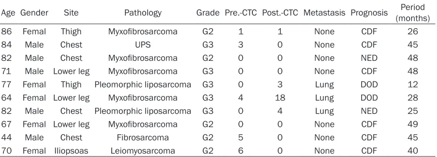
Table 1.
All clinical data and CTCs
Age Gender Site Pathology Grade Pre.-CTC Post.-CTC Metastasis Prognosis (months)Period
86 Femal Thigh Myxofibrosarcoma G2 1 1 None CDF 26
84 Male Chest UPS G3 3 0 None CDF 45
82 Male Chest Myxofibrosarcoma G2 0 0 None NED 48
71 Male Lower leg Myxofibrosarcoma G3 0 0 None CDF 48
77 Femal Thigh Pleomorphic liposarcoma G3 0 3 Lung DOD 12
64 Femal Lower leg Myxofibrosarcoma G3 4 18 Lung DOD 28
82 Male Chest Pleomorphic liposarcoma G3 0 4 Lung NED 25
67 Femal Lower leg Myxofibrosarcoma G2 0 0 None CDF 49
44 Male Chest Fibrosarcoma G2 5 0 None CDF 45
70 Femal Iliopsoas Leiomyosarcoma G2 6 0 None CDF 40
Measurement of viable CTCs
Peripheral blood (7.5 ml) was obtained from
each patient before surgery and more than 2
weeks after surgery. Blood samples were drawn
into collection tubes containing
citrate-phos-phate-dextrose solution and incubated with
lysis buffer containing ammonium chloride to
remove erythrocytes. After centrifugation, the
remaining white blood cells were washed twice
with Dulbecco’s modified Eagle’s medium
con-taining 10% fetal bovine serum (FBS). White
blood cell pellets were mixed with 2.3 × 10
8OBP-401 or 1 × 10
9OBP-1101 viral particles
and incubated for 24 h at 37°C with gentle
rota-tion. Following centrifugation, cells were
incu-bated with primary antibodies at room
temper-ature for 30 min, fixed with 4%
paraformalde-hyde, and permeabilized with 0.15% Triton
X-100. The following primary antibodies were
used: anti-CD14 (1:200 325601; BioLegend,
San Diego, US), anti-CD45 (1:100 304002;
BioLegend), and anti-vimentin (1:200 ab45939;
cent microscopy (IX71; Olympus, Tokyo, Japan),
and cell images were acquired on Metamor-
ph (Molecular Devices; Sunnyvale, US) using
each filter set (DAPI/FITC/RFP/CY5; Olympus)
(
Figure 1
).
Statistical analysis
Correlations between clinical factors and
num-bers of CTCs were calculated with unpaired
t-tests. Each prognostic factor was divided into
two groups based on average values. Data are
presented as mean ± standard deviation. In all
analyses, values of
P
<0.05 were considered
significant. All analyses were performed using
the Statview version 5.0 statistical package
(Abacus Concepts, Berkley, CA).
Results
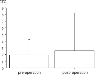
[image:3.612.88.376.71.143.2]The mean number of CTCs was 1.9 (range: 0-6)
preoperatively and 2.6 (range: 0-18)
postopera-tively (
Table 1
). No significant difference in
numbers of CTCs were evident between pre-
and postoperatively (P=0.704;
Figure 2
). All
patients showing increased numbers of CTCs
postoperatively compared to preoperatively
also displayed lung metastases. The number of
preoperative CTCs did not appear significantly
related to each clinical factor (sex, P=0.714;
tumor location, P=0.254; metastasis, P=0.650;
age, P=0.153; histological grade, P=0.538;
tumor size, P=0.882; life prognosis, P=0.952).
The number of postoperative CTCs appeared
unrelated to following clinical factors (sex, P=-
0.338; tumor location, P=0.338; age, P=
0.603; histological grade, P=0.190; tumor size,
P=0.243). However, the number of
postopera-tive CTCs was significantly related to
metasta-sis (P=0.022;
Figure 3
). The number of CTCs
postoperatively was significantly higher in
patients with metastasis (8.3 ± 8.4) than in
Figure 1. After 7.5-ml peripheral blood samples were infected with viral agent, each sample was treated with a surface-active agent to degrade red blood cells. The remaining cells were scraped from slides. Glass slides were then immunostained with CD45, vimentin and DAPI. The number of CTCs (GFP+, CD45-, vimentin + cells) was counted under fluorescence microscopy.
Figure 2. The mean number of CTCs was 1.9 in pre-operative patients and 2.6 in postpre-operative patients. No significant difference in number of CTCs was evi-dent between pre- and postoperatively (P=0.704).
[image:3.612.94.286.227.376.2]fluores-those without metastasis (0.14 ± 0.38). The
number of postoperative CTCs correlated
sig-nificantly with life prognosis (P=0.014;
Figure
4
). The number of postoperative CTCs was
sig-nificantly higher in dead patients (10.5 ± 10.6)
than in living patients (0.63 ± 1.41).
Discussion
Cancer metastasis is the process of cancer cell
dissemination to establish new growth in
differ-ent organs from the primary lesion via the blood
circulation [23, 24]. CTCs can be detected in
peripheral blood, may play an important role in
the biology of metastasis, and offer clinically
useful biomarkers for several carcinomas [5-8,
25-27]. Detection of CTCs in the blood of
patients with carcinoma offers a less-invasive
method for detecting cancer in the early
stag-es, assessing tumor progression, and
deter-mining prognosis. CTCs also serve as a
bio-marker of therapeutic efficacy.
In contrast to carcinoma, little data has been
published on CTCs in sarcomas. CTCs may be
detectable in sarcomas, but their clinical value
is unknown [3, 4]. The spread of sarcoma cells
to the lungs occurs mainly via the vasculature,
although angiosarcoma, clear cell sarcoma,
and epithelioid sarcoma more frequently spr-
ead to lymph nodes [2]. CTCs represent an
important event in metastatic relapse via the
vascular system and may have the potential
to form metastases. CTCs may therefore be of
great importance for the analysis of metastasis
in almost all types of sarcoma. However, the
prognostic significance of CTCs remains
contro-versial. In some reports, detection of CTCs has
indicated oncological aggressiveness and
cor-related with metastasis of the sarcoma
[28-32]. In contrast, another report found that CTC
detection did not correlate with tumor
progres-sion or metastasis [33]. In the present study, all
patients with an increased number of CTCs
postoperatively compared to preoperatively
had lung metastases. The number of CTCs
postoperatively correlated significantly with
both metastasis (P=0.022) and life prognosis
(P=0.014).
Tumor cell dormancy may be due to the
micro-environment, response to treatments, and the
immune system [34-36]. CTCs are present in
peripheral blood; this cell type present in bone
marrow is referred to as a disseminated tumor
cell (DTC). CTCs and DTCs can persist as
dor-mant cells in the absence of cell division and
apoptosis [37-39], or may persist due to a
bal-ance between proliferation and apoptosis [40,
41]. DTCs may exist in specific organ niches
with enhanced survival, and may remain
dor-mant before growing into clinically detectable
metastases [42, 43]. We therefore
hypothe-sized that sarcomas with DTCs in the course of
metastatic growth may exhibit a time lag before
metastatic relapse, as indicated by an increase
in the number of CTCs after surgery in relation
to metastasis.
[image:4.612.91.287.70.221.2]Previous reports have shown that CTCs can
be shed into the vasculature during tumor
resection, and manipulation has also been
shown to correlate with micrometastases
[44-47]. A previous report demonstrated that CTCs
are increased during surgery, but the numbers
Figure 3. Patients with metastasis (8.3 ± 8.4) showed significantly higher CTC numbers than those without metastasis (0.14 ± 0.38).
[image:4.612.91.286.283.428.2]recover 7 days after surgery [44]. In the current
study, peripheral blood was obtained from
patients more than 2 weeks after surgery,
removing the influence of surgical
manipula-tion. Our data showed no significant difference
in numbers of CTCs between pre- and
postop-eratively (P=0.704).
In conclusion, we demonstrated that an incre-
ase number of CTCs postoperatively compared
to preoperatively was related to lung
metasta-sis, although the small sample size represents
a limitation of this study. The number of
post-operative CTCs correlated significantly with
metastasis and prognosis. This assay is ex-
tremely simple and uses an
ex vivo
method
able to detect viable human CTCs in peripheral
blood. Detection of CTCs using this method
may be useful for prognostic evaluation of
sar-coma patients.
Acknowledgements
This work was supported by JSPS KAKENHI
Grant Number 26462280.
Disclosure of conflict of interest
None.
Address correspondence to: Toshihiro Matsuo, De- partment of Orthopaedic Surgery, Aichi Medical University, 1-1 Yazakokarimata, Nagakute, Aichi 480-1195, Japan. Tel: +81561-62-3311; E-mail: [email protected]
References
[1] van Geel AN, Pastorino U, Jauch KW, Judson IR, van Coevorden F, Buesa JM, Nielsen OS, Boudinet A, Tursz T and Schmitz PI. Surgical treatment of lung metastases: the european organization for research and treatment of cancer-soft tissue and bone sarcoma group study of 255 patients. Cancer 1996; 77: 675-682.
[2] Pennacchioli E, Tosti G, Barberis M, De Pas TM, Verrecchia F, Menicanti C, Testori A and Mazzarol G. Sarcoma spreads primarily through the vascular system: are there bio-markers associated with vascular spread? Clin Exp Metastasis 2012; 29: 757-773.
[3] Chang L, Asatrian G, Dry SM and James AW. Circulating tumor cells in sarcomas: a brief re-view. Med Oncol 2015; 32: 430.
[4] Nicolazzo C and Gradilone A. Significance of circulating tumor cells in soft tissue sarcoma. Anal Cell Pathol (Amst) 2015; 2015: 697395.
[5] Cristofanilli M, Budd GT, Ellis MJ, Stopeck A, Matera J, Miller MC, Reuben JM, Doyle GV, Allard WJ, Terstappen LW and Hayes DF. Circulating tumor cells, disease progression, and survival in metastatic breast cancer. N Engl J Med 2004; 351: 781-791.
[6] Cristofanilli M, Hayes DF, Budd GT, Ellis MJ, Stopeck A, Reuben JM, Doyle GV, Matera J, Allard WJ, Miller MC, Fritsche HA, Hortobagyi GN and Terstappen LW. Circulating tumor cells: a novel prognostic factor for newly diagnosed metastatic breast cancer. J Clin Oncol 2005; 23: 1420-1430.
[7] Devriese LA, Voest EE, Beijnen JH and Schellens JH. Circulating tumor cells as phar-macodynamic biomarker in early clinical onco-logical trials. Cancer Treat Rev 2011; 37: 579-589.
[8] Economos C, Morrissey C and Vessella RL. Circulating tumor cells as a marker of re-sponse: implications for determining treat-ment efficacy and evaluating new agents. Curr Opin Urol 2012; 22: 190-196.
[9] Zakian VA. Structure and function of telo-meres. Annu Rev Genet 1989; 23: 579-604. [10] Lingner J and Cech TR. Telomerase and
chro-mosome end maintenance. Curr Opin Genet Dev 1998; 8: 226-232.
[11] de Lange T. Activation of telomerase in a hu-man tumor. Proc Natl Acad Sci U S A 1994; 91: 2882-2885.
[12] Nakamura TM, Morin GB, Chapman KB, Weinrich SL, Andrews WH, Lingner J, Harley CB and Cech TR. Telomerase catalytic subunit ho-mologs from fission yeast and human. Science 1997; 277: 955-959.
[13] Nakayama J, Tahara H, Tahara E, Saito M, Ito K, Nakamura H, Nakanishi T, Tahara E, Ide T and Ishikawa F. Telomerase activation by hTRT in human normal fibroblasts and hepatocellu-lar carcinomas. Nat Genet 1998; 18: 65-68. [14] Wright WE, Piatyszek MA, Rainey WE, Byrd W
and Shay JW. Telomerase activity in human germline and embryonic tissues and cells. Dev Genet 1996; 18: 173-179.
[15] Shay JW and Bacchetti S. A survey of telomer-ase activity in human cancer. Eur J Cancer 1997; 33: 787-791.
[16] Hiyama E and Hiyama K. Clinical utility of telomerase in cancer. Oncogene 2002; 21: 643-649.
[17] Matsuo T, Shay JW, Wright WE, Hiyama E, Shimose S, Kubo T, Sugita T, Yasunaga Y and Ochi M. Telomere-maintenance mechanisms in soft-tissue malignant fibrous histiocytomas. J Bone Joint Surg Am 2009; 91: 928-937. [18] Matsuo T, Shimose S, Kubo T, Fujimori J,
[19] Matsuo T, Sugita T, Shimose S, Kubo T, Yasunaga Y and Ochi M. Telomeres: function, shortening and lengthening. telomere mainte-nance mechanisms in bone and soft tissue tumors. New York: Nova Science Publishers; 2009. pp. 391-401.
[20] Fujiwara T, Kagawa S, Kishimoto H, Endo Y, Hioki M, Ikeda Y, Sakai R, Urata Y, Tanaka N and Fujiwara T. Enhanced antitumor efficacy of telomerase-selective oncolytic adenoviral agent OBP-401 with docetaxel: preclinical evaluation of chemovirotherapy. Int J Cancer 2006; 119: 432-440.
[21] Kojima T, Hashimoto Y, Watanabe Y, Kagawa S, Uno F, Kuroda S, Tazawa H, Kyo S, Mizuguchi H, Urata Y, Tanaka N and Fujiwara T. A simple biological imaging system for detecting viable human circulating tumor cells. J Clin Invest 2009; 119: 3172-3181.
[22] Guillou L, Coindre JM, Bonichon F, Nguyen BB, Terrier P, Collin F, Vilain MO, Mandard AM, Le Doussal V, Leroux A, Jacquemier J, Duplay H, Sastre-Garau X and Costa J. Comparative study of the National Cancer Institute and French Federation of Cancer Centers Sarcoma Group grading systems in a population of 410 adult patients with soft tissue sarcoma. J Clin Oncol 1997; 15: 350-362.
[23] Fidler IJ. The relationship of embolic homoge-neity, number, size and viability to the inci-dence of experimental metastasis. Eur J Cancer 1973; 9: 223-227.
[24] Liotta LA, Kleinerman J and Saidel GM. Qu- antitative relationships of intravascular tumor cells, tumor vessels, and pulmonary metasta-ses following tumor implantation. Cancer Res 1974; 34: 997-1004.
[25] Alix-Panabieres C and Pantel K. Challenges in circulating tumour cell research. Nat Rev Cancer 2014; 14: 623-631.
[26] Fusi A, Metcalf R, Krebs M, Dive C and Black- hall F. Clinical utility of circulating tumour cell detection in non-small-cell lung cancer. Curr Treat Options Oncol 2013; 14: 610-622. [27] Joosse SA, Gorges TM and Pantel K. Biology,
detection, and clinical implications of circulat-ing tumor cells. EMBO Mol Med 2015; 7: 1-11. [28] Schleiermacher G, Peter M, Oberlin O, Philip T,
Rubie H, Mechinaud F, Sommelet-Olive D, Landman-Parker J, Bours D, Michon J, Delattre O; Société Française d’Oncologie Pédiatrique. Increased risk of systemic relapses associated with bone marrow micrometastasis and circu-lating tumor cells in localized ewing tumor. J Clin Oncol 2003; 21: 85-91.
[29] Avigad S, Cohen IJ, Zilberstein J, Liberzon E, Goshen Y, Ash S, Meller I, Kollender Y, Issakov J, Zaizov R and Yaniv I. The predictive potential of molecular detection in the nonmetastatic
Ewing family of tumors. Cancer 2004; 100: 1053-1058.
[30] Hashimoto N, Myoui A, Araki N, Asai T, Sonobe H, Hirota S and Yoshikawa H. Detection of SYT-SSX fusion gene in peripheral blood from a pa-tient with synovial sarcoma. Am J Surg Pathol 2001; 25: 406-410.
[31] Kelly KM, Womer RB and Barr FG. Minimal dis-ease detection in patients with alveolar rhab-domyosarcoma using a reverse transcriptase-polymerase chain reaction method. Cancer 1996; 78: 1320-1327.
[32] Gallego S, Llort A, Roma J, Sabado C, Gros L and de Toledo JS. Detection of bone marrow micrometastasis and microcirculating disease in rhabdomyosarcoma by a real-time RT-PCR assay. J Cancer Res Clin Oncol 2006; 132: 356-362.
[33] Zoubek A, Ladenstein R, Windhager R, Amann G, Fischmeister G, Kager L, Jugovic D, Ambros PF, Gadner H and Kovar H. Predictive potential of testing for bone marrow involvement in Ewing tumor patients by RT-PCR: a preliminary evaluation. Int J Cancer 1998; 79: 56-60. [34] Michelson S and Leith JT. Dormancy,
regres-sion, and recurrence: towards a unifying theory of tumor growth control. J Theor Biol 1994; 169: 327-338.
[35] Stewart TH. Immune mechanisms and tumor dormancy. Medicina (B Aires) 1996; 56 Suppl 1: 74-82.
[36] Osisami M and Keller ET. Mechanisms of met-astatic tumor dormancy. J Clin Med 2013; 2: 136-150.
[37] Aguirre-Ghiso JA. Models, mechanisms and clinical evidence for cancer dormancy. Nat Rev Cancer 2007; 7: 834-846.
[38] Luzzi KJ, MacDonald IC, Schmidt EE, Kerkvliet N, Morris VL, Chambers AF and Groom AC. Multistep nature of metastatic inefficiency: dormancy of solitary cells after successful ex-travasation and limited survival of early micro-metastases. Am J Pathol 1998; 153: 865-873. [39] Naumov GN, MacDonald IC, Weinmeister PM,
Kerkvliet N, Nadkarni KV, Wilson SM, Morris VL, Groom AC and Chambers AF. Persistence of solitary mammary carcinoma cells in a sec-ondary site: a possible contributor to dorman-cy. Cancer Res 2002; 62: 2162-2168. [40] Holmgren L, O’Reilly MS and Folkman J. Dor-
mancy of micrometastases: balanced prolifer-ation and apoptosis in the presence of angio-genesis suppression. Nat Med 1995; 1: 149-153.
[41] Murray C. Tumour dormancy: not so sleepy af-ter all. Nat Med 1995; 1: 117-118.
[43] Ono M, Kosaka N, Tominaga N, Yoshioka Y, Takeshita F, Takahashi RU, Yoshida M, Tsuda H, Tamura K and Ochiya T. Exosomes from bone marrow mesenchymal stem cells contain a microRNA that promotes dormancy in meta-static breast cancer cells. Sci Signal 2014; 7: ra63.
[44] Ge MJ, Shi D, Wu QC, Wang M and Li LB. Observation of circulating tumour cells in pa-tients with non-small cell lung cancer by real-time fluorescent quantitative reverse tran-scriptase-polymerase chain reaction in perop-erative period. J Cancer Res Clin Oncol 2006; 132: 248-256.
[45] Sergeant G, Roskams T, van Pelt J, Houtmeyers F, Aerts R and Topal B. Perioperative cancer cell dissemination detected with a real-time RT-PCR assay for EpCAM is not associated with worse prognosis in pancreatic ductal adeno-carcinoma. BMC Cancer 2011; 11: 47.
[46] Hashimoto M, Tanaka F, Yoneda K, Takuwa T, Matsumoto S, Okumura Y, Kondo N, Tsubota N, Tsujimura T, Tabata C, Nakano T and Hase- gawa S. Significant increase in circulating tu-mour cells in pulmonary venous blood during surgical manipulation in patients with primary lung cancer. Interact Cardiovasc Thorac Surg 2014; 18: 775-783.