Original Article
Research on expression and effect of transforming
growth factor β
2and vascular endothelial growth
factor in filtering bleb after trabeculectomy
Yuan Wang1*, Yuebing Lu2*, Dongmei Zhu1, Xiaoping Sun1
1Department of Ophthalmology, Zhengzhou Central Hospital Affiliated to Zhengzhou University, Zhengzhou,
Henan, P. R. China; 2Department of Ophthalmology, Children’s Hospital of Zhengzhou City, Zhengzhou City,
Henan, P. R. China. *Co-first authors.
Received June 12, 2016; Accepted June 20, 2016; Epub September 15, 2016; Published September 30, 2016
Abstract: Objective: To observe the expression changes of Transforming growth factor beta-2 (TGF-β2) and Vascular
endothelial growth factor (VEGF) in filtering bleb tissues after trabeculectomy on rabbit eyes, and to discuss their correlationship with the post-operative scarring of filtering bleb. Methods: Chinchilla rabbits were selected for the experiment; each rabbit had one eye undergoing Glaucoma trabeculectomy and the other without surgery, the oper
-ated eyes were tre-ated as the experiment group while the un-oper-ated eyes were regarded as the control group. The dynamic expression of VEGF and TGF-β2 in the formation of filtering bleb scar after trabeculectomy in rabbit eyes was detected by ELISA and immunohistochemical staining. By subconjunctival injection of Bevacizumab, ELISA and immunohistochemical staining were adopted for detection of the expression changes of VEGF and TGF-β2 in Filtering
bleb one week after the operation. Results: After trabeculectomy, the content of VEGF and TGF-β2 in the filtering bleb tissues changed, and both reached the peak one week after the surgery; the comparison of the contents between the experiment group and the control group was statistically significant, and the differences at different time points were also with statistical significance. Bevacizumab could inhibit fibrosis of the filtering bleb tissues one week after the operation, as well as the content of VEGF and TGF-β2 in filtration tissue. The postoperative contents of VEGF and TGF-β2 in filtration tissues were relevant to each other (0.775, P=0.012). Conclusions: VEGF and TGF-β2 had
synergistic effects in the process of filtering bleb scarring after trabeculectomy operation. Both of them promoted
the occurrence and development of scar together.
Keywords: Glaucoma trabeculectomy, vascular endothelial growth factor, transforming growth factor β2, filtering
bleb
Introduction
Glaucoma trabeculectomy is the most com-mon surgical procedure for the treatment of glaucoma at present [1]. However, the postop-erative filtering bleb scarring obstructs the aqueous humor and increases intraocular pres-sure, which is one of the main reasons for the failure of trabeculectomy [2, 3]. Although, clini -cal application of anti-scarring drugs can effec-tively improve the early filtering function after glaucoma surgery, the side effects of these drugs cannot be ignored. Therefore, how to inhibit scar formation with high efficiency and low toxicity has become a hot spot in the research of glaucoma.
In recent years, the research has shown that
promot-Laboratory animals
30 healthy male chinchilla rabbits were pro-vided by the Experimental Animal Center of Zhengzhou University. The rabbits weighted about 2.5 kg, and were raised under room tem-perature. Slit-lamp microscope examination, fundus and intraocular pressure examination were performed to exclude eye disease; chlor -amphenicol eye drops was applied three times a day to prevent infection. Each of the 30 chinchilla rabbits had one eye undergoing tra-beculectomy, and the other eye was used as blank control; then the 54 rabbits were ran-domly divided into three groups with 18 in each group: bevacizumab group with inject-ion of 0.1 ml of bevacizumab (at the concen-tration of 100 mg/4 ml) under conjunctival fil -tering bleb after trabeculectomy; normal sa-line group with injection of 0.1 ml of normal saline under conjunctival filtering bleb after trabeculectomy; and control group without any treatment after operation.
Main reagents and instruments
Mouse anti rabbit VEGF monoclonal antibody and mouse anti rabbit TGF-β2 monoclonal anti-body (Santa Cruz company, USA); Rabbit VEGF and TGF-β2 ELISA Kit (R&D company, USA); Rebound tonometer (TIOLATOY, Finland); Slit-lamp microscope (Zeiss company, Germany); RT-6100 type enzyme-labeled instrument (Sh-enzhen Rayto Life Science incorporated com -pany, China), Bevacizumab (Geneteeh compa -ny, USA).
Methods
Operation method: The rabbits were anesthe-tized by intravenous injection of 30 g/L sodium pentobarbital (1 ml/kg), and one was randomly
ferent time points (pre-operation and postop-erative 1 day, 3 days, 1 week, 2 weeks and 3 weeks) to harvest the eyes; conjunctivas of fil -tering bleb area, subconjunctival tissues and scleral tissues were cut off for subsequent experiments (3 rabbits at each time point of each group).
Detection of VEGF and TGF-β2 content: the tis-sues were fully grinded to form homogenate, then centrifugated at 3000 r/min for 15 min, and supernatant was obtained for ELISA test according to the instructions of ELISA kit; final -ly, the absorbance value of each hole were determined at 450 nm wavelength. The mea-surement was carried by the same researcher in accordance with double blind principle. Immunohistochemical staining: All specimens were paraffin embedded and sectioned, then de-waxed by gradient ethanol to water. The specimen were incubated at room temperature for 10 min with 3% H2O2 to eliminate the acti- vity of endogenous peroxidase. After being washed with PBS, the specimens were then closed with 10% normal goat serum and in- cubated at room temperature for 10 min; gen-teelly removed serum and add Mouse anti rab -bit VEGF monoclonal antibody or Mouse anti rabbit TGF-β2 monoclonal antibody to incu-bate in a wet box at 4°C for overnight; after washed with PBS for 5 min × 3 times, add HRP marked sheep-anti mouse IgG antibodies and incubated at 37°C for 30 min, then washed with PBS for 5 min × 3 times; DAB was used for color showing, which was controlled under light microscope; after completely colored, the specimens were rinsed with distilled water to terminate the color rendering, and re-dyed with Hematoxylin and mounted.
Statistical treatment
by mean ± standard deviation; single factor analysis of variance was used for the compari -son between different groups, and Spearman correlation analysis was used for correlation analysis. The difference was statistically signifi -cant with P<0.05.
Results
To detect the expression of VEGF and TGF-β2
by immunohistochemical staining
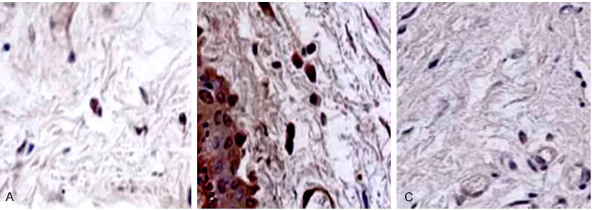
VEGF was expressed in the cytoplasm, and the nucleus was not stained. VEGF was expressed in the conjunctival epithelial cells in both con-trol group and blank concon-trol group; and in experiment group, VEGF was post-operatively expressed in conjunctival epithelial cells, fibro -blast cells and vascular endothelial cells, with the highest expression (showing dark brown) at postoperative 1 week. As time went on, the
number of positive staining cells reduced grad-ually, as shown in Figure 1.
TGF-β2 weakly expressed in the fibroblasts and conjunctival epithelial cells of the control group and blank group, located in cytoplasm. In experiment group, TGF-β2 showed the highest expression in positive staining cells at postop-erative 1 week, which representing bulky nu-clear and various shape. As time went on, the number of TGF-β2 positive staining cells re- duced gradually, and the nucleus was elong-ated, and the shapes tend to be unified, as shown in Figure 2.
The expressions of VEGF and TGF-β2 in filtering bleb determined by ELISA
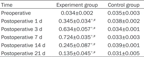
[image:3.612.90.525.70.224.2]After the trabeculectomy, the content of VEGF and TGF-β2 in the eyes of experiment group began to increase on the postoperative day 1 Figure 1. Immunohistochemical staining of VEGF in filtration tissues after operation. A: Postoperative 3 d; B: Post -operative 7 d; C: Post-operative 14 d.
[image:3.612.92.523.273.426.2]and reached the peak at postoperative day 7, then decreased gradually. The content change of VEGF and TGF-β2 showed in the peak pat-tern. Compared with the control group, the con-tent of VEGF and TGF-β2 in the experiment group was significantly higher, and the di-fference was statistically significant (P<0.05). Compared with preoperative value, the con- tent of VEGF and TGF-β2 was various at differ-ent time points, the differences between each time point were statistically significant (P< 0.05), See Tables 1, 2.
Correlation analysis of content of VEGF and TGF-β2
Pearson Correlation analysis showed that the expressions of VEGF and TGF-β2 in filtering bleb was correlated with each other after trabe- culectomy, and the correlation coefficient was 0.775, P=0.012, indicating the relevance
be-Effect of bevacizumab on filtering bleb’s VEGF and TGF-β2
One week after surgery, the expressions of VEGF (0.212±0.020 pg/μL) and TGF-β2 (3.470± 0.39 pg/μL) in Filtering bleb of bevacizumab injection group were lower than those of saline injection group (VEGF: 0.578±0.054 pg/μL, TGF-β2: 6.890±0.41 pg/μL), and the difference between the two groups was statistically signifi -cant (P<0.05), shown in Figure 5.
Discussion
Glaucoma is one of the major causes of blind-ness in the world [6, 7]. Trabeculectomy plays an important role in the treatment of glaucoma, but the filtration bleb scarring is one of the main reasons for the failure of trabeculectomy [8, 9]. The result of the research shows that postoperative fiber scarring is a complex bio
-Table 2. The contents of TGF-β2 in filtering bleb tissues in two groups at different time points (pg/μL)
Time Experiment group Control group
Preoperative 1.343±0.120 1.365±0.030
Postoperative 1 d 4.345±0.270*,# 1.343±0.002 Postoperative 3 d 5.634±0.157*,# 1.356±0.001 Postoperative 7 d 7.524±0.352*,# 1.366±0.003 Postoperative 14 d 3.245±0.871*,# 1.376±0.001 Postoperative 21 d 3.035±0.452*,# 1.386±0.005
*P<0.05, compared with control group; #P<0.05, compared with
preoperative.
Table 3. Pearsoncorrelation analysis on the correla -tion between VEGF and TGF-β2
Variable Correlation coefficient (r) Statistic (P)
VEGF and TGF-β2 0.775 0.012
was mild hyperplasia. 2 weeks after the operation, a large number of collagen fibers were observed in the saline injection group, the collagen fibers were enlarged and in a disordered arrangement, and scars formed. But in bevacizumab injec -tion group, only a small quantity of fibro -blast cells were observed in filtering bleb, there was no large number of collagen fibers formed, and most of the filtration channels kept unobstructed, shown in
[image:4.612.91.323.96.189.2] [image:4.612.91.325.257.352.2]logical process that involves various cytokines [10, 11]. VEGF and TGF-β2 play a very impor-tant role in this process [12, 13].
During the operation, many factors may pro -mote the local chemotaxis of inflammatory cells, stimulate the migration and proliferation of fibroblasts, as well as blood vessel growth and the synthesis of the extracellular matrix; these factors include: blood vessel injury, out -flow of aqueous humor, and a large quantity of
growth factors and various inflammatory cyto -kines emerged in the surgical area, in which VEGF and TGF-β2 are the most important fac-tors [14-16]. Therefore, the role of VEGF and TGF-β2 in the formation of scarring after trab-eculectomy attracts more and more attention [17, 18]. In the present study, we used enzyme linked immunosorbent assay (ELISA) to detect the expression of VEGF and TGF-β2 in the filtra -tion bleb of rabbits that underwent trabeculec-tomy. The results of the present study showed, with the formation of filtering bleb scars, the expression of VEGF in the filtration bleb obvi -ously changed - it highly expressed in the fol -lowing 21 days after operation with a peak at postoperative 1 week. On the first day after operation, TGF-β2 increased significantly, show -ing high expression in the postoperative 3 weeks with a peak at postoperative 1 week. In the control groups, the contents of VEGF and TGF-β2 in the conjunctiva and scleral tissues were very low. Foreign experimental results showed that the most active period of the migration and proliferation of fibroblasts was 4-7 d, which is consistent with our experimental results in the peak of VEGF and TGF-β2 in filtra -tion bleb [19, 20].
Bevacizumab is a humanized VEGF monoclonal antibody, which can bind to all isomers of VEGF to inhibit VEGF function. In this study, the results showed that bevacizumab can restrain Figure 3. 20 follicles in each group were observed for
the morphology under light microscope at postopera -tive 1 week, *P<0.05, compared with saline group.
Figure 4. Comparison of the number of fibroblasts in filtering bleb in 2 groups at postoperative 7 days.
*P<0.05.
expression of VEGF, promoting the formation of scars. The possible mechanisms are as follows [21-23]: (1) triggering an immune response: after tissue trauma, VEGF can combine with endothelial cells, which release a variety of growth factors and cytokines with biological activity, inducing the migration of neutrophil and mononuclear cells to the traumatized part and promoting the formation of granulation tis-sue; (2) promoting the formation of neovessels: vascular endothelial cells could proliferate, dif-ferentiate, and migrate under the action of VEGF to form new blood vessels; (3) increasing the vascular permeability: VEGF is one of the strongest known vascular permeability agents; it can enhance the permeability of postcapillary venules, and macromolecular substance in the blood, such as plasma protein and white blood cells, can migrate to the traumatized site to form fibrin gel, which is conducive to the growth of angiogenesis and stromal cells; (4) maintain-ing vascular structure.
In summary, VEGF and TGF-β2 have synergistic effect in the process of filtration bleb scarring; they co-promote the occurrence and develop -ment of scarring. To inhibit the process of scar-ring, combined treatment should be applied in order to achieve a better effect.
Disclosure of conflict of interest
None.
Address correspondences to: Xiaoping Sun, De-
partment of Ophthalmology, Zhengzhou Central
Hospital Affiliated to Zhengzhou University, No. 195 Tongbai Road, Zhongyuan District, Zhengzhou
450000, Henan Province, P. R. China. Tel: +86-0371-55966339; E-mail: xiaopingsun2016@163. com
370371.
[4] Lama PJ and Fechtner RD. Antifibrotics and wound healing in glaucoma surgery. Surv
Ophthalmol 2003; 48: 314-346.
[5] Fan N, Wang P, Tang L and Liu X. Ocular Blood Flow and Normal Tension Glaucoma. Biomed
Res Int 2015; 2015: 308505.
[6] Choi J and Kook MS. Systemic and Ocular Hemodynamic Risk Factors in Glaucoma.
Bio-med Res Int 2015; 2015: 141905.
[7] Ah-Kee EY, Egong E, Shafi A, Lim LT and Yim
JL. A review of drug-induced acute angle closure glaucoma for non-ophthalmologists. Qatar Med J 2015; 2015: 6.
[8] Nafissi N and Foldvari M. Neuroprotective
therapies in glaucoma: II. Genetic
nanotech-nology tools. Front Neurosci 2015; 9: 355.
[9] Al Habash A, Aljasim LA, Owaidhah O and
Edward DP. A review of the efficacy of mito-mycin C in glaucoma filtration surgery. Clin
Ophthalmol 2015; 9: 1945-1951.
[10] Zetterberg M. Age-related eye disease and
gender. Maturitas 2016; 83: 19-26.
[11] Flammer J and Konieczka K. Retinal venous
pressure: the role of endothelin. EPMA J 2015; 6: 21.
[12] Khaimi MA. Canaloplasty: A Minimally Invasive and Maximally Effective Glaucoma Treatment.
J Ophthalmol 2015; 2015: 485065.
[13] Maggio F. Glaucomas. Top Companion Anim Med 2015; 30: 86-96.
[14] Beamer G, Reilly CM and Pizzirani S. Micros-copic Lesions in Canine Eyes with Primary
Glaucoma. Vet Clin North Am Small Anim Pract 2015; 45: 1213-1233, vi.
[15] Abdolrahimzadeh S, Fameli V, Mollo R,
Con-testabile MT, Perdicchi A and Recupero SM. Rare Diseases Leading to Childhood Glau-
coma: Epidemiology, Pathophysiogenesis, and Management. Biomed Res Int 2015; 2015:
781294.
[16] Cascella R, Strafella C, Germani C, Novelli G, Ricci F, Zampatti S and Giardina E. The Gene-
Congeni-tal Glaucoma. Biomed Res Int 2015; 2015:
321291.
[17] Jain S and Aref AA. Senile Dementia and Glaucoma: Evidence for a Common Link. J Ophthalmic Vis Res 2015; 10: 178-183. [18] Jordan JF, Engels BF, Dinslage S, Dietlein TS,
Ayertey HD, Roters S, Esser P, Konen W and
Krieglstein GK. A novel approach to supracho-roidal drainage for the surgical treatment of intractable glaucoma. J Glaucoma 2006; 15: 200-205.
[19] Chan JE and Netland PA. EX-PRESS Glaucoma
Filtration Device: efficacy, safety, and predict
-ability. Med Devices (Auckl) 2015; 8: 381-388.
[20] Mantravadi AV and Vadhar N. Glaucoma. Prim Care 2015; 42: 437-449.
[21] Park HY, Kim JH and Park CK. VEGF induces
TGF-beta1 expression and myofibroblast trans
-formation after glaucoma surgery. Am J Pathol
2013; 182: 2147-2154.
[22] Takai Y, Tanito M and Ohira A. Multiplex cyto
-kine analysis of aqueous humor in eyes with primary open-angle glaucoma, exfoliation glau -coma, and cataract. Invest Ophthalmol Vis Sci 2012; 53: 241-247.