Original Article
Identification of genes
associated with apoptosis-sensitive
acute lymphoblastic leukemia responsive
to ionising radiation by bioinformatics analyses
Ying Sun, Meng Huo, Chunyan Zhang, Rengui Wang, Tingguo Wen
Radiation Center, Beijing Shijitan Hospital of Capital Medical University, Beijing, China
Received May 25, 2016; Accepted August 2, 2016; Epub November 15, 2016; Published November 30, 2016
Abstract: Objective: This study was aimed to characterize radiosensitivity for apoptosis sensitive acute lympho-blastic leukemia (ALL) by identifying differential genes using bioinformatics analysis. Materials and Methods: The microarray data of GSE13280 were downloaded from the Gene Expression Omnibus database. The differentially expressed genes (DEGs) between apoptosis-sensitive B-precursor ALL tumors responsive to ionising radiation (IR)
and not responsive to IR were identified. Then, biological process (BP) and pathway enrichment analyses of DEGs
were performed, and protein-protein interaction (PPI) network was constructed. Results: Total 59 up-regulated and 48 down-regulated DEGs were selected in IR samples. Besides, 109 PPI relationships were obtained from the 107 DEGs. The up-regulated DEGs, such as BCL2-associated X protein (BAX) and Fas cell surface death receptor (FAS), were mainly enriched in the BP terms related to apoptosis and p53 signaling pathway. The down-regulated DEGs including cyclin D3 (CCND3) were mainly enriched in the BP terms related to cell proliferation. Additionally, FBJ Murine Osteosarcoma Viral Oncogene Homolog and CD40 molecule, TNF receptor superfamily member 5 (CD40) were found to be hub genes in the PPI network. Conclusions: DEGs including BAX, FAS, CCND3 and CD40 may be associated with the radiosensitivity of ALL.
Keywords: Acute lymphoblastic leukemia, differentially expressed genes, functional enrichment analysis, protein-protein interaction network
Introduction
Acute lymphoblastic leukemia (ALL) is the most common pediatric malignancy with a peak inci-dence at age 2 to 5 years, and remains the leading cause of cancer-related death in chil-dren and adolescents [1]. It is characterized by the overproduction and accumulation of cancerous, immature lymphoblasts [2]. Despi- te considerable progress in cure rate over the past 10 to 15 years, 20% of children with B-precursor ALL still experience disease pro-gression with current treatment [3].
Generally, tumor is caused by the damage of DNA which leads to uncontrolled cell prolifera-tion [4]. Study has found that in animals and humans, ALL is associated with exposure to radiation and chemical radiation which is con-sidered to cause damage to cellular DNA [5].
Presently, ionising radiation (IR) is an estab-lished DNA-damaging agents in ALL treatment [6]. Weston et al. [7] have reported that some ALL exhibits defective induction of apoptosis following IR. Kruyt [8] and Gong et al. [9] have reported that in B-precursor ALL cell lines, IR can activate the tumor necrosis factor-related apoptosis-inducing ligand signaling pathway, which has been found to cooperate synergis- tically with the cytotoxic effect of radiation. De- spite progresses achieved in exploring the pa- thophysiology of IR-related ALL, there remains a lack of understanding of the molecular basis behind this disease.
apoptosis-resistant response in the remaining 36% of leukaemias. Importantly, they deposited all mi- croarray data of these patients in Gene Ex- pression Omnibus (GEO, http://www.ncbi.nlm. nih.gov/geo/) under accession number GSE- 13280. Data above suggest that most of ALL are apoptosis-sensitive. Therefore, we down-loaded the gene expression microarray data GSE13280 associated with apoptosis-sensi-tive response to explored the roles of IR in apo- ptosis-sensitive ALL. In the present study, we identified the differentially expressed genes (DEGs) between apoptosis-sensitive B-precur- sor ALL tumors responsive to IR and not res- ponsive to IR. Then functional enrichment an- alysis and protein-protein interaction (PPI) net-work construction for the DEGs were perform- ed. We aimed to characterize radiosensitivity for apoptosis-sensitive ALL by identifying dif- ferential genes.
Materials and methods
Affymetrix microarray data
The exon array data of GSE13280 [10] were downloaded from GEO database. Twenty-two B-precursor ALL tumors including 11 apopto-sis-sensitive responsive to 5 Gy IR (cobalt Co60) for 8 h and 11 apoptosis-sensitive not
responsive to IR were analyzed based on the platform of [HG-U133A] Affymetrix Human Genome U133A Array (Affymetrix Inc., Santa Clara, California, USA).
Data preprocessing and differential expression analysis
The original array data were preprocessed with background correction, quartile data normal-ization and probe summarnormal-ization by robust multi array average (RMA) [11], then they were converted into expression measures by the algorithm in R affy [12] package. The paired t-test based on the limma package [13] in R was used to identify the DEGs between the two groups of samples. The genes with and adjusted p-value < 0.05 and |log2FC| > 1 were regarded as DEGs.
PPI relationship prediction
The Search Tool for the Retrieval of Interact- ing Genes (STRING) database (http://string-db. org/) [14] is a precomputed global resource
which has been designed to evaluate the PPI information. In this paper, the STRING online tool was applied to predict the PPI relation- ships of DEGs and only those experimentally validated interactions with a combined score > 0.4 was selected as significant.
Functional enrichment analysis
The Database for Annotation, Visualization and Integrated Discovery (DAVID) [15] is a compre-hensive set of functional annotation tool and has been developed for relating the functional terms with gene lists by clustering algorithm. In this study, the DEGs involved in the PPI re- lationships were performed functional enrich-ment analysis, including Gene ontology (GO) biological process (BP) terms and Kyoto Ency- clopedia of Genes and Genomes (KEGG) [16] pathways analyses, using the DAVID online tool. PPI network construction
Based on the obtained PPI relationships, the PPI network was constructed using the Cyto- scape software [17] which was used for visual-ization and analysis of biological networks. From the previous obtained biological network we found that most of the PPI networks obey- ed the scale-free attribution. Thus, the connec-tivity degree was analyzed by statistics to ob- tain the important nodes (hub proteins) [18] in the PPI network.
Results
Identification of DEGs
For the dataset of GSE13280, 107 DEGs in samples responsive to IR were selected, in- cluding 59 up-regulated DEGs and 48 down-regulated DEGs.
PPI relationship prediction and functional en-richment analysis
cyclin D3 (CCND3), CD79a molecule, immuno-globulin-associated alpha (CD79A) and CD79B were mainly enriched in the BP terms related to cell proliferation and immune (Table 2). PPI network construction
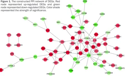
Based on the STRING database, the PPI net-work was constructed (Figure 1). Fourteen nodes were selected as hub genes (degree ≥ 5), such as FBJ murine osteosarcoma viral oncogene homolog (FOS, degree = 17), CD40 molecule, TNF receptor superfamily member 5 (CD40, degree = 12), and growth arrest and DNA-damage-inducible, alpha (degree = 10). Discussion
In the present study, a total of 59 up-regulated and 48 down-regulated DEGs were identified
between IR and control samples through gene expression profile of GSE13280. The up-regu -lated DEGs, such as BAX and FAS, were mainly enriched in the BP terms related to apoptosis and p53 signaling pathway. The down-regu- lated DEGs were mainly enriched in the BP terms related to cell proliferation and immune response. Additionally, FOS and CD40 were found to be hub gene in the PPI network. The results suggested that IR might affect the pro-gression of ALL by regulating these genes or pathway.
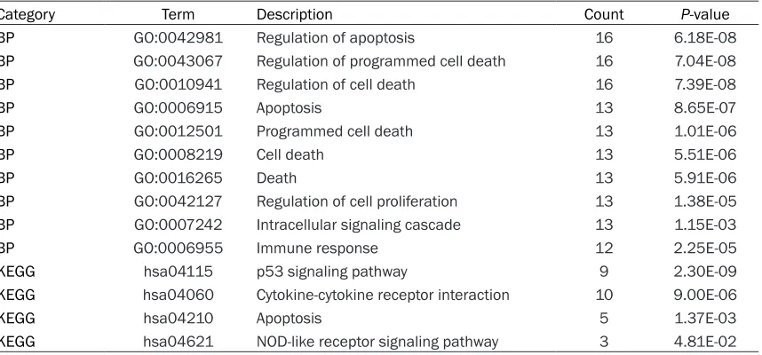
[image:3.612.95.524.84.284.2]Apoptosis is an active process that can be induced through signal transduction by DNA-damaging agents including IR. The regulation of apoptosis is delicately balanced by signaling pathways between apoptosis-promoting fac-tors such as p53 [19]. In the present study, BP terms related to apoptosis and p53 signaling Table 1. Functional enrichment analysis for the up-regulated differentially expressed genes (DEGs)
Category Term Description Count P-value
BP GO:0042981 Regulation of apoptosis 16 6.18E-08
BP GO:0043067 Regulation of programmed cell death 16 7.04E-08
BP GO:0010941 Regulation of cell death 16 7.39E-08
BP GO:0006915 Apoptosis 13 8.65E-07
BP GO:0012501 Programmed cell death 13 1.01E-06
BP GO:0008219 Cell death 13 5.51E-06
BP GO:0016265 Death 13 5.91E-06
BP GO:0042127 Regulation of cell proliferation 13 1.38E-05
BP GO:0007242 Intracellular signaling cascade 13 1.15E-03
BP GO:0006955 Immune response 12 2.25E-05
KEGG hsa04115 p53 signaling pathway 9 2.30E-09
KEGG hsa04060 Cytokine-cytokine receptor interaction 10 9.00E-06
KEGG hsa04210 Apoptosis 5 1.37E-03
KEGG hsa04621 NOD-like receptor signaling pathway 3 4.81E-02
BP: Biological process; KEGG: Kyoto Encyclopedia of Genes and Genomes.
Table 2. Functional enrichment analysis for the down-regulated differentially expressed genes (DEGs)
Category Term Description Count P-value
BP GO:0008283 Cell proliferation 7 1.25E-03
BP GO:0006955 Immune response 6 4.25E-02
BP GO:0007610 Behavior 5 4.16E-02
BP GO:0002684 Positive regulation of immune system process 4 2.89E-02
BP GO:0022409 Positive regulation of cell-cell adhesion 2 3.05E-02
BP GO:0045730 Respiratory burst 2 3.59E-02
BP GO:0051412 Response to corticosterone stimulus 2 4.40E-02
KEGG hsa04662 B cell receptor signaling pathway 4 1.50E-03
[image:3.612.91.524.329.451.2]pathway were found enriched by some up-regu-lated DEGs, such as BAX and FAS. BAX encod-ing protein belongs to the BCL2 protein family which regulates and contributes to programmed cell death or apoptosis [20]. Bax has the ability to form heterodimers with the other BCL2 pro-tein family members and acts as a positive regulator of apoptosis [21]. Additionally, BAX has been found to be expressed in a variety of acute myelogenous leukemia and ALL cell line [21]. For the other DEGs FAS, its encoding pro-tein is a member of the TNF-receptor superfam-ily which has been shown to play an important role in the physiological regulation of apoptosis, and has been implicated in the pathogenesis of various malignancies [22]. Lenardo et al. [23] revealed that the apoptosis of B- and T- lymphocyte was initiated by the binding of the FAS ligand to FAS. Taken together, IR may pro-mote apoptosis through up-regulating the DEGs such as BAX and FAS.
In addition, the down-regulated DEGs were mainly enriched in BP terms related to cell proliferation. In other words, the up-regulated DEGs in samples not responsive to IR were mainly associated with functions of cell prolif-eration, such asCCND3. CCND3 belongs to the highly conserved cyclin family, which is a key regulator of the progression from G1- to S-phase
of the cell cycle [24, 25]. Doglioni et al. [26] have suggested that CCND3 expression is as- sociated with cell proliferation in lymphoid tis-sues. Filipits et al. [27] also reported that high CCND3 expression was an prognostic factor associated with poor clinical outcome in pa- tients with diffuse B-cell lymphoma. In our study, CCND3 was up-regulated in the ALL tu- mors B-precursor not responsive to IR, which was in accordance with the findings above. Th-erefore, the down-regulation of CCND3 in the IR samples may be due to the influence of IR.
[image:4.612.101.519.79.334.2]Furthermore, the down-regulated DEGs were mainly enriched in GO terms associated with immune response as well. Study has suggest- ed that host genetic variation within immune response genes may contribute to risk of child-hood ALL [28]. For instance, in our study, CD79A and CD79Bwere found down-regulated in term of immune response. The two genes play mul-tiple and diverse roles in B cell development and function [29]. Lai et al. [30] indicated that CD79A was a reliable marker for ALL of B cell lineage. Additionally, CD79B methylation has been found in leukemic cells and has been suggested to be a critical determinant of lin-eage specification in ALL [31]. Taken together, immune response may play important roles in the development of ALL when tumor cells of Figure 1. The constructed PPI network of DEGs. Red
node represented up-regulated DEGs and green node represented down-regulated DEGs. Color shade
ALL are not responsive to IR. In the PPI net-work, CD40 was a up-regulated hub gene with a higher degree. CD40 belongs to the tumor necrosis factor receptor superfamily and is expressed on a wide variety of cell types [32]. This receptor has been found to be essential in regulating a broad variety of immune and in- flammatory responses [33]. Additionally, CD40 has been found to have a widespread expres-sion on tumor cells, including lymphomas, my- eloma and some carcinomas [34]. Importantly, some evidence suggests that CD40-CD154 interactions may play a role in the control of B cell haematopoiesis. In the present study, CD40 was up-regulated in the IR group, which might indicate that CD40 was a gene respon- se to IR in ALL.
In conclusion, our data provide a comprehen-sive bioinformatics analysis of DEGs and func-tions which may be responsive to IR. DEGs including BAX, FAS, CCND3 and CD40 in ALL may be involved in the radiosensitivity of ALL. However, further genetic studies with larger sample size are still needed to confirm our observation.
Disclosure of conflict of interest
None.
Address correspondence to: Rengui Wang and Tingguo Wen, Radiation Center, Beijing Shijitan Hos- pital of Capital Medical University, Beijing 100038, China. E-mail: [email protected] (RGW); [email protected] (TGW)
References
[1] Gaynon PS. Childhood acute lymphoblastic leukaemia and relapse. Br J haematol 2005; 131: 579-587.
[2] Inaba H, Greaves M and Mullighan CG. Acute lymphoblastic leukaemia. Lancet 2013; 381: 1943-1955.
[3] Marston E, Weston V, Jesson J, Maina E, McConville C, Agathanggelou A, Skowronska A, Mapp K, Sameith K and Powell JE. Strati-
fication of pediatric ALL by in vitro cellular re -sponses to DNA double-strand breaks pro-vides insight into the molecular mechanisms underlying clinical response. Blood 2009; 113: 117-126.
[4] Sperka T, Wang J and Rudolph KL. DNA dam-age checkpoints in stem cells, dam-ageing and can-cer. Nat Rev Mol Cell Biol 2012; 13: 579-590.
[5] Ward J, Limoli C, Calabro-Jones P and Evans J. Radiation vs chemical damage to DNA. Anti- carcinogenesis and radiation protection. Spr- inger; 1988. pp. 321-327.
[6] Preston DL, Kusumi S, Tomonaga M, Izumi S, Ron E, Kuramoto A, Kamada N, Dohy H, Matsui T and Nonaka H. Cancer incidence in atomic bomb survivors. Part III: Leukemia, lymphoma and multiple myeloma, 1950-1987. Radiat Res 1994; 137 Suppl: S68-97.
[7] Weston VJ, Austen B, Wei W, Marston E, Alvi
A, Lawson S, Darbyshire PJ, Griffiths M, Hill
F and Mann JR. Apoptotic resistance to ioniz-ing radiation in pediatric B-precursor acute lymphoblastic leukemia frequently involves
increased NF-κB survival pathway signaling.
Blood 2004; 104: 1465-1473.
[8] Kruyt FA. TRAIL and cancer therapy. Cancer Lett 2008; 263: 14-25.
[9] Gong B and Almasan A. Apo2 ligand/TNF-related apoptosis-inducing ligand and death receptor 5 mediate the apoptotic signaling in-duced by ionizing radiation in leukemic cells. Cancer Res 2000; 60: 5754-5760.
[10] Marston E, Weston V, Jesson J, Maina E, McConville C, Agathanggelou A, Skowronska A, Mapp K, Sameith K, Powell JE, Lawson S, Kearns P, Falciani F, Taylor M and Stankovic
T. Stratification of pediatric ALL by in vitro cel -lular responses to DNA double-strand bre- aks provides insight into the molecular me- chanisms underlying clinical response. Blood 2009; 113: 117-126.
[11] Irizarry RA, Hobbs B, Collin F, Beazer-Barclay YD, Antonellis KJ, Scherf U and Speed TP. Exploration, normalization, and summaries of high density oligonucleotide array probe level data. Biostatistics 2003; 4: 249-264.
[12] Gautier L, Cope L, Bolstad BM and Irizarry RA. affy-analysis of Affymetrix GeneChip data at the probe level. Bioinformatics 2004; 20: 307-315.
[13] Ritchie ME, Phipson B, Wu D, Hu Y, Law CW, Shi W and Smyth GK. limma powers differen-tial expression analyses for RNA-sequencing and microarray studies. Nucleic Acids Res 2015; 43: e47.
[14] Franceschini A, Szklarczyk D, Frankild S, Kuhn M, Simonovic M, Roth A, Lin J, Minguez P, Bork P and von Mering C. STRING v9. 1: protein-protein interaction networks, with increased coverage and integration. Nucleic Acids Res 2013; 41: D808-D815.
[15] Alvord G, Roayaei J, Stephens R, Baseler MW, Lane HC and Lempicki RA. The DAVID Gene
[16] Kanehisa M and Goto S. KEGG: kyoto encyclo-pedia of genes and genomes. Nucleic Acids Res 2000; 28: 27-30.
[17] Smoot ME, Ono K, Ruscheinski J, Wang PL and Ideker T. Cytoscape 2.8: new features for data integration and network visualization. Bioinformatics 2011; 27: 431-432.
[18] He X and Zhang J. Why do hubs tend to be essential in protein networks? PLoS Genet 2006; 2: e88.
[19] Thompson CB. Apoptosis in the pathogenesis and treatment of disease. Science 1995; 267: 1456-1462.
[20] Findley HW, Gu L, Yeager AM and Zhou M. Expression and regulation of Bcl-2, Bcl-xl, and Bax correlate with p53 status and sensitivity to apoptosis in childhood acute lymphoblastic leukemia. Blood 1997; 89: 2986-2993. [21] Kaufmann SH, Karp JE, Svingen PA, Krajewski
S, Burke PJ, Gore SD and Reed JC. Elevated expression of the apoptotic regulator Mcl-1 at the time of leukemic relapse. Blood 1998; 91: 991-1000.
[22] Wang M, Wu D, Tan M, Gong W, Xue H, Shen H and Zhang Z. FAS and FAS ligand poly- morphisms in the promoter regions and risk of gastric cancer in Southern China. Biochem Genet 2009; 47: 559-568.
[23] Lenardo M, Chan KM, Hornung F, McFarland H, Siegel R, Wang J and Zheng L. Mature T lymphocyte apoptosis-immune regulation in a dynamic and unpredictable antigenic environ-ment 1. Annu Rev Immunol 1999; 17: 221-253.
[24] Sherr CJ and Roberts JM. Living with or without cyclins and cyclin-dependent kinases. Genes Dev 2004; 18: 2699-2711.
[25] Bartkova J, Lukas J, Strauss M and Bartek J. Cyclin D3: requirement for G1/S transition and high abundance in quiescent tissues sug-gest a dual role in proliferation and differentia-tion. Oncogene 1998; 17: 1027-1037.
[26] Doglioni C, Chiarelli C, Macrí E, Dei Tos AP, Meggiolaro E, Dalla Palma P and Barbareschi M. Cyclin D3 expression in normal, reactive and neoplastic tissues. J Pathol 1998; 185: 159-166.
[27] Filipits M, Jaeger U, Pohl G, Stranzl T, Simoni- tsch I, Kaider A, Skrabs C and Pirker R. Cyclin D3 is a predictive and prognostic factor in dif-fuse large B-cell lymphoma. Clin Cancer Res 2002; 8: 729-733.
[28] Urayama KY, Chokkalingam AP, Metayer C, Ma X, Selvin S, Barcellos LF, Wiemels JL, Wiencke JK, Taylor M and Brennan P. HLA-DP genetic variation, proxies for early life immune modula-tion and childhood acute lymphoblastic leuke-mia risk. Blood 2012; 120: 3039-47.
[29] Herzog S, Reth M and Jumaa H. Regulation of B-cell proliferation and differentiation by pre- B-cell receptor signalling. Nat Rev Immunol 2009; 9: 195-205.
[30] Lai R, Juco J, Lee SF, Nahirniak S and Etches WS. Flow cytometric detection of CD79a ex-pression in T-cell acute lymphoblastic leuke-mias. Am J Clin Pathol 2000; 113: 823-830. [31] Figueroa ME, Chen SC, Andersson AK, Phillips
LA, Li Y, Sotzen J, Kundu M, Downing JR, Me- lnick A and Mullighan CG. Integrated genetic and epigenetic analysis of childhood acute lymphoblastic leukemia. J Clin Invest 2013; 123: 3099.
[32] van Kooten C and Banchereau J. CD40-CD40 ligand. J Leukoc Biol 2000; 67: 2-17.
[33] Grewal IS and Flavell RA. CD40 and CD154 in cell-mediated immunity. Annu Rev Immunol 1998; 16: 111-135.
[34] Pype S, Declercq W, Ibrahimi A, Michiels C, Van Rietschoten JG, Dewulf N, de Boer M, Vandenabeele P, Huylebroeck D and Remacle JE. TTRAP, a novel protein that associates with CD40, tumor necrosis factor (TNF) recep-tor-75 and TNF receptor-associated factors
(TRAFs), and that inhibits nuclear factor-κB