warwick.ac.uk/lib-publications
Original citation:
Fu, Ying, Romero, Maria, Salassa, Luca, Cheng, Xi, Habtemariam, Abraha, Clarkson, Guy J.,
Prokes, Ivan, Rodger, Alison, Costantini, Giovanni and Sadler, P. J.. (2016) Os2-Os4 switch
controls DNA knotting and anticancer activity. Angewandte Chemie International Edition .
Permanent WRAP URL:
http://wrap.warwick.ac.uk/80284
Copyright and reuse:
The Warwick Research Archive Portal (WRAP) makes this work of researchers of the
University of Warwick available open access under the following conditions.
This article is made available under the Creative Commons Attribution 4.0 International
license (CC BY 4.0) and may be reused according to the conditions of the license. For more
details see:
http://creativecommons.org/licenses/by/4.0/
A note on versions:
The version presented in WRAP is the published version, or, version of record, and may be
cited as it appears here.
German Edition: DOI: 10.1002/ange.201602995
Anticancer Complexes
International Edition: DOI: 10.1002/anie.201602995Os
2–Os
4Switch Controls DNA Knotting and Anticancer Activity
Ying Fu
+, Mara J. Romero
+, Luca Salassa, Xi Cheng, Abraha Habtemariam, Guy J. Clarkson,
Ivan Prokes, Alison Rodger, Giovanni Costantini, and Peter J. Sadler*
Abstract: Dinuclear trihydroxido-bridged osmium–arene complexes are inert and biologically inactive, but we show here that linking dihydroxido-bridged OsII–arene fragments by
a bridging di-imine to form a metallacycle framework results in strong antiproliferative activity towards cancer cells and distinctive knotting of DNA. The shortened spacer length reduces biological activity and stability in solution towards decomposition to biologically inactive dimers. Significant differences in behavior toward plasmid DNA condensation are correlated with biological activity.
P
latinum drugs are used in over 50 % of all chemother-apeutic regimens.[1]The basis for their activity is believed tobe mainly due to DNA binding, in particular to changes in DNA conformation.[2] Resistance to Pt drugs is a clinical
drawback that might be overcome by designing new drugs that induce distinctly different conformational changes in DNA.[3]Multinuclear metal complexes provide a promising
strategy for such an approach.[4]
Herein, we consider the design of multinuclear “piano-stool” organo-osmium complexes. Half-sandwich OsII arene
complexes exhibit antitumor activity both in vitro and in vivo.[5]O,O-chelated OsIIcomplexes, such as [(h6
-arene)Os-(acac)Cl], undergo rapid hydrolysis to produce not only the aqua adduct, [(h6-arene)Os(acac)(OH
2)]
+
, but also the hydroxido-bridged dimer, [(h6-arene)Os(m2-OH)
3Os(h 6
-arene)]+
, which is inactive against cancer cells.[6]Early work
by Fujita et al., Chi et al., and Therrien et al. demonstrated that macrocyclic polynuclear Pt and Ru complexes can have anticancer activity comparable to cisplatin, possibly through targeting DNA.[7]However, little is known about the aqueous
stability of polynuclear metallacycles and its effect on biological activity.
In this work, we link inert biologically inactive dinuclear hydroxido-bridged OsII arene units to form active
tetra-nuclear complexes that can induce DNA knotting. We show that the length of the linker is critical for maintaining stability of the tetranuclear assembly in solution, inducing DNA binding and enhancing antiproliferative activity towards human cancer cells. We compare 4,4’-azopyridine (pap) as a linker with the shorter pyrazine (prz).
Direct addition of either the pap or prz linkers to the hydroxido intermediate afforded tetranuclear OsII products
[Os4(h6-p-cym)4(m2-OH)4(pap)2][PF6]4 (1·[PF6]4) and [Os4(h6
-p-cym)4(m2-OH)4(prz)2][PF6]4 (2·[PF6]4). Recrystallization
from CH2Cl2/CH3OH solutions gave single crystals of
1·[PF6]4·2 CH2Cl2·CH3OH and 2·[PF6]4·6 CH3OH·2 H2O,
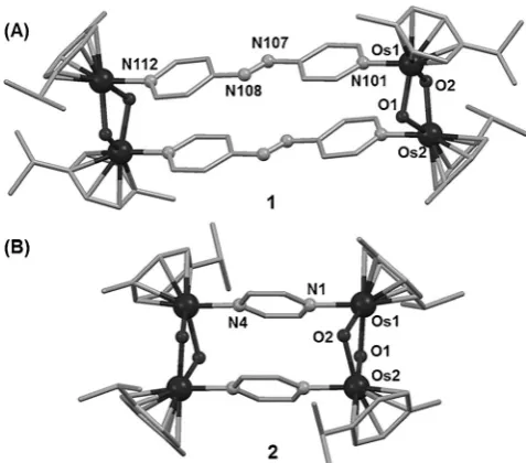
respectively. The X-ray crystal structures of 1 and 2 are shown in Figures 1 and S1 (Supporting Information). Crystal data and selected bond lengths and angles are listed in Tables S1, S2.
[image:2.595.303.542.505.715.2]Very few X-ray structures of hydroxido-bridged osmium-(II) arene complexes have been reported since the early study
Figure 1. X-ray crystal structures of1(A) and2(B). [*] Dr. Y. Fu,[+]Dr. M. J. Romero,[+]Dr. L. Salassa, Dr. X. Cheng,
Dr. A. Habtemariam, Dr. G. J. Clarkson, Dr. I. Prokes, Prof. A. Rodger, Prof. G. Costantini, Prof. P. J. Sadler
Department of Chemistry, University of Warwick Coventry, CV4 7AL (UK)
E-mail: [email protected] Dr. Y. Fu[+]
Present address: Laboratory of Molecular Biology Center for Cancer Research, NCI
Building 37, Room 5011, Bethesda, MD 20892-4264 (USA) Dr. M. J. Romero[+]
Present address: Departamento de Qumica Inorgnica Facultade de Qumica
Universidade de Santiago de Compostela 15782 Santiago de Compostela (Spain) Dr. L. Salassa
Present address: CIC biomaGUNE
Paseo de Miramn 182, 20009 Donostia-San Sebastin (Spain) [+] These authors contributed equally to this work.
Data generated during this research are available from the University of Warwick Research Archive Portal at http://wrap.warwick.ac.uk. Supporting information and the ORCID identification number(s) for the author(s) of this article can be found under
http://dx.doi.org/10.1002/anie.201602995.
of oxo/hydroxido OsIIbenzene complexes.[8]The structures of
1·[PF6]4and2·[PF6]4(Figure 1) appear to be the first examples
of organometallic OsII complexes containing arene, m2
-hydroxido, and aromatic N-donor ligands simultaneously. A detailed description of the structures is in Figure S1. The distance between the Os atoms bridged by pap is 13.175 (1), and almost half (6.995 ) with the prz bridge (2). DFT-optimized geometries are in good agreement with these structures (Tables S3–S5). UV/Vis absorption spectra were recorded for1and2in acetone (Figure S2). Time-dependent Density Functional Theory (TDDFT) singlet excited state calculations for both complexes showed that the absorption band at 435–450 nm has 1MLCT (metal-to-ligand
charge-transfer) character and is composed of two major transitions involving Os-based occupied orbitals and ligand-based LUMO and LUMO+1 (Figure S2). Notably, MLCT transi-tions at wavelengths higher than 600 nm are found for1, in agreement with the experimental spectrum. The intensity of such TDDFT transitions is overestimated (particularly the singlet electronic transition S3), as expected for highly delocalized systems displaying charge-transfer bands.[9]
The stability of the tetramer1in solution was investigated first in [D6]acetone. The1H NMR spectra of1·[PF6]4at 298 K
showed two singlets at 6.68 and 6.66 ppm assignable as OH peaks (Figure S3A). This appears to be the first detection of peaks for bridging-OH groups in organometallic tetranuclear complexes.[10]To confirm this assignment,1H NMR at various
temperatures and 2D1H DOSY NMR spectra were recorded.
The two OH singlets shifted downfield reversibly at lower temperature (Figure S3 and S4) with a linear temperature dependence (Dd/DT= 0.009 ppm K 1). Interestingly, two
peaks assignable to H2O and HOD were also observed in
these [D6]acetone solutions (Figure S5).[11] When H2O
(10mL) was added, the H2O and OH bridge peaks shifted
downfield (Figure S6). The two low-field OH peaks disap-peared upon addition of D2O (Figure S7). The
diffusion-ordered spectroscopy (DOSY) 2D 1H NMR spectrum of
1·[PF6]4supported assignment of the two singlets (6.68 and
6.66 ppm) to OH bridges (Figure S8). These studies suggested that both OH bridges are involved in H-bond interactions, which are strongly dependent on temperature and water concentration.
The aqueous stabilities of1·[PF6]4 and 2·[PF6]4 in 10 %
MeOD-d4/90 % D2O phosphate buffer (1 mm, pH*=7.4)
were investigated by 1H NMR spectroscopy. After 24 h at
310 K, 63 % of cationic 1 was still present as the intact tetramer, whereas2decomposed completely under the same conditions and the solution became colorless (Figure S9). 2D
1H DOSY experiments on2·[PF
6]4(Figure 2) confirmed the
release of the aromatic linker with a concomitant breakdown of the tetranuclear assembly and generation of an hydroxido-bridged OsII–arene dimer [Os
2(h6-p-cym)2(m2-OH)3]
+
for both
1and2. The formation of this biologically inert hydroxido dimer[6]was confirmed by ESI-MS (Figure S9).
The antiproliferative activity of1 and2towards cancer cells was determined. The IC50 value for A2780 human
ovarian cancer cells decreased dramatically from>100mmto
10mmas the linker length increased from pyrazine in complex 2·[PF6]4to 4,4’-azopyridine in complex1·[PF6]4(Table 1). An
analogous trend in antiproliferative activity was observed for A549 and H596 human non-small-cell lung cancer (NSCLC) cell lines (Table 1). In these two cell lines, 1·[PF6]4showed
similar anticancer potency to cisplatin and was approximately 2-times more potent than 2·[PF6]4. No antiproliferative
activity was observed for the free linker ligands. The highest antiproliferative activity is therefore associated with the cationic complex that more readily retains its tetranuclear structure in solution (complex1). The anticancer activity of
2·[PF6]4 is comparable to some reported dinuclear RuII
cylinders.[12]
Metallosupramolecules are also known to bind in the grooves of DNA.[13]Therefore, binding of complexes1and2
to calf thymus DNA (ct-DNA) was also investigated (Fig-ures S11 and S13). The characteristic circular dichroism (CD) bands of ct-DNA (Figure S11) and the negative linear dichroism (LD) band arising from the DNA bases (Fig-ure S13) decreased in intensity with increasing concentrations of 1 or 2, even at low Os loading. Red fibers, likely corresponding to DNA-tetramer adducts, were observed at high Os loading (Figure S12). These data suggest that both OsII tetramers bind to ct-DNA and induce DNA
[image:3.595.304.545.54.300.2]condensa-tion, facilitated by their 4+charge, but more significantly for tetramer1.
Figure 2. 2D DOSY1
H NMR study of the stability of complex2·[PF6]4
in buffered aqueous solution (see the Supporting Information); t=time point at which spectra were recorded.
Table 1: Cytotoxicity (IC50,mm) of1·[PF6]4and2·[PF6]4towards A549
(lung), H596 (lung), and A2780 (ovarian) human cancer cell lines.
Complex A549 H596 A2780
CDDP 6.7(0.6) 5.9(1.4) 1.8(0.1)
1 5.2(0.3) 4.8(2.0) 10.1(0.1)
2 12.9(2.2) 10.8(1.3) >100
2
www.angewandte.org 2016 The Authors. Published by Wiley-VCH Verlag GmbH & Co. KGaA, Weinheim Angew. Chem. Int. Ed.2016,55, 1 – 5 [image:3.595.304.543.382.432.2]Further insight into OsIItetramer–DNA interactions was
obtained using tapping mode atomic force microscopy (AFM) (Figures 3 and S14). AFM images were recorded at two different time points, 30 min after preparing the samples and 24 h after incubation at 310 K. Figure 3 a, b shows images of the control sample where the free pBR322 plasmid was deposited onto freshly cleaved mica treated with Mg2+ to
ensure firm adsorption of the negatively charged DNA. Mainly relaxed open circular (OC) and linear (L) forms were observed on the surface after the free plasmid relaxation procedure (Supporting Information). Analysis of cross sec-tions of the different forms resulted in comparable heights of 0.700.08 nm (OC) and 0.700.10 nm (L; Figure 3 a,b). These values are smaller than the theoretical DNA diameter (ca. 2 nm), probably owing to electrostatic interactions with the positively charged mica surface that tend to compress the plasmids.[14]
Figures 3 c, d and S14 show the effect of interactions between plasmid DNA and complexes 1 or 2 at a DNA base:Os4ratio of 5:1. No plasmids were detected on any of the
analyzed samples after mixing with the more biologically active tetramer1·[PF6]4(Figure S14a, b). This was interpreted
as an indication that the DNA forms an adduct with cationic tetramer1, thus becoming less negatively charged and less strongly bound to the positively charged mica substrate.
Interaction with the less biologically active tetramer
2·[PF6]4gave rise to the formation of knotted aggregates of
plasmid DNA, appearing as plasmid loops emerging from a condensed nucleus, probably generated by the linking of several DNA strands mediated by2(Figure 3 c). The DNA adducts with complex2were strongly attached to the Mg2+
-treated surface. This suggests a larger negative charge density of DNA-2with respect to DNA-1adducts, probably owing to a reduced number of bound molecules of2with respect to1. A cross-sectional height analysis confirmed this conclusion (Figure S14, detailed discussion in the Supporting Informa-tion). This is the first report on the formation of this type of DNA aggregate with organo-osmium anticancer complexes. The unusual behavior of1and2differs considerably from the DNA cross-linking caused by RuII–arene m-Hoxonato
tet-ramers[15] or the DNA full-coiling induced by
metallocylin-ders.[12]
Experiments carried out at a lower DNA:2 mol ratio (2.5:1) showed the formation of similar knotted aggregates (Figure S14f), characterized by a higher surface density and a slightly larger cross-sectional height (Supporting Informa-tion), confirming a higher linear density of 2bound to the DNA strands. Conversely, a lower density of bridging tetramers2was observed on incubating 5:1 DNA:2samples for 24 h at 310 K (Figures 3 d and S14e, detailed discussion in the Supporting Information).1H NMR experiments
demon-strated that incubation caused cleavage of the tetranuclear structure of these OsIIcomplexes into dimers (Figures 2 and
S9), and can thus be expected to partially release plasmids from the central core in the DNA-2adducts, resulting in the larger loops observed in Figure 3 d. Both AFM experiments were consistent with the dichroism measurements, indicating that the changes in the CD and LD signals observed upon increasing concentrations of2(Figures S11 and S13, respec-tively) or incubating the 5:1 DNA:2samples (Figure S13) are probably due to a different extent of DNA condensation.
In conclusion, our studies reveal how the choice of spacer length in tetranuclear organo-osmium metallacycles can control their stability in solution and their biological activity, which correlates with their ability to induce plasmid DNA condensation. The more cytotoxic tetramer 1 reduces the DNA negative charge significantly more than tetramer 2, although the latter also exerts a considerable effect on DNA structure. The different modes of interaction of1and2with DNA seem likely to contribute to the large difference in cytotoxicity exhibited by these candidate metallodrugs and may play a role in their anticancer mechanism of action.
Acknowledgements
[image:4.595.52.292.393.667.2]We thank the ERC (grant no. 247450 BIOINCMED), EPSRC (grant no. EP/F034210/1), BBSRC (grant no. BB/F011199/1), and Science City/EU ERDF/AWM for funding. G. C. acknowledges financial support through the ERC Grant “VISUAL-MS”. M.J.R. thanks Fundacin Barri fellowship. L.S. thanks the MC CIG fellowship UCnanomat4iPACT (grant no. 321791) and the MINECO grants (CTQ2012-39315 and RYC-2011-07787). We thank colleagues in the EC COST Action CM1105 for stimulating discussions, and Ben Moreton for his help with AFM.
Figure 3. AFM images of pBR322 plasmid DNA (a, b) in the absence, and (c, d) the presence of2·[PF6]4in a DNA base:tetramer ratio of 5:1.
Keywords: cancer · DNA · organometallic · osmium · supramolecular
[1] a) R. H. Fish, G. Jaouen,Organometallics2003,22, 2166 – 2177; b) T. W. Hambley,Dalton Trans.2007, 4929 – 4937; c) J. Reedijk,
Eur. J. Inorg. Chem.2009, 1303 – 1312; d) G. Gasser, I. Ott, N. Metzler-Nolte,J. Med. Chem.2011,54, 3 – 25; e) C. G. Hartinger, P. J. Dyson,Chem. Soc. Rev.2009,38, 391 – 401; f) Y. W. Jung, S. J. Lippard,Chem. Rev. 2007,107, 1387 – 1407; g) C. Orvig, M. J. Abrams,Chem. Rev.1999,99, 2201 – 2204.
[2] P. M. Takahara, A. C. Rosenzweig, C. A. Frederick, S. J. Lip-pard,Nature1995,377, 649 – 652.
[3] a) E. Hillard, A. Vessieres, L. Thouin, G. Jaouen, C. Amatore,
Angew. Chem. Int. Ed.2006,45, 285 – 290;Angew. Chem.2006,
118, 291 – 296; b) Q. L. Zhang, J. H. Liu, J. Z. Liu, P. X. Zhang, X. Z. Ren, Y. Liu, Y. Huang, L. N. Ji,J. Inorg. Biochem.2004,98, 1405 – 1412; c) O. R. Allen, L. Croll, A. L. Gott, R. J. Knox, P. C. McGowan,Organometallics2004,23, 288 – 292; d) H. M. Chen, J. A. Parkinson, S. Parsons, R. A. Coxall, R. O. Gould, P. J. Sadler,J. Am. Chem. Soc.2002,124, 3064 – 3082.
[4] a) E. Almaraz, Q. A. de Paula, Q. Liu, J. H. Reibenspies, M. Y. Darensbourg, N. P. Farrell,J. Am. Chem. Soc.2008,130, 6272 – 6280; b) Z. Li, A. David, B. A. Albani, J. P. Pellois, C. Turro, K. R. Dunbar, J. Am. Chem. Soc. 2014, 136, 17058 – 17070; c) S. N. Georgiades, N. H. Abd Karim, K. Suntharalingam, R. Vilar, Angew. Chem. Int. Ed. 2010, 49, 4020 – 4034; Angew. Chem.2010,122, 4114 – 4128; d) M. J. Hannon,Chem. Soc. Rev.
2007,36, 280 – 295; e) G. Li, R. Guan, L. Ji, H. Chao,Coord. Chem. Rev. 2014, 281, 100 – 113; f) J. Wang, J. Newman, S. Higgins, K. M. Brewer, B. S. J. Winkel, K. J. Brewer, Angew. Chem. Int. Ed.2013,52, 1262 – 1265;Angew. Chem.2013,125, 1300 – 1303; g) D. A. Lutterman, P. K.-L. Fu, C. Turro,J. Am. Chem. Soc.2006,128, 738 – 739.
[5] a) V. B. Arion, A. Dobrov, S. Goschl, M. A. Jakupec, B. K. Keppler, P. Rapta,Chem. Commun.2012,48, 8559 – 8561; b) A. Bergamo, A. Masi, A. F. Peacock, A. Habtemariam, P. J. Sadler,
G. Sava,J. Inorg. Biochem.2010,104, 79 – 86; c) H. Kostrhunova, J. Florian, O. Novakova, A. F. A. Peacock, P. J. Sadler, V. Brabec,
J. Med. Chem.2008,51, 3635 – 3643.
[6] A. F. A. Peacock, M. Melchart, R. J. Deeth, A. Habtemariam, S. Parsons, P. J. Sadler,Chem. Eur. J.2007,13, 2601 – 2613. [7] a) M. Fujita, J. Yazaki, K. Ogura,J. Am. Chem. Soc.1990,112,
5645 – 5647; b) V. Vajpayee, Y. J. Yang, S. C. Kang, H. Kim, I. S. Kim, M. Wang, P. J. Stang, K. W. Chi,Chem. Commun.2011,47, 5184 – 5186; c) T. R. Cook, V. Vajpayee, M. H. Lee, P. J. Stang, K. W. Chi,Acc. Chem. Res.2013,46, 2464 – 2474; d) N. P. Barry, F. Edafe, P. J. Dyson, B. Therrien,Dalton Trans.2010,39, 2816 – 2820.
[8] R. O. Gould, C. L. Jones, T. A. Stephenson, D. A. Tocher, J. Organomet. Chem.1984,264, 365 – 378.
[9] A. Dreuw, M. Head-Gordon,J. Am. Chem. Soc.2004,126, 4007 – 4016.
[10] D. Carmona, A. Mendoza, J. Ferrer, F. J. Lahoz, L. A. Oro,J. Organomet. Chem.1992,431, 87 – 102.
[11] H. E. Gottlieb, V. Kotlyar, A. Nudelman,J. Org. Chem.1997,62, 7512 – 7515.
[12] a) G. I. Pascu, A. C. Hotze, C. Snchez-Cano, B. M. Kariuki, M. J. Hannon, Angew. Chem. Int. Ed. 2007, 46, 4374 – 4378;
Angew. Chem. 2007, 119, 4452 – 4456; b) I. Meistermann, V. Moreno, M. J. Prieto, E. Moldrheim, E. Sletten, S. Khalid, P. M. Rodger, J. C. Peberdy, C. J. Isaac, A. Rodger, M. J. Hannon,
Proc. Natl. Acad. Sci. USA2002,99, 5069 – 5074.
[13] a) A. Oleksi, A. G. Blanco, R. Boer, I. Uson, J. Aymami, A. Rodger, M. J. Hannon, M. Coll,Angew. Chem. Int. Ed.2006,45, 1227 – 1231;Angew. Chem.2006,118, 1249 – 1253; b) A. M. Pyle, J. P. Rehmann, R. Meshoyrer, C. V. Kumar, N. J. Turro, J. K. Barton,J. Am. Chem. Soc.1989,111, 3051 – 3058.
[14] F. Moreno-Herrero, J. Colchero, A. M. Bar,Ultramicroscopy
2003,96, 167 – 174.
[15] F. Linares, M. A. Galindo, S. Galli, M. A. Romero, J. A. Navarro, E. Barea,Inorg. Chem.2009,48, 7413 – 7420.
Received: March 26, 2016 Revised: May 1, 2016
Published online:&& &&,&&&&
4
www.angewandte.org 2016 The Authors. Published by Wiley-VCH Verlag GmbH & Co. KGaA, Weinheim Angew. Chem. Int. Ed.2016,55, 1 – 5Communications
Anticancer Complexes
Y. Fu, M. J. Romero, L. Salassa, X. Cheng, A. Habtemariam, G. J. Clarkson, I. Prokes, A. Rodger, G. Costantini,
P. J. Sadler* &&&&—&&&&
Os2–Os4Switch Controls DNA Knotting
and Anticancer Activity
![Table 1: Cytotoxicity (IC50, mm) of 1·[PF6]4 and 2·[PF6]4 towards A549(lung), H596 (lung), and A2780 (ovarian) human cancer cell lines.](https://thumb-us.123doks.com/thumbv2/123dok_us/9470590.453451/3.595.304.545.54.300/table-cytotoxicity-lung-ovarian-human-cancer-cell-lines.webp)
![Figure 3. AFM images of pBR322 plasmid DNA (a,b) in the absence,plasmid DNA. Conditions: 2 mand (c,d) the presence of 2·[PF6]4 in a DNA base:tetramer ratio of 5:1.Images were recorded 30 min after preparing the samples (a,c) and24 h after incubation at 310](https://thumb-us.123doks.com/thumbv2/123dok_us/9470590.453451/4.595.52.292.393.667/figure-conditions-presence-tetramer-recorded-preparing-samples-incubation.webp)
