warwick.ac.uk/lib-publications
A Thesis Submitted for the Degree of PhD at the University of Warwick
Permanent WRAP URL:
http://wrap.warwick.ac.uk/101423
Copyright and reuse:
This thesis is made available online and is protected by original copyright. Please scroll down to view the document itself.
Please refer to the repository record for this item for information to help you to cite it. Our policy information is available from the repository home page.
Chromatin Remodelling During
Plant-Pathogen Interactions
by
Emmanouil Mastorakis
A thesis submitted in partial fulfilment of the
requirements for the degree of
Doctor of Philosophy in Life Sciences
2013-2017
School of Life Sciences, University of Warwick
Supervisor: Dr. Vardis Ntoukakis
of
Dedication
I dedicate this work to my family who have provided me with
unparalleled support for as long as I can remember
of
Table of Contents
List of Figures 6
List of Tables 8
Author’s declaration 9
Acknowledgements 10
Abbreviations 11
Summary 12
Chapter 1 - Introduction 13
1.1. Biotechnology promises to tackle global food security problems 13 1.2. The Arabidopsis thaliana - Pseudomonas syringae pathosystem 17 1.3. Current understanding of the plant immune system 18 1.4. PTI outputs contribute towards a robust defence response 22
1.5. Effector-triggered immunity 26
1.6. Phytohormone signalling in plant immunity 27 1.7. Massive transcriptional changes occur during infection 29 1.8. Chromatin remodelling in regulation of gene expression 31 1.9. Histone acetylation and GCN5 in other systems 40 1.10. Chromatin remodelling in plant defence 43
1.11. Histone acetylation in plant immunity 44
1.12. HAG1 is an important co-activator of transcription 49
1.13. SAGA complex in plants 50
1.14. HAG1 is involved in abiotic and biotic stresses in plants 51 1.15. Chromatin remodelling mechanisms targeted by pathogens 54
1.16. Context of this work 56
Chapter 2 - Materials & Methods 59
2.1. Material and Growth conditions 59
2.2. Molecular Biology 61
2.3. Biochemistry 64
2.4. Physiological assays 70
Chapter 3 - HAG1 is required for plant immunity 76
3.1. Context of this chapter 76
3.2. Main findings 81
3.3. Results 82
3.4. Discussion 95
Chapter 4 - HAG1 complex in plant immunity 103
4.1. Context of this Chapter 103
4.2. Main findings 106
4.3. Results 106
4.4. Discussion 121
Chapter 5 - Identifying bacterial effectors with roles in chromatin
remodel-ling 134
5.1. Context of this chapter 134
5.2. Main findings 137
5.3. Results 137
5.4. Discussion 161
Chapter 6 - General Discussion 171
6.1. HAG1 is responsible for the flg22-induced histone acetylation 171 6.2. HAG1 complex has core and PTI/ETI-specific interactors 172 6.3. HopO1-1 is involved in various molecular processes 174 Chapter 7 - Conclusions & Looking Forward 177 7.1. Epigenetics and chromatin remodelling across organisms 177 7.2. Benefiting from research in other model organisms 177 7.3. From model plant organisms to commercially important crops 178
Appendix - Supplementary Information 181
Bibliography 192
of
List of Figures
Figure 1.1. The plant immune system
Figure 1.2. Post-translational histone modifications in plants
Figure 1.3. Yeast SAGA complex has multiple modules with different functions Figure 1.4. Acetylation/De-acetylation reaction of Lysine/Acetyl-Lysine
Figure 1.5. Sequence alignment of HAT domains in different model organisms
Figure 1.6. Proposed model of the function of the interaction of TPL with HATs/HDACs in the context of auxin-mediated transcription
Figure 1.7. Developmental phenotypes of AtGCN5, AtADA2a and AtADA2b mutants Figure 3.1. Principles of Fluorescence Recovery After Photobleaching (FRAP) Figure 3.2. Perception of flg22 leads to opening of the chromatin
Figure 3.3. Global histone acetylation levels increase after flg22 perception Figure 3.4. Arabidopsis thaliana HAT mutants
Figure 3.5. Screen of HAT mutants for altered susceptibility to Pst DC3000 Figure 3.6.HAG1 gene and HAG1 protein structures
Figure 3.7. Morphological phenotypes of HAG1 gene T-DNA insertion mutants Figure 3.8. Bacterial growth in various HAG1 mutants including known interactors of HAG1
Figure 3.9. Histone H3 acetylation levels are reduced in hag1-5 and hag1-6 mutants Figure 3.10. Impaired flg22-induced histone acetylation in hag1-6 mutant
Figure 3.11. Reactive oxygen species (ROS) production in hag1 mutants is not signifi-cantly impaired
Figure 3.12. MAPK activation is not affected in hag1-6 mutant
Figure 3.13. Morphological phenotypes of hag1-6 mutants crossed to sid2-1, NahG and
hda19 plants
Figure 3.14. HAG1 is not involved in SA responses
Figure 4.1. Transiently-expressed HAG1 interacts with histone H3 Figure 4.2. Protoplast-expressed HAG1 localises to the nucleus Figure 4.3. Stably-expressed HAG1 is successfully purified
Figure 4.4. Stably-expressed HAG1 is purified and detected successfully by mass spec-trometry using in-gel digestion protocol
Figure 4.5. Experimental design for the identification of HAG1 complex Figure 4.6. Volcano plot of HAG1 interactors
Figure 4.7. Zoomed-in volcano plot of HAG1 interactors
Figure 4.9. TOPLESS and TPR1 expression Figure 4.10. TOPLESS immunoprecipation Figure 4.11. Coverage of TPL protein sequence
Figure 4.12. Human GCN5, ADA2 and ADA2B can be modified by multiple types of PTMs
Figure 4.13. ADA2 proteins sequence coverage and post-translational modifications Figure 4.14. Domains identified in A. thaliana Chromatin Remodeller 5
Figure 5.1.Pst DC3000 effectors with putative ADP-ribosyltransferase domain Figure 5.2. Identification of nuclear localised effectors by nuclear isolation and im-munoblotting
Figure 5.3. Nuclear localised effectors transiently expressed in N. benthamiana Figure 5.4. Nuclear localised effectors in Arabidopsis protoplasts
Figure 5.5. Fluorescence recoveries for effectors expressed in N. benthamiana Figure 5.6. Chromatin binding properties of Pst DC3000 nuclear effectors in N. ben-thamiana
Figure 5.7. A comprehensive screening methodology towards identifying chromatin-associated effectors
Figure 5.8. Chromatin mobility is reduced in the presence of HopO1-1
Figure 5.9. HopO1-1 is purified successfully after DEX induction, but does not interact with H3
Figure 5.10. HopO1-1 is detected by mass spectrometry in A. thaliana
Figure 5.11. HopO1-1 expression and purification from N. benthamiana prior to mass spectrometry
Figure 5.12. HopO1-1 is detected by mass spectrometry in N. benthamiana Figure 5.13. Deletion of HopO1-1 does not reduce Pst DC3000 virulence
Figure 5.14. Photosynthetic activity is the same after infection with Pst DC3000 and Pst
DC3000 hopO1-1
Figure 5.15. StKRBP1 specifically relocalises Pi04089 to nuclear speckles Figure 6.1. HAG1 is responsible for flg22-induced histone acetylation
Figure 6.2. Confirmed components of HAG1 complex in plants compared to yeast SAGA
Figure 6.3. HAG1 acts in a multi-protein complex, which consists of core interactors and potentially PTI/ETI-specific interactors
Figure 6.4. HopO1-1 is involved in various molecular processes
of
List of Tables
Table 1.1. Effect of histone marks on gene expression
Table 1.2. Major HAT complexes identified in different model organisms
Table 1.3. List of histone acetyltransferases (HATs) and histone deacetylases (HDACs) in Arabidopsis
Table 1.4. Important protein domains in chromatin-associated proteins Table 2.1. Plant genotypes used in this project
Table 2.2. Antibodies used in this project
Table 4.1. Identified peptides and post-translational modifications on HAG1 Table 4.2: Major interactors of HAG1
Table 4.3. Major interactors of TPL
Author’s declaration
I hereby certify that this thesis is presented in accordance with the regula-tions for the degree of Doctor of Philosophy. It has been composed by myself and has not been submitted in any previous application for any degree. The work in this thesis has been undertaken by myself except where otherwise stated.
Dr. Sophie Piquerez from the Ntoukakis group (University of Warwick, UK) has been closely involved in many of the experiments presented here, as an advisor and collaborator.
Dr. Selena Gimenez-Ibanez (University of Warwick, Coventry, UK & Na-tional Centre of Biotechnology CSIC, Madrid, Spain) cloned all of the
Pseudomonas syringae effectors presented in Chapter 5 and performed the effector screen using the FRAP method under my guidance and as-sistance. Additional data from Dr. Gimenez-Ibanez have been used in this chapter as indicated. The project presented in Chapter 5 also benefited from work conducted by undergraduate students such as Charles Green-spon, Oscar Guerra and James Coleman.
Emmanouil Mastorakis
PhD candidate Student ID: 0920695 University of Warwick Coventry
United Kingdom
of
Acknowledgements
A four year-long journey could not be complete without the help of sever-al fellow travellers.
To my awesome friends in the lab Bea, Chrysa, Alonso, Arsheed, Steph, Ruth, Jack and Ranjith I owe massive gratitude for being amazing friends and colleagues. I learned many things from each and every one of them.
To Vardis I would like to say a big ‘Thank you’ for all the support and for giving me the chance as his first PhD student to work on extremely inter-esting, challenging and ambitious projects.
Abbreviations
Term Definition
35S Cauliflower Mosaic Virus (strong promoter)
AcCoA Acetyl Coenzyme A
bp Base pair
Br/Bromo Bromodomain
CBB Coomassie Brilliant Blue
Chr/Chromo Chromodomain
cDNA Complementary DNA
dpi days post infection
EMS Ethyl Methane Sulfonate
ETI Effector-Triggered Immunity
FA Formaldehyde
FRAP Fluorescence Recovery After Photobleaching
g Gram
g Centrifugal force unit
HAT Histone Acetyltransferase
HDAC Histone Deacetylase
kb Kilobase
kDa kilo Daltons
PAMPs Pathogen Associated Molecular Patterns
PCR Polymerase Chain Reaction
PEG Polyethylene Glycol
PRR Pattern Recognition Receptor
PTI PAMP-Triggered Immunity
ROS Reactive Oxygen Species
rpm rounds per minute
RPM Revolutions per minute
SA Salicylic Acid
SAGA Spt-Ada-Gcn5 acetyltransferase
SWI/SNF SWItching-defective/Sucrose Non-Fermenting
T-DNA Transfer DNA
of
Summary
Chapter 1 - Introduction
1.1. Biotechnology promises to tackle global food security problems
Food and agriculture globally are estimated to be a $5 trillion in-dustry experiencing continuous growth (Denis, 2015). The socioeconom-ic and environmental impact of agribusiness represents 10% of global consumer spending, 20% of employment as well as 30% of greenhouse-gas emissions. By current trends, with an exponentially growing world population expected to reach 10 billion people by 2050, caloric demands for both human and animal feed will double. In order to meet this grow-ing demand, food production must continue to increase, despite a sizeable improvement in productivity improvement observed over the past 50 years (Fuglie et al., 2012). However, agriculture today faces important challenges, which render this objective a difficult task. The increased agri-cultural activity suggests that 40% of water demand in 2030 is unlikely to be met, whilst the continuous loss of arable land and changing climates are some of the most highlighted and well-studied problems. In addition, approximately 26% of the worldwide crop production each year is lost due to pests and pathogens even before harvest. With rapidly advancing global trades, changing climates and agricultural intensification, the spread of plant diseases is expected to increase further. Different geogra-phies may face some of these problems to different levels, nevertheless, the above considerations represent global issues (Bebber et al., 2013). Al-together, this means that increases in food production will largely rely not only on increasing current agricultural efforts, but also on the develop-ment of existing technologies to improve output from the same amount of arable land. The four staples, which feed more than half the population includes wheat (Triticum aestivum), maize (Zea mays), banana (Musa acumi-nata) and rice (Oryza sativa). Importantly, the challenges facing these
of
jor crops includes emerging infectious diseases throughout both develop-ing and developed countries (Fisher et al., 2012).
Crop plants are exposed to a wide-range of pests and pathogens, such as bacteria, fungi, oomycetes, viruses, nematodes, and insects, but only in certain interactions does this culminate in disease. For example, wheat faces a threat by the virulent Ug99 strain of wheat stem rust fun-gus (Puccinia grimness g. sp. tritici) (Singh et al., 2015), while Panama dis-ease (fungus Fusarium oxysporum) is an important issue in bananas (Churchill, 2011). Potato and tomato are challenged by late blight disease caused by the oomycete Phytopthora infestans, which isknown for the 1.25 million deaths during the Irish potato famine in 1845, but also for being a modern-day problem for farmers (Haverkort et al.,2008; Haas et al., 2009).
In the absence of genetic resistance in crops, food production heav-ily relies on chemical control of pathogens, a critical element in pest man-agement. Although pesticides are a testament of the improved productivi-ty in the past few decades allowing the quadrupling of food grain produc-tion from the 1950s to 2000s, there are many reported hazards of these chemicals, which are driving the requirement for more sustainable ap-proaches. Initially, lime sulfur was introduced in the 1800s and fungicides interfering with the metabolic processes of fungi were introduced in the 1900s. Since then, use of fungicides has seen a continuous rise in all mar-kets of the world, and has further increased during pathogen outbreaks in different parts of the world. Rice blast, for example, the most economical-ly important disease in Japan faces recurrent outbreaks once to twice every decade leading to the introduction of multiple types of fungicides for the protection of blast-susceptible rice varieties (Hirooka et al., 2013). This highlights the inevitable vicious cycle of chemical control and pathogen resistance in modern-day agriculture.
extensively showing the detrimental effects of pesticides on human health and environment. In fact, the World Health Organization (WHO) has re-ported that there is no population segment that is completely protected against exposure to pesticides and the associated health effects, although there are high-risk countries shouldering a disproportionate level of this burden (WHO, 1990).
Briefly, certain chemicals have been found to mimic or antagonise natural hormones in humans and have been linked to immune suppres-sion, reproductive abnormalities and cancer (Brouwer et al., 1999; Crisp et al., 1998). Meanwhile, such pesticides have also been found to contam-inate ground and surface water through runoff from treated plants and soil, while their effect on heavily treated soils can cause beneficial soil mi-croorganisms to decline. Pesticide sprays can also directly affect non-tar-get venon-tar-getation and non-tarnon-tar-get organisms damaging wildlife overall (Aktar et al., 2009).
Furthermore, limitations in conventional breeding approaches for a large group of crops is an additional factor driving the necessity for genet-ic engineering of crops. For example, banana, whgenet-ich is the staple food in more than 50 countries exists as a sterile triploid selected from the wild making its breeding a challenging process and is, therefore, propagated vegetatively. This lack of genetic diversity makes banana trees highly sus-ceptible to changing biotic and abiotic stresses (Cronauer-Mitra and Krikorian, 1987).
All the above highlight the need for a more sustainable approach, which would allow for durable endogenous resistance to reduce depen-dence on chemical-based approaches. There is already a significant amount of evidence to support the potential of biotechnological ap-proaches in meeting this challenge. In general, biotechnology is capable of overcoming production constraints that are more difficult or intractable
of
by conventional breeding. Thus, it can provide farmers with disease-free planting materials by creating inherently resistant crops.
provitamin A (Paul et al., 2017).
There is also a plethora of well-characterised examples of gene transfer into crops translating research in model organisms to crops. For example, the EF-Tu receptor (EFR), which is restricted to the Brassicaceae
genus, was shown to confer broad spectrum bacterial resistance in the
Solanaceae genus (Lacombe et al., 2010), whilst ongoing projects involve the same strategy applied in potato, lettuce, apple and citrus (2Blades-Foundation, 2004). Narusaka et al (2013) also demonstrated the activity of nucleotide binding and leucine rich repeat (NB-LRRs) proteins in tax-onomically distinct families by transferring Brassicaceae-specific RPS4 and
RRS1 to tomato (Solanum lycopersicum) and tobacco (Nicotiana benthamiana). These examples show that the downstream elements of R genes are highly conserved and this supports the current strategies of re-sistance gene transfer between different crop species. Lastly, the applica-tion of such strategies has relied for years on the extensive study of plant immunity by employing model patho-systems such as the one consisting of the model plant Arabidopsis thaliana and the Gram-negative bacterium
Pseudomonas syringae.
1.2. The Arabidopsis thaliana - Pseudomonas syringae pathosystem
Arabidopsis thaliana has proved to be an indispensable tool in char-acterising biological processes in plants, empowered by a huge library of well-described Arabidopsis mutants generated by T-DNA insertion or EMS mutagenesis. This system lends itself to both forward and reverse genetics, which have led to the identification of important proteins in-volved in defence such as FLS2 (FLAGELLIN SENSING2) (Gomez-Gomez and Boller, 2000) and BAK1 (BRASSINOSTEROID INSENSITIVE1-AS-SOCIATED KINASE1) (Chinchilla et al., 2007). Arabidopsis can be in-fected by multiple pathogens, which has allowed the study of plant-pathogen interactions including plant-bacterium (e.g. Pseudonomas
of
ringae) and plant-fungus (e.g. Magnaporthe oryzae) and plant-oomycete (e.g. Phytophthora infestans) interactions.
P. syringae, specifically, can infect different plant species thus fur-ther strain classification into pathovars (pv.) is based on host specificity. As an example, P. syringae DC3000 pv. tomato was first identified from tomatoes in California and is known to infect this plant causing bacterial speck disease and resulting in great yield and financial losses (Scofield et al., 1996). Interestingly, P. syringae has been found to have a dual lifestyle whereby the pathogen initially grows at the leaf surface (epiphytically), gradually entering the leaf tissue through stomata or wounds. This is fol-lowed by aggressive multiplication at which stage host cell death is ob-served, also known as necrosis. The pathogen is thus described as a hemibiotroph as it avoids killing the host until the later stages of infec-tion, which is in contrast to necrotrophic pathogens known to kill the host for the purposes of obtaining nutrients (Xin and He, 2013). Addi-tionally, Pst DC3000 pv. tomato is able to establish infection in Arabidop-sis, which resulted in its popularisation as a model species across multi-ple research groups allowing researchers to expand our knowledge of plant-bacteria interactions (Koornneef and Meinke, 2010).
Research on Pst DC3000 has revealed that its virulence relies on the production of toxins, phytohormones and protein effectors encoded in the Pst DC3000 genome, which are delivered through a Type III secretion systems (Lindeberg et al., 2008). Overall, this pathosystem has aided in the discovery of defence mechanisms on the host side as well as virulence mechanisms on the pathogen side, both of which form the basis of the evolutionary arms-race between host and pathogen.
1.3. Current understanding of the plant immune system
entirely on an innate system and the ability of individual cells to mount immune responses upon pathogen detection by cell surface or intracellu-lar immune receptors (Jacob et al., 2013).In this way, the plant immune system has evolved to protect the host against a variety of pathogens. However, the diverse strategies employed by pathogens make it a bigger challenge for plants to successfully respond to infection. Our current un-derstanding of a plant-pathogen interaction and co-evolution is sum-marised in the zig zag model, proposed by Dangl and Jones, which de-scribes the quantitative output of the plant immune system throughout the course of the infection. This model is heavily based on the Arabidopsis thaliana-P.syringae interaction. Specifically, in Phase 1, the presence of a pathogen is rapidly recognised by the plant. Recognition takes place through pattern recognition receptors (PRRs) on the surface of the plant plasma membrane. The ligands of PRRs are highly conserved pathogen structures known as pathogen-associated molecular patterns (PAMPs) and some of the most famous studied examples include bacterial flagellin or its synthetic derivative, the 22-amino acid epitope, flg22 (Felix et al., 1999b); elongation factor Tu (EF-Tu) or its synthetic derivative, the 18-amino acid epitope, elf18 (Kunze et al., 2004). Notably, different recep-tors are responsible for the recognition of different PAMPs. Experiments involving immunological techniques and binding assays have shown that a 175kDa transmembrane leucine rich receptor (LRR) kinase known as FLS2 is essential for the recognition of flg22 in Arabidopsis thaliana and tomato (Felix et al., 1999a). Similarly, EF-Tu is recognised by the LRR-ki-nase called EFR leading to converged downstream PTI responses (Kunze et al., 2004). The formation of immune receptor complexes between pro-teins and PRRs is necessary for normal perception of PAMPs and signal transduction. In vivo phospho-labelling experiments showed the de novo
phosphorylation of both FLS2 and the co-receptor BAK1 within 15 sec-onds of flg22 elicitation, suggesting that extracellular receptors are poised
of
to detect the presence of PAMPs and can do so in a rapid and robust manner (Schulze et al., 2010). Pattern recognition receptors (PRRs) are receptor kinases (RKs), which also contain a cytosolic kinase domain and a variable ectodomain with leucine-rich repeats (LRRs) as in the case of FLS2 and EFR. Others such as CERK1 (CHITIN ELICITOR RECEPTOR KINASE 1) contain LysM motifs or other ligand-binding domains. Down-stream RKs are proteins known as plant receptor-like cytoplasmic kinases (RLCKs), which have been recognised as important regulators of immuni-ty (Lin et al. 2013). For example, the RLCK BIK1 (BOTRYTIS-INDUCED KINASE 1) is a common downstream interactor for RKs including FLS2, EFR, CERK1 and BAK1. Following phosphorylation and activation, BAK1 subsequently phosphorylates its two known substrates RBOHD and CDPKs (Lin et al., 2014). Hyperphosphorylation is observed upon PAMP perception, which is likely to further activate BIK1, facilitating the onset of PAMP-triggered responses such as production of reactive oxygen species (ROS) and phosphorylation of RBOHD (Kadota et al., 2014). Ad-ditional PTI (PAMP-triggered immunity) responses include but are not limited to the strengthening of the cell wall by callose deposition (Luna et al., 2011), the production of reactive oxygen species (ROS), Ca2+
influx-es, MAPK (mitogen-activated protein kinases) and CDPK (cyclin-depen-dent protein kinases) cascade activation leading to further downstream gene activation (Boller and Felix, 2009; Boudsocq et al., 2010; Ma and Berkowitz, 2007; Tena et al., 2011).
In phase 2, effector-triggered susceptibility (ETS) ensues when pathogens deliver into the host cell and the apoplast multiple effector proteins. Effectors interfere with PTI processes and can thus enhance the pathogen’s virulence and colonisation (Jones and Dangl 2006).
NB-LRRs (Nulceotide-Binding-Leucine Rich Repeat) and resemble Nod-Like Receptors (NLRs) found in mammals (Matzinger, 2007). Upon intracellu-lar detection of pathogen effectors, ETI ensues, leading to increased dis-ease resistance and involving a hypersensitive cell death response (HR) at the site of infection. This aims to effectively prevent the pathogen from spreading to other tissues (Jones and Dangl 2006).
Figure 1.1. The plant immune system. PAMPs such as flagella or its epitope, flg22, are recognised by plasma membrane-associated receptors such as FLS2. Dimerisation with BAK1 is known to occur immediately after PAMP perception. Downstream events in-clude interaction and trans-phosphorylation with BIK1. The latter together with CDPKs further phosphorylates RBOHD protein to facilitate the release of ROS at the extracellu-lar space. Meanwhile, calcium influx takes place leading to the activation of CDPKs, which mediates nuclear processes such as gene activation. Genes such as NHL10 and
PHI1 are known to be partially or completely dependent upon CDPKs, respectively. At the same time, MAPKs are activated leading to gene activation. FRK1 is an example of MAPK-dependent gene. It is unknown whether MAPKs and CDPKs are able to directly phosphorylate histones, however, in the case of MAPKs, direct targets such as histone deacetylases (HDACs) or transcription factors have been reported. Different enzymes are involved in chromatin remodelling such as histone acetyltransferases (HATs), HDACs, histone methyltransferases (HMTs) and chromatin remodelling complexes (CRCs). These responses are part of PAMP-triggered immunity, which can be targeted by effectors delivered into the cell via a Type III Secretion System (T3SS) found in bacterial pathogens such as Pseudomonas syringae DC3000 pv. tomato. Suppression of PTI results in effector-triggered susceptibility (ETS). The figure does not include nucleotide-binding leucine-rich repeat (NB-LRR) proteins, which are involved in the recognition of effectors leading to effector-triggered immunity (ETI). Adapted from multiple sources referenced in the main text.
In phase 4 of the proposed zig-zag model, natural selection on both sides of this evolutionary arms race drives the formation of diversified
ef-nucleus Pseudomonas s
yringae
BAK1
BIK1 MAPKs
Defence-gene expression
Ca2+
O2 O2
.-
CDPKs
ROS FLS2
P Flagella,
flg22
P P
P
CaCa2+ 2+ Ca2+
Ca2+
Nucleus Cytosol
Type 3 effectors
HDACs HATs
HMTs
CRCs T3SS
of
fector genes capable of avoiding or inhibiting ETI and the subsequent evo-lution of new resistance genes to trigger ETI once again. It is believed that microbial pathogens evolve faster than plants, however, defence mecha-nisms evolved by plants have been found to be effective allowing the host to keep up with the fast-evolving virulence strategies of pathogens.
1.4. PTI outputs contribute towards a robust defence response
PAMP-triggered immunity (PTI) is usually effective at stopping pathogen growth due to a rapid and robust set of responses, which includes the production of ROS, increases in intracellular Ca2+, CDPK and MAPK
sig-nalling cascades, callose deposition, closing of stomata and downstream activation of defence genes, among other mechanisms (Nicaise et al., 2009; Tena et al., 2011).
Ca2+ influx is another PTI response with multiple known
func-tions. A Ca2+ gradient exists across the plasma membrane whereby
extra-cellular Ca2+ is higher in resting plant cells. Upon elicitation of plant
cells, cytosolic Ca2+ rapidly increases and is perceived by calmodulin and
calcium dependent protein kinases resulting in their activation. In addi-tion, Ca2+ is believed to function as a potentiator of reactive oxygen
in-termediates (ROI) and nitric oxide (NO) production thus contributing to the hypersensitive response (HR) overall. In relation to this, CDPKs phosphorylate RBOHD and contribute to ROS production (Dubiella et al., 2013; Lecourieux et al., 2002; Lecourieux et al., 2006). Another impor-tant role of Ca2+ influx is to activate downstream calcium-dependent
pro-tein kinases (CDPKs). Specifically, Boudoscq et al (2010) showed that four CDPKs were critical for the transcriptional reprogramming that oc-curs in plant immune signalling. Interestingly, in the same study, using an in-gel kinase assay with histone H3 as a substrate, activation of CDPKs was observed as early as 15 minutes after flg22 elicitation. This was solely dependent upon Ca2+ ions as the phosphorylation was not seen in the
presence of a Ca2+ chelator.
Interestingly, CPK4, 5, 6 and 11 were found to re-localize to the nucleus, interacting with WRKY TFs, resulting in the phosphorylation of WRKY8, 28 and 48 during ETI induced by RPS2 or RPM1 R proteins (Gao et al., 2013).
Signalling of PAMP perception can also be relayed downstream by MAPKs. These enzymes are involved collectively in signalling cascades whereby they phosphorylate downstream kinases giving rise to a nomen-clature such as MAPKs, MAPK kinases (MAPKKs) and MAPKK kinases (MAPKKKs)(Pitzschke et al., 2009). A phosphorylation cascade that con-sists of MEKK1 (MAPKKK), MKK4 & MKK5 (MAPKK) and MPK3 & 6 (MAPK) is activated following elicitation by flg22 (Asai et al 2002). Knockout mutations in mpk3 and mpk6 mutants result in increased
of
tis cinerea (necrotrophic fungus) susceptibility (Kliebenstein et al., 2005), although MPK3 was found to be a negative regulator of inducible de-fences in the interaction with P. syringae (Frei dit Frey et al., 2014). A number of genes have been found to be induced in a MAPK-dependent manner such as FRK1 (FLG22-INDUCED RECEPTOR KINASE1) (Asai et al., 2002; Boudsocq et al., 2010). Interestingly, the activation of some de-fence genes is dependent on MAPKs as well as CDPKs, as for example,
As mentioned earlier, pathogens are able to subvert immune pro-cesses such as PTI responses of the host, leading to enhanced susceptibili-ty. Numerous examples of this have been recorded.
HopAI1 was shown to inactivate MPK3 and MPK6 by de-phospho-rylating threonines through a unique phosphothreonine lyase activity leading to the suppression of two independent downstream evens such as the re-inforcement of cell wall defences as well as PAMP-induced tran-scriptional activation of specific genes. Pst DC3000 pv. tomato lacking HopAI1 was less virulent, whilst the MAPK-dependent FRK1 gene among others was not induced as strongly in transgenic plants expressing HopAI1 (Zhang et al., 2007)
AvrPto and AvrPtoB are two very well-studied effectors capable of suppressing PTI responses by targeting the PRR complex consisting of FLS2 and BAK1 that forms after flg22 perception. CERK1 is also targeted by AvrPtoB (Gimenez-Ibanez et al., 2009).
HopM1 is an effector from Pst DC3000 that alters proteasome specificity in Arabidopsis MIN7 in a post-translational manner resulting in the destabilization of MIN7. AtMIN7 is an ADP-ribosylation factor-guanine nucleotide exchange factor (ARF-GEF) regulating vesicle traffick-ing and is believed to recycle or mobilise immunity-related plasma-mem-brane proteins (e.g. FLS2) allowing plants to perceive or respond to the pathogen. In min7 mutants lacking AtMIN7, susceptibility is increased, but also rescues the growth of Pst DC3000 lacking HopM1 (Nomura et al., 2006; Ustun et al., 2016).
Another example of effector-mediated interference of immune sig-nalling is HopAO1. Following elf18 perception by the EF-TU RECEPTOR (EFR) the receptor is phosphorylated in multiple tyrosine residues. Phos-phorylation of Y836 is necessary to induce downstream signalling in re-sponse to P. syringae infection, however, HopAO1 effector, a tyrosine phosphatase was found to reduce EFR phosphorylation (Bretz et al.,
of
2003; Espinosa et al., 2003). HopAO1 expression led to 50% reduction in the phosphorylation of EFR upon elf18 treatment. As a result, in the presence of HopAO1, dampening of immune responses such as MAPK activation and ROS production was observed and was followed by in-creased bacterial growth (Macho et al., 2014).
1.5. Effector-triggered immunity
In response to these virulence strategies, plants have developed mechanisms allowing the recognition of effectors and the establishment of a hypersensitive response (HR), which aims to effectively stop the spread of the pathogen. HR development is part of ETI and relies on NB-LRRs, which are particularly important in plant immunity, as exemplified by a significantly larger group of NB-LRRs in plants amounting to 125 in Arabidopsis (Col-0 ecotype) as opposed to approximately 20 in mammals (Jones and Dangl, 2006). In addition, the NB-LRR gene family is one of the largest within plants and contains genes that are polymorphic and evolve rapidly due to the presence of transposable elements within NB-LRR gene clusters (Baumgarten et al., 2003). NB-NB-LRRs are predicted to be cytoplasmic allowing the monitoring of plant proteins that are poten-tial targets of effector proteins (McHale et al., 2006). NB-LRR classifica-tion is based on their N-terminal structural features, whereby, TIR-NB-LRRs contain N-terminus with homology to the Toll and interleukin re-ceptors, whereas non-TIR-NB-LRRs are also known as CC-NB-LRRs due to the presence of a coiled-coil domain in their N-terminus (Dangl and Jones, 2001; Van der Biezen and Jones, 1998).
A well-known example of TIR-NB-LRR pair consists of RRS1-R and RPS4. Briefly, the bacterial effectors PopP2 (from the Gram-negative root pathogen Ralstonia solanacearum) and AvrRps4 (from P. syringae pv.
solanacearum 1). The latter is atypical in that it consists of an additional C-terminal WRKY DNA-binding domain, which is traditionally known to exist in WRKY proteins that are transcription factors binding DNA at specific sequences (Deslandes et al., 2003). In addition, Pop2, which be-longs to the YopJ family and has acetyltransferase activity is able to acety-late RRS1 in leucine 1221 thus weakening its DNA-binding affinity and leading to the activation of cell death. AvrRps4 on the other hand does not acetylate RRS1, but leads to cell death in a yet unknown mechanism (Deslandes et al., 2003; Sarris and Jones, 2015).
1.6. Phytohormone signalling in plant immunity
During plant defence, different plant hormones contribute towards effective defence against the pathogens. Classic phytohormones include abscisic acid (ABA), auxins (AUX), cytokinins (CKs), ethylene (ET) and gibberellins (GAs), but also smaller signalling molecules including brassinosteroids (BRs), jasmonates (JAs) and salicylic acid (SA) (Pieterse et al., 2012). Crosstalk exists between the different hormonal pathways. For example, jasmonates appear to play a role in conferring resistance to necrotrophic pathogens, whilst SA is more important against (hemi)biotrophs (Glazebrook, 2005). The active JA-isoleucine is per-ceived by a coreceptor complex between the F-box protein CORONA-TINE-INSENSITIVE 1 (COI1) and JAZ proteins (Sheard et al., 2010). Arabidopsis mutants lacking COI1 have increased susceptibility to necrotrophic fungi such as Alternaria brassicicola and Botrytis cinerea (Loren-zo et al., 2003; Thomma et al., 1998), while resistance to the hemibiotrioph P. syringae is higher. This is consistent with elevated SA levels in the same mutant supporting an antagonistic relationship be-tween the two pathways (Kloek et al., 2001). Furthermore, failure to ac-cumulate SA, either by means of reduced biosynthesis, such as in the case of the sid2 mutants (SALICYLIC ACID INDUCTION DEFICIENT 2), a gene
of
encoding an isochorismate synthase or by means of constitutive degrada-tion of SA in transgenic plants expressing the bacterial gene nahG, leads to increased susceptibility to hemibiotrophic pathogens such as P. syringae
(Abreu and Munne-Bosch, 2009).
Salicylic acid is a small phenolic compound that acts as a signalling molecule (Vlot et al., 2009). There is a tight interplay between growth and immunity, in which SA is known to play an important role consider-ing that accumulation of SA results in stunted growth but increased resis-tance e.g. in mpk4 mutants, whereas failure to accumulate SA results in decreased resistance and sometimes increased growth (Ishihara 2008). Therefore, SA is an underpinning molecule for the trade-off between growth and resistance to pathogens.
The isochorismate (IC) and phenylalanine ammonia-lyase (PAL) pathways use chromate to produce SA (Dempsey 2011) in species such as tobacco, tomato, pepper and others (Catinot et al., 2008; Kim and Hwang, 2014; Sadeghi et al., 2013; Uppalapati et al., 2007; Wildermuth et al., 2001). Mutants in either of these pathways show impaired induction of SA accumulation after pathogen infection (Huang 2010). Salicylic acid production takes place in the chloroplasts as isochorismate synthase (ICS) converts chorismate into isochorismate (IC) (Wildermuth et al., 2001) followed by conversion to SA (Dempsey et al., 2011). SA percep-tion is achieved through SA receptors such as the recently identified NPR (NON-EXPRESSOR of PATHOGENESIS-RELATED GENES) family members, NPR1, 3 and 4 (Fu et al., 2012; Wu et al., 2012). NPR1 is also a transcrip-tional coactivator (Pajerowska-Mukhtar et al., 2013) regulating about 95% of the genes that are responsive to the SA-analog compound known as benzothiadiazole (BTH) (Mou et al., 2003; Wang et al., 2010b).
can be achieved even with exogenous application of salicylic acid or its analogs (Durrant and Dong, 2004b). This ‘priming’ phenomenon allows faster response to subsequent challenge. For example, npr1 mutants are deficient in SAR and cannot become primed for enhanced gene expres-sion, whereas mutants such as sni1 and edr1 show constitutive priming along with the developmental cost associated with continuously up-regu-lated defence responses, such as smaller plant size (Conrath, 2011; Frye and Innes, 1998; Mosher et al., 2006).
SA is also involved in preventing bacterial entry into plant tissue via stomatal closure, which occurs soon after the pathogen has been in contact with the plant (Melotto et al., 2006). This is a process that P. sy-ringae DC3000 pv. tomato can hijack, leading to the reopening of closed stomata, which is mediated by the phytotoxin coronatine. Specifically, coronatine is used as a mimic of JA-Ile that binds COI1/JAZ leading to 26S-mediated proteasomal degradation of the JAZ transcriptional repres-sors. Repression of JA responsive genes occurs at resting state by JAZ re-pressor proteins together with the adaptors and co-rere-pressors NINJA and TOPLESS, interacting with and suppressing MYC TFs. Upon infection and release of coronatine into the host cells, expression of JA-dependent genes is no longer blocked and at the same time the SA pathway is inhib-ited. The affinity of COR for the COI1/JAZ complex is in fact greater than JA-Ile making it a very potent virulence factor. The end result is suppres-sion of SA-responses and increased bacterial colonization (Chini et al., 2007; Sheard et al., 2010; Thines et al., 2007).
1.7. Massive transcriptional changes occur during infection
One of the most notable outputs in response to infection is the rapid transcriptional reprogramming leading to differential expression of a large number of genes involved in a plethora of cellular processes. At the transcriptomic level, it has been shown that approximately 1/10 of
of
the Arabidopsis genome exhibits differential expression within an hour after perception of flg22 (Zipfel et al., 2004) and up to 1/3 of the Ara-bidopsis genome during infection with the necrotrophic fungus Botrytis cinerea within the first 48 hours (Windram et al., 2012). These massive transcriptomic changes are known to be accompanied by the activity of transcription factors and the RNA polymerase machinery as well as changes in the architecture of chromatin, which is a highly dynamic macromolecule (Lusser, 2002; Smale, 2014).
In Arabidopsis, RNA polymerase II is known to be activated by cy-clin-dependent kinase (CDK)-mediated phosphorylation at the C-termi-nal domain (CTD). Thus, phosphorylation of RNA pol II is recognised as an important mechanism for the rapid induction of defence gene expres-sion (Li et al., 2014b).
with MPK4 and upon activation by PAMPs the complex dissociates allow-ing the WRKY-dependent activation of PAD3 (phytoalexin deficient 3), nec-essary for the production of camalexin, a secondary metabolite with an-timicrobial activity.
Part of the massive transcriptional reprogramming upon PAMP perception involves suppression of nuclear encoded chloroplast-targeted genes (NECGs) and is followed by production of photosynthesis-derived ROS in the chloroplast. The transcriptional responses within the first two hours after PAMP perception were related to chloroplast functions such as ROS production, photosynthesis and synthesis of salicylic acid (de Tor-res Zabala et al., 2015; Lewis et al., 2015). In addition, photosynthetic process were shown to be important in the expression of flg22-inducible NECGs (Sano et al., 2014).
1.8. Chromatin remodelling in regulation of gene expression
Transcription is a complex process that involves multiple proteins acting on the DNA, however, the DNA macromolecule is not always read-ily accessible to all these factors mentioned above. Specifically, the genetic material of eukaryotic cells is tightly packed inside the nucleus to a mil-lionth of its length into a hierarchical structure consisting of proteins and DNA, known as chromatin. Chromatin consists of DNA organised around proteins known as histones that assemble into octameric structures called nucleosomes. This structure is important for nuclear processes involving DNA such as repair, recombination, replication as well as transcription. Chromatin is very dynamic and its structure at the macro- and micro-scale may vary significantly to accommodate the processes that are taking place at any time (Lusser et al., 2001). The histones are highly conserved throughout eukaryotic organisms and are characterised by a small size ranging from 11 to 21 kDa. Histones H2A, H2B, H3 and H4 are very small, have a globular shape and are very basic. Two copies of H2A and
of
serve a multitude of roles in nuclear processes, while a major advantage is their reversibility allowing rapid responses to changing environmental cues (Hamon and Cossart, 2008). Most notably, these modifications can strongly and rapidly affect the packaging of DNA into nucleosomes and subsequently influence all processes where DNA acts as a template.
Acetylation of histones, the addition of acetyl groups onto lysine residues on histone tails, is known to affect chromatin structure and gene expression. Acetylation is associated with a loosened interaction between histones and DNA and increased levels of transcription (Sterner and Berger, 2000). This can be explained by various mechanisms. First, at a smaller scale, the negatively charged DNA interacts strongly with the pos-itively charged histone tails, and the addition of a negatively charged acetyl group can neutralise the charge difference and weaken this interac-tion leading to a more accessible DNA. At a larger scale, the same effect of acetylation can lead to higher-order conformational changes of chro-matin. Third, acetylation can also occur in non-histone proteins. Exam-ples include the HMG (High Mobility Group) proteins, other transcrip-tional activators and even HATs and HDACs (Spange et al., 2009). In these cases, non-histone acetylation affects protein-protein interactions. Fourth, acetyl groups have been found to act as docking sites for proteins interacting with histones. For example, several HATs (Gcn5, PCAF, CBP/ p300) contain bromodomains, a conserved domain that binds acetylated lysines and can mediate the recruitment and propagation of acetylation on the same nucleosomes. Overall, it has been established that in animals and plants, through these mechanisms, acetylation almost always is asso-ciated with more ‘open’ chromatin and increased gene activation (Kouzarides, 2007). Lysines found at the core region of histones are also post-translationally modified. For example, lysine H3K56 was recently found to be acetylated. This modification is facing towards the major
of
groove of DNA, thus is in an optimal conformation to influence the his-tone-DNA interaction (Xu et al., 2005).
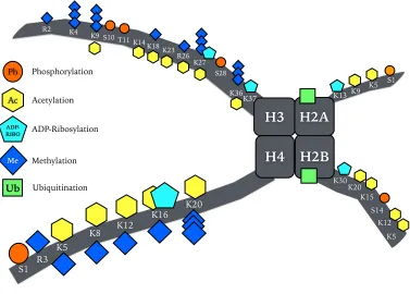
Figure 1.2. Post-translational histone modifications in plants. Histone modifications occur at the N-terminal tails and the core region of histones H2A, H2B, H3 and H4. Two copies of each create an octameric structure known as the nucleosome. For simplicity only one copy of each histone is shown. Modifications such as phosphorylation, meth-ylation (mono-, di- and tri-methmeth-ylation), ADP-ribosmeth-ylation, acetmeth-ylation and ubiquitina-tion can occur on different or on the same residues, but not simultaneously. Ubiquitina-tion is mostly identified at the C-ter part of the histones H2A(K120) and H2B(K119).
Phosphorylation is found on Tyrosines (Tyr, T) and Serines (Ser, S). These modifications have been identified in planta using methods such as mutagenesis, in vitro reactions, mass spectrometry, western blotting and others. Adapted from multiple sources (Earley et al., 2007; Kouzarides, 2007; Lusser, 2002; Messner et al., 2010)
The remarkable property of histones being post translationally modified to a larger extent than any other known protein by the addition of many different PTMs (Kouzarides, 2007) raises the question of what is the function of these in gene expression as well as how these PTMs are interpreted by the transcription machinery upon different stresses. The balance of different histone modifications is governed by multiple en-zymes that can deposit them, recognise or bind them, and remove them, which gives rise to the colloquial terms: ‘writers’, ‘readers’ and ‘erasers’
H3
H4 H2B K4
K9 S10 T11 K14 K18 K23
K27 R26
S28
K36
H2A
K5 R3
K8
S1
K12 K16
K20
K5 K12 S14 K15 K20
S1 K5 K9
R2
K13
K30 K37
Ph
Ac
ADP-RIBO
Me
Ub
Phosphorylation
Acetylation
ADP-Ribosylation
Methylation
of chromatin, respectively (Strahl and Allis, 2000). Genetic and biochemi-cal evidence has elucidated the roles of histone PTMs in chromatin-relat-ed processes. The histone code, proposchromatin-relat-ed by Strahl and Allis (2000) sup-ports that transcription is in part regulated by the modifications men-tioned before and that these are critical in recruiting other proteins with specific protein domains and a variety of functions involved in altering chromatin structure and/or altering transcription (Jenuwein and Allis, 2001). In other words, the histone code describes the complex language of how the different PTMs act in concert and may affect each other direct-ly (e.g. by steric hindrance) or indirectdirect-ly through the action of ‘writers’, ‘readers’ and ‘erasers’ of chromatin. Specifically, in vitro acetylation of H3 by GCN5 at lysine 14 (H3K14ac) is dependent upon specific consensus motifs in the same way as protein kinases, such as MAPKs are proline-di-rected Serine/Threonine (Ser/Thr) kinases obeying the S/T-P motif (Pereira et al., 2011). Crystal structures of GCN5 enzyme with its sub-strate H3 has revealed that GCN5 acetylation of H3K14 is dependent on the G-K14*-X-P motif where the acetylated lysine is preceded by a glycine and followed by a proline (Rojas et al., 2004; Trievel et al., 1999). It is also important to note that the close proximity of most of these modifica-tions could positively or negatively affect the ability of these enzymes to further modify their target residues. In support of this, it was found that H3S10 phosphorylation (H3S10ph) enhanced the acetylation of H3K14 by GCN5 (Trievel et al., 1999). Furthermore, H3K9 methylation is a known repressive mark and can affect adjacent H3S10ph negatively, thus indirectly inhibiting H3K14ac and excluding acetylation of H3K9 at the same time. Deacetylation of H3K9ac could in the same way promote methylation of the same site.
The histone code is an appealing hypothesis as it can explain the opposing roles of many modifications. For example, although acetylation is normally associated with a more relaxed chromatin conformation and
of
increased gene expression, in several cases, heterochromatic areas in flies and silent loci in yeast are rich in acetylated H4K12 (Braunstein et al., 1996; Turner et al., 1992). Similarly, H3S10ph is associated with chromo-some condensation as well as immediate early gene induction after mito-genic stimulation (Sawicka and Seiser, 2012). A possible explanation to these paradoxical examples could be that these marks are read by distinct sets of histone modifying enzymes leading to a variety of possible down-stream responses. In addition, the presence of a modification does not al-ways imply an immediate function, rather it could imply transcriptional priming. Bivalent chromatin marks are such an example, which are present at the promoters of the developmental HOX genes that specify the anteroposterior development in vertebrates. In this example, activat-ing marks (H3K4me3) are co-present with repressive marks (H3K27me3), suggesting a model of transcriptional regulation where the genes are primed for activation but held in check by a negative mark and the fate of transcription depends on upstream signals such as develop-mental or environdevelop-mental cues. Similarly, robust regulation of transcrip-tion can be ensured by the co-localisatranscrip-tion of different activating marks as in the case of acetylated forms of H3 and H3K4me3 (Bernstein et al., 2006; Tee et al., 2014).
multiple sources (Alvarez et al., 2010; Berger, 2007; Jenuwein and Allis, 2001; Kouzarides, 2007)
Chromatin largely exists in two states - euchromatin and hete-rochromatin; the first is described by a looser architecture, associated with increased transcriptional activity and the latter by a more condensed one, which does not favor transcription. Generally, highly active genes are found in euchromatin, while genes under repression are buried in hete-rochromatin, inaccessible to transcription factors and the RNA poly-merase machinery (Lusser, 2002). Many genes may be activated upon a particular signal and thus their chromatin context can change significant-ly. Modification of histones has been shown to influence the activity of a gene, for example, acetylation is important for the activation of gene ex-pression (Durrin et al., 1991; Yoshida et al., 1995). Generally, the post-translational addition of acetyl groups onto proteins can have a variety of roles. For example, (i) the affinity of a protein for DNA is reduced, (ii) the localisation of a protein may change upon acetylation, (iii) the stabili-ty of a protein may increase i.e. through prevention of ubiquitin-mediated degradation and (iv) the interaction with other proteins may change. In the case of histones, which are basic proteins and thus have high affinity for the negatively charged DNA macromolecule, (v) this affinity may be reduced upon addition of the positively charged acetyl groups on lysine residues of histones (Glozak et al., 2005). This leads to a loosening of the histone-DNA interaction and a more relaxed chromatin structure.
Eukaryotic nuclei have been found to contain transcriptional hotspots also known as transcription factories. These were found using fluorescence recovery after photobleaching (FRAP) experiments, whereby the kinetics of a GFP-tagged protein of interest are measured after photo-bleaching a fluorescent area with high-power laser at the same wave-length of fluorescence. The rate of fluorescence recovery in this area is then calculated as adjacent GFP-tagged molecules naturally diffuse into
of
that space. Proteins involved in chromatin-interacting processes, like gene transcription, DNA replication, DNA repair and histones, bind directly or indirectly to DNA leading to slow recovery (Day et al., 2012). FRAP analysis has been shown to be a strong in vivo approach to study the mo-bility and transient immobilisation of nuclear proteins associated with DNA (Stasevich and McNally, 2011). A study monitored RNAPII in living cells using FRAP showing that at least two populations of RNAPII can be observed, the majority described by a very short recovery time, whilst a transcriptionally engaged pool of RNAPII was found to have a longer re-covery time (Dundr et al., 2002). This finding pointed towards the exis-tence of distinct transcription centers (‘hotspots’) inside the nucleus, which are characterised by the enrichment of transcription factors and RNAPII to allow more efficient transcription.
As a consequence, acetylation of histones appears to allow easier loss of nucleosome ahead of the polymerase machinery and their re-incorpora-tion behind the polymerase machinery (Svejstrup, 2003).
DNA methylation is a well-established epigenetic mechanism with a key role in gene expression in response to stresses. It involves the cova-lent binding of a methyl group to the fifth carbon in a cytosine nucleotide ring of a DNA molecule and often occurs in cytosines linked to guanine via a phosphodiester bond denoted altogether as CpG. Specifically in plants it can also be found in the context of CpG, CpHG (whereby H rep-resents A,C or T) and CpHH and it is particularly abundant representing approximately 30% of the nucleotides in plants (Gruenbaum et al., 1981). In a similar way to the other mechanisms, DNA methylation can be high-ly dynamic in response to environmental changes and different meth-ylation patterns are observed throughout a plant’s development (Finnegan et al., 2000). In A. thaliana, the centromeric regions and other loci were found to have reduced levels of DNA methylation following in-fection with P. syringae (Pavet et al., 2006).
Another level of regulation of gene expression is the enrichment of genomic areas with a distinct set of histone variants. For example, Stroud
et al (2012) showed that the histone variant H3.1 is strongly enriched in areas of the genome that were more silent and were co-present with re-pressive modifications such as H3K27me3, H3K9me and DNA meth-ylation. By contrast the histone variant H3.3 is more commonly found in actively transcribed regions, correlating with modifications that promote gene expression such as H3K4me and H2Bub as well as transcriptionally engaged RNA polymerase II. Crucial enzymes in the displacement and deposition of histone variants are ATP-dependent chromatin remodellers.
The discovery of the first ATP-dependent chromatin remodellers was made in yeast mutant screens. Specifically, some of the swi (switch) and snf (sucrose non-fermenting) mutations resulted in impaired gene
of
tivation in many different pathways (Sudarsanam and Winston, 2000; Workman and Kingston, 1998). Later studies revealed that those genes encoded subunits of the same SWI/SNF complex and their mutation re-sulted in phenotypes similar to those observed in histone and other chromatin mutants. Now, SWI/SNF is known to be a major player in chromatin remodelling across species. After the biochemical purification of the complex it was revealed that the remodelling mechanism requires energy from ATP in order to displace nucleosomes resulting in increased accessibility of DNA-binding sites.
1.9. Histone acetylation and GCN5 in other systems
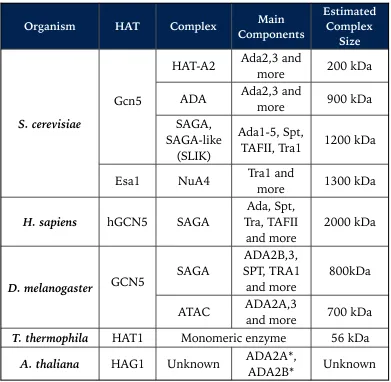
Following the discovery of chromatin remodelling enzymes in yeast, the other main class of co-activators, Gcn5 (homolog of plant HAG1), was also discovered through similar genetic screens in yeast and was linked to chromatin-related pathways after it was shown to be identi-cal with the p55 subunit of a histone acetyltransferase complex initially purified from Tetrahymena (Cote et al., 1998; Georgakopoulos and Thireos, 1992; Kruger et al., 1995). Gcn5 complexes preferentially acetylate his-tone H3 N-terminal tails. Two biochemically distinct complexes were first identified in yeast, where Gcn5 is the catalytic component. These include the Ada and SAGA (Spt-Ada-Gcn5 Acetyltransferase) complexes (Grant et al., 1997). Other complexes in yeast and other organisms are shown in
Table 1.2. Major HAT complexes identified in different model organisms. In the case of HAG1 in A. thaliana, although the full complex has not been described yet, inter-action with ADA2A and ADA2B has been confirmed. Collated from multiple sources referenced in the main text.
In yeast, highly optimised biochemical approaches allowed researchers to purify and characterize the various modules of the SAGA complex giving rise to a 21-protein complex with four main modules (Fig. 1.3). These in-clude the histone acetylation (HAT) module that consists of Ada3, Ada2, Sgf29 and Gcn5. The de-ubiquitination (DUB) module is composed of Sgf11, Sgf73, Sus1 and Ubp8, which performs the de-ubiquitination reac-tion. The SPT module is responsible for the assembly of the pre-initiation complex during transcription and consists of Spt3, Spt7, Spt20, Ada1, Tra1 and Chd1/Chr5. TAF is a co-activation module and is known to have roles in the structural integrity of the complex (Koutelou et al., 2010; Samara and Wolberger, 2011).
Organism HAT Complex ComponentsMain Estimated Complex
Size
S. cerevisiae
Gcn5
HAT-A2 Ada2,3 and more 200 kDa
ADA Ada2,3 and more 900 kDa SAGA,
SAGA-like (SLIK)
Ada1-5, Spt,
TAFII, Tra1 1200 kDa
Esa1 NuA4 Tra1 and more 1300 kDa
H. sapiens hGCN5 SAGA Tra, TAFII Ada, Spt,
and more 2000 kDa
D. melanogaster GCN5 SAGA
ADA2B,3, SPT, TRA1
and more 800kDa
ATAC ADA2A,3 and more 700 kDa
T. thermophila HAT1 Monomeric enzyme 56 kDa
A. thaliana HAG1 Unknown ADA2A*, ADA2B* Unknown
of
Figure 1.3. Yeast SAGA complex has multiple modules with different functions. SAGA is a multi-protein complex with multiple functions performed by its different modules. The HAT module is responsible for the histone acetyltransferase activity with Gcn5 being the catalytic component. The DUB module removes ubiquitin molecules from histone H2B, while SPT and TAF modules play important roles in facilitating tran-scription, through assembly of the pre-initiation complex and co-activation processes. Modified from (Koutelou et al., 2010; Samara and Wolberger, 2011)
Figure 1.4. Acetylation/De-acetylation reaction of lysine/acetyl-lysine. HATs catal-yse the transfer of an acetyl group from acetyl-CoA onto a lysine residue forming the byproduct coenzyme A with a thiol group (CoASH). HDACs catalyse the reverse reac-tion with the input of a water molecule. It should be noted that different HATs use dif-ferent amino acids to catalyse the transfer of the acetyl group. Modified from Kim and Yang (2011). HAG1 homologs Tra1 Spt7 Spt8 Spt3 Spt20 Ada1 Taf12 Taf10 Taf9 Taf6 Taf5 Sgf29 Sgf73 Ubp8 Sgf11 Sus1 HAT (Histone Acetylation) SPT (Pre-Initiation Complex assembly) DUB (De-ubiquitination of H2B) TAF (Co-activation) Gcn5 Ada2a Ada2b Ada3 Chd1/ Chr5
YEAST SAGA
MODEL
NH3 CH2 (CH2)3CH
HATs
HDACs
NH CH2 (CH2)3
CH + CH3 C=O CoA S CH3 C=O CoA S Acetyl Group Acetyl-Lysine (AcK) Lysine (K)
H2O CH3
-In yeast, it has been shown that E173 of GCN5 acts as the general base responsible for de-protonating the lysine of a histone. This is a re-quirement for the transfer of an acetyl moiety onto the target lysine. Upon completion of the reaction coenzyme-A with a thiol group (CoASH) is formed (Tanner et al., 1999) (Fig. 1.5).
Although there is no study in HAG1 showing which amino acid is responsible for the catalytic activity, the existing literature on GCN5 from other eukaryotes suggested that the highly conserved 8-amino acid se-quence highlighted in Figure 1.4 was identified as part of a core region suf-ficient for catalysis of the acetylation reaction in yeast (Gregory et al., 1998; Wang et al., 1998). Further studies using mutagenesis and in vitro
and in vivo HAT reactions also pinpointed that the glutamic acid (E173 in yeast, E121 in Tetrahymena, E582 in human) inside the highly conserved HAT domain of GCN5 is responsible for the enzymatic activity. This cor-responds to E289 in A. thaliana HAG1 within the catalytic HAT domain as shown in Figure 1.5.
Figure 1.5. Sequence alignment of HAT domains in different model organisms. Yeast GCN5, human P/CAF, human GCN5, Tetrahymena GCN5, Arabidopsis GCN5, Drosophila GCN5 protein sequences are shown. The catalytic domain for each protein is highlighted with a red square and is highly conserved across all organisms (Modified from (Trievel et al., 1999) .
1.10. Chromatin remodelling in plant defence
Several examples of proteins involved in chromatin remodelling have been found to be important for biotic stress responses in plants. SPLAYED is an ATP-dependent chromatin remodeller, and HISTONE MONOUBIQUITINATION1 is a RING-finger E3 ligase and both were re-quired for resistance to necrotrophic fungal pathogens (Dhawan et al., 2009; Walley et al., 2008). In addition, sdg8 mutants impaired in H3K36me3 deposition also displayed lower resistance to necrotrophic
of
fungal pathogen infection (Berr et al., 2010). Furthermore, Arabidopsis Trithorax 1 (ATX1), another histone methyltransferase, directly regulates the transcriptional activity of WRKY70, a positive regulator of salicylic acid (SA)-mediated defence signalling (Alvarez-Venegas et al. 2007). Components of the Arabidopsis SWR1-like complex, which is responsible for the replacement of histone H2A with the histone variant H2A.Z, are required for the repression of SA-dependent defence genes (March-Diaz et al., 2008).
In another example, histone monoubiquitination, histone variants and a chromatin remodeller act together on the same pathway ensuring that defence genes are activated only after infection. For example, in rice, defence genes are kept silent in the absence of pathogen attack and this is held in check by the presence of histone variants at the promoters of the pathogenesis-related gene OsPBZc and the LRR OsSIRK1 gene. The mech-anism by which these genes are kept silent is explained by the action of SWI/SNF ATPase BRHIS1, the expression of which is downregulated upon infection by the rice blast fungus. Therefore, the interaction be-tween BRHIS1 and histone variants of H2A and H2B is lost allowing the expression of the underlying genes. Given the enrichment in histone mo-noubiquitination, these genes are poised for expression until BRHIS1 re-pression is relieved (Li et al., 2015).
1.11. Histone acetylation in plant immunity