Design of a Simulation-Based Training for Flexible Bronchoscopy
Julia Kania
Department of Cognitive Psychology and Ergonomics
University of Twente
Supervisors:
Dr. Martin Schmettow
Dr. Marleen Groenier
Enschede, Netherlands
Abstract
Background/aim: Medical training based on simulators is a patient-safe and cost-effective
addition to the traditional apprenticeship model. Instead of letting patients bear the burden of beginner's mistakes, trainees can acquire skills for flexible bronchoscopy (FB) before the first patient contact on simulators. This study's aim was to gain insight into the required skills for FB, their underlying cognitive aspects (e.g. cues, goals, difficulties) and to what extent they can be trained on simulators.
Method: (1) Qualitative Document Analysis of four established FB guidelines and a
simulation-based curriculum. (2) Qualitative Interview Study: Four Dutch pulmonologists, one resident and one technical physician performed a diagnostic FB on a virtual reality (VR) simulator (10-22 min.) which was video-recorded. Participants engaged in retrospective think-aloud while watching their performance on video. A semi-structured interview helped clarify concepts and opinions about simulation-based training.
Results: Seven skills were identified which are crucial for performing a FB. Participants
agreed that only two of them can be trained on the VR simulator: (1) Handling the bronchoscope and (2) Inspecting the airway. For the other skills, the VR simulator was considered too inaccurate to serve as a training modality. The results provide detailed descriptions of a diagnostic FB performance (e.g. cues, goals, difficulties) and the VR simulator’s (in)adequacy for training and assessing these skills.
Conclusion: Simulation-based training should combine different types of simulators. The VR
simulator’s inaccuracies should be improved to optimize learning experiences. Disagreements about execution of skills should be resolved among experts to reach consensus about what to train and thus, how to assess the level of skill of a trainee. Often discarded but worth
Table of Contents
1. INTRODUCTION ... 4
1.1 TIME FOR CHANGE: SUPPLEMENTING THE APPRENTICESHIP MODEL ... 5
1.2 SIMULATION-BASED TRAINING ... 7
1.3OVERVIEW OF CURRENT RESEARCH PAPER ... 7
2. DOCUMENT ANALYSIS STUDY ... 9
2.1 METHOD ... 9
Design. ... 9
Selection of documents. ... 9
Analysis of documents. ... 10
2.2RESULTS ... 11
Comparison of FB guidelines/curricula. ... 11
Simulation-based curriculum. ... 11
Basic skills and knowledge required for FB. ... 12
2.3CONCLUSION AND DISCUSSION ... 17
3. INTERVIEW STUDY... 19
3.1 METHOD ... 19
Design. ... 19
Participants. ... 19
Materials. ... 19
Simulator environment.... 19
Simulator task: Diagnostic Bronchoscopy. ... 20
Interview guide. ... 22
Procedures. ... 24
Preparation. ... 24
On-site. ... 24
Analysis. ... 25
3.2RESULTS ... 26
Skills. ... 29
1) Mastering diagnostic procedures. ... 29
2) Handling the bronchoscope. ... 34
3) Inspecting the airway. ... 37
4) Preventing and dealing with complications. ... 39
5) Administering anaesthetics or sedatives. ... 40
6) Patient monitoring. ... 41
Context ... 41
7) Working with an assistant. ... 41
3.3DISCUSSION ... 42
REFERENCES ... 58
1.
Introduction
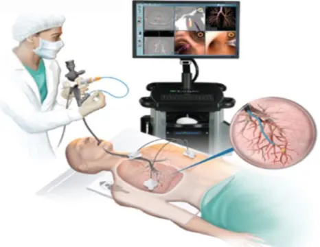
Medical training based on simulators has proven to be an efficient, if not superior, way of acquiring basic skills for flexible bronchoscopy compared to the traditional apprenticeship model (Blum, Powers, Sundaresan, 2004). Flexible bronchoscopy is a medical, invasive procedure which is used to examine the airways of a patient (Naur, Nilsson, Pietersen, Clementsen and Konge, 2017; American Thoracic Society, 2015). Figure 1 illustrates a flexible bronchoscopy. By viewing the breathing passages of the lungs and acquiring samples of its mucus or tissue, pulmonologists can diagnose patients with various diseases and
[image:4.595.77.310.363.543.2]complications such as lung cancer, infections and inflammation (American Thoracic Society, 2015; Naur et al., 2017). A bronchoscope which is a thin instrument resembling a tube, is placed in either the mouth or nose of a patient and is inserted into the lungs. The instrument carries a camera which transfers pictures of the airway onto a video screen, enabling a visual exam of the airways. (American Thoracic Society, 2015).
Figure 1: A flexible bronchoscopy procedure (Sydney Respiratory & Sleep Physician, n.d., Retrieved from: https://www.sydneyrespiratoryspecialist.com.au/flexible-bronchoscopy.html)
Traditionally, novice bronchoscopists are trained based on the apprenticeship model: letting trainees observe how experts perform the procedure and then have them gradually gain own experience by practicing on fully awake or consciously sedated patients under
minimum of 100 flexible bronchoscopies under supervision. However, demanding a fixed number of performed procedures is controversial as trainees differ greatly in their dexterity and confidence. Assessing competency based on performance seems to be a more useful alternative (Konge, Arendrup, Buchwald & Ringsted, 2011).
An essential rationale for the inclusion of simulator is the concern for patient safety: on simulators, trainees can learn surgical skills and make mistakes without harming human patients (Bjerrum, Thomsen, Nayahangan, & Konge, 2018). The simulators’ cost-effective, patient-safe characteristics as well as empirically proven efficacy render it vital to include simulation-based training in medical education for flexible bronchoscopy. In order to maximize productivity of this training, it needs to be tailored to the specific needs of the trainees. Simulation-based training is often based on personal opinions of educators or surgeons and on what is feasible and available. An objective needs assessment is needed in order to shift the focus from personal opinions and feasibility to relevance; to identify which skills need to be trained (Nayahangan, Stefanidis & Konge, 2018). Although different flexible bronchoscopy curricula exist, differences in geographical locations, specialities and regional differences in resources render it necessary to explore specific, local learning needs of
pulmonologists in the Netherlands in order to contribute to the effort of establishing a national curriculum (Nayahangan, Stefanidis & Konge, 2018). This study aims to explore the
procedures, processes, concepts and decision-making of Dutch pulmonologists of different experience levels in order to design a simulation-based training.
1.1 Time for Change: Supplementing the Apprenticeship Model
The traditional apprenticeship model has been a subject of criticism. Practicing on human beings in order to gain experience and bronchoscopy skills puts risks on patients’ safety and lacks efficiency: there are neither objective measures of skills nor constant
feedback (Davoudi & Colt, 2009). More specifically, Colt et al. (2001) assert that training on real patients may lead to extended invasive procedures, false diagnoses and patient discomfort and morbidity. Konge et al. (2011) note that during the first trimester of training, novice bronchoscopists’ procedures have an increased complication rate.
obstructions of airway secretions (Colt, 2001). Furthermore, practicing on animal subjects raises ethical and monetary concerns (Blum, 2004).
Numerous studies show that bronchoscopy education can be enhanced by including simulator training (Davoudi & Colt, 2009). Simulators, or virtual reality (VR), are computer-based environments characterized by advanced hard- and software, graphics and perhaps most importantly, tactile, auditory and visual feedback. Through its characteristics, simulators are able to provide users a close-to-reality experience (Colt, Crawford & Galbraith, 2001). For example, the study by Colt (2001) describes the simulator as follows: including a proxy flexible bronchoscope with close resemblance to a conventional one and showing virtual anatomy and realistic images when navigating the proxy bronchoscope through the virtual patient. The images of the airway are based on CT sets. Motions of the proxy bronchoscope are detected by an interface device and the forces normally felt during such movements by a surgeon are reproduced and felt by the user of the simulator. The simulated patient is
represented realistically as well by changes in vital signs, breathing, bleeding and coughing. Studies on the efficacy of simulators have demonstrated simulators to be effective, patient-safe and cost-effective (Davoudi & Colt, 2009). Simulator training allow trainees to practice their skills at their own pace without harming a patient, are less expensive than the costs of supervision needed for real-life procedures (Colt et al., 2001) and ease resident and patient anxiety since trainees can familiarize themselves with the instrument, technique and anatomy before the first patient contact (Ost, DeRosiers, Britt, 2001; Blum, Powers,
Sundaresan, 2004). Its objective assessment of trainee skills make it an efficient training device, addressing the issue of time contraints for residents and instructors. Naur et al. (2017) conducted a systematic review of articles on flexible bronchoscopy and simulator-training and concluded that simulation-based training is more efficient than the traditional apprenticeship model. A study by Colt, Crawford and Galbraith (2001) showed that after their specific curriculum of training with a virtual reality bronchoscopy simulator, novice trainees had fewer contact with the bronchial wall and less neglect of segments. Moreover, novice trainees performed equally well after the simulation training as skilled physicians who were trained in the traditional way. Additionally, compared to skilled physicians, novices trained with the simulator executed more meticulous procedures and neglected significantly less segments (Colt et al., 2001). Moreover, simulators offer the opportunity to practice case management skills: learning how to deal with complications which arise only rarely in real-life.
Vergis and Hardy (2017) note that technical expertise (the focus of most simulation-based programs) requires multiple abilities in addition to technical dexterity such as decision-making skills. This study aims at uncovering different decision-decision-making points of pulmonologists during a FB.
1.2 Simulation-based Training
According to Blum et al. (2004), residency training is characterized by eighty-hour work weeks and reduced time at the hospital, demonstrating the urgency of maximum training productivity. Although simulators are efficient, each trainee with a different experience level has different bronchoscopy skill acquisition needs. Therefore, it needs to be specified which skills are needed and how they can be practiced in order to become a competent
bronchoscopist (Davoudi & Colt, 2009). Training curricula have already included simulators for learning surgical procedures such as bronchoscopy (Konge, Bjerrum, Nayahangan,
Schroeder, 2015) and this study aims to gain more insight into the different features needed to achieve a successful simulation-based training. A similar study has been conducted by Tijam, Schout, Hendrikx, Scherpbier, Witjes, Van Merriënboer & Van Merriënboer (2012) which identified tasks, sub-tasks and blueprints for the execution of a nephrostomy procedure which could be used to design a simulator-based training. While psychomotor skills and theoretical, and procedural knowledge is vital to perform a bronchoscopy, studies which evaluate
simulators usually ignore the importance of appropriate decision-making points (Tijam et al., 2012). In addition, uniformly designed training programs are inappropriate as trainees with different experience levels may aim to acquire distinct skills (Konge et al., 2011).
Cognitive Task Analysis is an ideal method to gain more insight into cognitive processes and events that emerge when pulmonologists perform flexible bronchoscopy on a simulator. It is an ideal method since it specifies all the cognitive aspects involved in a performance and how they could be elicited and analysed. It helps to extract their knowledge, perception, thoughts regarding performing a flexible bronchoscopy. Thus, by means of a Cognitive Task Analysis, this study aims to explore the following research question: What are the learning needs of trainees in pulmonology in a simulation-based training program?
1.3 Overview of Current Research Paper
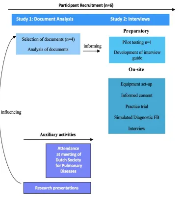
qualitative study addresses the questions of how simulators are currently incorporated in flexible bronchoscopy training and in what manner they can be optimally included in a FB curriculum. Figure 1 shows an overview of the organisation of this research. Therefore, in the first part of this research, the starting point is a review of literature and established curricula, gaining an overview of and integrating information from different sources on basic skills and knowledge needed for FB. In the second part of this study, interviews with expert
pulmonologists are conducted which allow to extract more detailed ideas of the specific learning needs and objectives of pulmonologists in the Netherlands and thus, reach
[image:8.595.105.476.303.721.2]preliminary consensus of experts about the contents of a simulation-based FB curriculum in the Netherlands.
2. Document Analysis Study
2.1 Method
Design.
As a first step in conducting the needs assessment, five documents were qualitatively content-analysed to serve multiple purposes: (1) to identify basic skills and knowledge required for performing a flexible bronchoscopy, (2) to compare flexible bronchoscopy curricula and by that, to (3) inform the development of interview guidelines and guide the researcher in extracting relevant information during the interviews. The research question guiding the analyses was: “What are the processes, steps, concepts and decision points
involved in a flexible bronchoscopy?”.
Selection of documents.
Documents that were considered admissible were already developed (inter)national training curricula for FB training and a textbook on flexible bronchoscopy. The latter was considered necessary to include in order to understand the general concepts involved in flexible bronchoscopy and to make sense of the curricula. Exclusion criteria for the curricula were documents that were not written in English and published prior to 2011. One exception to the latter exclusion criterion was the European Respiratory curriculum which was included after suggestion of pulmonologists during presentations of the research proposal in hospitals. Based on Bowen’s (2009) listing of functions of documents, the documents used in this study have the following functions: they suggest questions and processes to be asked and observed during the interviews and they also contextualise the data that results from the interviews.
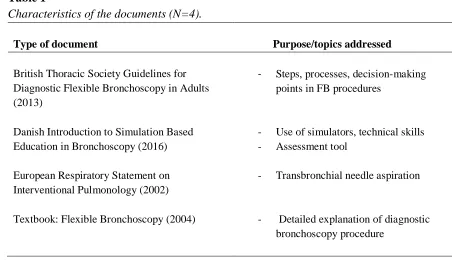
Documents were sampled online (the first three) and from the University of Twente library (the textbook). Table 1 outlines the characteristics of the four selected documents. One document used was the British Thoracic Society Guideline for diagnostic flexible
bronchoscopy in adults (2013). This document’s target audience are respiratory practitioners in the UK which will perform FB. It underlines specific steps, processes and decision-making points involved in FB procedures. Another document is an established, Danish, simulation-based training curriculum for flexible bronchoscopy (2016). This curriculum specifies the use of simulators as well as important skills and knowledge vital for performing a FB. Moreover, it provides an assessment tool based on bronchoscopy simulators. The European Respiratory Statement on Interventional Pulmonology (2002) includes short descriptions of necessary equipment, techniques, indications, complications and training requirements for
Table 1
Characteristics of the documents (N=4).
Type of document
Purpose/topics addressed
British Thoracic Society Guidelines for Diagnostic Flexible Bronchoscopy in Adults (2013)
- Steps, processes, decision-making points in FB procedures
Danish Introduction to Simulation Based Education in Bronchoscopy (2016)
- Use of simulators, technical skills - Assessment tool
European Respiratory Statement on Interventional Pulmonology (2002)
Textbook: Flexible Bronchoscopy (2004)
- Transbronchial needle aspiration
- Detailed explanation of diagnostic bronchoscopy procedure
Analysis of documents.
Document analysis consists of an iterative process of superficially skimming, thoroughly examining and interpreting documents (Bowen, 2009). During this process, aspects of content analysis and thematic analysis are incorporated; meaningful text passages are identified, coded and then categorized into similar themes (Bowen, 2009). After skimming through the first document (guidelines), the researcher started inductively developing
preliminary sets of codes. The researcher’s initial strategy consisted in finding answers (codes) to the rather general question: “What are important steps, processes, concepts and decision-making points in performing flexible bronchoscopy?”.
Guiding the development of codes and categories was the goal of not summarizing existent declarative knowledge but focusing on the perspective of the doctor with regard to his or her cognition: What does the doctor need to know and do generally? Information that was too detailed and technical was skimmed; the approach to coding was taking a “birds-eyes” view with basic information of concepts. For example, when information as given about the variations in anatomy, anatomical details of those variations were skipped and a code relating to the knowing the anatomy was given.
the code “handling the bronchoscope” was applied. After reviewing the different portions of text, an all-inclusive definition was established for the specific code. Codes were revised, deleted and added during the process if appropriate.
After coding all four documents, the resulting coding scheme was examined and the codes were categorised into larger themes, into aspects of FB prior to, during, after the procedure and FB’s contextual, non-procedural aspects (see Table 2). The last step consisted in comparing the three documents, identifying gaps, similarities and differences.
2.2 Results
The document analysis elucidated several aspects of flexible bronchoscopy including steps, required knowledge, skills and decision points, chronologically separated into those relevant prior to the procedure, for the procedure itself and after the procedure. Moreover, contextual aspects of FB were identified. In the following, first, the different guidelines are compared in the topics and detail they include. Afterwards, all the identified basic skills and knowledge from the documents are presented.
Comparison of FB guidelines/curricula.
The British and European curricula were similar in the amount of detail they provided but were conceptually different, as the European curricula focused on a specific aspect of FB, Tranbronchial Needle Aspiration. In contrast, the British guidelines included a more
encompassing guide on FB from “A to Z”: precautions, complications, patient considerations, diagnostic accuracy, sedation, anesthesia, disinfection, staff, patient satisfaction. The Danish curriculum focused more on the technical details of performing FB and how to train those on different simulators. In general, the documents complemented each other well, while the textbook and Danish curriculum sometimes respectively included information not found in other documents, due to their specific purpose: providing details of FB and incorporating simulators to train FB. Table 2 summarizes the emergent themes.
Simulation-based curriculum.
The Danish Practical Handbook for Bronchoscopy (2016) was the single document which demonstrated a simulation-based training program. The program includes four basic aspects:
1) basic theoretical knowledge
4) an assessment
The theoretical knowledge contained in the curriculum is rather concise and the authors recommend self-study with additional literature. In this case, the British and European
curricula as well as the textbook would seem to provide more detailed theoretical knowledge. Moreover, the British and European guidelines and the textbook describe knowledge and processes that were not mentioned in the Danish curriculum (e.g. improving diagnostic yield). While the Danish curriculum mentions similar topics as the other documents (e.g. treatment of bleedings), not all concepts described in the Danish curriculm are also translated into the practical simulation-based training (e.g. dealing appropriately with complications such as bleeding).
The second aspect is that trainees are introduced to both the flexible bronchoscopy procedure in general and the simulators. This is executed by experts from pulmonary medicine.
Later, the simulators and phantoms will be used to transfer knowledge and practice different skills. The VR simulators are mainly used for technical skill acquisition: handling the bronchoscope, learning to navigate through the anatomy and mastering sampling techniques. However, those skills are not exclusively trained on the VR simulators. Instead, lung models phantom models, a real bronchoscope and sampling equipment are used as well for the same purposes. Although this is self-training, it will be supervised by other medical students and nurses familiar with the simulators and equipment.
Lastly, after self-training, an assessment is conducted. The assessment tool given in the document is based on scores from either the VR CAE simulator or the VR Symbionix simulator. Scores are given by simulators for correct localization of pathology and
performance of sampling techniques (divided into sub-aspects such as at least one lavage performed in middle lobe, correct amount of suction). In addition to the scores, simulators record the time and the test is passed when 28 points are reached in maximum 70 minutes. After passing, trainees are considered ready to start with the clinical training on patients.
Basic skills and knowledge required for FB.
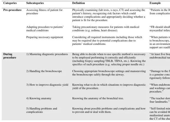
Table 2 shows the results of the document analysis; the basic skills and knowledge that are paramount for a flexible bronchoscopy. Pre-procedure and aftercare aspects were
The majority of aspects of FB that were identified were for the use during the procedure. As the basis, it is important to be familiar with the 4) anatomy of the bronchial tree. Here, the textbook was very specific regarding the aspects to be known. These include the anatomic orientation, the normal anatomy but also anatomic variations which are not clinically relevant. Pathological abnormalities, and knowledge of resectabilities of lesions should be known as well.
In addition to knowing the anatomy, bronchoscopists should be skilled in the 1) different diagnostic techniques such as Transbronchical Lung Biopsy (TBLB), Transbronchical Needle Aspiration (TBNA), Bronchial Brushing and Biopsy and Bronchoalveolar Lavage. Details to each procedure (e.g. selecting the proper needle, not damaging biopsy specimens) should be known, also taking into account the context of the patient. For example, in one document, it is noted:
“At least five biopsy samples should be taken when endobronchial tumor is visible […].” (British Thoracic Society, 2013, p.2).
Another important skill relates to the 2) handling of the bronchoscope. The bronchoscope should be chosen appropriately in terms of selecting the appropriate diameter of the
bronchoscope for each case. The bronchoscope should be manoeuvred correctly without causing any damage to the bronchoscope. A lot of detail about the bronchoscope was included in the Danish simulation-based curriculum. Another important aspect was mentioned
exclusively in the textbook which is the 9) systematic inspection of airways. Since the bronchoscopist is immediately drawn toward the abnormalities of the airways, the inspection should be started in the airways which are considered to not be involved in any pathology.
Another recurrent theme was 3) improving diagnostic yield, the probability that the techniques used result in a clear-cut diagnosis. Bronchoscopists should be concerned with knowing which techniques could improve the diagnostic yield in which procedure, also taking into account the context of the patient’s condition. For example:
“When endobronchial tumour is visible, brushings and washings can increase the diagnostic yield of the procedure.” (British Thoracic Society, 2013, p.2).
drug/solution, the adequate amount and the preference of the patient should be taken into account. It is not only sufficient to engage only in preparations for ensuring the patients’ tolerance; bronchoscopists also need to 7) constantly monitor the patients’ tolerance, symptoms, vital signs and act appropriately in case of crucial changes.
Moreover, bronchoscopists should be prepared to know of 5) possible problems and complications which could occur during the procedure. Problems relate to confinement issues (having impaired vision of the airway due to mucus or blood), irritation, decreased toleration of the patient and losing anatomical orientation. A complication which could occur is
bleeding which can be caused my malignant diseases 25% of the time and also chronic inflammatory processes, being the source of bleeding 50% of the time. Thus, bronchoscopists should know how to prevent problems and complications, if possible. For instance:
“Irritation by frequent passage of airways under local anesthesia can be avoided if a tube is inserted over the bronchoscope.” (Wang, Mehta & Turner, 2004).
Table 2
Results of the document analysis: Aspects of flexible bronchoscopy.
Categories Subcategories Definition Example
Pre-procedure Assessing fitness of patient for procedure
Adapting procedure to patients’ medical conditions
Preparing necessary equipment
Physically examining (lab tests, x-rays, CT) and assessing the patient’s history, recognizing risk factors which could introduce complications and appropriately deciding whether a patient is fit for the procedure.
Taking precautionary measures for patients with medical conditions (e.g. asthma, heart disease).
Considering all required instruments including those which may be required due to potential complications due to patients’ medical conditions.
“Patients in the ICU should be considered at high risk from complications when undergoing bronchoscopy.”
“FB should ideally be delayed for 4 weeks after myocardial infarction.”
“When patients require non-invasive ventilation prior to bronchoscopy, the procedure should be conducted in an environment where intubation and ventilatory support are readily accessible.”
During procedure
1) Mastering diagnostic procedures
2) Handling the bronchoscope
3) How to improve diagnostic yield
4) Knowing anatomy
5) Handling problems and complications
Being able to decide when to use specific method is necessary to be employed performing it correctly and efficiently (including biopsy sampling/TBLB, TBNA, etc.). Knowing the specifics of each procedure (e.g. selecting proper needle etc.)
Choosing appropriate bronchoscope settings and manoeuvring the bronchoscope safely through the airway.
Knowing what to do in which situations to improve diagnostic yield of the procedure.
Knowing the anatomy of the bronchial tree.
Knowing about possible problems and complications and how to prevent and/or deal with them.
“At least five biopsy samples should be taken when endobronchial tumour is visible […]”
“Damage to the flexible bronchoscope during TBNA is a genuine concern and can be avoided by
rigorously following optimal procedures.”
“When endobronchial tumour is visible, brushings and washings can increase the diagnostic yield of the procedure.”
“The teacher demonstrates the anatomy including the four landmarks.”
6) Deciding (how) to sedate
7) Patient monitoring during procedure
8) Giving topical anaesthesia
Choosing an appropriate drug, an adequate amount of the drug and incorporating patient preference.
Observing the patients’ heart rate, respiratory rate, blood pressure, oxygen saturation and symptoms during the
procedure, recognizing changes and responding appropriately.
Deciding which is the appropriate way of giving topical anaesthesia.
“Intravenous midazolam is the preferred drug for sedation, … no more than 5 mg midazolam for patients under age of 70, 2mg for patients over 70
…”.
“Patients should be monitored by continuous pulse oximetry during bronchoscopy”.
“1% lidocaine solution should be used for spray-as-you-go administration”.
9) Systematic inspection of airways Navigating through the airway in a systematic way. “[…] first examine airways that are apparently uninvolved in pathology.”
Aftercare All immediate and remote activities after the procedure from preparing cytological specimens to monitoring patients and informing patients.
“Patients should be advised of the potential for delayed complications following TBLB and provided with written information regarding likely symptoms and action required.”
Context and non-procedural aspects
Including an instructor for teaching purposes
Assessing performance
Having an assistant
Disinfecting instruments
Staff
Letting an instructor demonstrating relevant skills and guiding trainees.
Performance should be assessed periodically according to specified criteria (efficacy, record of personal diagnostic accuracy for FB, complications, patient satisfaction surveys).
Performing the procedure together with a helping assistant.
Having cleaned, decontaminated bronchoscopes and storing instruments appropriately.
Having staff with appropriate training.
“The instructor will guide the student as the student brushes and biopsies an airway abnormality”.
“The participant should describe the pathology and its exact location in the bronchial tree.”
“In addition to the bronchoscopist, the procedure requires a dedicated assistant.”
“Bronchoscopes must be cleaned and disinfected before and after placing in carrying cases as these cases cannot be disinfected […].”
2.3 Conclusion and Discussion
The purpose of the document analysis study was to extract from existent FB curricula, (and a textbook) the basic knowledge and skills that trainees should learn to perform FB. This study is the basis for a subsequent interview study, specifically the interview scheme used to interview pulmonologists about performing FB on a simulator. In general, during the
procedure different technical skills, knowledge and decision-making skills are required. Technical skills include the mastery of diagnostic techniques, handling of the bronchoscope and systematic inspection of airways which in turn requires a thorough knowledge of the anatomy. Decision-making skills are handling problems and complications, sedating, anaesthetising, improving diagnostic yield and patient monitoring. Important contextual aspects of a training environment for FB are experienced staff, assistants, instructors, disinfected instruments and assessment of performance.
In light of the empirically supported efficiency of simulators for training of
endoscopic skills (Blum, Powers, Sundaresan, 2004), it is a perhaps surprising finding that it was rather difficult to find curricula which incorporate simulators for training FB. Though the Danish curriculum focused on the use of simulators for technical skill acquisition, it was not exclusively restricted to VR simulators; inanimate lung models were included as well for the training of the same technical skills. This may point to a potential inadequacy of VR
simulators as a sufficient training modality in itself. Similarly, Bjerrum et al. (2018) note that a holistic skill acquisition can only be achieved through integrating different simulators.
Thus, the use of the VR simulator in the Danish simulation-based curriculum was restricted to the acquisition of skills regarding handling the bronchoscope, exploring the anatomy and mastering diagnostic techniques which are more technical skills. Little attention was given in general to using any kind of simulator for acquiring decision-making skills to improve diagnostic yield, handling problems and complications or responding to crucial changes in the patient’s vital signs and symptoms. This finding is in line with Tijam et al. (2012) who claim that studies which evaluate simulators often discard the importance of decision-making points in favour of training psychomotor skills and theoretical, procedural knowledge.
complications are rare, Konge et al. (2001)’s finding that novice bronchoscopists have an increased complication rate during the first trimester of training, underlines exactly the point that those decision-making points are important to train as well, in addition to purely technical skills.
Nonetheless, it must be pointed out that the Danish curriculum indeed included
theoretical knowledge about treatment of complications, for instance. This kind of knowledge was recommended to be supplemented by self-study of further literature. Although study of potential complications and how to handle them increases theoretical knowledge, applying this knowledge in real-life scenarios may require appropriate decision-making skills. Hence, it may be worthwhile to inspect the possibility of training and assessing decision-making skills on VR simulators.
Next, the Danish curriculum put emphasis on having a teacher who demonstrates the anatomy to students (on lung models and the simulator). According to Colt (2001), such instructors are rather costly. The question may arise whether the use of simulators could completely waive the presence of teachers. However, not only are instructors included in the Danish simulation-based training, assistants and staff were considered essential by the British and European guidelines for a FB procedure. Since assistants and staff are crucial during a real bronchoscopy, a simulated environment might need company too.
Regarding the goal of the document analysis study to inform the development of interview guidelines for the interview study, it has to be said that the goal was achieved sufficiently, giving the researcher a basic theoretical background of a flexible bronchoscopy and FB training. However, more information on incorporation of the simulator would have helped the researcher to get a better sense of what the simulator is capable of and how it can be used.
All in all, the documents provided an encompassing view of FB but for the purpose of identifying learning needs involved during a flexible bronchoscopy, the pre-procedure and aftercare codes were discarded for the interview guide. The contextual aspects however, may be important to discuss with pulmonologists in the interview study. Thus, all “during
3. Interview Study
3.1 Method
Design.
A semi-structured interview was conducted in English with six Dutch professionals in order to identify the learning needs of trainees in pulmonology for a simulation-based flexible bronchoscopy training. Participants performed a diagnostic bronchoscopy task on a simulator (10-22 min.) during which they freely inspected the airway and sampled tissue, using tools of choice. Their performance was video-recorded. Participants were asked to retrospectively think aloud while watching the video-recording and specific questions regarding their
performance, the FB procedure in general and the usefulness of the simulator were asked. The interviews were audio-recorded and transcribed. Cognitive Task Analysis of the interviews resulted in a list of learning needs of Dutch pulmonologists which was combined with the list of aspects of FB resulting from the document analysis study.
Participants.
A purposive sample of four Dutch pulmonologists, one pulmonology resident and one Technical Physician in training was included in this study based on their prior experience in performing a flexible bronchoscopy. In the Netherlands, according to law, both
pulmonologists and technical physicians are allowed to perform flexible bronchoscopies. At the time of the study, the participating Technical Physician has performed over 100 flexible bronchoscopies under supervision. Moreover, the participant has developed a surgical simulator before. Thus, by including a Technical Physician, insight into the procedures and especially simulators, could be gained from a technical perspective as well.
Participants had varying experiences and skills which is determined by the amount of years in their profession and the number of performed flexible bronchoscopies (self-reported). Excluding trainees, years spent in profession ranged from 2 to over 30 years. The number of performed flexible bronchoscopies varied from 100 to 7000. Participants also varied in their degree of familiarity with the simulator. While some participants (n=3) were familiar with the simulator due to frequent or recent use, other participants have used a simulator before but were less familiar with it (n=3).
Materials.
Simulator environment.
clinical experience for clinical procedures. It is a combined simulator for both GI endoscopy and flexible bronchoscopy. It includes basic skill tasks as well as complete clinical procedures (3D Systems, 2017). Regarding flexible bronchoscopy, the BRONCH Mentor features an authentic bronchoscope which can be inserted into the mouth of the plastic mannequin and a master tool and a syringe (3D Systems, n.d.b) which are inserted into the scope. After insertion of the master tool, users can choose a method for tissue sampling by touching the screen. Pulling and pushing the master tool results in opening and closing of the tool selected. Similarly, the syringe must be drawn back before inserting it into the scope and pressed down when inserted in order to draw up and distribute fluid into the virtual airway, respectively.
[image:20.595.120.496.281.527.2]
Figure 3: 3D Systems Bronch Mentor simulator (left) and master tools (right).
Simulator task: Diagnostic Bronchoscopy.
The 3D Systems BRONCH Mentor offers a diagnostic bronchoscopy module which comprises six cases with different patient histories and conditions (3D Systems, n.d.a). The user has to perform a visual airway inspection and can choose between several methods for endobronchial and transbronchial tissue sampling including biopsy forceps, cytology brush, aspiration needle and bronchi alveolar lavage. During the procedure, the virtual patient’s condition is displayed on the screen, allowing the user to check the vital signs, topical
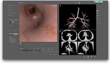
Prior to starting the diagnostic bronchoscopy, the user reads a short patient history, looks at measured vital signs, test and imaging results and selects pre-medication including Lidocaine 4% nebulize 200mg, Midazolam, 2mg and Meperidine, 50mg. Figure 4 shows the information displayed on the screen during the procedure including the main view (scope’s video), main view controls (e.g. full screen or anatomical compass), the tool panel, patient monitoring and management and complementary displays (e.g. X-rays, anatomy atlas or 3D map of the scope within the bronchial tree).
Figure 4: Screenshot of VR simulator display during a participant’s performance on
“diagnostic bronchoscopy” task. The tool panel “Fluids Endobronchially” on the left appears, suggesting that the participant has inserted the syringe into the scope and is attempting to anaesthetise the airway.
symptoms of cough, severe dyspnoea and tiredness without fever or night sweats. A chest CT showed diffused reticulonodular infiltrates and multiple enlarged lymph nodes.
Interview guide.
An open-ended, semi-structured interview guide (see Table 3) was developed based on open-ended, cognitive probing questions (Crandall, Klein and Hoffman, 2006) and the results of the document analysis. In addition to the interview guide, during the interview, the
tabulated results of the document analysis were used to suggest possible content areas of questioning (e.g. questions related to the bronchoscope or anaesthesia), depending on the flow of the individual conversation.
Crandall et al.’s (2006) Working Minds was browsed and relevant cognitive probing questions that were considered appropriate regarding the goals of the study were included in the interview guide. Areas of cognitive probing constituted sensory cues (What were you seeing, hearing, feeling?), goals (What were you trying to inspect?), alternative actions (How was another possible course of action chosen and others rejected?), common mistakes, helpful prior experience, aids, mental models, decision making, difficulty and used information. In addition, two specific probing areas relating to the accuracy of the simulator have been formulated. These probes pertained to (1) necessary skills and knowledge for FB which may not have been included in the simulator and (2) the helpfulness and accuracy of the
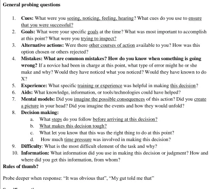
Table 3
Interview guide.
Interview guide
General probing questions
1. Cues: What were you seeing, noticing, feeling, hearing? What cues do you use to ensure that you were successful?
2. Goals: What were your specific goals at the time? What was most important to accomplish at this point? What were you trying to inspect?
3. Alternative actions: Were there other courses of action available to you? How was this option chosen or others rejected?
4. Mistakes: What are common mistakes? How do you know when something is going wrong? If a novice had been in charge at this point, what type of error might he or she make and why? Would they have noticed what you noticed? Would they have known to do X?
5. Experience: What specific training or experience was helpful in making this decision? 6. Aids: What knowledge, information, or tools/technologies could have helped?
7. Mental models: Did you imagine the possible consequences of this action? Did you create a picture in your head? Did you imagine the events and how they would unfold?
8. Decision making:
a. What steps do you follow before arriving at this decision? b. What makes this decision tough?
c. What let you know that this was the right thing to do at this point? d. How much time pressure was involved in making this decision? 9. Difficulty: What is the most difficult element of the task and why?
10. Information: What information did you use in making this decision or judgment? How and where did you get this information, from whom?
Rules of thumb?
Probe deeper when response: “It was obvious that”, “My gut told me that”
Specific questions
1. Skills/Knowledge: Are there skills or knowledge that you feel are important to perform a FB but are not included in the simulation-based task?
2. Assessing performance: Do you think that simulation-based assessment metrics are appropriate to evaluate a trainee’s FB skills? How would you assess whether a student has sufficient basic FB skills? Which metrics do you think could distinguish between
novice/intermediate/expert performance?
Procedures.
Preparation.
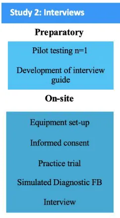
[image:24.595.228.349.295.514.2]A summary of the procedures of this study is displayed in Figure 5. Ethical approval for the study was obtained from the BMS Ethics Committee at the University of Twente. A pilot test was conducted with a volunteer student in order to test the accuracy of the video- and audio-recording. Participants were purposefully sampled through professional contact networks both before and throughout the whole data collection process. The researcher wrote emails to both potential participants and those who already agreed to participate during presentations of the research at hospitals. Due to participants’ tight schedules and the limited availability of the simulator, some participants could not partake in the study.
Figure 5. Flowchart of procedures in the interview study including preparatory and on-site procedures.
On-site.
On site, the equipment was set up including a video-camera, tripod, laptop and smartphone (for audio-recording). Participants were first asked to sign the informed consent form. During or afterwards, participants were asked about the number of years in their profession and the number of performed flexible bronchoscopies. Depending on whether the participant was familiar with the simulator, they were offered a test trial to familiarize themselves with the simulated procedure.
Due to moving during the procedure, sometimes, the view of either the display or hand movements was entirely or partially obstructed.Participants were shown the case history and imaging results related to the specific patient case on the simulator and they could choose a pre-medication. The researcher started the video-recording and the participants started to perform the diagnostic bronchoscopy task on the simulator which took 10-21 minutes. For the first two participants, the researcher stood behind the camera and helped whenever necessary.
Afterwards, the video-recording was displayed on a laptop, the participant and researcher sat down to start with the retrospective think aloud which was followed by a semi-structured interview if additional information was needed. Participants were told to mention everything that was going through their minds during their performance on the simulator parallel to what the video was showing. The interview was audio-recorded and occasionally, notes were taken of the participants’ utterances. Whenever a participant wanted to explain something in more detail, the video-recording was stopped in order not to miss important parts of the video that could have been missed otherwise. After the video ended, the researcher focused on asking participants specific questions, letting them explain the steps they took and clarify concepts which were not clear to the researcher. With respect to the flow of the conversation, appropriate cognitive probing questions and specific questions as outlined in the interview guide were asked. Whenever necessary, the researcher skimmed through the printed tabulated results of the document analysis in order to get ideas for further areas of probing. The interview duration was about an hour.
Analysis.
The audiotaped interviews were transcribed verbatim. If participants also explained their considerations during the procedure, videotapes were transcribed too. Cognitive task analysis of the textual data was conducted in two main phases with the help of the ATLAS.ti program, an analysis software program ATLAS.ti which is particularly useful for coding textual data (ATLAS.ti, n.d.). A first analysis was done with two interview transcriptions and a second with all six interviews.
First, transcripts were read freely. The coding of the data was done inductively as codes emerged from the data. Coding units were themes, a new code was applied to a text when it represented a new information related to the cognition, process or context of performing a FB. The experience from coding different FB documents facilitated the creation of codes to
aspects involved during the procedure of a diagnostic bronchoscopy that are relevant to develop a simulator training. More specifically, to find cognitive aspects, the following questions adapted from Crandall et al. (2006) were guides:
1) What is the participant paying attention to and ignoring? 2) Which senses is the participant using?
3) What is the participant thinking about?
4) What information is the participant seeking, from where?
These questions allowed to focus on how exactly the general processes of FB come about and hence, to extract the skill-set needed to perform a diagnostic bronchoscopy. However, attention was also paid to the opinions of participants towards the accuracy of the simulator and its potential use for a FB training program. When a concept has been mentioned by several participants but in slightly different or complementing ways, the same code has been applied to those textual parts in order to easily review the variation within the code between participants (through Atlas.ti). After coding all the transcriptions, codes were reviewed and compared to the codes that emerged from the document analysis study which were merged together into higher categories of the same general concepts. For example, the general
concept of “dealing with complications” was created to include the sub-codes of complication cues, dealing with bleedings and dealing with infections, to name a few.
3.2 Results
This study’s aim was to identify the learning needs that trainees in pulmonology have for learning flexible bronchoscopy in a simulation-based environment. The following sections describe the skill-set which needs to be acquired for performing a diagnostic flexible
bronchoscopy (see Table 4). For each main skill, cues, goals and difficulties are mentioned, whenever pointed out by participants. Moreover, remarks of participants on the VR
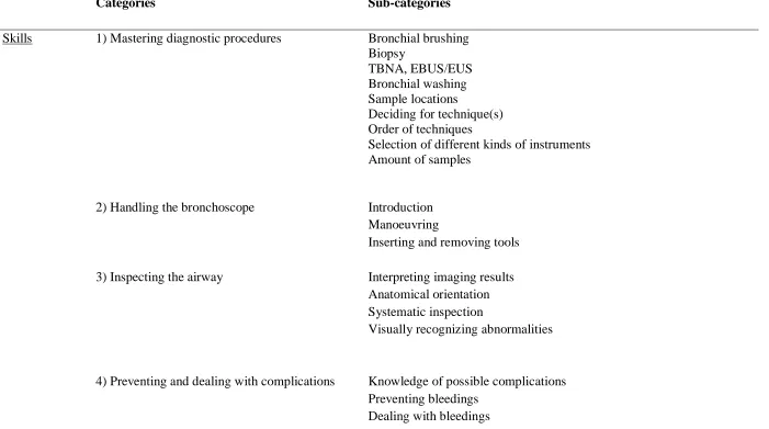
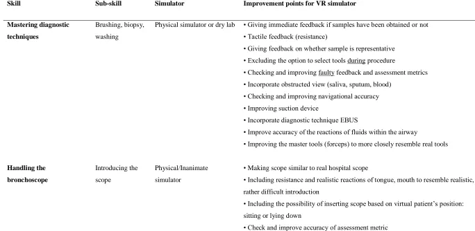
Table 4
Training requirements for simulation-based flexible bronchoscopy.
Categories Sub-categories
Skills 1) Mastering diagnostic procedures Bronchial brushing Biopsy
TBNA, EBUS/EUS Bronchial washing Sample locations
Deciding for technique(s) Order of techniques
Selection of different kinds of instruments Amount of samples
2) Handling the bronchoscope Introduction Manoeuvring
Inserting and removing tools 3) Inspecting the airway Interpreting imaging results
Anatomical orientation Systematic inspection
Visually recognizing abnormalities
4) Preventing and dealing with complications Knowledge of possible complications Preventing bleedings
5) Administering anaesthetics and sedatives Cues
Systematic administration Amount
6) Patient monitoring Paying attention to vital signs, oxygen saturation Auditory cues
Context 7) Working with an assistant Teamwork
Skills.
1
) Mastering diagnostic procedures.
Four main diagnostic techniques to be used during a FB were identified: Bronchial brushing, biopsy, Transbronchial Needle Aspiration (TBNA), Endobronchial Ultrasonography (EBUS) and bronchial washing. The VR simulator was considered inadequate by participants for training of these skills mainly due to missing tactile feedback. According to a participant, physical simulators and dry labs are more accurate due to providing haptic feedback and seeing the “real effects” (P6, p.12).
Bronchial brushing.
One technique to be mastered is bronchial brushing. As one participant puts it, the purpose of brushing is to collect, with a brush, cells from the abnormality (e.g. a tumor) (P1, p.1). Another participant adds that a brush is rubbed against the mucosa, making cells loose which are collected on the brush (P3, p.3). She adds that the brush is to be examined by the pathologist (P3, p.3).
It is important to ensure that samples are successfully collected. Ensuring this is not possible during brushing: a participant mentions that while brushing, cells cannot be seen in the brush (P4, p.3). However, in a real bronchoscopy, after the brushing, another participant describes that the brush is taken out and the samples are put onto a glass which is when he would be able to see if samples have indeed been obtained or not (P6, p.9). In the VR simulator, such samples could not be seen directly (neither during nor after the brushing), leading participants unsure about whether to continue obtaining samples or not. The only cue participants were given by the simulator is bleeding. Although a bleeding could be a reaction to obtained samples (P3, p.3), one participant says that in real life, it does not necessarily have to bleed afterwards (P6, p.9). Only after the complete simulated bronchoscopy, assessment metrics gave information about the amount of samples that were obtained successfully. However, one participant mentioned that those metrics may not be accurately reflecting the actual performance. Moreover, a participant mentions that during a real bronchoscopy, while brushing, resistance can be felt sometimes too (P5, p.7). The VR simulator in this study did not give any tactile feedback.
Biopsy.
mentions that while doing a biopsy in real-life, it is not known whether the obtained sample is representative; the obtained sample must firstly be put into a glass container. Again, bleeding was the sole cue given by the VR simulator. During the simulation, one participant noticed that a little bleeding occurred, concluding that “you definitely know you didn’t grab air, but you grabbed tissue” (P5, p.2). After seeing the assessment metrics, one participant added: “it was very handy to know that I did take some samples.” (P3, p.10).
A participant describes the general processes involved in taking a biopsy. As soon as resistance is felt from the forceps, it can be closed in order to take a sample. Thereafter, the forceps is removed from the scope and the sample secured “in the unit with the saline so you can see if there is a biopsy” (P3, p.3). However, the participant did not feel any resistance in the VR simulation (P3, p.3). Another problem encountered with the VR simulator is when selecting the tools: one participant wanted to change the selected tool, but it stayed the same (P1, p.2). It has to be added that the tool selection panel shows on the display only for a short amount of time, before disappearing (P2, p.5). When taking off the sample from a tumor, normally you feel some kind of resistance, according to a participant, which was absent in the VR simulator (P2, p.6). One participant mentions that given feedback during the procedure from the VR simulator was faulty when the display said “wrong sample location” (P4, p.3).
Difficulties.
Another participant also explains how to take a proper biopsy from a tumor which can be rather difficult according to him:
“[…] You look at the abnormal place, where is the most important abnormality, if you have the top of the tumor, it’s often necrosis, so the tumor has gone, has died because of lack of blood and so on. The top, it’s just debris, just all kind of rubbish so you should go deeper to
be in the real-life tumor. […] but not too deep that you can get bleeding.” (P2, p.8).
Another difficulty lies in taking biopsies from specific types of tumors. An experienced participant mentions that biopsies are “normally not a problem” (P1, p.2). However, he had a case with a wall-sided tumor which according to him, is rather difficult to biopsy because the “scope slides off it” (P1, p.1). Similarly, a resident mentioned that it is more difficult to get biopsies from wall-sided tumors because of the angle that needs to be made to the wall. Moreover, one participant also mentioned that doing a biopsy (in general) is hampered by the virtual patient’s respiration which causes a movement of the tumor inside the body (P1, p.1).
Transbronchial Needle Aspiration (TBNA) and EBUS.
Cells can also be collected from lymph nodes which are hidden underneath the surface/mucosa (P3, p.4; P3, p.9). According to a participant, a TBNA is performed at the location of the head carina (P4, p.7). During a TBNA, cells are collected from lymph nodes through pinching a needle through the wall and then using a suction device (i.e. aspiration). During the simulation, participants could not use the suction device (e.g. P3, p.5). Moreover, one participant explains:
“in real-life you get to wall and can always put the forceps on wall and open and then it stays there and then you can take a sample. [Here,] I tried to get needle in and take biopsy but it didn’t do it.” (P4, p.2).
Bronchial washing.
By conducting bronchial washings, one participant describes that cells are collected as well (P1, p.5). Another participant described that one part of the sample is sent to the
pathologist (cytology) to examine if there are malignant cells, and another part is sent to the microbiologist to investigate whether there is tuberculosis, infection or bacteria (P5, p.3).
For the washing of the airway, the participant describes that “a physiological saline solution” (P1, p.3) is added into the airway because saline is also contained within human bodies (P1, p.3). Moreover, a younger participant adds that water cannot be used because it is not sterile, containing bacteria (P3, p.4). She also explains that after introducing saline into the airway, coughing is a normal reaction:
“It is not normal to have fluid over there other than your own saliva. So, if I put […] fluid in, the patient will cough.” (P3, p.4).
The participant adds that the fluid then has to be aspirated and collected in order to allow for cytology and microbiology analyses (P1, p.3). The fluid /saline which is aspirated contains cells (P3, p.3). The amount of fluid which can be aspirated depends on the location within the anatomy: in a smaller bronchus, more fluid can be aspirated while the participant was in a bigger bronchus during the simulation, to which she adds “I think I put some fluid and then it didn’t come back” (P3, p.3). However, another participant makes clear that this is a fault on side of the VR simulator:
“in real life, you see the saline going into the bronchus and you have to swallow it up to get it back, here you put some saline, and then it’s gone.” (P4, p.3).
Sample locations.
Samples (e.g. from biopsies, brushings) are taken from the area that is abnormal (e.g. red and swollen) (P3, p.2). However, bronchoscopists may still have to decide where to sample. For example, a participant who had a patient case in which abnormality was present in both the right and the left lung interpreted it as the same disease and was free to choose where to sample, mentioning that “it doesn’t matter if I do the biopsies and the brushing at
could be that there are different problems so then you have to do it on both sides.” (P3, p.5). Thus, it is important to be able to interpret abnormalities and judge, taking into account risks, where to obtain samples.
Deciding for technique(s).
According to one of the experienced pulmonologists, bronchial washings are always part of the procedure, even when nothing abnormal is to be seen (P1, p.5). When no
abnormality is recognized, he explains, merely washings (for cells & bacteria) should be conducted (P1, p.5). However, in case of seeing abnormal tissue, in addition to washings, one has to biopsy and brush (P1, p.5).
The bronchoscopist has to decide which technique to use depending on the specifics of each patient case. As an experienced participant puts it:
“When you have […] patients coming with coughing, and there’s no other alarming signs, […] you can get a saline sample, for example. When you have a patient with a tumor, you are searching for, are there abnormalities in the airway that, […] you can get some material to examine from a pathology, microbiology […]. When you have a subcarinal notice, just behind the head carina, you know, this possibility that you can get a needle aspiration. So then you have to [do] […] a biopsy and […] brush and […] saline sample.” (P4, p.6).
Order of techniques.
One participant mentions that using biopsies, brushing and flushing with saline together increases the sensitivity for a diagnosis for the patient (P5, p.3). There may be an order of performing different diagnostic techniques. An experienced participant mentions to always start with 1. brushing followed by a 2. biopsy and then 3. bronchial washing (P1, p.1). For a younger pulmonologist, the preferred action was to start with a biopsy and in case a “good” sample cannot be obtained, she considered brushing “to have something” (P3, p.7). Another more experienced participant takes biopsies, then brushes and then flushes with saline (P4, p.1).
Selection of different kinds of instruments.
p.2). He adds that the type of forceps chosen also depends on the abnormality which needs to be sampled (P2, p.2). For example, given the tumor in the simulated case, he mentions that a “big forceps” will increase the chances of causing heavy bleedings (P2, p.4). However, another experienced participant mentioned that in real life, there is no choice between different kinds of forceps’, he mentions that “we only have the forceps without the needle” (P4, p.2). Similarly, he mentions that “we do have only one brush” (P4, p.3).
In addition to that, the VR simulator instruments are a bit wider and larger than in real life. Due to being out of plastic, one participant also mentioned how those instruments do not feel realistic (P6, p.7).
Amount of samples.
An experienced P mentioned that although he usually happens to take two samples, it is advised for students to take 4-6 samples because more samples facilitate the process of making a diagnosis for the pathologist who examines the samples (P1, p.9). During a real bronchoscopy, obtained samples are put into a container (P1, p.9). He explains that depending on the size of the samples that are being obtained, the required quantity of samples may vary: if samples are large, less/two samples can be enough to obtain a “perfect diagnosis” (P1, p.9). Another experienced P usually takes up to seven biopsies (P2, p.6). However, the required quantity of samples varies for him depending on the abnormality (e.g. tumor) and the extent of bleeding. For example, another experienced participant mentions that a tumor-like abnormality’s vascularity is high, meaning that it may bleed heavily (P4, p.2). Therefore, he adds, the first sample must be good, while also aiming to obtain 6 or 7 more samples if possible (P4, p.2). A younger participant mentions that the goal is to get as big a sample as possible since “that’s the most useful for the pathologist to examine” (P3, p.7).
2) Handling the bronchoscope.
That is one of the two main skills that participants emphasized that should be trained on the VR simulator.
Introduction.
bronchoscopist to choose to enter through the mouth or the nose which both have benefits and drawbacks.
Some participants mentioned that during the simulation, entering the airway was very easy:
“There is no resistance. Always open. The tongue is out, the whole mouth, larynx is open. In one second, split second, I’m in. I have seen colleagues, sweating.” (P1, p.9)
One participant describes that in reality, it is “bleak” (P6, p.13), so one has to “find your way around” in order to end up near the vocal chords (P6, p.13). Similarly, asanother participant describes, during a real bronchoscopy, for beginners, entering the airway is very difficult because patients are coughing or vomiting and moving around, causing the
bronchoscope to diverge from the track (P2, p.5). This in turn, he adds, may cause novices to accidentally enter the stomach instead of the airway (P2, p.5). Another younger participant adds that one has to “take the right route” in order not to enter into the Esophagus (stomach) (P6, p.13). The experienced participant mentions three ways to overcome this challenge of introduction: proper anaesthesia (which is not always working), having anatomical orientation and working fast (P2, p.5).
The simulator also provides an assessment metric, referring to whether the user tried to introduce the scope while the vocal chords were closed, which according to one participant is important (P6, p.13). However, he also noticed that the VR simulator does not register this information correctly (P6, p.13), referring to a dissociation between actual and recorded performance.
Manoeuvring.
While one participant mentioned to have no specific technique for handling the scope, he adds that he holds the scope in the left hand and uses the right hand for taking biopsies (P5, p.4). Other people, he adds, may hold the scope in the right hand instead. One participant uses his whole body to manoeuvre the scope inside the airway:
The participant explains that the benefits of this technique are that the scope does not have to be twisted, it is easier to enter the airway and that one is always positioned in the middle (P2, p.2). Another participant mentions that most of the time, one stands behind the patient at the head side. However, he adds, when manoeuvring through the right upper lobe which is very sharp, he turns:
“a little bit to the left, next to the patient and then it’s a little bit more straight and not around the corner so that can be nice for your hands” (P4, p.4).
When attempting to take biopsies, one participant mentions that “you have to try to
use all possible positions […] with the scope and there’s a wheel on the scope so you can […] look up or down and by moving the scope to the right or the left, you can move it also” (P3, p.6).
Regarding the VR simulator, one participant mentioned that it was more difficult to “manipulate” the scope (or forceps) in the simulator than it is with a real bronchoscope (P1, p.3). In contrast, the participant prefers the physical inanimate simulator for handling the scope which includes a real hospital scope (P1, p.3).
The VR simulator provided an assessment metric related to the amount of wall contact. While more experienced participants disregarded its importance, mentioning that it does not hurt the patient because the bronchial tree is anaesthetised (e.g. P1, p.5,6), younger participants added that wall contact could cause irritation and then coughing, making the procedure more uncomfortable and difficult for both the patient and bronchoscopist. In that sense, he adds, wall contact is important. Moreover, one participant mentioned that in the VR simulation, it was more difficult to stay in the middle than it would have been realistically (P5, p.5). One participant mentions that the scope should be held as straight as possible and not flex it in a 90 degree angle in order to prevent damage to the scope (P4, p.8).
Inserting and removing tools.
“normally, I go back centrally, then you insert the tool so that it’s just protruding so that […] it cannot damage the working channel when you’re navigating afterwards. Then you navigate
towards your place of destination, and […] only then you open the tool (needle, forceps, brush).” (P6, p.2).
Similarly, one participant says that damage to the scope could be done while using the forceps, brush or needle for the TBNA. It is important, according to him, to close the tools before removing them from the scope and to communicate well with the assistant who opens and closes the tools (P5, p.5).
The VR simulator was not accurate in this one because as one participant put it: “you
insert it like five centimetres, then it recognizes the tool already, then you insert it one
centimetre more, then it’s already visible.” (P6, p.3). Because of this unrealistic procedure, he adds, the normal workflow is disrupted, causing dissociations between how he would act during a real bronchoscopy and how he acts during the simulation (P6, p.3).
3) Inspecting the airway.
In addition to handling the bronchoscope, participants mentioned the VR simulator is Also adequate for learning this skill.
Interpreting imaging results.
CT scans of the lungs may guide bronchoscopists in inspection of the airway. One participant described that based on the scan which showed an abnormality on the left side, she decided to firstly inspect the right side in order to ensure that “everything is seen” (P3, p.1). CT scans also allow to see the condition of the lymph nodes which are usually covered
underneath the mucosa (P3, p.2). One participant describes that “on the CT scan, […], you see there is a grey area and that is too large, that should be smaller” (P3, p.4). Thus, this skill is paramount also to the decision-making regarding which techniques to use (e.g. TBNA for enlarged lymph nodes).
Anatomical orientation.